Embed Size (px)
Citation preview

“Mitral Regurgitation in the MitraClip Era”
Patient management during and after MitraClip Therapy
Eric Brochet, MD University Hospital Bichat
Paris, France

Faculty disclosure Eric Brochet, MD
I disclose the following financial relationships:
Consultant for General Electrics, Philips Heathcare, Abbott Vascular Paid speaker for Abbott Vascular

Procedural management of MitraClip Therapy
– Multidisciplinary team interaction: Interventional
cardiologists, cardiac surgeons, echocardiographers, cardiac anaesthesiologists, proctors…
– General anaesthesia, TEE and fluoroscopy guidance – Role of echocardiography (2D and 3D TEE) central to the
successful performance of the procedure
Boekstegers P et al. Clin Res Cardiol (2014) 103:85–96

Arrangements in the cath lab
Boekstegers P et al. Clin Res Cardiol (2014) 103:85–96
Procedure performed in the cardiac Cath lab or Hybrid OR
Patient management during and after MitraClip Therapy
• Anaesthesia • Imaging: TEE guidance • ICU • Anticoagulation Therapy • Follow-up

Patient management during and after MitraClip Therapy
• Anaesthesia • Imaging: TEE guidance • ICU • Anticoagulation Therapy • Follow-up

Anaesthesia during Mitraclip therapy • Specific anesthetic considerations
– Same level of expertise as anaesthesiology in cardiac surgery – General anaesthesia required for continuous TEE monitoring – Local anaesthesia and conscious sedation in few centers – Prolonged procedure, generally well tolerated hemodynamically – Monitoring :
• Invasive arterial blood pressure, central venous line , pulse oximetry
• ± IABP in patients with severely reduced LV function – Few drugs used during intervention (Vasopressor agents , Adenosine,
Dobutamine)
Kothandan H et al. Ann Card Anaesth 2014;17:17-22

Anaesthesia during Mitraclip therapy • Close cooperation between interventional cardiologists and
cardiac anaesthetists – Blood pressure control supported by the use of vasopressor agents – Help during difficult grasping attempts
• ventilator breath hold • use of adenosine (after pacing wire insertion ++)
– Anticoagulation: • heparin given before or immediately after transseptal puncture. • ACT maintained between 250 and 300s , checked every 30 mn
– Assessment of result – Early detection of complications
Kothandan H et al. Ann Card Anaesth 2014;17:17-22

Anaesthesia during Mitraclip therapy Acute problems during MitraClip placement : • Procedural failure • Hypotension: check TEE
– Tamponade (TS puncture) Importance of baseline echo ++ – LV dysfunction – Worsening of MR – Creation of mitral stenosis
• Left atrial shunt
Kothandan H et al. Ann Card Anaesth 2014;17:17-22

Right to left atrial shunt

Right to left atrial shunt

Patient management during and after MitraClip Therapy
• Anaesthesia • Imaging: TEE guidance • ICU • Anticoagulation Therapy • Follow-up

Mitraclip echo-guided procedure • TEE guidance
– Importance of communication between interventionists and echocardiographers
– Common language – Reliance on specific views for each procedural step – Standardization of orientation of 2D/ Xplane and 3DTEE
images

Mitraclip echo-guided procedure • TEE guidance
– Importance of communication between interventionists and echocardiographers
– Common language – Reliance on specific views for each procedural step – Standardization of orientation of 2D/ Xplane and 3DTEE
images

Mitraclip echo-guided procedure Spatial orientation (2D or Xplane TEE)
MED LAT POST ANT
Intercommissural view LVOT view

Mitraclip echo-guided procedure Spatial orientation (2D or Xplane TEE)
MED LAT POST ANT
Intercommissural view LVOT view

Mitraclip echo-guided procedure Spatial orientation (3D TEE)
Ao
POST
ANT
LAA MED LAT
Ao
LAA

Mitraclip echo-guided procedure Spatial orientation (3D TEE)
Ao
POST
ANT
LAA MED LAT

Mitraclip procedure 1. Trans-septal puncture 2. Introduction of the Steerable Guide Catheter (SGC) into the LA 3. Advancement of the Clip Delivery System (CDS) into the LA 4. Steering and positioning of the MitraClip above the mitral valve 5. Advancing the MitraClip into the LV 6. Grasping of the leaflets 7. Assessment of proper leaflet insertion and reduction of MR 8. MitraClip release

Mitraclip procedure 1. Trans-septal puncture
– Echo-guided puncture Superior and Posterior - Mid fossa
– 3 echo steps
1.tenting in the Bi-caval view
2.Observe tenting in SAX at Base
3.Observe tenting in 4-CH view (0°) (Height)

Mitraclip procedure 2 . Introduction of the Steerable Guide Catheter (SGC) into the LA

Mitraclip procedure 3. Steering and positioning of the MitraClip above the mitral valve

Mitraclip procedure 3. Steering and positioning of the MitraClip above the mitral valve

Mitraclip procedure 4. positioning and perpendicularity of the MitraClip
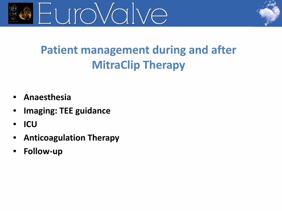
Mitraclip procedure 6. Grasping of the leaflets

Mitraclip procedure 6. Assessment of proper leaflet insertion and reduction of MR

Mitraclip procedure 6. Assessment of proper leaflet insertion and reduction of MR

Mitraclip procedure
• Immediate result
• Performed while the patient is still under anaesthetic ! – BP raised at the level prior intervention using inotropes – Manipulate preload and afterload – Sometimes use dobutamine to test for MS

• If Clip position and decrease in MR are satisfactory, the Clip is released
• In case of unsatisfactory MR reduction, repositioning of the Clip and/or the implantation of a second clip may be considered
Mitraclip procedure • Immediate result

• Second clip

Mitraclip procedure • Immediate result

Assessment of result
• After clip 1
• After clip 2
MVA 2 cm²
MVA 1,4 cm²
MG 2 mmHG
MG 5 mmHG

Assessment of result Dobutamine 5 mcg/kg/mn
MG: 12 mmHG

Patient management during and after MitraClip Therapy
• Anaesthesia • Imaging: TEE guidance • ICU • Anticoagulation Therapy • Follow-up
Periprocedural management
• ICU monitoring (12-24hours) – Clinical assessment / monitoring – Detection of complications – Bedside TTE
• LV function • Presence of the clip • Filling pressures/RV function/ sPAP • Pericardial effusion

Patient management during and after MitraClip Therapy
• Anaesthesia • Imaging: TEE guidance • ICU • Anticoagulation Therapy • Follow-up

Periprocedural management
• Anticoagulation – During the procedure
• IV UF Heparin started after trans-septal puncture • Patients not already being treated with anti-platelets
should be started on aspirin and clopidogrel immediately after the procedure
– After the procedure • no standard regimen defined • most commonly : ASA (100 mg/day) for 3 months and
clopidogrel (75 mg/day) without a loading dose for 4 weeks • Maintain anticoagulation if interrupted before the
procedure

Patient management during and after MitraClip Therapy
• Anaesthesia • Imaging: TEE guidance • ICU • Anticoagulation Therapy • Follow-up

Periprocedural management
• Post-procedural follow-up : – Clinical and echocardiographic assessment
• Functional status • BNP • Echocardiographic assessment
– TTE generally sufficient – TEE in case there is an abnormality that needs
clarification

Periprocedural management • TTE at discharge (Baseline TTE)
– Clip assessment • Presence of the clip ; attachment to both leaflets • Double orifice morphology
– MV function • MV inflow gradient, • MV area • Residual MR
– LV dimensions and function in systole and diastole, and LVEF. (2D- or 3D- echo)
– residual ASD evaluation, RV function , SPAP – Pericardial effusion
Foster et al Am J Cardiol 2001;100:1577–83 Wunderlich et al Eur Heart Journal – Cardiovasc Imaging 2013; 14, 935–949

Periprocedural management • TTE at discharge
sPAP

Periprocedural management • Residual MR
Multiparametric assessment Qualitative assessment Quantitative Doppler ?

Conclusion • Periprocedural management of mitraclip therapy
– Multidisciplinary team interaction and cooperation – Central role of echocardiography for guidance of the
procedure and assessment of procedural success – High success rate in experienced teams – Sage and well tolerated procedure in the majority of cases.
Procedure time can be reduced with experience – Careful clinical and echocardiographic follow up