Embed Size (px)
Citation preview

Mitral Stenosis
Dr Annette Vegas FRCPC, FASE
Professor Anesthesiology
Outline• Etiology
• Physiology
• Echocardiography
– Valve
– Secondary
• Assessment
http://pie.med.utoronto.ca/TEE/TEE_content/TEE_assessment_cardiacValves.html
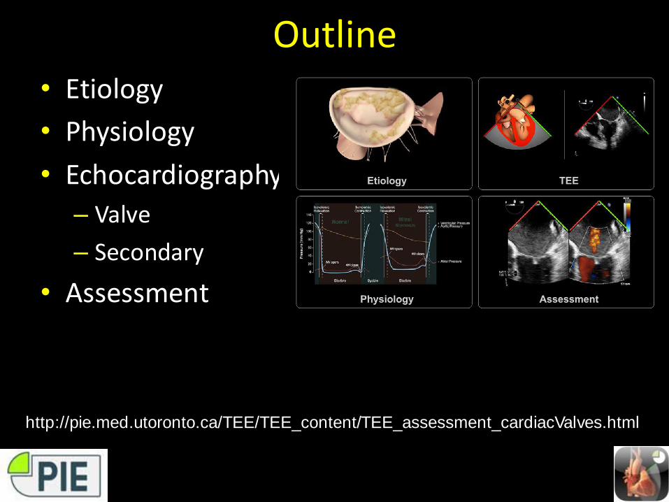
Etiology
• Rheumatic (75%)• Calcific• Congenital
– Parachute MV– Shone complex
• Inflammatory– Lupus– Rheumatoid arthritis
• Mass
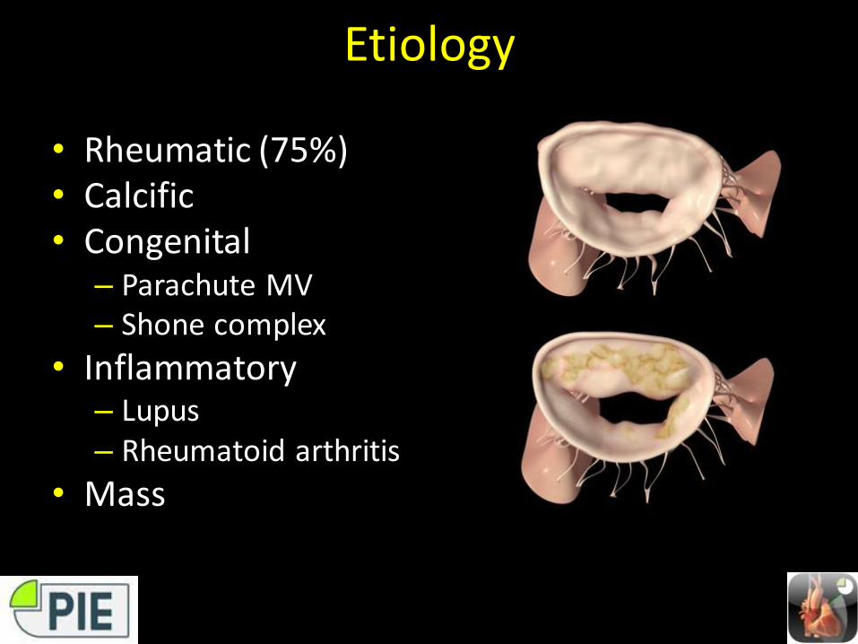
Rheumatic MV
• Leaflet thickening + calcification
• Commissural fusion
• Chordal shortening + fusion = funnel shape
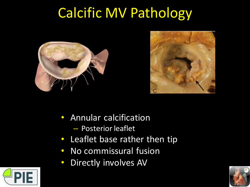
Calcific MV Pathology
• Annular calcification– Posterior leaflet
• Leaflet base rather then tip• No commissural fusion• Directly involves AV
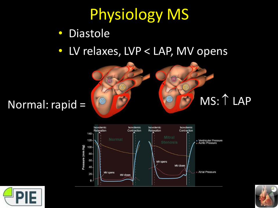
Physiology MS• Diastole
• LV relaxes, LVP < LAP, MV opens
MS: LAPNormal: rapid =
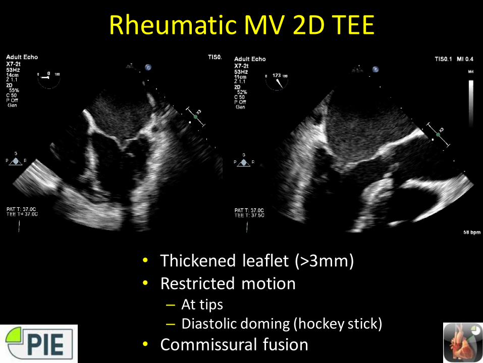
Rheumatic MV 2D TEE
• Thickened leaflet (>3mm)• Restricted motion
– At tips– Diastolic doming (hockey stick)
• Commissural fusion
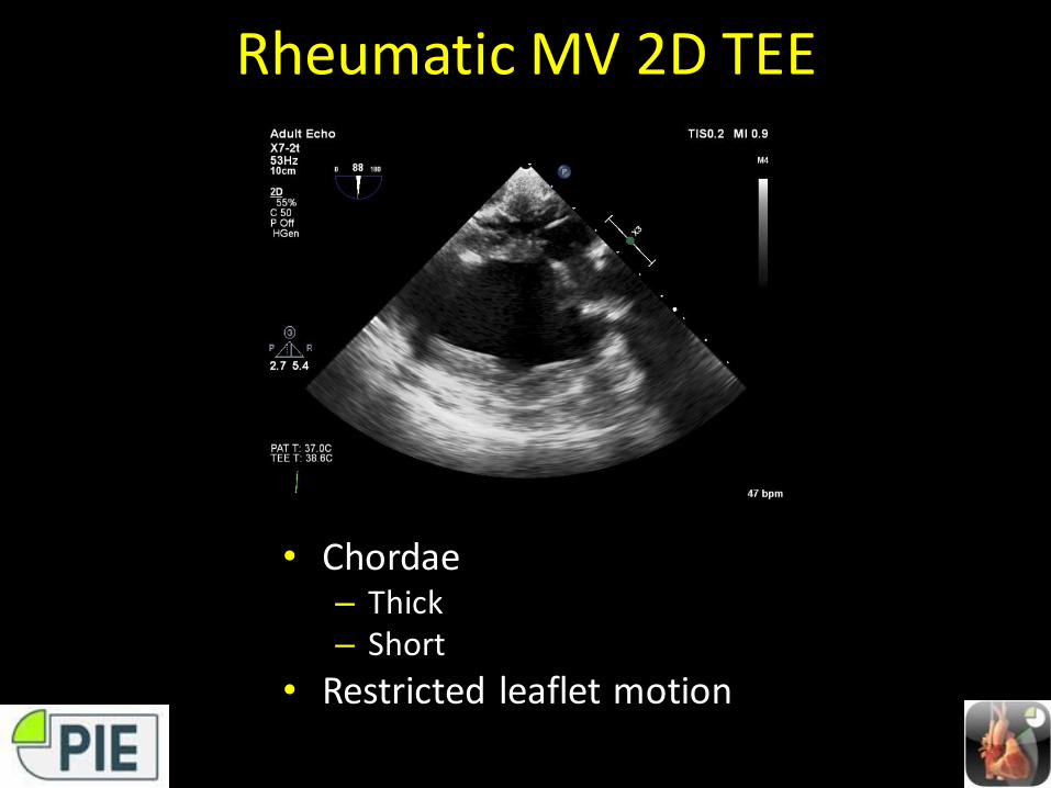
Rheumatic MV 2D TEE
• Chordae – Thick– Short
• Restricted leaflet motion

Rheumatic MV 3D TEE
LA LV
Commissural fusion
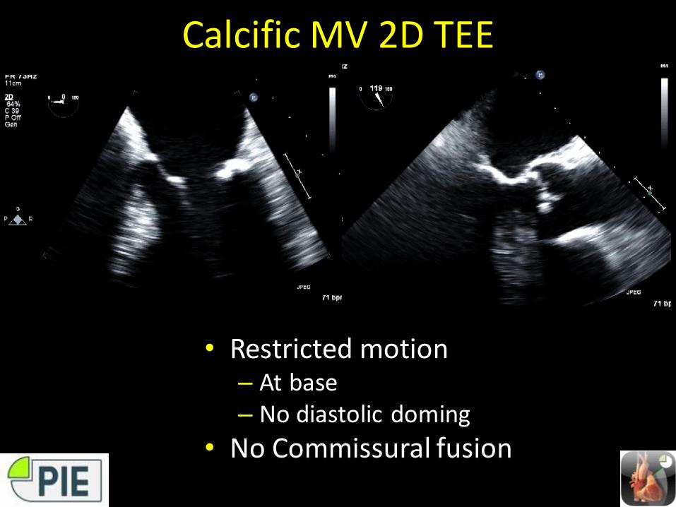
Calcific MV 2D TEE
• Restricted motion– At base– No diastolic doming
• No Commissural fusion
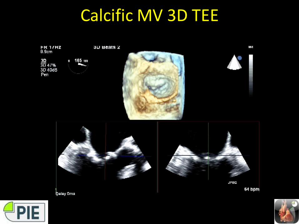
Calcific MV 3D TEE
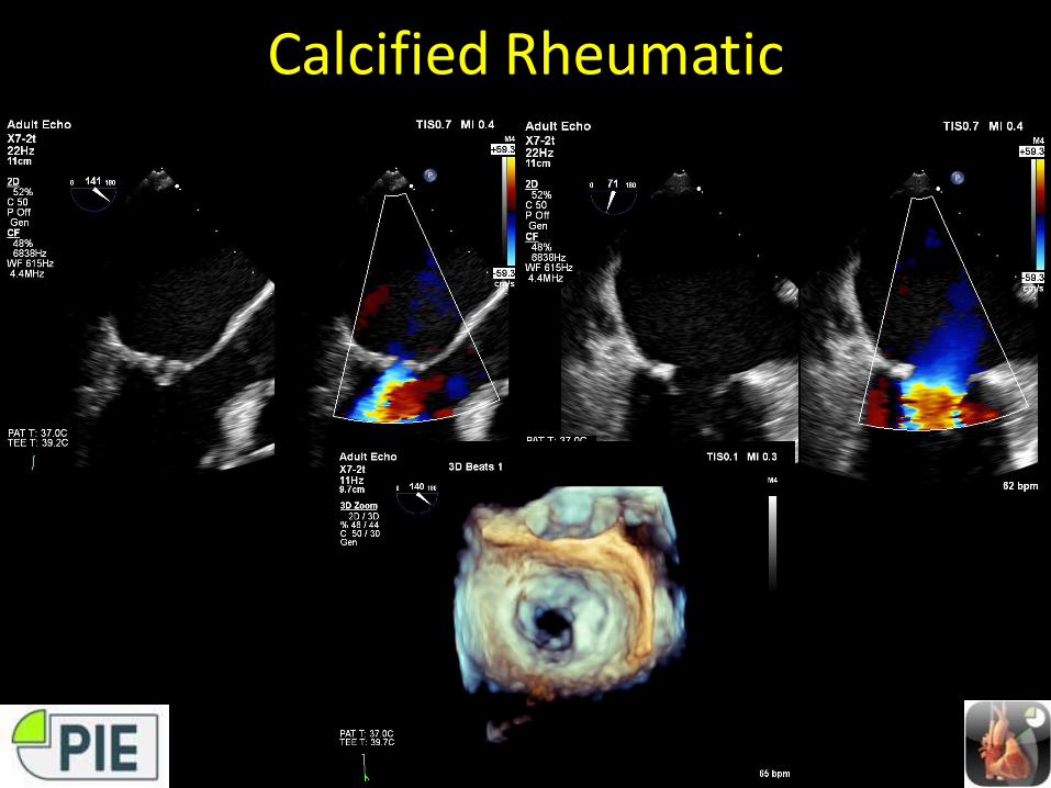
Calcified Rheumatic
MS Secondary Changes• Left Atrium
– Enlarged
– Spontaneous echo contrast (SEC)
– Thrombus
• MV (MR)
• Pulmonary hypertension
– RV dilatation
– RVSP
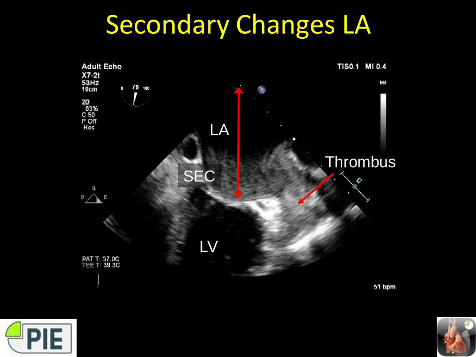
Secondary Changes LA
LA
LV
SECThrombus
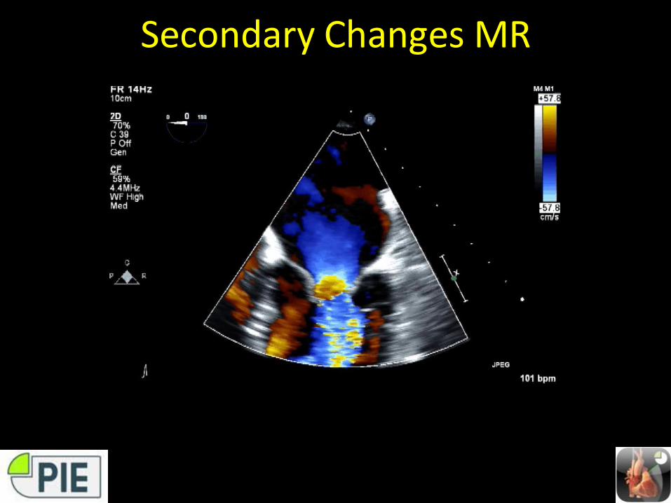
Secondary Changes MR

Secondary Changes PAP
• IAS shift to R• RV dilatation• TR
– Estimate RVSP
TR
PG 60
RVSP 70
↑ PAP• Not measure of MS severity• Does reduce survival
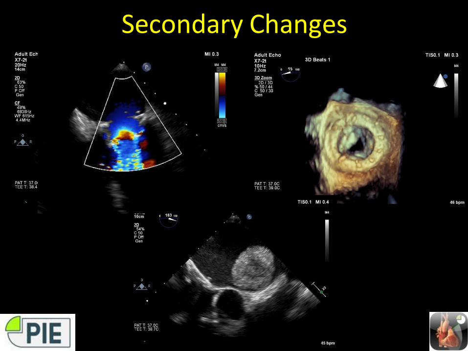
Secondary Changes

MS Assessment
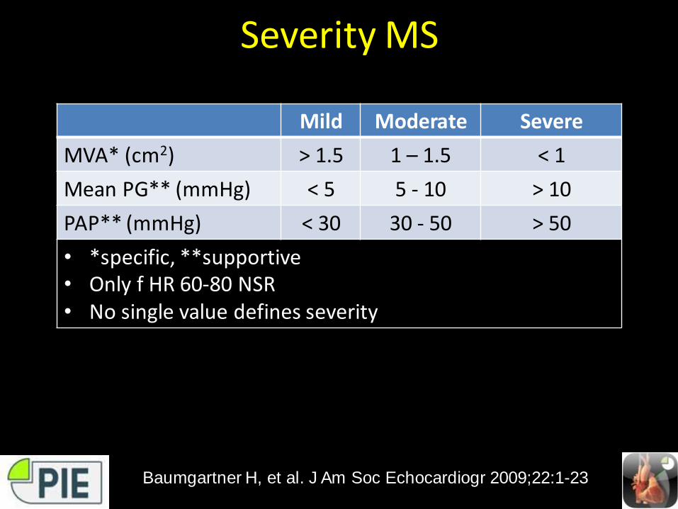
Severity MS
Mild Moderate Severe
MVA* (cm2) > 1.5 1 – 1.5 < 1
Mean PG** (mmHg) < 5 5 - 10 > 10
PAP** (mmHg) < 30 30 - 50 > 50
• *specific, **supportive• Only f HR 60-80 NSR• No single value defines severity
Baumgartner H, et al. J Am Soc Echocardiogr 2009;22:1-23
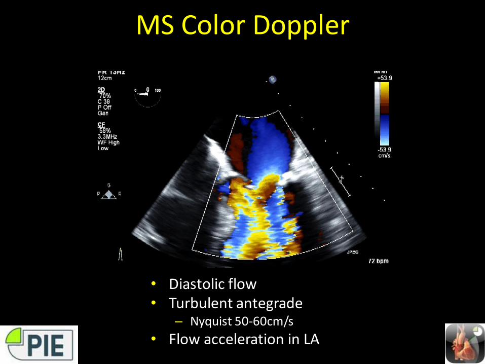
MS Color Doppler
• Diastolic flow• Turbulent antegrade
– Nyquist 50-60cm/s
• Flow acceleration in LA
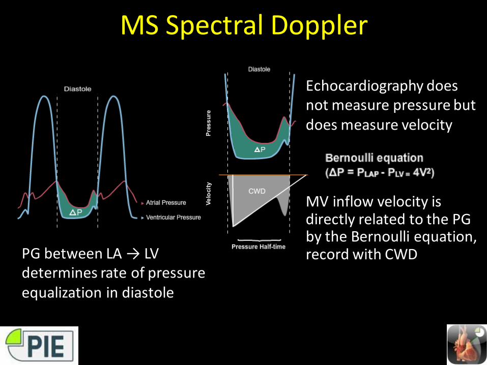
MS Spectral Doppler
PG between LA → LV determines rate of pressure equalization in diastole
Echocardiography does not measure pressure but does measure velocity
MV inflow velocity is directly related to the PG by the Bernoulli equation, record with CWD
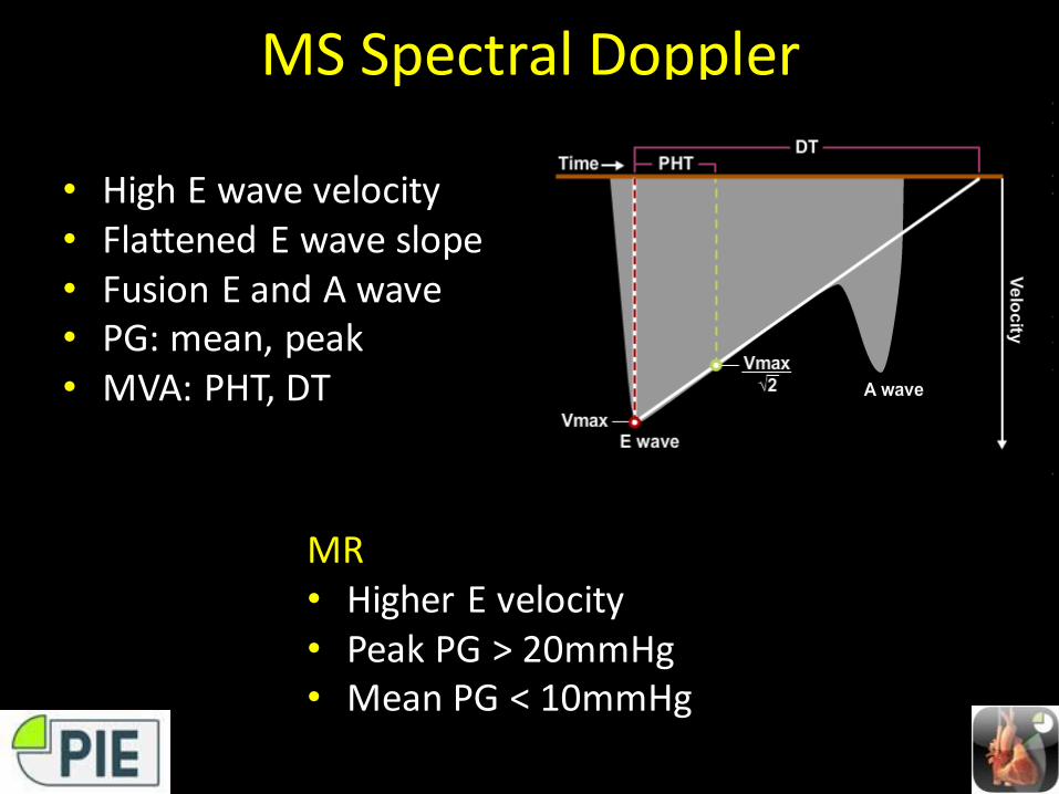
MS Spectral Doppler
• High E wave velocity• Flattened E wave slope• Fusion E and A wave• PG: mean, peak• MVA: PHT, DT
MR• Higher E velocity• Peak PG > 20mmHg• Mean PG < 10mmHg
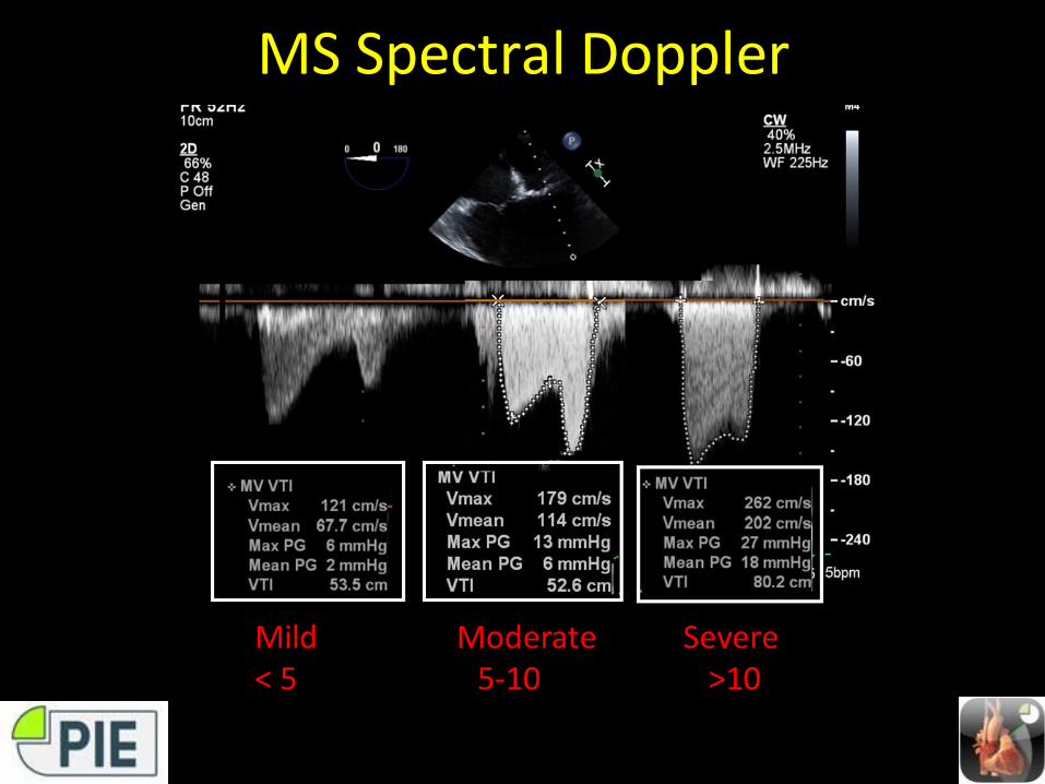
MS Spectral Doppler
Mild Moderate Severe< 5 5-10 >10
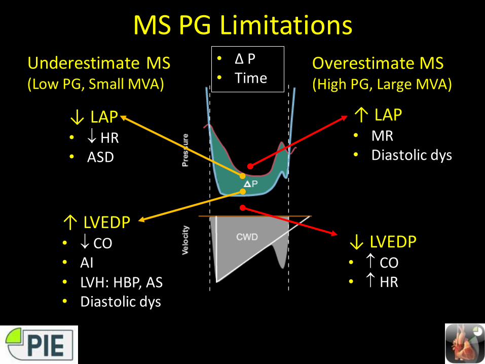
MS PG LimitationsUnderestimate MS(Low PG, Small MVA)
Overestimate MS(High PG, Large MVA)
↓ LVEDP• CO• HR
↑ LAP• MR• Diastolic dys
↑ LVEDP• CO• AI• LVH: HBP, AS• Diastolic dys
↓ LAP• HR• ASD
• ∆ P• Time

MVAAnatomic
Planimetry 2D TG Basal SAX, 3D
Functional
Pressure Half Time 220 ÷ PHT (ms)
Deceleration Time 759 ÷ deceleration time (ms)
Continuity Equation r2 x VTILVOT
VTImitral
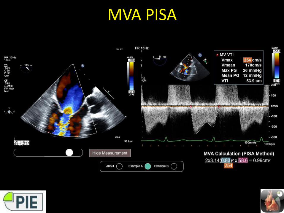
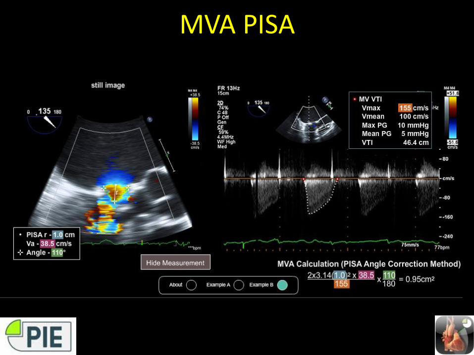
PISA 2r2 x Valiasing x /180Peak Vmitral
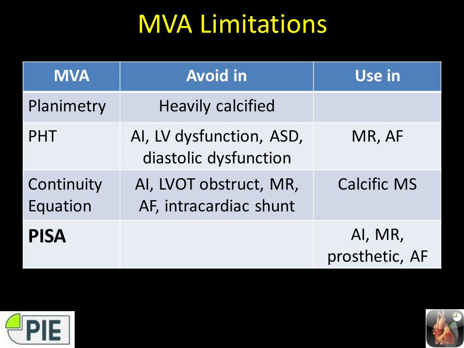
MVA Limitations
MVA Avoid in Use in
Planimetry Heavily calcified
PHT AI, LV dysfunction, ASD, diastolic dysfunction
MR, AF
Continuity Equation
AI, LVOT obstruct, MR, AF, intracardiac shunt
Calcific MS
PISA AI, MR, prosthetic, AF
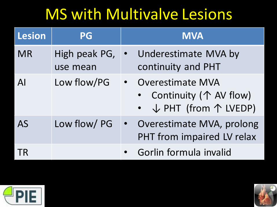
MS with Multivalve LesionsLesion PG MVA
MR High peak PG, use mean
• Underestimate MVA by continuity and PHT
AI Low flow/PG • Overestimate MVA• Continuity (↑ AV flow) • ↓ PHT (from ↑ LVEDP)
AS Low flow/ PG • Overestimate MVA, prolong PHT from impaired LV relax
TR • Gorlin formula invalid
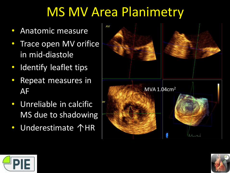
MS MV Area Planimetry• Anatomic measure
• Trace open MV orifice in mid-diastole
• Identify leaflet tips
• Repeat measures in AF
• Unreliable in calcific MS due to shadowing
• Underestimate ↑HR
MVA 1.04cm2
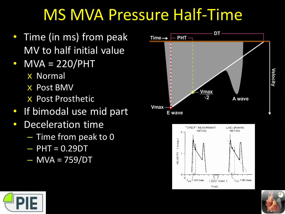
MS MVA Pressure Half-Time• Time (in ms) from peak
MV to half initial value• MVA = 220/PHT
x Normalx Post BMVx Post Prosthetic
• If bimodal use mid part• Deceleration time
– Time from peak to 0– PHT = 0.29DT– MVA = 759/DT
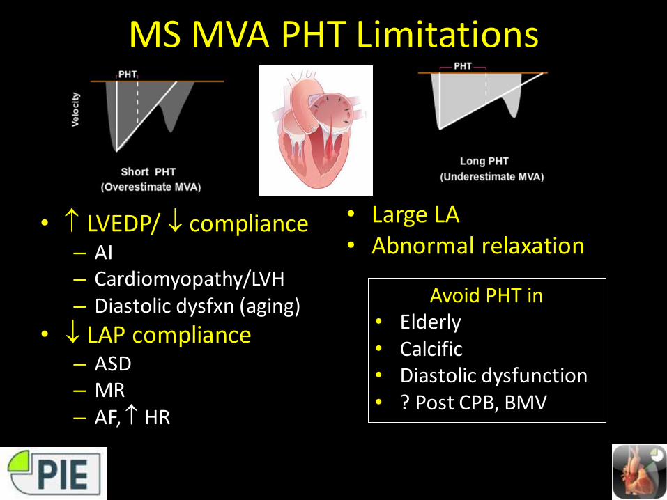
MS MVA PHT Limitations
• LVEDP/ compliance– AI– Cardiomyopathy/LVH– Diastolic dysfxn (aging)
• LAP compliance– ASD– MR– AF, HR
• Large LA• Abnormal relaxation
Avoid PHT in• Elderly• Calcific • Diastolic dysfunction• ? Post CPB, BMV
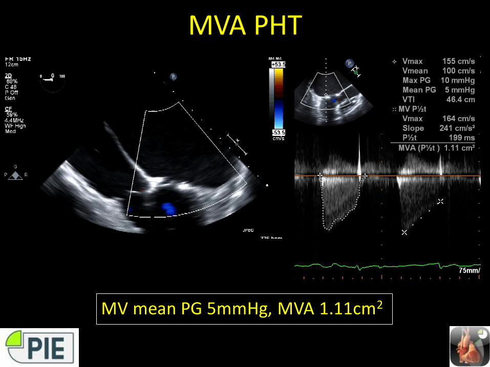
MVA PHT
MV mean PG 5mmHg, MVA 1.11cm2
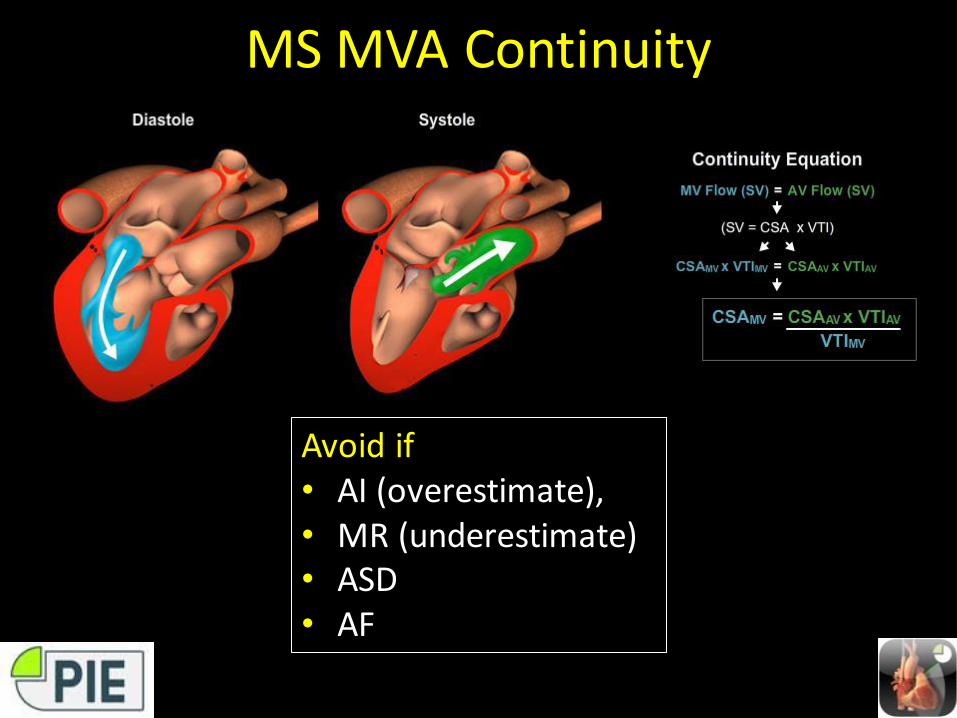
MS MVA Continuity
Avoid if• AI (overestimate), • MR (underestimate)• ASD• AF
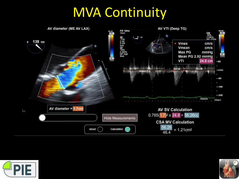
MVA Continuity
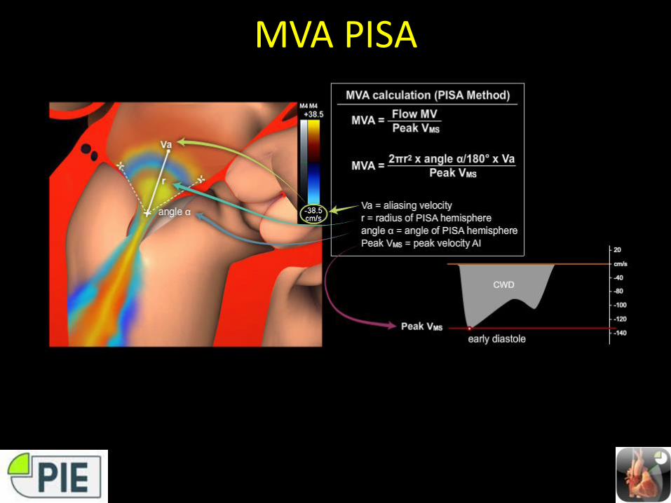
MVA PISA
MVA PISA
MVA PISA
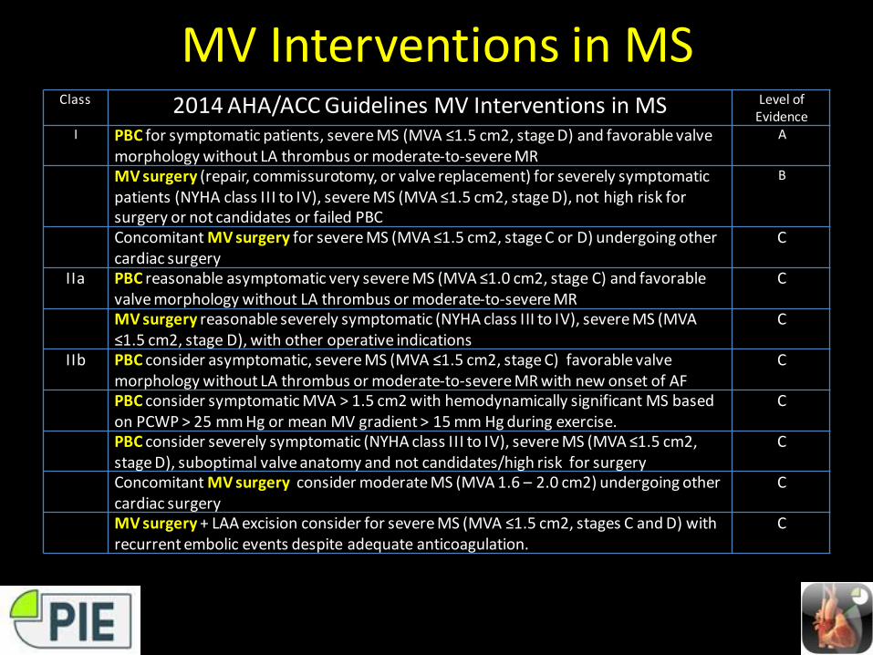
MV Interventions in MSClass 2014 AHA/ACC Guidelines MV Interventions in MS Level of
Evidence I PBC for symptomatic patients, severe MS (MVA ≤1.5 cm2, stage D) and favorable valve
morphology without LA thrombus or moderate-to-severe MR
A
MV surgery (repair, commissurotomy, or valve replacement) for severely symptomatic patients (NYHA class III to IV), severe MS (MVA ≤1.5 cm2, stage D), not high risk for surgery or not candidates or failed PBC
B
Concomitant MV surgery for severe MS (MVA ≤1.5 cm2, stage C or D) undergoing other cardiac surgery
C
IIa PBC reasonable asymptomatic very severe MS (MVA ≤1.0 cm2, stage C) and favorable valve morphology without LA thrombus or moderate-to-severe MR
C
MV surgery reasonable severely symptomatic (NYHA class III to IV), severe MS (MVA ≤1.5 cm2, stage D), with other operative indications
C
IIb PBC consider asymptomatic, severe MS (MVA ≤1.5 cm2, stage C) favorable valve morphology without LA thrombus or moderate-to-severe MR with new onset of AF
C
PBC consider symptomatic MVA > 1.5 cm2 with hemodynamically significant MS based on PCWP > 25 mm Hg or mean MV gradient > 15 mm Hg during exercise.
C
PBC consider severely symptomatic (NYHA class III to IV), severe MS (MVA ≤1.5 cm2, stage D), suboptimal valve anatomy and not candidates/high risk for surgery
C
Concomitant MV surgery consider moderate MS (MVA 1.6 – 2.0 cm2) undergoing other cardiac surgery
C
MV surgery + LAA excision consider for severe MS (MVA ≤1.5 cm2, stages C and D) with recurrent embolic events despite adequate anticoagulation.
C
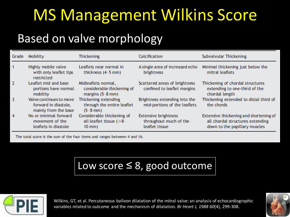
MS Management Wilkins ScoreBased on valve morphology
Wilkins, GT, et al. Percutaneous balloon dilatation of the mitral valve: an analysis of echocardiographic variables related to outcome and the mechanism of dilatation. Br Heart J, 1988 60(4), 299-308.
Low score ≤ 8, good outcome
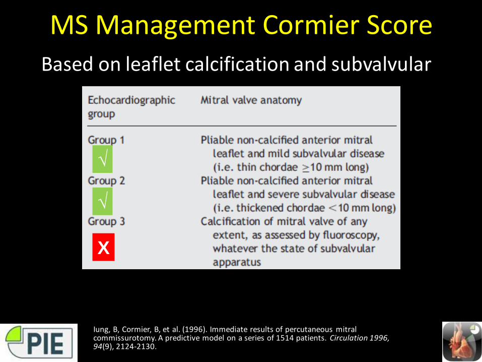
MS Management Cormier ScoreBased on leaflet calcification and subvalvular
Iung, B, Cormier, B, et al. (1996). Immediate results of percutaneous mitral commissurotomy. A predictive model on a series of 1514 patients. Circulation 1996, 94(9), 2124-2130.
X
√
√
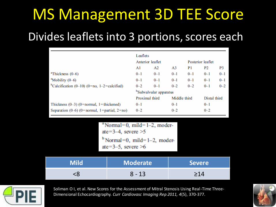
MS Management 3D TEE ScoreDivides leaflets into 3 portions, scores each
Mild Moderate Severe
<8 8 - 13 ≥14
Soliman O I, et al. New Scores for the Assessment of Mitral Stenosis Using Real-Time Three-Dimensional Echocardiography. Curr Cardiovasc Imaging Rep 2011, 4(5), 370-377.

Selected Readings• Cherry AD, Maxwell CD, Nicoara A. Intraoperative Evaluation of Mitral Stenosis by
Transesophageal Echocardiography. Anesth Analg 2016;123:14-20.• Baumgartner H, et al. Echocardiographic assessment of valve stenosis: EAE/ASE
recommendations for clinical practice. J Am Soc Echocardiogr2009;22:1-23.• Wunderlich NC, Beigel R, Siegel RJ. Management of Mitral Stenosis Using 2D and
3D Echo-Doppler Imaging. JACC: Cardiovasc Imaging 2013;6:1191-1205.• Alaa Mabrouk SO, Tanaka H, et al. Comparison of mitral valve area by pressure
half-time and proximal isovelocity surface area method in patients with mitral stenosis: effect of net atrioventricular compliance. Eur J Echocardiogr2011;12:283–290.
• Wilkins, GT, et al. Percutaneous balloon dilatation of the mitral valve: an analysis of echocardiographic variables related to outcome and the mechanism of dilatation. Br Heart J, 1988 60(4), 299-308.
• Iung, B, Cormier, B, et al. (1996). Immediate results of percutaneous mitral commissurotomy. A predictive model on a series of 1514 patients. Circulation 1996, 94(9), 2124-2130.
• Soliman O I, et al. New Scores for the Assessment of Mitral Stenosis Using Real-Time Three-Dimensional Echocardiography. Curr Cardiovasc Imaging Rep 2011, 4(5), 370-377.
Question 1Which of the following is not a finding in rheumatic MS ?
1. Commissural fusion
2. Annular + leaflet base calcification
3. Chordal shortening + fusion
4. Leaflet tip calcification
5. Diastolic doming
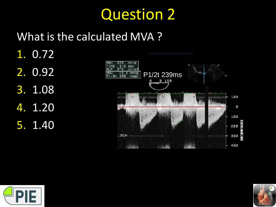
Question 2What is the calculated MVA ?
1. 0.72
2. 0.92
3. 1.08
4. 1.20
5. 1.40
P1/2t 239ms
Question 3Which of the following is true ?
A. Mean gradient is underestimated with tachycardia
B. MR underestimates MS severity by mean PG
C. Pressure half time is decreased with reduced cardiac output
D. AI overestimates MVA by PHT method
Question 4Severe MV stenosis is diagnosed when the normal MVA is reduced by at least ?
1. 25%
2. 33%
3. 50%
4. 66%
5. 75%
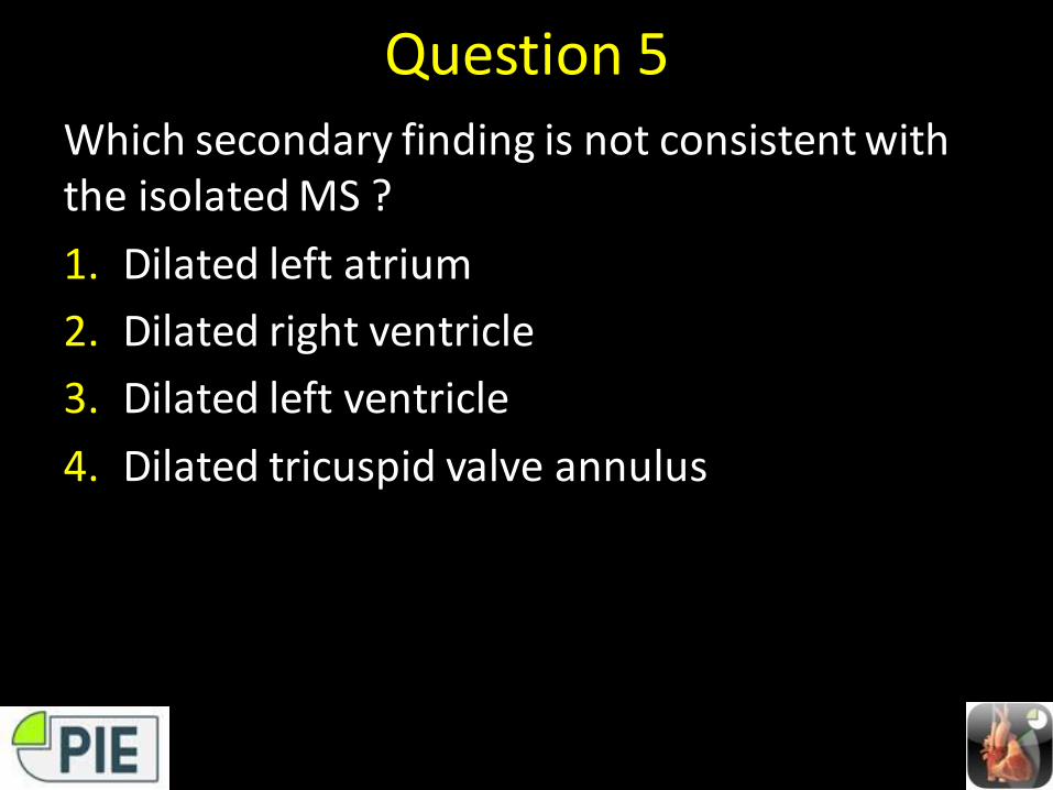
Question 5Which secondary finding is not consistent with the isolated MS ?
1. Dilated left atrium
2. Dilated right ventricle
3. Dilated left ventricle
4. Dilated tricuspid valve annulus
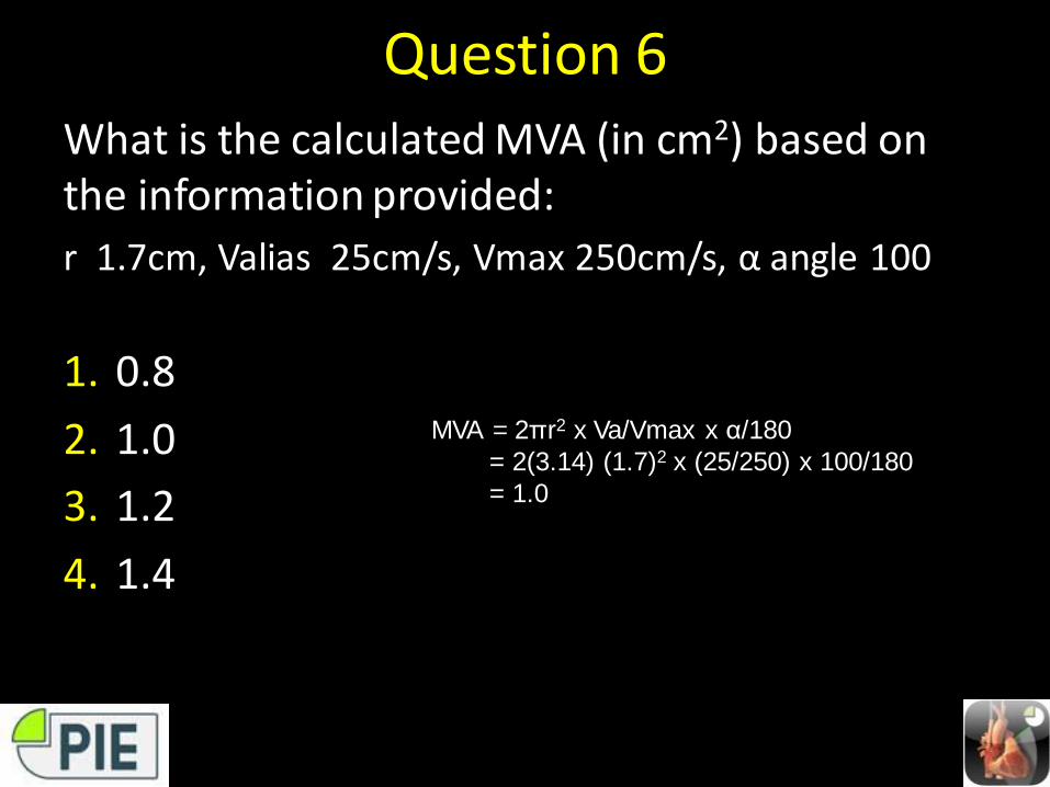
Question 6What is the calculated MVA (in cm2) based on the information provided:
r 1.7cm, Valias 25cm/s, Vmax 250cm/s, α angle 100
1. 0.8
2. 1.0
3. 1.2
4. 1.4
MVA = 2πr2 x Va/Vmax x α/180
= 2(3.14) (1.7)2 x (25/250) x 100/180
= 1.0