Embed Size (px)
Citation preview
61
© 2015 The Korean Society of Pathologists/The Korean Society for CytopathologyThis is an Open Access article distributed under the terms of the Creative Commons Attribution Non-Commercial License (http://creativecommons.org/licenses/by-nc/3.0) which permits unrestricted non-commercial use, distribution, and reproduction in any medium, provided the original work is properly cited.
pISSN 2383-7837eISSN 2383-7845
Malignant transformation of mature cystic teratoma is un-common, and occurs in approximately 2%–4% of cases.1 The most common tumor is squamous cell carcinoma, followed by mucinous carcinoma, carcinoid tumor, thyroid carcinoma, etc.; however, any of the tissues in mature teratoma may undergo malignant transformation.1
Goblet cell carcinoid (mucinous carcinoid) is a distinctive neoplasm with features of both carcinoid tumor and adenocarci-noma. Most cases described in the literature have occurred in the appendix and rarely, in the ovary. Although mature cystic teratomas occur in many extragonadal areas including the mes-entery2 and greater omentum, occurrence of goblet cell carci-noid arising in extragonadal teratoma has been rarely described, with only a single case in mediastinum being reported in the English literature.3 Moreover, a combination of mucinous ade-nocarcinoma, goblet cell carcinoid, carcinoid tumor, and mature teratoma in an extragenital organ has not been reported.
Herein, we present a rare case of combined mucinous adeno-carcinoma and goblet cell and typical carcinoid tumor arising in mature cystic teratoma of the mesentery in a 48-year-old wo-man.
CASE REPORT
A 48-year-old woman presented with a palpable abdominal mass in the periumbilical area with a vague abdominal pain of
one month duration. Past medical history was unremarkable. She had no symptoms of carcinoid syndrome. On physical ex-amination, the abdomen was markedly distended due to a solid movable mass. Abdominal and pelvic computed tomography scans revealed a huge heterogeneously enhanced mass in the ab-dominal cavity, which was associated with multiple smaller nodules throughout the entire abdominal and pelvic cavities and the right lobe of the liver. Bilateral ovaries were diffusely enlarged to form a semisolid mass. Laboratory findings demon-strated increased serum levels of carcinoembryonic antigen (264 ng/mL; normal<6 ng/mL) and carbohydrate antigen 125 (294 U/mL, normal<35 U/ml). Alpha-fetoprotein, beta-subunit of human chorionic gonadotropin and carcinohydrate antigen-19-9 were within normal range.
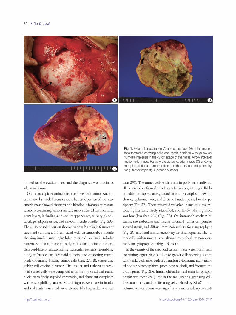
Exploratory laparotomy revealed a huge mass in the mesen-tery of the small intestine (Fig. 1A), measuring 20×18×11 cm, with coexisting multiple outbulging hepatic masses, bilateral ovarian masses, and diffusely seeding nodules on the peritoneal surface and omentum. On cut section, the mesenteric mass had solid and cystic portions. The cystic portion, occupying approx-imately 60% of the tumor, was filled with yellow-tan sebum-like materials, hair, mucoid and gelatinous substances, while the solid portion (40%) was composed of loose myxoid tissue (Fig. 1B). Bilateral ovaries were enlarged, measuring 12.5×8× 6 cm in the right ovary and 10×6×3 cm in the left ovary, and multiple gelatinous tumor nodules were observed on both ovar-ian surfaces and parenchyma (Fig. 1C). The cut surface of the ovarian masses was greyish white and gelatinous. The seeding nodules of the peritoneal surface and omentum had myxoid and gelatinous appearances. Excision of the mesenteric mass, bilat-eral salpingo-oophorectomy, total omentectomy and appendec-tomy were undertaken. Intraoperative frozen sections were per-
Journal of Pathology and Translational Medicine 2015; 49: 61-65http://dx.doi.org/10.4132/jptm.2014.09.17
▒ BRIEF CASE REPORT ▒
Corresponding AuthorKyu-Rae Kim, M.D.Department of Pathology, Asan Medical Center, University of Ulsan College of Medicine, 88 Olympic-ro 43-gil, Songpa-gu, Seoul 138-736, Korea Tel: +82-2-3010-4514, Fax: +82-2-472-7898, E-mail: [email protected]
Received: June 30, 2014 Revised: September 3, 2014 Accepted: September 17, 2014
Mixed Carcinoid-Mucinous Adenocarcinoma Arising in Mature Teratoma
of Mesentery
Su-Jin Shin · Eun-Mi Son · Chang Ohk Sung · Kyu-Rae Kim
Department of Pathology, Asan Medical Center, University of Ulsan College of Medicine, Seoul, Korea
http://jpatholtm.org/ http://dx.doi.org/10.4132/jptm.2014.09.17
62 • Shin S-J, et al.
formed for the ovarian mass, and the diagnosis was mucinous adenocarcinoma.
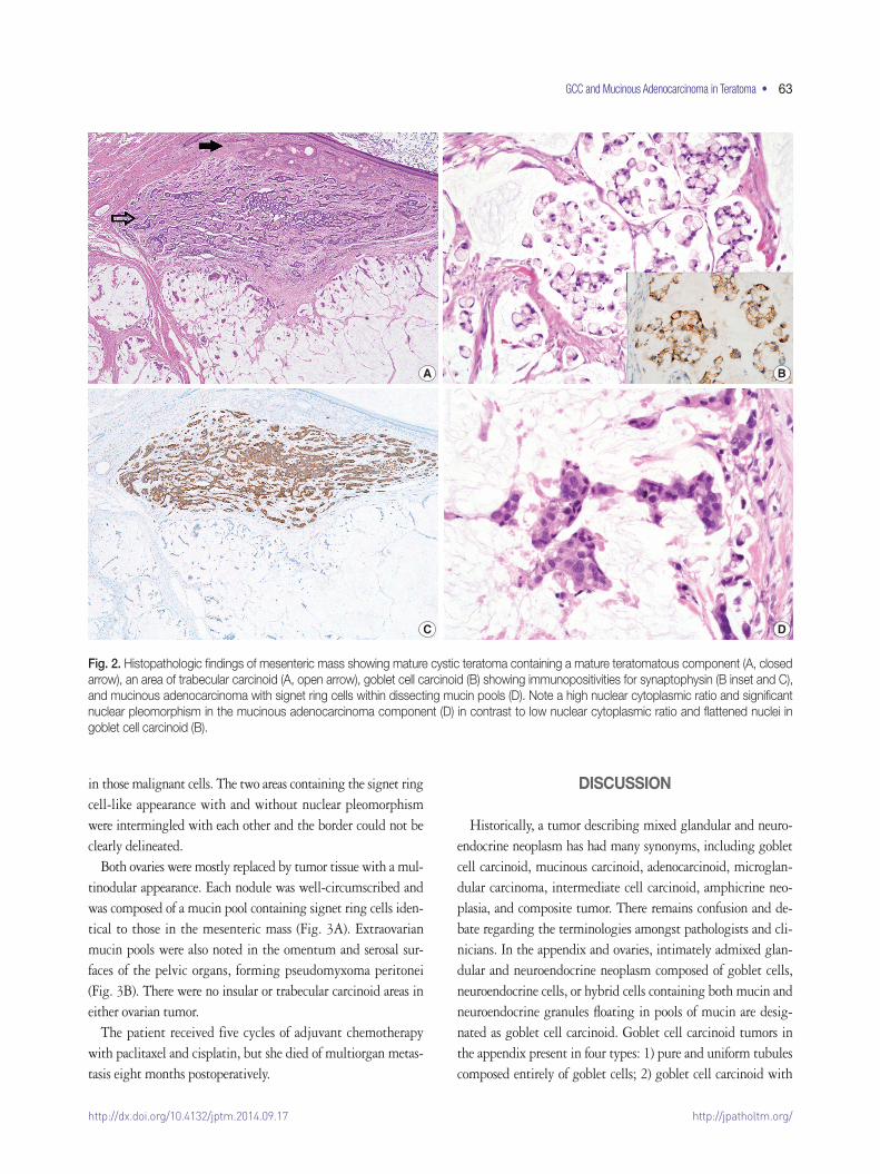
On microscopic examinations, the mesenteric tumor was en-capsulated by thick fibrous tissue. The cystic portion of the mes-enteric mass showed characteristic histologic features of mature teratoma containing various mature tissues derived from all three germ layers, including skin and its appendages, salivary glands, cartilage, adipose tissue, and smooth muscle bundles (Fig. 2A). The adjacent solid portion showed various histologic features of carcinoid tumors; a 1.5-cm sized well-circumscribed nodule showing insular, small glandular, rosettoid, and solid tubular patterns similar to those of midgut (insular) carcinoid tumors, thin cord-like or anastomosing trabecular patterns resembling hindgut (trabecular) carcinoid tumors, and dissecting mucin pools containing floating tumor cells (Fig. 2A, B), suggesting goblet cell carcinoid tumor. The insular and trabecular carci-noid tumor cells were composed of uniformly small and round nuclei with finely stippled chromatin, and abundant cytoplasm with eosinophilic granules. Mitotic figures were rare in insular and trabecular carcinoid areas (Ki-67 labeling index was less
than 2%). The tumor cells within mucin pools were individu-ally scattered or formed small nests having signet ring cell-like or goblet cell appearances, abundant foamy cytoplasm, low nu-clear cytoplasmic ratio, and flattened nuclei pushed to the pe-riphery (Fig. 2B). There was mild variation in nuclear sizes, mi-totic figures were rarely identified, and Ki-67 labeling index was low (less than 2%) (Fig. 2B). On immunohistochemical stains, the trabecular and insular carcinoid tumor components showed strong and diffuse immunoreactivity for synaptophysin (Fig. 2C) and focal immunoreactivity for chromogranin. The tu-mor cells within mucin pools showed multifocal immunoposi-tivity for synaptophysin (Fig. 2B inset).
In the vicinity of the carcinoid tumors, there were mucin pools containing signet ring cell-like or goblet cells showing signifi-cantly enlarged nuclei with high nuclear cytoplasmic ratio, mark-ed nuclear pleomorphism, prominent nucleoli, and frequent mi-totic figures (Fig. 2D). Immunohistochemical stain for synapto-physin was completely lost in the malignant signet ring cell-like tumor cells, and proliferating cells defined by Ki-67 immu-nohistochemical stains were significantly increased, up to 20%,
A B
C
Fig. 1. External appearance (A) and cut surface (B) of the mesen-teric teratoma showing solid and cystic portions with yellow se-bum-like materials in the cystic space of the mass. Arrow indicates mesenteric mass. Partially disrupted ovarian mass (C) showing multiple gelatinous tumor nodules on the surface and parenchy-ma (I, tumor implant; S, ovarian surface).
http://jpatholtm.org/http://dx.doi.org/10.4132/jptm.2014.09.17
GCC and Mucinous Adenocarcinoma in Teratoma • 63
in those malignant cells. The two areas containing the signet ring cell-like appearance with and without nuclear pleomorphism were intermingled with each other and the border could not be clearly delineated.
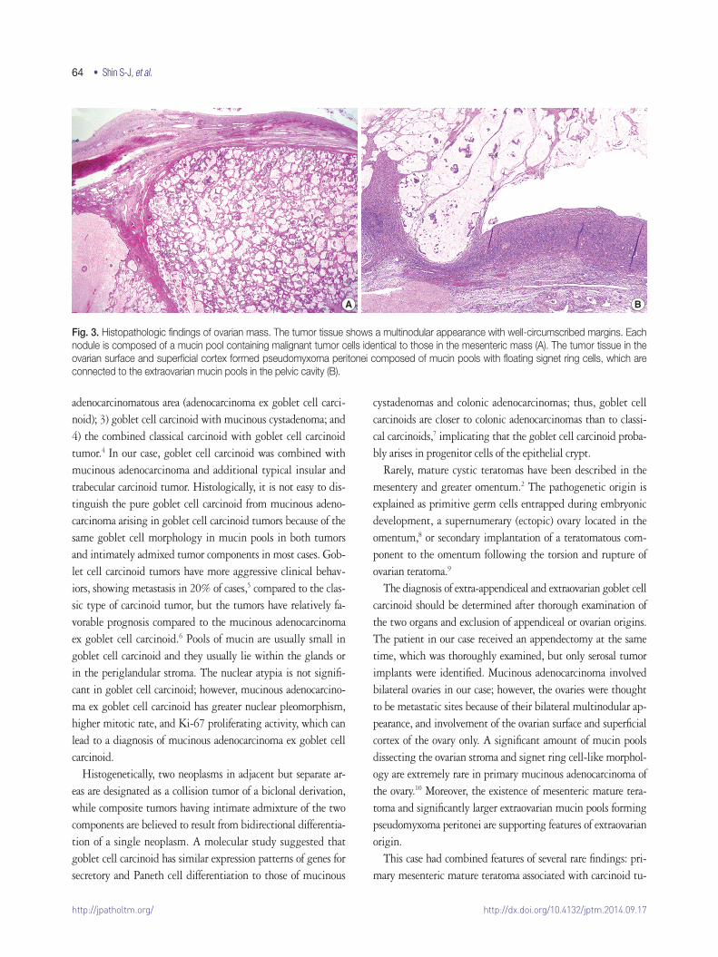
Both ovaries were mostly replaced by tumor tissue with a mul-tinodular appearance. Each nodule was well-circumscribed and was composed of a mucin pool containing signet ring cells iden-tical to those in the mesenteric mass (Fig. 3A). Extraovarian mucin pools were also noted in the omentum and serosal sur-faces of the pelvic organs, forming pseudomyxoma peritonei (Fig. 3B). There were no insular or trabecular carcinoid areas in either ovarian tumor.
The patient received five cycles of adjuvant chemotherapy with paclitaxel and cisplatin, but she died of multiorgan metas-tasis eight months postoperatively.
DISCUSSION
Historically, a tumor describing mixed glandular and neuro-endocrine neoplasm has had many synonyms, including goblet cell carcinoid, mucinous carcinoid, adenocarcinoid, microglan-dular carcinoma, intermediate cell carcinoid, amphicrine neo-plasia, and composite tumor. There remains confusion and de-bate regarding the terminologies amongst pathologists and cli-nicians. In the appendix and ovaries, intimately admixed glan-dular and neuroendocrine neoplasm composed of goblet cells, neuroendocrine cells, or hybrid cells containing both mucin and neuroendocrine granules floating in pools of mucin are desig-nated as goblet cell carcinoid. Goblet cell carcinoid tumors in the appendix present in four types: 1) pure and uniform tubules composed entirely of goblet cells; 2) goblet cell carcinoid with
A B
C D
Fig. 2. Histopathologic findings of mesenteric mass showing mature cystic teratoma containing a mature teratomatous component (A, closed arrow), an area of trabecular carcinoid (A, open arrow), goblet cell carcinoid (B) showing immunopositivities for synaptophysin (B inset and C), and mucinous adenocarcinoma with signet ring cells within dissecting mucin pools (D). Note a high nuclear cytoplasmic ratio and significant nuclear pleomorphism in the mucinous adenocarcinoma component (D) in contrast to low nuclear cytoplasmic ratio and flattened nuclei in goblet cell carcinoid (B).
http://jpatholtm.org/ http://dx.doi.org/10.4132/jptm.2014.09.17
64 • Shin S-J, et al.
adenocarcinomatous area (adenocarcinoma ex goblet cell carci-noid); 3) goblet cell carcinoid with mucinous cystadenoma; and 4) the combined classical carcinoid with goblet cell carcinoid tumor.4 In our case, goblet cell carcinoid was combined with mucinous adenocarcinoma and additional typical insular and trabecular carcinoid tumor. Histologically, it is not easy to dis-tinguish the pure goblet cell carcinoid from mucinous adeno-carcinoma arising in goblet cell carcinoid tumors because of the same goblet cell morphology in mucin pools in both tumors and intimately admixed tumor components in most cases. Gob-let cell carcinoid tumors have more aggressive clinical behav-iors, showing metastasis in 20% of cases,5 compared to the clas-sic type of carcinoid tumor, but the tumors have relatively fa-vorable prognosis compared to the mucinous adenocarcinoma ex goblet cell carcinoid.6 Pools of mucin are usually small in goblet cell carcinoid and they usually lie within the glands or in the periglandular stroma. The nuclear atypia is not signifi-cant in goblet cell carcinoid; however, mucinous adenocarcino-ma ex goblet cell carcinoid has greater nuclear pleomorphism, higher mitotic rate, and Ki-67 proliferating activity, which can lead to a diagnosis of mucinous adenocarcinoma ex goblet cell carcinoid.
Histogenetically, two neoplasms in adjacent but separate ar-eas are designated as a collision tumor of a biclonal derivation, while composite tumors having intimate admixture of the two components are believed to result from bidirectional differentia-tion of a single neoplasm. A molecular study suggested that goblet cell carcinoid has similar expression patterns of genes for secretory and Paneth cell differentiation to those of mucinous
cystadenomas and colonic adenocarcinomas; thus, goblet cell carcinoids are closer to colonic adenocarcinomas than to classi-cal carcinoids,7 implicating that the goblet cell carcinoid proba-bly arises in progenitor cells of the epithelial crypt.
Rarely, mature cystic teratomas have been described in the mesentery and greater omentum.2 The pathogenetic origin is explained as primitive germ cells entrapped during embryonic development, a supernumerary (ectopic) ovary located in the omentum,8 or secondary implantation of a teratomatous com-ponent to the omentum following the torsion and rupture of ovarian teratoma.9
The diagnosis of extra-appendiceal and extraovarian goblet cell carcinoid should be determined after thorough examination of the two organs and exclusion of appendiceal or ovarian origins. The patient in our case received an appendectomy at the same time, which was thoroughly examined, but only serosal tumor implants were identified. Mucinous adenocarcinoma involved bilateral ovaries in our case; however, the ovaries were thought to be metastatic sites because of their bilateral multinodular ap-pearance, and involvement of the ovarian surface and superficial cortex of the ovary only. A significant amount of mucin pools dissecting the ovarian stroma and signet ring cell-like morphol-ogy are extremely rare in primary mucinous adenocarcinoma of the ovary.10 Moreover, the existence of mesenteric mature tera-toma and significantly larger extraovarian mucin pools forming pseudomyxoma peritonei are supporting features of extraovarian origin.
This case had combined features of several rare findings: pri-mary mesenteric mature teratoma associated with carcinoid tu-
A B
Fig. 3. Histopathologic findings of ovarian mass. The tumor tissue shows a multinodular appearance with well-circumscribed margins. Each nodule is composed of a mucin pool containing malignant tumor cells identical to those in the mesenteric mass (A). The tumor tissue in the ovarian surface and superficial cortex formed pseudomyxoma peritonei composed of mucin pools with floating signet ring cells, which are connected to the extraovarian mucin pools in the pelvic cavity (B).

http://jpatholtm.org/http://dx.doi.org/10.4132/jptm.2014.09.17
GCC and Mucinous Adenocarcinoma in Teratoma • 65
mor, coexistence of all three types of carcinoid tumors including insular, trabecular and goblet cell carcinoid, overgrown muci-nous adenocarcinoma of goblet cell morphology in the back-ground of goblet cell carcinoid, metastasis of extragonadal gob-let cell carcinoid to the ovaries, and formation of pseudomyxo-ma peritonei from extra-appendiceal neoplasm, but the clinical course followed those of mucinous adenocarcinoma and high grade pseudomyxoma peritonei. Recognition of mucinous ade-nocarcinoma components seems to be closely correlated with the patient’s prognosis. Thus, careful histological observation is required to make a clinically relevant diagnosis.
Conflicts of InterestNo potential conflict of interest relevant to this article was
reported.
REFERENCES
1.KurmanRJ,EllensonLH,RonnettBM.Blaustein’spathologyofthefemalegenitaltract.6thed.NewYork:Springer,2011;877.
2.PapakonstantinouE,IavazzoC,HasiakosD,KleanthisCK,FotiouS,Kondi-PafitiA.Extraovarianmaturecysticteratomaofthemesen-tery:acasereportandliteraturereview.ClinExpObstetGynecol2011;38:291-3.
3.LancasterKJ,LiangCY,MyersJC,McCabeKM.Gobletcellcarci-noidarisinginamatureteratomaofthemediastinum.AmJSurgPathol1997;21:109-13.
4.ChettyR,KlimstraDS,HensonDE,Albores-SaavedraJ.Combinedclassicalcarcinoidandgobletcellcarcinoidtumor:anewmorpho-logicvariantofcarcinoidtumoroftheappendix.AmJSurgPathol2010;34:1163-7.
5.ToumpanakisC,StandishRA,BaishnabE,WinsletMC,CaplinME.Gobletcellcarcinoidtumors(adenocarcinoid)oftheappendix.DisColonRectum2007;50:315-22.
6.TuragaKK,PappasSG,GamblinT.Importanceofhistologicsub-typeinthestagingofappendicealtumors.AnnSurgOncol2012;19:1379-85.
7.vanEedenS,OfferhausGJ,HartAA,et al.Gobletcellcarcinoidoftheappendix:aspecifictypeofcarcinoma.Histopathology2007;51:763-73.
8.AbregoD,IbrahimAA.Mesentericsupernumeraryovary.ObstetGynecol1975;45:352-3.
9.KearneyMS.Synchronousbenignteratomasofthegreateromen-tumandovary:casereport.BrJObstetGynaecol1983;90:676-9.
10.LeeKR,YoungRH.Thedistinctionbetweenprimaryandmetastat-icmucinouscarcinomasoftheovary:grossandhistologicfindingsin50cases.AmJSurgPathol2003;27:281-92.