Embed Size (px)
Citation preview

Published OnlineFirst June 13, 2012.Cancer Res Jian Chen and Kathleen A. Gallo to drive metastasischemokine-mediated breast cancer cell migration and invasion MLK3 regulates paxillin phosphorylation in
Updated Version 10.1158/0008-5472.CAN-12-0655doi:
Access the most recent version of this article at:
MaterialSupplementary
http://cancerres.aacrjournals.org/content/suppl/2012/06/13/0008-5472.CAN-12-0655.DC1.htmlAccess the most recent supplemental material at:
ManuscriptAuthor
been edited.Author manuscripts have been peer reviewed and accepted for publication but have not yet
E-mail alerts related to this article or journal.Sign up to receive free email-alerts
SubscriptionsReprints and
[email protected] atTo order reprints of this article or to subscribe to the journal, contact the AACR Publications
To request permission to re-use all or part of this article, contact the AACR Publications
American Association for Cancer Research Copyright © 2012 on August 1, 2012cancerres.aacrjournals.orgDownloaded from
Author manuscripts have been peer reviewed and accepted for publication but have not yet been edited.Author Manuscript Published OnlineFirst on June 13, 2012; DOI:10.1158/0008-5472.CAN-12-0655
1
MLK3 regulates paxillin phosphorylation in chemokine-mediated breast cancer cell migration and invasion to drive metastasis Jian Chen1 and Kathleen A. Gallo2,3
Departments of 1Biochemistry and Molecular Biology, 2Physiology, 3Cell and Molecular Biology Program, Michigan State University, East Lansing, MI, USA. Correspondence: Dr. Kathleen A. Gallo, Department of Physiology, Michigan State University, 4164 Biomedical and Physical Sciences, East Lansing, MI 48824, USA Tel: (517) 884-5156; Fax: (517) 355-5125; Email: [email protected] Disclosure of Potential Conflicts of Interest: None. Word count: 5498 Abstract word count: 245 Figures: 7 Tables: 0 Running Title: MLK3-paxillin signaling in breast cancer metastasis Precis: Important mechanistic findings identify an MLK3-JNK-paxillin signaling axis as an important potential therapeutic target in metastatic breast cancer. Keywords: MLK3, JNK, migration, paxillin, FAK, breast cancer, metastasis
American Association for Cancer Research Copyright © 2012 on August 1, 2012cancerres.aacrjournals.orgDownloaded from
Author manuscripts have been peer reviewed and accepted for publication but have not yet been edited.Author Manuscript Published OnlineFirst on June 13, 2012; DOI:10.1158/0008-5472.CAN-12-0655
2
Abstract MLK3 kinase activates multiple MAPKs and plays a critical role in cancer
cell migration and invasion. In the tumor microenvironment, pro-metastatic
factors drive breast cancer invasion and metastasis, but their associated
signaling pathways are not well-known. Here, we provide evidence that MLK3 is
required for chemokine (CXCL12)-induced invasion of basal breast cancer cells.
We found that MLK3 induced robust phosphorylation of the focal adhesion
scaffold paxillin on Ser 178 and Tyr 118, which was blocked by silencing or
inhibition of MLK3-JNK. Silencing or inhibition of MLK3, inhibition of JNK, or
expression of paxillin S178A all led to enhanced Rho activity, indicating that the
MLK3-JNK-paxillin axis limits Rho activity to promote focal adhesion turnover and
migration. Consistent with this, MLK3 silencing increased focal adhesions and
stress fibers in breast cancer cells. MLK3 silencing also decreased the formation
of breast cancer lung metastases in vivo, and breast cancer cells derived from
mouse lung metastases showed enhanced Ser 178 paxillin phosphorylation.
Taken together, our findings suggest that the MLK3-JNK-paxillin signaling axis
may represent a potential therapeutic target and/or prognostic marker in breast
cancer metastasis.
Introduction
Recent decreases in breast cancer mortality are primarily due to improved
diagnosis and treatment. However, approximately 40,000 deaths annually in the
US are due to breast cancer(1), primarily from metastasis to distant organs.
Metastasis is a multistep process requiring tumor cell migration, intravasation,
American Association for Cancer Research Copyright © 2012 on August 1, 2012cancerres.aacrjournals.orgDownloaded from
Author manuscripts have been peer reviewed and accepted for publication but have not yet been edited.Author Manuscript Published OnlineFirst on June 13, 2012; DOI:10.1158/0008-5472.CAN-12-0655
3
survival in circulation, extravasation and colonization to a secondary site.
Interrupting the metastatic process is key to reducing breast cancer mortality.
Chemokines and growth factors drive breast cancer migration, invasion
and metastasis. The chemokine, CXCL12/SDF1-�, binds its G-protein-coupled
receptor, CXCR4, to promote cytoskeletal remodeling and migration in human
breast cancer cells (2); and CXCL12-CXCR4 signaling is critical for breast cancer
metastasis in mouse xenograft models (2, 3) . High levels of CXCR4 are found in
breast tumor cells isolated from pleural effusions (4), and correlate with lymph
node metastases (5) and poor overall survival in patients (5, 6). Hepatocyte
growth factor/scatter factor (HGF), through binding to its receptor, c-met,
promotes cell motility and invasion (7). Aberrant c-met signaling and the MET
oncogene are associated with basal breast cancer (8); and c-met overexpression
predicts poor outcome in breast cancer patients (9, 10).
MAPK signaling contributes to breast cancer cell migration, invasion, and
metastasis. MLK3 is a MAPKKK that regulates the three major MAPK pathways
(11). MLK3 contains an N-terminal SH3 domain, followed sequentially by a
serine/threonine kinase domain, leucine zippers, a Cdc42/Rac interactive binding
(CRIB) motif, and a C-terminal Pro-rich region. MLK3 is autoinhibited through its
SH3 domain. Binding of GTP-bound Rac or Cdc42 through MLK3’s CRIB motif
disrupts SH3-mediated autoinhibition and promotes zipper-mediated
homodimerization, resulting in transautophosphorylation within the kinase
domain, yielding the active kinase (12-14). MLK3 regulates cancer cell migration
and invasion (15-19). We recently demonstrated that MLK3 signaling through
American Association for Cancer Research Copyright © 2012 on August 1, 2012cancerres.aacrjournals.orgDownloaded from
Author manuscripts have been peer reviewed and accepted for publication but have not yet been edited.Author Manuscript Published OnlineFirst on June 13, 2012; DOI:10.1158/0008-5472.CAN-12-0655
4
JNK to the transcription factor AP-1 is required for migration and invasion in
human mammary basal epithelial and breast cancer cells. Activation of the
MLK3-JNK-AP1 signaling axis induces expression of several breast cancer
invasion genes (16).
The mechanics of cancer cell migration involve cytoskeletal remodeling
and focal adhesion dynamics (20). Paxillin is a multi-domain adaptor protein
which localizes to focal adhesions, the multiprotein complexes that bridge the
extracellular matrix and cytoskeleton. The dynamics of focal adhesion assembly
and disassembly are controlled by protein interactions and phosphorylation
events within the paxillin signaling hub. Paxillin, itself, is phosphorylated at
numerous sites (21). Recruitment of FAK to focal adhesions and subsequent
tyrosine phosphorylation of paxillin leads to focal adhesion disassembly (21-23).
Phosphorylation of paxillin on Ser 178 by JNK is required for focal adhesion
disassembly and migration in a basal breast cancer cell line (24).
Small Rho family GTPases include Rac, Cdc42 and Rho (25). Temporal
and spatial activation of Rho GTPases is tightly controlled in migrating cells (20).
Disrupting the activity cycle of Rho family GTPases results in inefficient cell
migration (26). Cdc42 regulates formation of filopodia, while Rac controls
formation of lamellipodia. Rho triggers formation of stress fibers and regulates
focal adhesion (25). Phosphorylation of paxillin on Tyr 31 and Tyr 118 leads to
enhanced Rac activity and decreased RhoA activity (27, 28).
Herein, we demonstrate that MLK3 is required for migration and invasion
in response to CXCL12 and both CXCL12 and HGF signal to JNK through MLK3.
American Association for Cancer Research Copyright © 2012 on August 1, 2012cancerres.aacrjournals.orgDownloaded from
Author manuscripts have been peer reviewed and accepted for publication but have not yet been edited.Author Manuscript Published OnlineFirst on June 13, 2012; DOI:10.1158/0008-5472.CAN-12-0655
5
We provide evidence that MLK3-activated JNK phosphorylates paxillin on Ser178
which, in turn, recruits FAK to paxillin, resulting in tyrosine phosphorylation.
Disruption of the MLK3-JNK-paxillin signaling pathway increases Rho activity,
focal adhesions, and stress fibers in basal breast cancer cells. MLK3 silencing
decreases formation of breast cancer lung metastases in a mouse xenograft
model. In addition, breast cancer cells derived from mouse lung metastases
show enhanced Ser 178 paxillin phosphorylation, which can be blocked by an
MLK inhibitor. Based on these findings, we propose that targeting MLK3-JNK-
paxillin signaling axis may be a useful strategy to combat breast cancer
metastasis.
Materials and Methods
Cell lines, Chemicals, Antibodies, DNA constructs and siRNAs
Human mammary epithelial and breast cancer cell lines were from ATCC.
MDA-MB-231-luc2-tdTomato cells were from Caliper Life Sciences. Cell line
authentication was performed using STR and amelogenin profiling. The p-
paxillin (S178) antibody was from Bethyl Laboratories. All other phospho-
antibodies were from Cell Signaling Biotechnology. Other antibody suppliers
were Santa Cruz Biotechnology (ERK, JNK and FAK); Sigma (Anti-Flag M2, HA,
vinculin and actin), Millipore (paxillin), Abcam (CD44), and Clontech (GFP).
MLK3 antibody was homemade or from Epitomics. Recombinant human CXCL12
was from R&D systems. Collagen I and Matrigel were from Becton Dickinson.
Pharmacological inhibitors SP600125, U0126 and SB203580 were from
American Association for Cancer Research Copyright © 2012 on August 1, 2012cancerres.aacrjournals.orgDownloaded from
Author manuscripts have been peer reviewed and accepted for publication but have not yet been edited.Author Manuscript Published OnlineFirst on June 13, 2012; DOI:10.1158/0008-5472.CAN-12-0655
6
Calbiochem. CEP-1347 was kindly provided by Cephalon. Flag-MLK3 or MLK3
K144R constructs were described (14). GFP-FAK was a gift from Dr. Jun-Lin
Guan (University of Michigan). HA–PaxS178A construct was generated from wild
type HA-Pax construct (a gift from Dr. Ravi Salgia, University of Chicago) using
site-directed mutagenesis (Stratagene) following manufacturer’s instructions. #1
Mlk3 siRNA (5’-GGGCAGUGACGUCUGGAGUUU-3’) and #2 Mlk3 siRNA (5’-
CUGGAGGACUCAAGCAAUG-3’) were from Dharmacon (11, 15). JNK1/2
siRNA (5’-AAAGAAUGUCCUACCUUCU-3’) was from Qiagen (29). AP21967
was provided by Ariad Pharmaceuticals.
Stable cell populations and transfections
MCF10A-MLK3 cells (16) were treated with -/+ 50 nM AP21967 to induce
MLK3 expression. MDA-MB-231 cells expressing pSuper or Mlk3 shRNA have
been described (16). Transfection of DNA constructs was performed using
Lipofectamine 2000 (Invitrogen). Transfection of siRNA (30-100 nM) was
performed using INTERFERin (Polyplus-transfection). 48 h posttransfection, cells
were subjected to migration, invasion assays, immunofluorescence or
immunoblotting.
Immunoblotting, co-immunoprecipitations, and Rho GTPase assays
Preparation of cellular lysates and immunoblotting was as previously
described (16). Western blots were developed by chemiluminescence or by
fluorescence using LI-COR Odyssey infrared imaging (LI-COR). Co-
American Association for Cancer Research Copyright © 2012 on August 1, 2012cancerres.aacrjournals.orgDownloaded from
Author manuscripts have been peer reviewed and accepted for publication but have not yet been edited.Author Manuscript Published OnlineFirst on June 13, 2012; DOI:10.1158/0008-5472.CAN-12-0655
7
immunoprecipitation experiments were performed as described (13). Rho-GTP
was measured using the Rhotekin-RBD pulldown assay (Cytoskeleton). Briefly,
cells were lysed in ice-cold Triton X-100 lysis buffer and cleared cellular extracts
(500 μg) were incubated with Rhotekin-RBD agarose beads (10 μg). Beads were
pelleted, washed and resuspended in 1.5x SDS sample buffer. GTP-bound Rho
was detected by immunoblotting.
Migration and invasion assays
Chemotactic migration was quantified using a Boyden chamber transwell
assay as described (16). The chemoattractant was 100 ng/ml CXCL12. For
invasion assays, chambers were coated with Matrigel (1:5 dilution in DMEM/F12).
Immunofluorescence
Formaldehyde-fixed cells were permeabilized with 0.5% Triton X-100,
blocked in 4% BSA, and stained with anti-vinculin antibody (1:200 dilution),
followed by Alexa Fluor-488-conjugated anti-mouse IgG (1:200 dilution). To
visualize stress fibers, cells were stained with Alexa Fluor-546-conjugated
phalloidin (1:50, Invitrogen); and nuclei were stained with 4'-6-diamidino-2-
phenylindole (DAPI, 0.5 μg/ml). Images were acquired using an Olympus
FV1000 confocal laser scanning microscope. Focal adhesions were quantified
using Image J software.
Spontaneous metastasis model
American Association for Cancer Research Copyright © 2012 on August 1, 2012cancerres.aacrjournals.orgDownloaded from
Author manuscripts have been peer reviewed and accepted for publication but have not yet been edited.Author Manuscript Published OnlineFirst on June 13, 2012; DOI:10.1158/0008-5472.CAN-12-0655
8
All experiments involving animals were performed in accordance with
standard protocols approved by All University Committee on Animal Use and
Care at MSU. Female athymic nu/nu mice (6 week old, Harlan Laboratory) were
maintained in microisolation cages under specific pathogen-free conditions.
MDA-MB-231 cells-pSuper or -shMlk3 (2x10⁶ cells/site) were resuspended in
PBS and Matrigel (1:1 v/v) and surgically inoculated bilaterally into No. 4
mammary glands. Tumors were measured using a caliper twice weekly. Mice
were euthanized after 7 weeks, primary tumors were excised and lysed using
RIPA buffer. Mouse lungs were fixed in formalin overnight and paraffin sections
were analyzed using anti-CD44 specific human antibody and Vectastain Elite
ABC kits and DAB Substrate (Vector laboratory). Metastatic nodules were
quantified in 10 lung sections per mouse and statistical analysis was performed
using GraphPad Prism 5.
Experimental metastasis model
MDA-MB-231-Luc2-tdTomato cells (106 in 100 μl saline) were injected into
tail vein of nude mice. Weekly, following i.p injection with D-luciferin (150 mg/kg)
mice were imaged using the Caliper IVIS Spectrum. After 12 weeks, lungs
containing MDA-MB-231 metastases were extracted, minced and cultured in
puromycin (2 μg/ml). Recovered MDA-MB-231 cells were designated as MDA-
MB-231 Lu cells.
Results
American Association for Cancer Research Copyright © 2012 on August 1, 2012cancerres.aacrjournals.orgDownloaded from
Author manuscripts have been peer reviewed and accepted for publication but have not yet been edited.Author Manuscript Published OnlineFirst on June 13, 2012; DOI:10.1158/0008-5472.CAN-12-0655
9
MLK3 is required for migration and invasion of basal breast cancer cells
towards CXCL12
CXCL12 is critical in breast cancer cell migration and metastasis (3, 30).
The highly invasive, basal-like breast cancer cell lines, MDA-MB-231 and BT549,
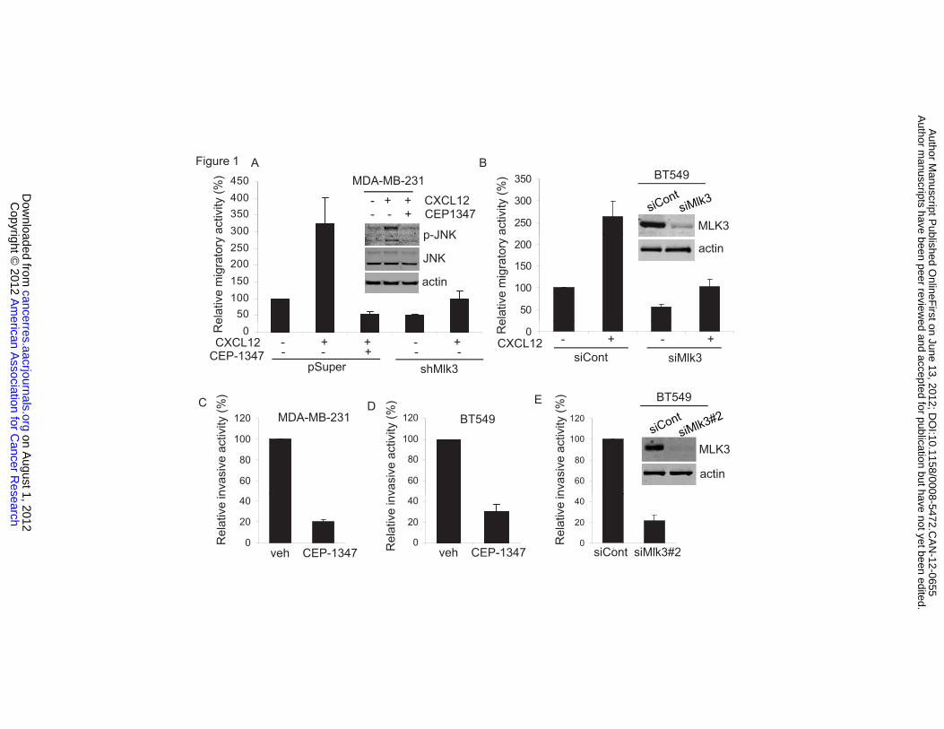
both express high levels of CXCR4 (30). In a transwell migration assay, CXCL12
increased migration of MDA-MB-231 cells expressing control vector by
approximately 3-fold. MLK3 expression is efficiently ablated in a stable
population of MDA-MB-231 cells (16). CXCL12-induced migration was
completely blocked in MDA-MB-231 cells stably expressing shMlk3 or treated
with CEP-1347, a selective MLK inhibitor (Figure 1A). As shown in Figure 1A,
CXCL12 activated JNK. To confirm the efficacy of CEP-1347, we used phospho-
JNK (p-JNK) as a readout for active MLK signaling. Immunoblotting using a p-
JNK antibody showed that CEP-1347 blocked CXCL12-induced JNK activation
(Figure 1A). In addition, transient silencing of MLK3 in BT549 cells reduced
CXCL12-induced migration (Figure 1B).
In a Matrigel invasion assay using CXCL12 as a chemoattractant, CEP-
1347 reduced invasion of MDA-MB-231 cells by approximately 5-fold (Figure 1C).
In addition, CEP-1347 blocked invasion of BT549 cells (Figure 1D). Silencing of
MLK3 in BT549 cells also largely inhibited invasion (Figure 1E). Mlk3 silencing
had negligible effect on proliferation of BT549 cells (Supplementary figure S1).
Taken together, these results support the idea that MLK3 signaling is required for
migration and invasion of invasive basal breast cancer cells in response to
CXCL12.
American Association for Cancer Research Copyright © 2012 on August 1, 2012cancerres.aacrjournals.orgDownloaded from
Author manuscripts have been peer reviewed and accepted for publication but have not yet been edited.Author Manuscript Published OnlineFirst on June 13, 2012; DOI:10.1158/0008-5472.CAN-12-0655
10
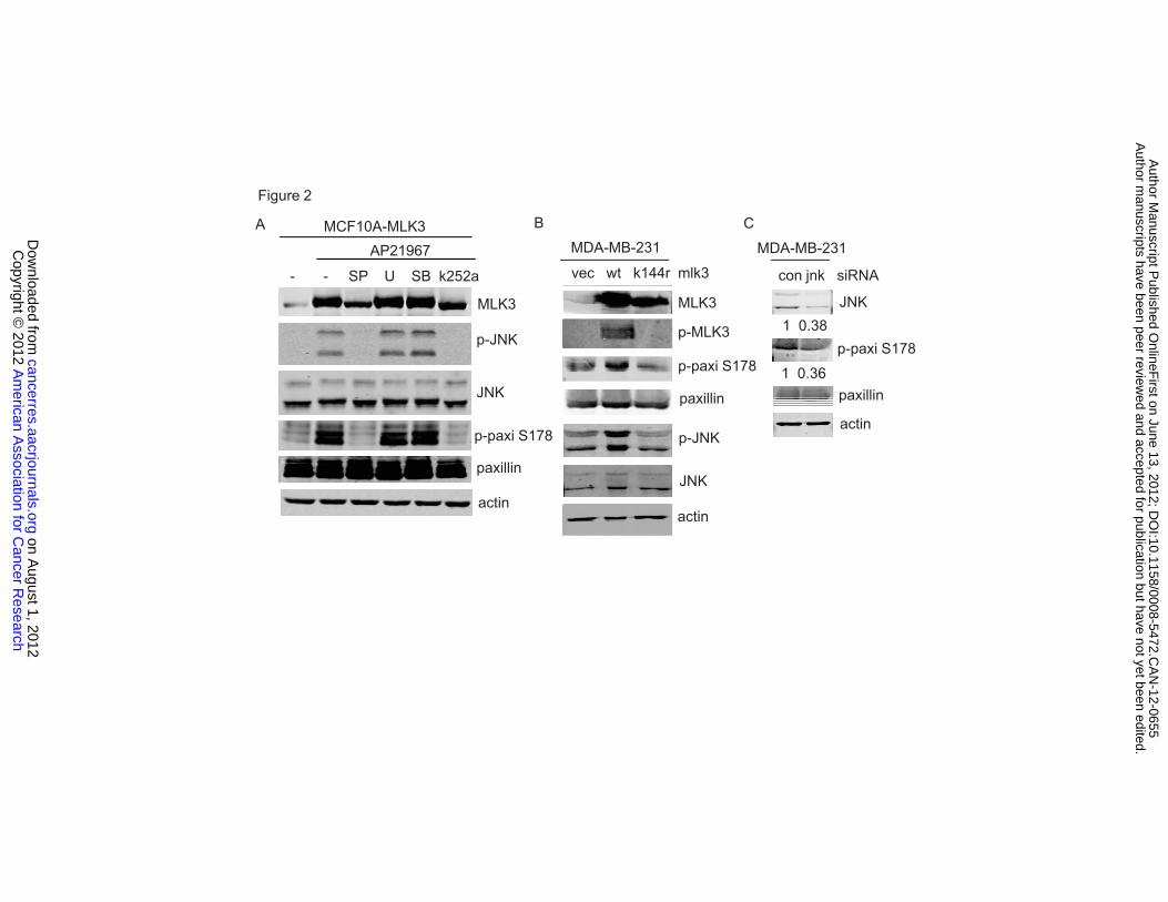
Active MLK3 promotes phosphorylation of paxillin through JNK
We recently showed that induced expression of MLK3 promotes migration
and invasion of MCF10A mammary epithelial cells, which requires JNK-AP1
(16). We hypothesized that, in addition to its impact on gene expression, MLK3-
JNK signaling might act upon cytoskeleton or focal adhesions to regulate cell
migration and invasion. Phosphorylation of Ser 178 of paxillin by JNK is
necessary for focal adhesion turnover and cell migration (24).
To investigate whether MLK3 can promote phosphorylation of paxillin,
MCF10A cells engineered to inducibly express MLK3 were used (16). Upon
MLK3 induction, JNK was activated and robust Ser 178 phosphorylation of
paxillin was observed. Inhibition of MLKs with K252a, or of JNK with SP600125,
blocked phosphorylation of paxillin at Ser 178. In contrast, inhibition of ERK
signaling with U0126, or p38 with SB203580, had no effect on MLK3-induced
paxillin phosphorylation (Figure 2A). Like K252a, its derivative CEP-1347 blocked
MLK3-induced paxillin phosphorylation (Supplementary figure S2). In MDA-MB-
231 cells, transient expression of wildtype MLK3, but not kinase inactive, MLK3
K144R, induced JNK activation and Ser 178 phosphorylation of paxillin (Figure
2B). Furthermore, silencing of JNK1/2 in MDA-MB-231 cells decreased Ser 178
phosphorylation of paxillin (Figure 2C), confirming the requirement for JNK in
paxillin phosphorylation. Thus the MLK3-JNK signaling axis promotes paxillin
phosphorylation at Ser 178.
American Association for Cancer Research Copyright © 2012 on August 1, 2012cancerres.aacrjournals.orgDownloaded from
Author manuscripts have been peer reviewed and accepted for publication but have not yet been edited.Author Manuscript Published OnlineFirst on June 13, 2012; DOI:10.1158/0008-5472.CAN-12-0655
11
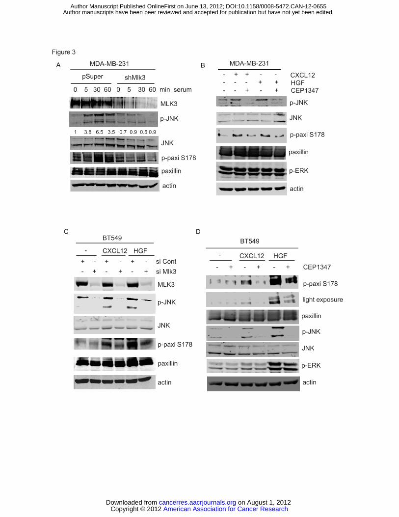
Chemokine and growth factor induce paxillin phosphorylation in an MLK3-
dependent manner
To determine whether MLK3 is required for phosphorylation of paxillin at
Ser 178, serum-deprived MDA-MB-231 cells stably expressing control or shMlk3
vector were treated with 10% serum and paxillin phosphorylation was
assessed. Serum treatment led to JNK activation and maximal phosphorylation
of paxillin at Ser 178 at 30 min, both of which were largely abrogated in cells
expressing shMlk3 (Figure 3A), suggesting MLK3 is a major mediator of JNK
signaling to paxillin. Since phosphorylation of paxillin on Ser 178 is associated
with breast cancer migration, we investigated the impact of CXCL12 and HGF on
paxillin phosphorylation. Both factors induced JNK activation and
phosphorylation of paxillin at Ser 178 in MDA-MB-231 cells, which was reduced
by CEP-1347 (Figure 3B).
Likewise, in BT549 cells, based on 4 independent experiments, both
CXCL12 and HGF induced JNK activation (3 and 3.4-fold, respectively) and
paxillin phosphorylation at Ser 178 (3.2 and 4.4-fold, respectively), which was
attenuated by silencing with Mlk3 siRNA (Figure 3C). MLK inhibition with CEP-
1347 also reduced both JNK activation and Ser 178 paxillin phosphorylation
(Figure 3D). The requirement of MLK3 in paxillin phosphorylation was confirmed
using a different siRNA sequence (Supplementary figure S3 A and B). In BT549
cells, HGF potently activated ERK, whereas only a small increase in ERK
activation was observed in response to CXCL12. ERK activation was refractory
to the MLK inhibitor, consistent with the proposed scaffolding role of MLK3 in
American Association for Cancer Research Copyright © 2012 on August 1, 2012cancerres.aacrjournals.orgDownloaded from
Author manuscripts have been peer reviewed and accepted for publication but have not yet been edited.Author Manuscript Published OnlineFirst on June 13, 2012; DOI:10.1158/0008-5472.CAN-12-0655
12
ERK activation (11). Since MDA-MB-231 cells harbor activating mutations in both
Ras and Raf (31), resulting in constitutive ERK activation, it is not too surprising
that CXCL12 and HGF have relatively little effect on ERK activity in these cells.
These data demonstrate a requirement for active MLK3 in JNK activation and
Ser 178 paxillin phosphorylation, triggered by either a prometastatic chemokine
or growth factor in basal breast cancer cells.
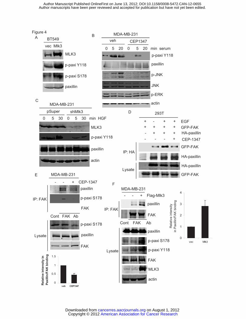
MLK3 controls Tyr 118 phosphorylation of paxillin and its association with
focal adhesion kinase
Cell migration requires efficient assembly and disassembly of focal
adhesion complexes. Paxillin undergoes phosphorylation at multiple sites to
modulate protein-protein interactions in focal adhesions (21). The role of Ser 178
phosphorylation of paxillin is not completely clear. In corneal epithelial cells, Ser
178 phosphorylation of paxillin recruits FAK to mediate tyrosine phosphorylation
of paxillin(32).
Since MLK3 controls Ser 178 phosphorylation of paxillin, we tested
whether MLK3 indirectly modulates tyrosine phosphorylation of paxillin and
regulates interactions among focal adhesion proteins. Ectopic expression of
MLK3 in BT549 cells promoted both Ser 178 and Tyr 118 paxillin phosphorylation,
demonstrating that active MLK3 drives Tyr 118 phosphorylation of paxillin (Figure
4A). In response to serum stimulation in MDA-MB-231 cells, CEP-1347 reduced
Tyr 118 phosphorylation of paxillin by 2-fold at 5 min and 3.5-fold at 20 min,
paralleling the effects of JNK inhibition (Figure 4B). Consistently, HGF-induced
American Association for Cancer Research Copyright © 2012 on August 1, 2012cancerres.aacrjournals.orgDownloaded from
Author manuscripts have been peer reviewed and accepted for publication but have not yet been edited.Author Manuscript Published OnlineFirst on June 13, 2012; DOI:10.1158/0008-5472.CAN-12-0655
13
Tyr 118 phosphorylation of paxillin was dramatically reduced in MDA-MB-231
cells expressing shMlk3 (Figure 4C). From these data we conclude that MLK3 is
critical for Tyr 118 phosphorylation of paxillin.
To test whether MLK3 influences association of paxillin with FAK, we took
advantage of 293T cells as an efficient cotransfection system. Ectopically
expressed GFP-FAK and HA-paxillin showed weak association in
coimmunoprecipitations from serum-deprived 293T cells (Figure 4D). EGF has
previously been shown to facilitate the interaction between FAK and paxillin in
293T cells (32). Our data confirm that the association between GFP-FAK and
HA-paxillin is enhanced by EGF (Figure 4D). However, pre-treatment with CEP-
1347 abrogated the EGF-induced association of GFP-FAK and HA-paxillin
(Figure 4D). We were able to detect endogenous paxillin in a FAK
immunoprecipitate from MDA-MB-231 cells in growth medium, which was
reduced by CEP-1347. In the immunoprecipitated FAK complex, levels of Ser
178 phosphorylated paxillin and total paxillin correlate directly, consistent with the
idea that Ser 178 phosphorylation drives association of FAK with paxillin (Figure
4E). Conversely, forced expression of active Flag-MLK3 in MDA-MB-231 cells
increased interaction of endogenous paxillin and FAK as well as phosphorylation
of paxillin at both Ser 178 and Tyr 118 (Figure 4 F). These data provide strong
evidence that MLK3 regulates both paxillin phosphorylation and FAK-paxillin
interactions.
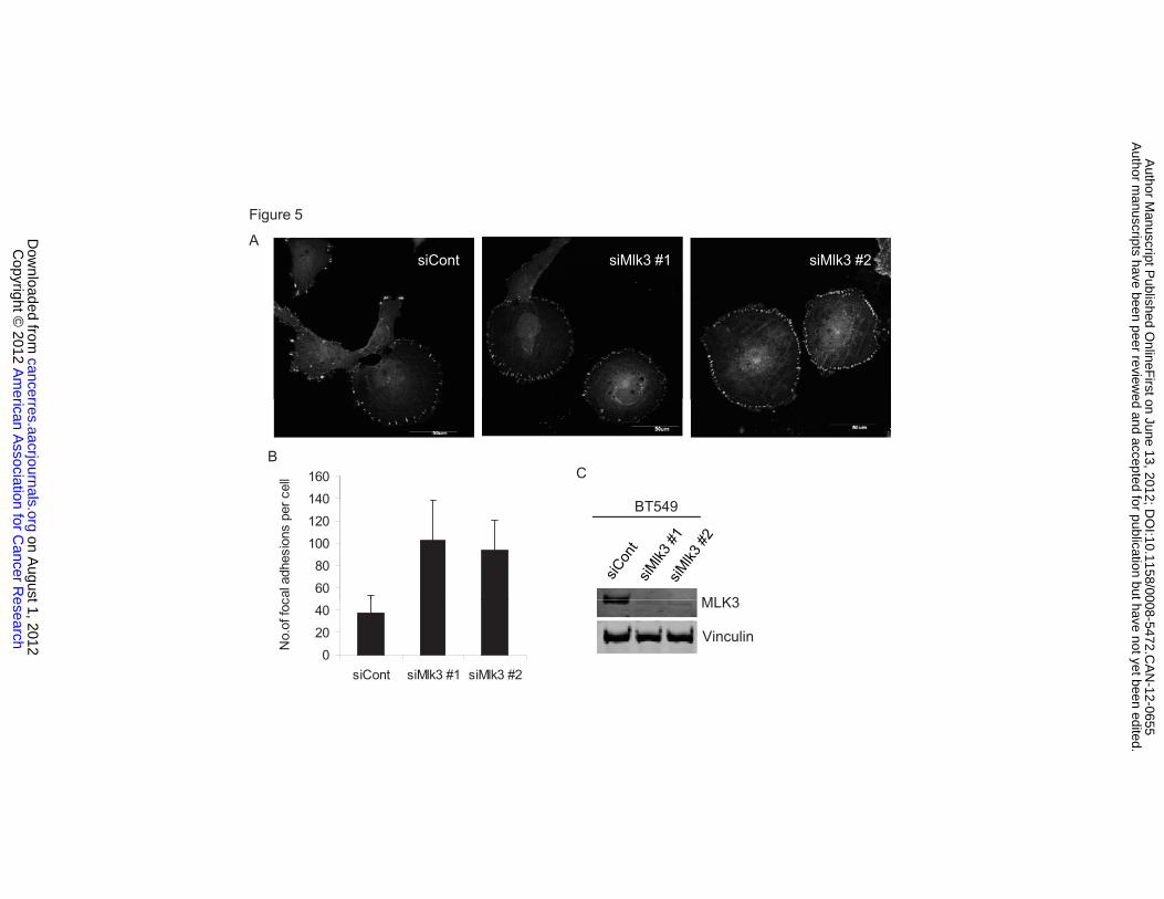
MLK3 silencing increases the number of focal adhesions
American Association for Cancer Research Copyright © 2012 on August 1, 2012cancerres.aacrjournals.orgDownloaded from
Author manuscripts have been peer reviewed and accepted for publication but have not yet been edited.Author Manuscript Published OnlineFirst on June 13, 2012; DOI:10.1158/0008-5472.CAN-12-0655
14
MLK3 modulates phosphorylation of Ser 178 and Tyr 118 of paxillin, which
is required for focal adhesion disassembly (23, 24). Consistent with this,
silencing of MLK3 in BT549 cells increased focal adhesions, which were
quantified as vinculin-staining focal adhesions, particularly at the cell periphery
(Figure 5 A and B). Similar effects were observed using two different Mlk3 siRNA
sequences. Silencing of MLK3 had no effect on total vinculin protein levels
(Figure 5 C). These data suggest that MLK3 is important for focal adhesion
turnover.
MLK3-JNK-paxillin signaling negatively regulates Rho activity
Tyr 118 phosphorylation of paxillin leads to decreased Rho activity,
enhancing focal adhesion turnover and cell migration (28). Ectopic expression of
MLK3 and wildtype paxillin in 293T cells resulted in robust Ser 178 paxillin
phosphorylation. As expected, no phospho-Ser 178-paxillin signal was detected
upon coexpression of the phosphorylation-defective mutant, paxillin S178A with
MLK3 (Supplementary figure S4A). In 293T cells, expressing control vector or
wildtype paxillin, Tyr 118 phosphorylation of paxillin was observed. However, the
paxillin mutant S178A was refractory to serum-induced Tyr 118 phosphorylation,
suggesting that Ser178 phosphorylation of paxillin is a prerequisite to Tyr 118
phosphorylation (Supplementary figure S4B). Likewise, in BT549 cells,
ectopically expressed wildtype paxillin, but not paxillin S178A, was
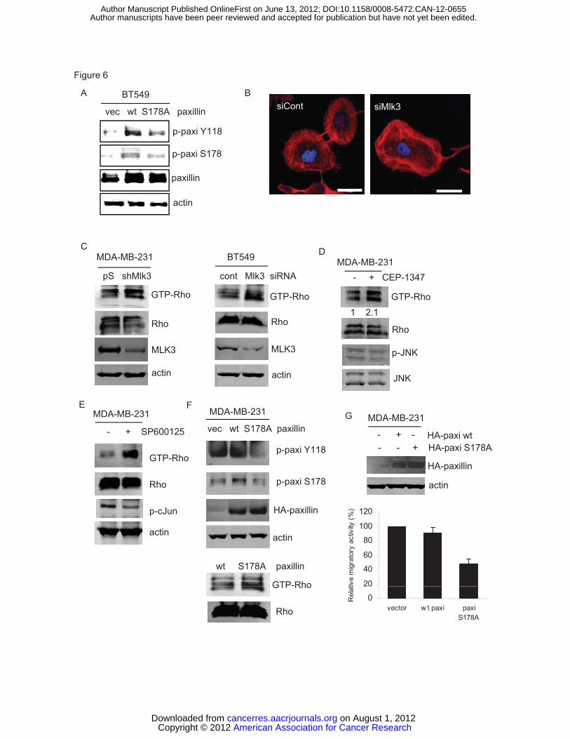
phosphorylated on Tyr 118 (Figure 6A).
American Association for Cancer Research Copyright © 2012 on August 1, 2012cancerres.aacrjournals.orgDownloaded from
Author manuscripts have been peer reviewed and accepted for publication but have not yet been edited.Author Manuscript Published OnlineFirst on June 13, 2012; DOI:10.1158/0008-5472.CAN-12-0655
15
Increased cellular stress fibers are observed in keratinocytes upon
expression of the paxillin S178A mutant (24). As shown in Figure 6B, a similar
phenotype was observed upon silencing of MLK3 in BT549 breast cancer
cells. Since Rho promotes stress fiber formation, we assessed whether
interruption of MLK3 and its signaling to JNK affects Rho activity in breast cancer
cells. In both MDA-MB-231 and BT549 cells, Rho activity was increased upon
silencing of MLK3 (Figure 6C). Furthermore, inhibition of MLK3,using CEP-1347,
as well as inhibition of downstream signaling to JNK with SP600125, increased
Rho activity in MDA-MB-231 cells (Figure 6D, E).
If MLK3-JNK suppresses Rho activity through Ser 178 phosphorylation of
paxillin, then expression of the paxillin S178A mutant should enhance Rho
activity. As shown in Figure 6F, expression of paxillin S178A, which fails to
undergo Tyr 118 phosphorylation, resulted in a marked increase of Rho activity
and decreased migration of MDA-MB-231 cells (Figure 6G). These data reveal
the MLK3-JNK-paxillin signaling axis as a negative modulator of Rho activity in
basal breast cancer cells.
MLK3 silencing decreases formation of lung metastases of human breast
cancer cells
To determine whether silencing of MLK3 is sufficient to prevent
metastases, MDA-MB-231 cells stably expressing control vector or Mlk3 shRNA
were introduced into the mammary gland of athymic nude mice. Both MDA-MB-
231-control and -shMlk3 formed primary tumors. MDA-MB-231-control tumors
American Association for Cancer Research Copyright © 2012 on August 1, 2012cancerres.aacrjournals.orgDownloaded from
Author manuscripts have been peer reviewed and accepted for publication but have not yet been edited.Author Manuscript Published OnlineFirst on June 13, 2012; DOI:10.1158/0008-5472.CAN-12-0655
16
were slightly larger than MDA-MB-231-shMlk3 tumors, but this did not reach
statistical significance (Figure 7A). Seven weeks post-inoculation, primary
tumors were excised. MLK3 silencing was maintained in primary tumors over the
course of the experiment as shown in immunoblots of tumor lysates
(Supplementary figure S5).
Lung micrometastases were detected by immunohistochemistry of lung
sections using a human-specific CD44 antibody. Numerous micrometastases
were observed in lung sections of mice inoculated with MDA-MB-231-control
cells, whereas few were found in those arising from MDA-MB-231-shMlk3 cells
(Figure 7B). Lung and liver tissue lysates from nude mice showed no human
CD44 immunoreactivity, confirming species specificity of the antibody.
Furthermore, silencing of MLK3 in MDA-MB-231 cells had no impact on CD44
protein levels (Supplementary figure S6). These data provide evidence for a
critical role of MLK3 in breast cancer metastasis.
Ser 178 phosphorylation of paxillin is associated with the metastatic
phenotype
In this study we have shown that prometastatic factors signal through
MLK3-JNK to promote Ser 178 phosphorylation of paxillin (Figure 7C) and
migration/invasion. Using an experimental metastasis model, MDA-MB-231 cells
expressing luciferase (luc2) were injected into the tail vein of nude mice.
Bioluminescence imaging revealed overt metastases 12 weeks post injection.
Tumor cells isolated from lung metastases (Lu) show higher paxillin
American Association for Cancer Research Copyright © 2012 on August 1, 2012cancerres.aacrjournals.orgDownloaded from
Author manuscripts have been peer reviewed and accepted for publication but have not yet been edited.Author Manuscript Published OnlineFirst on June 13, 2012; DOI:10.1158/0008-5472.CAN-12-0655
17
phosphorylation on Ser 178 compared with parental MDA-MB-231 cells (Pa),
both basally as well as in response to either CXCL12 or HGF (Figure 7D).
Furthermore, CEP-1347 inhibits both CXCL12- and HGF- induced Ser 178
phosphorylation of paxillin in Lu cells (Figure 7E), indicating these cells are still
sensitive to an MLK inhibitor.
Screening of a panel of human mammary epithelial and breast cancer cell
lines revealed a correlation between p-Ser 178 paxillin and metastatic potential
(Supplementary figure S7). These data, taken together, suggest that
phosphorylation of paxillin on Ser 178 may be a predictor of lung metastatic
potential.
Discussion
Deciphering key signaling pathways underlying breast cancer cell
migration and invasion may reveal novel therapeutic targets for effectively
treating or preventing metastatic breast cancer. We previously demonstrated
MLK3-JNK signaling upregulates multiple AP-1-driven invasion genes and
promotes a malignant phenotype in mammary epithelial cells (16). JNK is
important in breast cancer cell migration and invasion, and breast cancer
progression (24, 33, 34). Yet, how MLK3-JNK signaling regulates cell migration
machinery remains largely unknown.
In this study, we report, for the first time, that in response to CXCL12 and
HGF, MLK3 signals to JNK to control phosphorylation of paxillin on both Ser 178
and Tyr 118 (Figures 2-4), phosphorylation events that are essential in cell
American Association for Cancer Research Copyright © 2012 on August 1, 2012cancerres.aacrjournals.orgDownloaded from
Author manuscripts have been peer reviewed and accepted for publication but have not yet been edited.Author Manuscript Published OnlineFirst on June 13, 2012; DOI:10.1158/0008-5472.CAN-12-0655
18
migration (24, 35). MLK3 modulates interactions between two key focal adhesion
proteins, paxillin and FAK (Figure 4). MLK3-JNK-paxillin signaling negatively
regulates Rho activity to promote focal adhesion turnover in cell migration
(Figures 5, 6). Finally, MLK3 is critical for formation of breast cancer lung
micrometastases in a mouse xenograft model (Figure 7). The importance of
paxillin phosphorylation is highlighted by the finding that cells derived from MDA-
MB-231 lung metastases show higher phosphorylation of paxillin at Ser 178,
compared with parental MDA-MB-231 cells, implicating this phosphorylation site
in breast cancer metastasis. Taken altogether, our data reveal a novel MLK3-
JNK-paxillin signaling pathway that regulates breast cancer cell migration and
invasion.
Paxillin undergoes dynamic phosphorylation during cell migration (21). Ser
178 phosphorylation of paxillin is essential for cell migration (24). We show both
CXCL12 and HGF signal through MLK3 to paxillin, consistent with our data
showing that MLK3 is required for CXCL12-induced breast cancer cell migration
(Figures 1-3). Thus, MLK3 emerges as an important signal node that relays
extracellular cues to JNK to control paxillin phosphorylation. While it is possible
that other MAPKKKs contribute to paxillin phosphorylation through JNK, MLK3
appears to play a dominant role, at least in basal breast cancer cells. Because
CXCL12 and HGF are consistently linked with invasion and metastasis, our
findings provide a strong rationale for targeting MLK3 in the context of breast
cancer metastasis. In agreement with our findings, localized JNK activation and
Ser 178 phosphorylation of paxillin is observed during migration of rat kidney
American Association for Cancer Research Copyright © 2012 on August 1, 2012cancerres.aacrjournals.orgDownloaded from
Author manuscripts have been peer reviewed and accepted for publication but have not yet been edited.Author Manuscript Published OnlineFirst on June 13, 2012; DOI:10.1158/0008-5472.CAN-12-0655
19
epithelial cells, which involves the aPKC-Exocyst complex (36). Interestingly,
PKC is important for activation of MLK3 in response to free fatty acids
(37). Whether PKC plays a role in CXCL12- or HGF- induced MLK3 activation
remains to be determined.
Rapid assembly and disassembly of focal adhesions is a well-described
property of many migrating cancer cells. Experimental disruption of focal
adhesion turnover typically results in migratory defects in cancer cells (38).
Phosphorylation of paxillin at Ser 178 by JNK (24) and Tyr 118 by FAK/Src (39,
40) is critical for focal adhesion turnover and cell migration (24, 35, 41, 42). For
instance, a tyrosine phosphomimetic mutant of paxillin enhances focal adhesion
turnover, whereas a non-phosphorylatable mutant shows defective focal
adhesion turnover and migration (22, 23). Furthermore, phosphorylation of Tyr
118 on paxillin is implicated in cancer invasion and metastasis (43). Our results
support a model in which MLK3 is required for focal adhesion turnover in cell
migration through controlling Tyr 118 phosphorylation of paxillin (Figure 4).
Indeed, MLK3 silencing increases the number of focal adhesions in breast
cancer cells (Figure 5). Furthermore, experiments using a nocodazole-based
assay (44), in which nocodazole washout promotes microtubule formation and
focal adhesion turnover, reveal a defect in focal adhesion disassembly upon
MLK3 silencing in MDA-MB-231 cells (data not shown), supporting the necessity
of MLK3 in focal adhesion turnover.
Elevated levels and activity of FAK are found in high grade human
cancers, including breast cancer, and correlate with invasive phenotypes,
American Association for Cancer Research Copyright © 2012 on August 1, 2012cancerres.aacrjournals.orgDownloaded from
Author manuscripts have been peer reviewed and accepted for publication but have not yet been edited.Author Manuscript Published OnlineFirst on June 13, 2012; DOI:10.1158/0008-5472.CAN-12-0655
20
metastatic disease and poor prognosis (45). FAK inhibitors are currently in
clinical trials for treating human solid tumors (46). Activated FAK recruits Src to
form an active FAK/Src complex. Our data showing that MLK3 promotes
interaction of FAK with paxillin may explain how MLK3 controls Tyr 118
phosphorylation of paxillin and promotes focal adhesion turnover, since
association of FAK with paxillin promotes tyrosine phosphorylation of paxillin and
is correlated with less stable focal adhesions (47). Ablation of the Ser 178
phosphorylation site on paxillin decreased phosphorylation of Tyr 118 (Figure 6),
suggesting that, at least in this experimental context, Ser 178 phosphorylation is
a prerequisite for Tyr 118 phosphorylation. This is in agreement with the finding
that paxillin S178A has decreased affinity for FAK (32). In our working model,
MLK3-JNK-Ser 178 paxillin phosphorylation regulates association of FAK with
paxillin and indirectly controls subsequent tyrosine phosphorylation of paxillin
(Figure 7C).
Focal adhesion dynamics are tightly controlled by Rho GTPases (20, 25).
Active Rho increases stress fibers and focal adhesion maturation and decreases
focal adhesion turnover (48). While Rho activity is required for cell migration,
aberrantly high Rho activity also impairs cell migration (26). FAK promotes focal
adhesion turnover, in part, through suppression of Rho activity (48). In particular,
FAK/Src-mediated Tyr 118 phosphorylation of paxillin has been proposed to
release p190 Rho-GAP from its sequestration with Ras-GAP, leading to
downregulation of Rho activity (28). Our experimental evidence supports a model
in which the MLK3-JNK-paxillin Ser 178 signaling axis negatively regulates Rho
American Association for Cancer Research Copyright © 2012 on August 1, 2012cancerres.aacrjournals.orgDownloaded from
Author manuscripts have been peer reviewed and accepted for publication but have not yet been edited.Author Manuscript Published OnlineFirst on June 13, 2012; DOI:10.1158/0008-5472.CAN-12-0655
21
activity, through FAK-mediated tyrosine phosphorylation of paxillin. Indeed,
MLK3 silencing in breast cancer cells resulted in increased focal adhesions
(Figure 5) and stress fibers (Figure 6), two Rho-associated phenotypes. In A549
lung carcinoma cells, a role for MLK3 in limiting Rho activity through an
interaction with p63 RhoGEF has also been described (17). In summary, we
have identified in basal breast cancer cells, a distinct pathway involving an active
MLK3-JNK-paxillin axis that functions to negatively control Rho activity.
In a xenograft model in which MDA-MB-231 cells were introduced into
mammary fat pad of nude mice (49), MLK3 silencing inhibited spontaneous lung
micrometastases (Figure 7), in agreement with findings that MLK3 knockdown
reduced lymph node metastases of MDA-MB-231 cells (50). Although MLK3 has
been shown to promote cell survival (16, 50), which might contribute to formation
of metastases, we propose that an important mechanism through which MLK3
promotes metastasis is through facilitating cancer cell migration and invasion.
Our study demonstrates that MLK3 controls Ser 178 phosphorylation of
paxillin, which is required for cell migration. Interestingly, phospho-Ser 178
paxillin correlates with metastatic potential of breast cancer cells, suggesting that
phospho-Ser 178 paxillin might be a predictive biomarker for metastasis. Our
novel findings demonstrate that prometastatic factors found in the tumor
microenvironment converge on MLK3 to promote breast cancer cell migration
and invasion. Since the MLK inhibitor, CEP-1347, efficaciously blocks invasion
in response to such factors, we are currently testing the effect of this compound
in a preclinical study using a mouse xenograft model. Our findings indicate that
American Association for Cancer Research Copyright © 2012 on August 1, 2012cancerres.aacrjournals.orgDownloaded from
Author manuscripts have been peer reviewed and accepted for publication but have not yet been edited.Author Manuscript Published OnlineFirst on June 13, 2012; DOI:10.1158/0008-5472.CAN-12-0655
22
MLK3 regulates phosphorylation of paxillin and its interaction with FAK. We also
provide evidence that the MLK3-JNK-paxillin axis negatively regulates Rho
activity and focal adhesion turnover. Finally, we demonstrate a critical role of
MLK3 in breast cancer metastasis. Thus, targeting MLK3 could be a promising
therapeutic strategy for treatment or prevention of metastatic disease in breast
cancer.
Acknowledgements
This work was supported by grants to KAG from the Department of Defense
Breast Cancer Research Program (W81XWH-09-1-0049) and the Elsa U. Pardee
Foundation. JC was the recipient of the MSU-Barnett Rosenberg Fellowship in
Biological Sciences. We thank Eva Miller and Jonathan Kasper for assisting with
mouse surgery and Sandra O’Reilly (MSU-RTSF) for help with in vivo imaging.
References
1. DeSantis C, Siegel R, Bandi P, Jemal A. Breast cancer statistics. CA Cancer J Clin 2011;61:409-418.
2. Luker KE, Luker GD. Functions of CXCL12 and CXCR4 in breast cancer. Cancer Lett 2006;238:30-41.
3. Muller A, Homey B, Soto H, Ge N, Catron D, Buchanan ME, McClanahan T, et al. Involvement of chemokine receptors in breast cancer metastasis. Nature 2001;410:50-56.
4. Dupont VN, Gentien D, Oberkampf M, De Rycke Y, Blin NA. Gene expression signature associated with metastatic cells in effusions of breast carcinoma patients. Int J Cancer 2007;121:1036-1046.
5. Kato M, Kitayama J, Kazama S, Nagawa H. Expression pattern of CXC chemokine receptor-4 is correlated with lymph node metastasis in human invasive ductal carcinoma. Breast Cancer Res 2003;5:R144-150.
6. Li YM, Pan Y, Wei Y, Cheng X, Zhou BP, Tan M, et al. Upregulation of CXCR4 is essential for HER2-mediated tumor metastasis. Cancer Cell 2004;6:459-469.
American Association for Cancer Research Copyright © 2012 on August 1, 2012cancerres.aacrjournals.orgDownloaded from
Author manuscripts have been peer reviewed and accepted for publication but have not yet been edited.Author Manuscript Published OnlineFirst on June 13, 2012; DOI:10.1158/0008-5472.CAN-12-0655

23
7. Birchmeier C, Birchmeier W, Gherardi E, Vande Woude GF. Met, metastasis, motility and more. Nat Rev Mol Cell Biol 2003;4:915-925.
8. Gastaldi S, Comoglio PM, Trusolino L. The Met oncogene and basal-like breast cancer: another culprit to watch out for? Breast Cancer Res 2010;12:208.
9. Kang JY, Dolled-Filhart M, Ocal IT, Singh B, Lin CY, Dickson RB, et al. Tissue microarray analysis of hepatocyte growth factor/Met pathway components reveals a role for Met, matriptase, and hepatocyte growth factor activator inhibitor 1 in the progression of node-negative breast cancer. Cancer Res 2003;63:1101-1105.
10. Lengyel E, Prechtel D, Resau JH, Gauger K, Welk A, Lindemann K, et al. C-Met overexpression in node-positive breast cancer identifies patients with poor clinical outcome independent of Her2/neu. Int J Cancer 2005;113:678-682.
11. Chadee DN, Kyriakis JM. MLK3 is required for mitogen activation of B-Raf, ERK and cell proliferation. Nat Cell Biol 2004;6:770-776.
12. Gallo KA, Johnson GL. Mixed-lineage kinase control of JNK and p38 MAPK pathways. Nat Rev Mol Cell Biol 2002;3:663-672.
13. Zhang H, Gallo KA. Autoinhibition of mixed lineage kinase 3 through its Src homology 3 domain. J Biol Chem 2001;276:45598-45603.
14. Du Y, Bock BC, Schachter KA, Chao M, Gallo KA. Cdc42 induces activation loop phosphorylation and membrane targeting of mixed lineage kinase 3. J Biol Chem 2005;280:42984-42993.
15. Simpson KJ, Selfors LM, Bui J, Reynolds A, Leake D, Khvorova A, et al. Identification of genes that regulate epithelial cell migration using an siRNA screening approach. Nat Cell Biol 2008;10:1027-1038.
16. Chen J, Miller EM, Gallo KA. MLK3 is critical for breast cancer cell migration and promotes a malignant phenotype in mammary epithelial cells. Oncogene 2010;29:4399-4411.
17. Swenson-Fields KI, Sandquist JC, Rossol-Allison J, Blat IC, Wennerberg K, Burridge K,et al. MLK3 limits activated Galphaq signaling to Rho by binding to p63RhoGEF. Mol Cell 2008;32:43-56.
18. Zhan Y, Modi N, Stewart AM, Hieronimus RI, Liu J, Gutmann DH, et al. Regulation of mixed lineage kinase 3 is required for Neurofibromatosis-2-mediated growth suppression in human cancer. Oncogene 2011;30:781-789.
19. Mishra P, Senthivinayagam S, Rangasamy V, Sondarva G, Rana B. Mixed lineage kinase-3/JNK1 axis promotes migration of human gastric cancer cells following gastrin stimulation. Mol Endocrinol 2010;24:598-607.
20. Ridley AJ, Schwartz MA, Burridge K, Firtel RA, Ginsberg MH, Borisy G, et al. Cell migration: integrating signals from front to back. Science 2003;302:1704-1709.
21. Brown MC, Turner CE. Paxillin: adapting to change. Physiol Rev 2004;84:1315-1339.
American Association for Cancer Research Copyright © 2012 on August 1, 2012cancerres.aacrjournals.orgDownloaded from
Author manuscripts have been peer reviewed and accepted for publication but have not yet been edited.Author Manuscript Published OnlineFirst on June 13, 2012; DOI:10.1158/0008-5472.CAN-12-0655
24
22. Webb DJ, Donais K, Whitmore LA, Thomas SM, Turner CE, Parsons JT, et al. FAK-Src signalling through paxillin, ERK and MLCK regulates adhesion disassembly. Nat Cell Biol 2004;6:154-161.
23. Zaidel-Bar R, Milo R, Kam Z, Geiger BA. paxillin tyrosine phosphorylation switch regulates the assembly and form of cell-matrix adhesions. J Cell Sci 2007;120:137-148.
24. Huang C, Rajfur Z, Borchers C, Schaller MD, Jacobson K. JNK phosphorylates paxillin and regulates cell migration. Nature 2003;424:219-223.
25. Hall A. Rho GTPases and the actin cytoskeleton. Science 1998;279:509-514.
26. Nobes CD, Hall A. Rho GTPases control polarity, protrusion, and adhesion during cell movement. J Cell Biol 1999;144:1235-1244.
27. Valles AM, Beuvin M, Boyer B. Activation of Rac1 by paxillin-Crk-DOCK180 signaling complex is antagonized by Rap1 in migrating NBT-II cells. J Biol Chem 2004;279:44490-44496.
28. Tsubouchi A, Sakakura J, Yagi R, Mazaki Y, Schaefer E, Yano H, et al. Localized suppression of RhoA activity by Tyr31/118-phosphorylated paxillin in cell adhesion and migration. J Cell Biol 2002;159:673-683.
29. Li G, Xiang Y, Sabapathy K, Silverman RH. An apoptotic signaling pathway in the interferon antiviral response mediated by RNase L and c-Jun NH2-terminal kinase. J Biol Chem 2004;279:1123-31.
30. Holland JD, Kochetkova M, Akekawatchai C, Dottore M, Lopez A, McColl SR. Differential functional activation of chemokine receptor CXCR4 is mediated by G proteins in breast cancer cells. Cancer Res 2006;66:4117-4124.
31. Hollestelle A, Elstrodt F, Nagel JH, Kallemeijn WW, Schutte M. Phosphatidylinositol-3-OH kinase or RAS pathway mutations in human breast cancer cell lines. Mol Cancer Res 2007;5:195-201.
32. Huang Z, Yan DP, Ge BX. JNK regulates cell migration through promotion of tyrosine phosphorylation of paxillin. Cell Signal 2008;20:2002-2012.
33. Cui X, Kim HJ, Kuiatse I, Kim H, Brown PH, Lee AV. Epidermal growth factor induces insulin receptor substrate-2 in breast cancer cells via c-Jun NH(2)-terminal kinase/activator protein-1 signaling to regulate cell migration. Cancer Res 2006;66:5304-5313.
34. Yeh YT, Hou MF, Chung YF, Chen YJ, Yang SF, Chen DC, et al. Decreased expression of phosphorylated JNK in breast infiltrating ductal carcinoma is associated with a better overall survival. Int J Cancer 2006;118:2678-2684.
35. Petit V, Boyer B, Lentz D, Turner CE, Thiery JP, Valles AM. Phosphorylation of tyrosine residues 31 and 118 on paxillin regulates cell migration through an association with CRK in NBT-II cells. J Cell Biol 2000;148:957-970.
36. Rosse C, Formstecher E, Boeckeler K, Zhao Y, Kremerskothen J, White MD et al. An aPKC-exocyst complex controls paxillin phosphorylation and migration through localised JNK1 activation. PLoS Biol 2009;7:e1000235.
American Association for Cancer Research Copyright © 2012 on August 1, 2012cancerres.aacrjournals.orgDownloaded from
Author manuscripts have been peer reviewed and accepted for publication but have not yet been edited.Author Manuscript Published OnlineFirst on June 13, 2012; DOI:10.1158/0008-5472.CAN-12-0655
25
37. Jaeschke A, Davis RJ. Metabolic stress signaling mediated by mixed-lineage kinases. Mol Cell 2007;27:498-508.
38. Xu Y, Bismar TA, Su J, Xu B, Kristiansen G, Varga Z, et al. Filamin A regulates focal adhesion disassembly and suppresses breast cancer cell migration and invasion. J Exp Med 2010;207:2421-2437.
39. Bellis SL, Miller JT, Turner CE. Characterization of tyrosine phosphorylation of paxillin in vitro by focal adhesion kinase. J Biol Chem, 1995;270:17437-17441.
40. Schaller MD, Parsons JT. pp125FAK-dependent tyrosine phosphorylation of paxillin creates a high-affinity binding site for Crk. Mol Cell Biol 1995;15:2635-2645.
41. Su S, Li Y, Luo Y, Sheng Y, Su Y, Padia RN, et al. Proteinase-activated receptor 2 expression in breast cancer and its role in breast cancer cell migration. Oncogene 2009;28:3047-3057.
42. Vindis C, Teli T, Cerretti DP, Turner CE, Huynh-Do U. EphB1-mediated cell migration requires the phosphorylation of paxillin at Tyr-31/Tyr-118. J Biol Chem 2004;279:27965-27970.
43. Azuma K, Tanaka M, Uekita T, Inoue S, Yokota J, Ouchi Y, et al. Tyrosine phosphorylation of paxillin affects the metastatic potential of human osteosarcoma. Oncogene 2005;24:4754-4764.
44. Ezratty EJ, Partridge MA, Gundersen GG. Microtubule-induced focal adhesion disassembly is mediated by dynamin and focal adhesion kinase. Nat Cell Biol 2005;7: 581-590.
45. McLean GW, Carragher NO, Avizienyte E, Evans J, Brunton VG, Frame MC. The role of focal-adhesion kinase in cancer - a new therapeutic opportunity. Nat Rev Cancer 2005;5: 505-515.
46. Parsons JT, Slack-Davis J, Tilghman R, Roberts WG. Focal adhesion kinase: targeting adhesion signaling pathways for therapeutic intervention. Clin Cancer Res 2008;14:627-632.
47. Shan Y, Yu L, Li Y, Pan Y, Zhang Q, Wang F, et al. Nudel and FAK as antagonizing strength modulators of nascent adhesions through paxillin. PLoS Biol 2009;7:e1000116.
48. Ren XD, Kiosses WB, Sieg DJ, Otey CA, Schlaepfer DD, Schwartz MA. Focal adhesion kinase suppresses Rho activity to promote focal adhesion turnover. J Cell Sci 2000;113 (Pt 20):3673-3678.
49. Valastyan S, Reinhardt F, Benaich N, Calogrias D, Szasz AM, Wang ZC, et al. A pleiotropically acting microRNA, miR-31, inhibits breast cancer metastasis. Cell 2009;137:1032-1046.
50. Cronan MR, Nakamura K, Johnson NL, Granger DA, Cuevas BD, Wang JG, et al. Defining MAP3 kinases required for MDA-MB-231 cell tumor growth and metastasis. Oncogene 2011;10.1038/onc.2011.544.
Figure Legends
Figure 1. Silencing or inhibition of MLK3 blocks CXCL12-induced migration and invasion. (A) Serum-deprived MDA-MB-231-pSuper or –pSuper-shMlk3 were treated
American Association for Cancer Research Copyright © 2012 on August 1, 2012cancerres.aacrjournals.orgDownloaded from
Author manuscripts have been peer reviewed and accepted for publication but have not yet been edited.Author Manuscript Published OnlineFirst on June 13, 2012; DOI:10.1158/0008-5472.CAN-12-0655
26
with CXCL12 (100 ng/ml) -/+ CEP-1347 (400 nM) for 30 min. Total cellular lysates were analyzed by immunoblotting using indicated antibodies (upper panel). MDA-MB-231-pSuper or –pSuper-shMlk3 were allowed to migrate towards CXCL12 (100 ng/ml) for 24 h. Migrated cells were quantified as described (16).Column, mean of three experiments. Bar, SE. (B) BT549 cells were treated with control or Mlk3 siRNA for 48 h, serum-deprived and allowed to migrate towards CXCL12 (100 ng/ml) for 48 h. (C) MDA-MB-231 cells, pretreated -/+400 nM CEP-1347 for 6 h in serum-free medium, were allowed to invade towards CXCL12 (100 ng/ml) for 24 h. (D) BT549 cells were pretreated -/+ 400 nM CEP-1347 and CXCL12-induced transwell invasion was determined as in (B) . (E) BT549 cells were treated as in (B) and subjected to a transwell invasion assay. Column, mean of four experiments. Bar, SE. Figure 2. MLK3-JNK signaling promotes phosphorylation of paxillin at Ser 178. (A) MCF10A-MLK3 cells were treated -/+50 nM AP21967 for 20 h with indicated inhibitors: 15 μM SP600125, 10 μM U0126, 10 μM SB203580 or 400 nM K252a, for an additional 24 h. Cellular lysates were analyzed by western-blotting. (B) MDA-MB-231 cells were transfected wildtype or MLK3 K144R vector for 24 h. Cellular lysates were analyzed by western-blotting. (C) MDA-MB-231 cells were treated with control or JNK1,2 siRNA for 48 h. Cellular lysates were analyzed immunoblotting. Quantitation of blots normalized to actin was performed using LI-COR Odyssey software V3.0. Figure 3. MLK3 silencing or an MLK inhibitor impairs paxillin phosphorylation at Ser 178. (A) Serum-deprived MDA-MB-231-pSuper or pSuper-shMlk3 were treated with 10% serum for indicated times. Total cellular lysates were analyzed by immunoblotting. Quantitation of p-JNK/JNK determined by LI-COR Odyssey software V3.0 is shown. (B) Serum-deprived MDA-MB-231 cells treated -/+CEP-1347 (400 nM) for 6 h, followed by CXCL12 (100 ng/ml) or HGF (100 ng/ml) for 30 min. (C) BT549 cells were transfected with control or Mlk3 siRNA and treated for 30 min with 100 ng/ml CXCL12 or HGF. (D) Serum-deprived BT549 cells were treated -/+ CEP-1347 (400 nM) for 6 h, followed by 100 ng/ml CXCL12 or HGF for 30 min. Figure 4. MLK3 promotes Tyr 118 phosphorylation of paxillin and interaction of FAK with paxillin. (A) Immunoblots of cellular lysates from BT549 cells transfected with control or Flag-Mlk3 vector are shown. (B) Serum-deprived MDA-MB-231 cells were treated -/+400 nM CEP-1347 for 6 h followed by 10% serum for indicated times. Immunoblots are shown. (C) Serum-deprived MDA-MB-231-pSuper or pSuper-shMlk3 were treated with 100 ng/ml HGF. (D) 293T cells were cotransfected with indicated constructs, serum deprived, and treated -/+ CEP-1347 followed by 100ng/ml EGF (100 ng/ml) for 30 min. Lysates were immunoprecipitated using HA antibody and subjected to western-blotting. (E) After overnight treatment of MDA-MB-231 cells -/+CEP-1347, lysates were immunoprecipitated using control IgG or FAK antibody, followed by western-blotting. Ratios of relative intensities of FAK to paxillin with control (=1) are shown. Column, mean of three experiments. Bar, SE. (F) Immunoprecipitation and immunoblots from MDA-MB-231 cells transfected with control or Flag-MLK3 vector. FAK-paxillin association was quantified as in (E).
American Association for Cancer Research Copyright © 2012 on August 1, 2012cancerres.aacrjournals.orgDownloaded from
Author manuscripts have been peer reviewed and accepted for publication but have not yet been edited.Author Manuscript Published OnlineFirst on June 13, 2012; DOI:10.1158/0008-5472.CAN-12-0655
27
Figure 5. MLK3 knockdown increases vinculin-containing focal adhesions. (A) BT549 cells were transfected with control or two different Mlk3 siRNAs for 48 h. Fixed cells were stained with vinculin antibody and DAPI. Images were taken using Olympus FluoView confocal microscope. Bar, 50 μm. (B) Vinculin-positive focal adhesions were quantified from over 20 cells per group using Image J software. Column, mean of two experiments. Bar, SE. (C) Immunoblots corresponding to (A). Figure 6. MLK3-JNK-mediated Ser 178 phosphorylation of paxillin is necessary for Tyr 118 phosphorylation of paxillin and inhibits Rho activity. (A) Immunoblots from BT549 cells transiently expressing HA-paxillin wt or HA-paxillin S178A mutant after serum-deprivation and treatment with 10% serum for 30 min. (B) BT549 cells were treated with control or Mlk3 siRNA for 48 h, stained with phalloidin (F-actin) and DAPI (nucleus) and imaged using confocal microscopy. Bar, 50 μm. (C) Immunoblots from Rhotekin pulldown assay of MDA-MB-231-pSuper or -pSuper-Mlk3 (left panel) and BT549 cells treated with control or Mlk3 siRNA (right panel). (D) Immunoblots from Rhotekin pulldown assay of MDA-MB-231 cells treated -/+ CEP-1347 (E) Immunoblots from Rhotekin pulldown assay of MDA-MB-231 cells treated -/+ SP600125 for 6 h. (F) Rhotekin pulldown assay and immunoblots from MDA-MB-231 cells expressing HA-paxillin (Wt) or HA-paxillin S178A mutant. (G) Transwell migration assay of cells from (F) was performed with corresponding immunoblots shown. Figure 7. Depletion of MLK3 prevents formation of lung metastases. (A) Tumor growth curve of MDA-MB-231-pSuper or -pSuper-shMlk3 inoculated into mouse mammary fat pads. (B) Immunohistochemistry of lung sections using a human-specific CD44 antibody. Magnification, 400x. CD44-positive nodules were quantified in 10 sections per mouse. Statistical analysis was done using GraphPad Prism 5. (C) Schematic model showing MLK3-JNK-pSer 178 paxillin signaling axis, activated through prometastatic factors CXCL12 and HGF, leading to FAK-mediated Tyr118 phosphorylation of paxillin, and suppression of Rho activity. (D) Cultured cells derived from lung metastases of MDA-MB-231-Luc2-tdTomato were designated as Lu. Immunoblots from serum-deprived parental MDA-MB-231-Luc2-tdTomato cells (designated as Pa) and Lu cells treated with CXCL12 or HGF for 20 min. (E) Serum-deprived Lu cells treated -/+CEP-1347 overnight, and treated with CXCL12 or HGF as in (D).
American Association for Cancer Research Copyright © 2012 on August 1, 2012cancerres.aacrjournals.orgDownloaded from
Author manuscripts have been peer reviewed and accepted for publication but have not yet been edited.Author Manuscript Published OnlineFirst on June 13, 2012; DOI:10.1158/0008-5472.CAN-12-0655
250
300
350
MLK3
BT549
300350400450
activ
ity (%
)
activ
ity (%
)
A B
p JNK
- - + CEP1347- + + CXCL12
MDA-MB-231Figure 1
50
100
150
200 actin
50100150200250
elat
ive
mig
rato
ry
elat
ive
mig
rato
ry
actin
JNK
p-JNK
00Re
Re
%)%)
%)
- + + - +
pSuper
CXCL12- - + - -
shMlk3CEP-1347
CXCL12 - + - +siCont siMlk3
C D E BT549
60
80
100
120
60
80
100
120
60
80
100
120
vasi
ve a
ctiv
ity (%
nvas
ive
activ
ity (%
nvas
ive
activ
ity (%MDA-MB-231 BT549
D
MLK3
actin
0
20
40
0
20
40
1 20
20
40
Rel
ativ
e in
v
Rel
ativ
e in
Rel
ativ
e in
veh CEP-1347 veh CEP-1347 siCont siMlk3#2
A
merican A
ssociation for Cancer R
esearch C
opyright © 2012
on August 1, 2012
cancerres.aacrjournals.orgD
ownloaded from
Author m
anuscripts have been peer reviewed and accepted for publication but have not yet been edited.
Author M
anuscript Published O
nlineFirst on June 13, 2012; D
OI:10.1158/0008-5472.C
AN
-12-0655
- - SP U SB k252a con jnk siRNA
AP21967MCF10A-MLK3
vec wt k144r mlk3
MDA-MB-231 MDA-MB-231A B C
Figure 2
MLK3
p-JNK
JNK
MLK3
p-MLK3
p-paxi S178
paxillin
JNK
p-paxi S178
paxillin
1 0.38
1 0.36
p-paxi S178
paxillin
actinJNK
p-JNK
paxillin paxillin
actin
actinactin
A
merican A
ssociation for Cancer R
esearch C
opyright © 2012
on August 1, 2012
cancerres.aacrjournals.orgD
ownloaded from
Author m
anuscripts have been peer reviewed and accepted for publication but have not yet been edited.
Author M
anuscript Published O
nlineFirst on June 13, 2012; D
OI:10.1158/0008-5472.C
AN
-12-0655
MLK3
0 5 30 60 0 5 30 60 min serum
pSuper shMlk3
MDA-MB-231
p-JNK
- - + - +
- + + - -
CEP1347
CXCL12- - - + + HGF
MDA-MB-231A B
Figure 3
p-paxi S178
JNK
p-JNK
p-paxi S178
paxillin
JNK
1 3.8 6.5 3.5 0.7 0.9 0.5 0.9
actin
paxillin
actin
p-ERK
p-paxi S178
- + - + - + CEP1347
CXCL12 HGF
BT549
-
D
si Mlk3 si Cont+ - + - + -
- + - + - +
- CXCL12 HGF
BT549C
MLK3
p-JNK
paxillin
JNK
light exposure
p-paxi S178
p-JNK
JNK
actin
p-ERK
JNK
actin
paxillin
American Association for Cancer Research Copyright © 2012 on August 1, 2012cancerres.aacrjournals.orgDownloaded from
Author manuscripts have been peer reviewed and accepted for publication but have not yet been edited.Author Manuscript Published OnlineFirst on June 13, 2012; DOI:10.1158/0008-5472.CAN-12-0655
MLK3
p-paxi Y118
vec Mlk3
BT549A
p-paxi Y118
0 5 20 0 5 20 min serum
veh CEP1347
paxillin
MDA-MB-231BFigure 4
paxillin
p paxi Y118
p-paxi S178 p-JNK
JNK
p-ERK
actin
p
MDA MB 231C actin
p-paxi Y118
MLK3
0 5 30 0 5 30 min HGF
pSuper shMlk3
MDA-MB-231
CEP-1347
GFP-FAKEGF+ - + +
+ + + +- + + +- - - +
HA-paxillin
293TD
actin
paxillin
CEP 1347
HA-paxillin
IP: HA
LysateGFP-FAK
HA-paxillin
GFP-FAK
MDA-MB-231E
2
3
4
inte
nsity
AK
bin
ding
IP: FAK
Cont FAK Ab
- - + CEP-1347
FAK
p-paxi S178
paxillin
paxillin
FAKIP: FAK
- - + Flag-Mlk3MDA-MB-231
F
0
1
2
vec Mlk3
Rel
ativ
e in
Pax
illin
/F
FAK
paxillin
p-paxi S178
Lysate
Cont FAK Ab FAK
paxillin
p-paxi S178
p-paxi Y118Lysate
Cont FAK Ab
0
0.5
1
1.5
veh CEP1347Rel
ativ
e in
tens
ity in
Pa
xilli
n/FA
K b
indi
ng
FAK
MLK3
actin
American Association for Cancer Research Copyright © 2012 on August 1, 2012cancerres.aacrjournals.orgDownloaded from
Author manuscripts have been peer reviewed and accepted for publication but have not yet been edited.Author Manuscript Published OnlineFirst on June 13, 2012; DOI:10.1158/0008-5472.CAN-12-0655
siCont siMlk3 #1 siMlk3 #2A
Figure 5
140
160
cell
BC
60
80
100
120
140
cal a
dhes
ions
per
BT549
MLK3
0
2040
siCont siMlk3 #1 siMlk3 #2
No.
of fo
c
Vinculin
MLK3
A
merican A
ssociation for Cancer R
esearch C
opyright © 2012
on August 1, 2012
cancerres.aacrjournals.orgD
ownloaded from
Author m
anuscripts have been peer reviewed and accepted for publication but have not yet been edited.
Author M
anuscript Published O
nlineFirst on June 13, 2012; D
OI:10.1158/0008-5472.C
AN
-12-0655
A
p-paxi Y118
p pa i S178
vec wt S178A paxillin
BT549siMlk3siCont
B
Figure 6
p-paxi S178
paxillin
actin
MDA-MB-231
GTP-Rho
Rho
pS shMlk3
BT549
GTP-Rho
Rho
cont Mlk3 siRNA
GTP-Rho
MDA-MB-231
1 2.1
- + CEP-1347
C D
Rho
MLK3
actin
MLK3
actin
MDA MB 231
JNK
p-JNK
Rho
MDA-MB-231E F
G
GTP-Rho
Rho
- + SP600125
MDA-MB-231
p-paxi S178
p-paxi Y118
vec wt S178A paxillin
MDA MB 231
HA-paxillin
actin
HA-paxi wt- + -- - + HA-paxi S178A
MDA-MB-231G
p-cJun
actin actin
HA-paxillin
GTP-Rho
wt S178A paxillin
20
40
60
80
100
120
tive
mig
rato
ry a
ctiv
ity (%
)
GTP Rho
Rho0
vector w t paxi paxiS178A
Rel
at
American Association for Cancer Research Copyright © 2012 on August 1, 2012cancerres.aacrjournals.orgDownloaded from
Author manuscripts have been peer reviewed and accepted for publication but have not yet been edited.Author Manuscript Published OnlineFirst on June 13, 2012; DOI:10.1158/0008-5472.CAN-12-0655

AFigure 7
B
pSuper shMlk3
C
MDA-MB-231-Luc2-tdTomatoDMET
HGF
CXCR4
CXCL12
paxillin
p-paxi Ser178
Pa Lu Pa Lu Pa Lu
- CXCL12 HGF
MLK3
actin
FAK JNK
PY118 P S178+ + + CEP 1347
- CXCL12 HGF
MDA-MB-231-Luc2-tdTomato-Lu
E
Paxillin
Rho paxillin
p-paxi Ser178
- + - + - + CEP-1347
American Association for Cancer Research Copyright © 2012 on August 1, 2012cancerres.aacrjournals.orgDownloaded from
Author manuscripts have been peer reviewed and accepted for publication but have not yet been edited.Author Manuscript Published OnlineFirst on June 13, 2012; DOI:10.1158/0008-5472.CAN-12-0655