Embed Size (px)
Citation preview
LYMPHOID NEOPLASIA
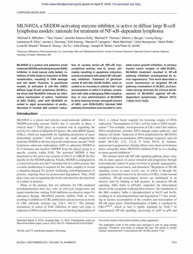
MLN4924, a NEDD8-activating enzyme inhibitor, is active in diffuse large B-celllymphoma models: rationale for treatment of NF-�B–dependent lymphoma*Michael A. Milhollen,1 *Tary Traore,1 Jennifer Adams-Duffy,1 Michael P. Thomas,1 Allison J. Berger,1 Lenny Dang,1
Lawrence R. Dick,1 James J. Garnsey,1 Erik Koenig,1 Steven P. Langston,1 Mark Manfredi,1 Usha Narayanan,1 Mark Rolfe,1
Louis M. Staudt,2 Teresa A. Soucy,1 Jie Yu,1 Julie Zhang,1 Joseph B. Bolen,1 and Peter G. Smith1
1Discovery, Millennium Pharmaceuticals, Inc, Cambridge, MA; and 2Metabolism Branch, Center for Cancer Research, National Cancer Institute, NationalInstitutes of Health, Bethesda, MD
MLN4924 is a potent and selective smallmolecule NEDD8-activating enzyme (NAE)inhibitor. In most cancer cells tested, in-hibition of NAE leads to induction of DNArereplication, resulting in DNA damageand cell death. However, in preclinicalmodels of activated B cell–like (ABC)diffuse large B-cell lymphoma (DLBCL),we show that MLN4924 induces an alter-native mechanism of action. Treatmentof ABC DLBCL cells with MLN4924 re-sulted in rapid accumulation of pI�B�,decrease in nuclear p65 content, reduc-
tion of nuclear factor-�B (NF-�B) tran-scriptional activity, and G1 arrest, ulti-mately resulting in apoptosis induction,events consistent with potent NF-�B path-way inhibition. Treatment of germinal-center B cell–like (GCB) DLBCL cells re-sulted in an increase in cellular Cdt-1 andaccumulation of cells in S-phase, consis-tent with cells undergoing DNA rereplica-tion. In vivo administration of MLN4924to mice bearing human xenograft tumorsof ABC- and GCB-DLBCL blocked NAEpathway biomarkers and resulted in com-
plete tumor growth inhibition. In primaryhuman tumor models of ABC-DLBCL,MLN4924 treatment resulted in NF-�Bpathway inhibition accompanied by tu-mor regressions. This work describes anovel mechanism of targeted NF-�Bpathway modulation in DLBCL and pro-vides strong rationale for clinical devel-opment of MLN4924 against NF-�B–dependent lymphomas. (Blood. 2010;116(9):1515-1523)
Introduction
MLN4924 is a potent and selective small-molecule inhibitor ofNEDD8-activating enzyme (NAE) that is currently in phase 1clinical trials.1-3 NAE plays an essential role in regulating theactivity of a subset of ubiquitin E3 ligases, the cullin-RING ligases(CRLs), which are responsible for regulating destruction of manyintracellular proteins.4 NAE activates the small ubiquitin-likemolecule NEDD8 as the first step in the neddylation cascade.5 NAEhydrolyzes adenosine triphosphate (ATP) to adenylate NEDD8 atits C-terminus and transfers NEDD8 from the adenyl group to aspecific cysteine within NAE. The activated NEDD8 is thentransferred to the active-site cysteine of Ubc12 or UBE2F the E2sspecific for the NEDD8 pathway. Finally, NEDD8 is conjugated ona conserved lysine near the C-terminal end of a cullin protein; thiscovalent modification is required for the cullin complex to recruita ubiquitin-charged E2 protein facilitating polyubiquitination ofproteins, targeting them for proteasomal degradation. Thus, NAEplays a key role in regulating the levels (and therefore the function)of a subset of proteins.
Many of the proteins that are substrates for CRL-mediatedpolyubiquitination have key roles in cell-cycle progression andsignal transduction, making NAE inhibition an attractive target foranticancer therapy. MLN4924 potently inhibits NAE in vitro,resulting in inhibition of CRL neddylation and an increase in levelsof CRL substrate proteins (eg, Cdt-1, Nrf-2).3 The primarymechanism of action of NAE inhibition in many cell types isinduction of DNA rereplication because of blocking degradation of
Cdt-1, a critical factor required for licensing origins of DNAreplication.3 Dysregulation of Cdt-1 activity leads to DNA rerepli-cation.6,7 For example, overexpression of Cdt-1 and Cdc6 inducesDNA rereplication, activates DNA damage repair pathways, andinduces cell death.7 Induction of DNA rereplication by MLN4924results in S-phase accumulation, DNA-damage responses, and celldeath3 (M.A.M., U.N., T.A.S., P. Veiby, P.G.S., B. Amidon,manuscript in preparation). Similar effects were observed in humantumor xenografts where MLN4924 inhibited NAE in vivo leadingto tumor growth inhibition.3
The nuclear factor-�B (NF-�B) signaling pathway plays a keyrole in many aspects of cancer initiation and progression throughtranscriptional control of genes involved in growth, angiogenesis,antiapoptosis, invasiveness, and metastasis.8 Regulation of NF-�Bsignaling occurs at many levels, one of which is through theregulation of protein turnover by the action of CRLs. Under normalconditions, NF-�B transcription factors are maintained in aninactive state by binding to I�B proteins. In canonical NF-�Bsignaling, I�B� binds to p50-p65, sequesters the transcriptionfactors in the cytoplasm rendering them inactive. On stimulation ofthe IKK complex, I�B� is phosphorylated at Ser32 and Ser36,resulting in its polyubiquitination and degradation,9-11 thus result-ing in nuclear accumulation of the complex and transcription ofNF-�B target genes. Polyubiquitination of I�B� is regulated byCRL1�TRCP, which in turn is tightly regulated by NAE.12 Innoncanonical NF-�B signaling, processing of p105 to p50 and
Submitted March 5, 2010; accepted May 14, 2010. Prepublished online asBlood First Edition paper, June 4, 2010; DOI 10.1182/blood-2010-03-272567.
*M.A.M. and T.T. contributed equally to this study.
The online version of this article contains a data supplement.
The publication costs of this article were defrayed in part by page chargepayment. Therefore, and solely to indicate this fact, this article is herebymarked ‘‘advertisement’’ in accordance with 18 USC section 1734.
1515BLOOD, 2 SEPTEMBER 2010 � VOLUME 116, NUMBER 9
For personal use only.on April 9, 2019. by guest www.bloodjournal.orgFrom
p100 to p52 is also controlled by CRL1�TRCP.13,14 Therefore, bothcanonical and noncanonical NF-�B signaling requires NAE activ-ity. Thus, NAE inhibition by MLN4924 represents a novelapproach to inhibit NF-�B and may provide a targeted therapeuticstrategy for the treatment of cancers in which NF-�B signaling isimportant.
One such cancer is diffuse large B-cell lymphoma (DLBCL), anon-Hodgkin lymphoma that has been classified into severaldistinct genetic subgroups based on cDNA microarray analysis.15
Activated B cell–like (ABC)–DLBCL has the poorest prognosisand remains an indication with unmet medical needs where patientshave elevated NF-�B gene expression signature.16 Inhibition ofNF-�B using shRNA, expression of an I�B-super repressor, ortreatment with small-molecule IKK inhibitors has been shown tobe selectively toxic to ABC-DLBCL but not germinal centerB-cell-like (GCB)–DLBCL cells.16-18 Inhibition of IKK in thesecells leads to G1-arrest and induction of apoptosis. These studiesdemonstrate the dependence of ABC-DLBCL on NF-�B signalingfor survival and offer a compelling clinical rationale for the use ofNF-�B inhibitors in this disease.
In this report, we investigate the effect of MLN4924 as a novelNF-�B targeted therapy in DLBCL cells and mouse xenograftmodels. We show that MLN4924 is a potent inhibitor of NF-�Bpathway in vitro and in vivo in NF-�B–dependent DLBCLs,resulting in induction of apoptosis and tumor regressions. Inaddition, we show that MLN4924 also has potent antitumor activityin non–NF-�B–dependent DLBCLs through induction of DNArereplication, independent of NF-�B status. These studies supportthe potential for broad activity of MLN4924 in hematologicmalignancies, with a rationale for targeted therapy in NF-�B–dependent lymphomas, and serve as the basis for the ongoing phase1 evaluation of MLN4924 in hematologic malignancies.
Methods
Cell viability assay
OCI-Ly3, OCI-Ly7, OCI-Ly10, OCI-Ly19, HBL1, L1236, and U2932 cellswere a kind gift from Lou Staudt (National Cancer Institute, Bethesda,MD). All other cell viability assays were performed by Southern Research.Cell suspensions were seeded at 3000 to 8000 cells per well in 96-wellculture plates and incubated overnight at 37°C. Compounds were added tothe cells in complete growth media and incubated for 72 hours at 37°C. Cellnumber was quantified using the ATPlite assay (PerkinElmer Life andAnalytical Sciences).
Western blot analysis of cultured cells
DLBCL cells grown in 6-well cell culture dishes were treated with 0.1%dimethyl sulfoxide (DMSO; control) or MLN4924 for the times indicated.Whole-cell extracts were prepared and analyzed by immunoblotting. Foranalysis of E2-UBL thioester levels, lysates were fractionated by nonreduc-ing sodium dodecyl sulfate–polyacrylamide gel electrophoresis and immu-noblotted with polyclonal antibodies to Ubc12 (generated by Millennium).For analysis of other proteins, lysates were fractionated by reducing sodiumdodecyl sulfate–polyacrylamide gel electrophoresis and probed with pri-mary antibodies as follows: Cdt-1, NEDD8, and Chk1 (Ser 317, Millen-nium), pI�B� (Ser 32), pp105 (Ser 933), p105/50, pH3 (Ser 10), pH2AX(Ser 139), cleaved PARP and cleaved caspase-3 (Cell Signaling), and Nrf-2(Epitomics). Secondary horseradish peroxidase–labeled antibodies to rabbitimmunoglobulin G (IgG) or mouse IgG (Santa Cruz Biotechnology) wereused as appropriate. Blots were developed with enhanced chemilumines-cence reagent (GE Healthcare).
Cell-cycle analysis
Logarithmically growing DLBCL cells were incubated with either MLN4924or DMSO for the times indicated. Analysis was performed as describedpreviously.3
Nuclear p65 quantitation
Logarithmically growing DLBCL cells were incubated with increasing concen-trations of MLN4924 or DMSO as indicated for 4 hours. The nuclear extract wasprepared (Nuclear Extract Kit, Active Motif) and protein concentration deter-mined by the Bradford method. After incubating nuclear extract or p65 standardin oligonucleotide-coated assay plate (TransAM kit, Active Motif) for 1 hour atroom temperature, the plate was washed 3 times with wash buffer, and anti-p65antibody (Santa Cruz Biotechnology) was added to each well. The plate wasincubated for 1 hour at room temperature, washed 3 times with wash buffer andonce with Delfia Wash Buffer (PerkinElmer Life andAnalytical Sciences). DelfiaEu-N1–labeled secondary antibody (PerkinElmer Life and Analytical Sciences)was then added to each well with Delfia Assay Buffer (PerkinElmer Life andAnalytical Sciences). The plate was incubated for 1 hour; and after repeatedwashes, Delfia Enhancement Solution (PerkinElmer Life and Analytical Sci-ences) was added. The plate was measured using a time-resolved fluorometer,and relative fluorescent units were converted to picograms of p65 using thestandard curve and then normalized by total amount of protein loaded per well.
BrdU incorporation assay
Logarithmically growing DLBCL cells were incubated with increasingconcentrations of MLN4924 or DMSO as indicated for 3.5 hours andwere pulsed with 10�M bromodeoxyuridine (BrdU; BD Biosciences) for30 minutes (total MLN4924 incubation time of 4 hours). Cells were thencollected and fixed at 4°C in 70% ethanol. Cells were washed in coldphosphate-buffered saline (PBS), pelleted, and resuspended in 1 mL of 2NHCl for 20 minutes and with PBS. Cell pellets were then resuspended in0.5 mL 0.1M sodium borate (Na2B4O7), pH 8.5, for 2 minutes, washed inPBS, and the resultant cell pellet was resuspended in 20 �L of anti-BrdUmonoclonal antibody (BD Biosciences) and incubated in the dark for20 minutes at room temperature. After incubation, cells were washed withPBS and resuspended in 0.5 mL PBS containing 10 �g/mL propidiumiodide for 1 hour protected from light. Labeled cells were analyzed forpropidium iodide and BrdU staining on a BD Biosciences FACSCaliburflow cytometer using Winlist software v5.0 (Verity).
Quantitative RT-PCR (in vitro/in vivo)
DLBCL cells grown in 6-well cell culture dishes were treated with 0.1% DMSO(control) or MLN4924 for the times indicated. Cells were pelleted and snap-frozen on dry ice before extraction of DNAase-treated RNA using QIAGENreagents. cDNA synthesis and quantitative reverse-transcribed polymerase chainreaction (RT-PCR) were performed using ABI Gene Expression Assays (supple-mental Table 1, available on the Blood Web site; see the Supplemental Materialslink at the top of the online article), reagents, andABI PRISM 7900HT SequenceDetection Systems (Applied Biosystems) using the following cycle conditions:hold at 50°C for 2 minutes for AmpErase UNG activation, then 95.0°C for10 minutes to activate DNA polymerase, and then run 40 two-part cycles of95.0°C for 15 seconds and 60.0°C for 1 minute. Relative mRNA expressionquantitation was derived using the comparative Ct method (Applied Biosystems).The �Ct (dCt) was calculated using the average Ct of control genes B2M(Hs99999907_m1) and 18S (Hs99999901_s1). The ��Ct (ddCt) was calculatedby subtracting the dCt value for the treated sample from the dCt value ofthe experimental control sample following equation: ddCt � dCt(control) �dCt(treated). For analysis of mRNA levels in xenograft samples, tumor frag-ments were harvested, snap-frozen on dry ice, and subjected to RT-PCR analysisas described in this section.
Tumor xenograft efficacy experiments
Female CB.17 SCID mice (Taconic Farms) were used for in vivo studies withOCI-Ly10, OCI-Ly19, and OCI-LY19-luc cells. Female SCID NOD mice(Taconic Farms) were used for in vivo studies with PHTX22Lmodel.All animalswere housed and handled in accordance with the Guide for the Care and Use of
1516 MILHOLLEN et al BLOOD, 2 SEPTEMBER 2010 � VOLUME 116, NUMBER 9
For personal use only.on April 9, 2019. by guest www.bloodjournal.orgFrom
Laboratory Animals. For subcutaneous xenograft studies, mice were inoculatedwith 4 � 106 OCI-Ly10 or OCI-Ly19 cells with Matrigel (BD Biosciences) in theright flank, and tumor growth was monitored with caliper measurements. Whenthe mean tumor volume reached approximately 200 mm3, animals were dosedsubcutaneously with vehicle (10% cyclodextrin) or MLN4924. Inhibition oftumor growth (T/C, average treated tumor volume/average control tumorvolume) was calculated on the last day of treatment. For disseminated xenograftstudies, mice were inoculated with 1 � 106 OCI-Ly19-luc cells (OCI-Ly19-luccells are a stable cell line generated after retroviral infection of OCI-Ly19 cellswith the luciferase transgene under the control of the cytomegalovirus promoter)intravenously, and tumor burden was monitored with xenogen imaging (TaconicFarms).
Pharmacodynamic marker analysis
Mice bearing DLBCL tumors of 300 to 500 mm3 were administered a singleMLN4924 dose; and at the indicated times, tumors were excised andextracts prepared. The relative levels of NEDD8-cullin, pI�B�, and Nrf-2were estimated by quantitative immunoblot analysis (Li-Cor Odysseysystem) using Alexa 680–labeled anti-IgG (Invitrogen) as the secondaryantibody. Cleaved caspase 3 was detected using enhanced chemilumines-cence immunoblotting.
Results
MLN4924 inhibits NAE, prevents cell growth, and generatesdistinct cell-cycle profiles in DLBCL cells
Two cell lines representative of ABC-DLBCL (OCI-Ly10, OCI-Ly3) and 2 representative of GCB-DLBCL (OCI-Ly19, OCI-Ly7)were used.15,16
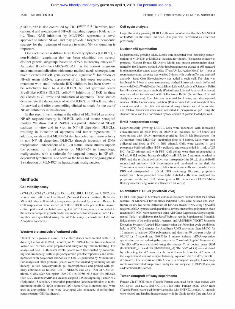
Potent inhibition of cell viability was observed across17 lymphoma cell lines tested in an ATPlite viability assay, withEC50 values of 10 to 244nM (supplemental Table 2). Interestingly,MLN4924 inhibited viability in GCB-DLBCL cell lines (OCI-Ly19, OCI-Ly7), which have been previously shown to be insensi-tive to small-molecule IKK inhibitors.17 Because there was nodiscrimination in sensitivity of ABC- and GCB-DLBCL cells, wehypothesized that in ABC-DLBCL NAE inhibition may primarilyresult in NF-�B pathway inhibition, whereas in GCB-DLBCL themechanism of action may be the result of DNA rereplication.3 Toconfirm NAE inhibition in both types of DLBCL cells, ananti-NEDD8 antibody was used to detect NEDD8 conjugation toUbc12 (an E2 for NEDD8) and the cullin proteins. Cells wereexposed to increasing concentrations of MLN4924 for 24 hours; adose-dependent decrease of Ubc12-NEDD8 thioester and NEDD8-cullin conjugate levels was observed in all cells (Figure 1A). Thesedata show that the neddylation pathway was inhibited to similarlevels in ABC- and GCB-DLBCL cells.
I�B� superrepressor transfection and small-molecule IKKinhibition induce G1-phase cell-cycle arrest in OCI-Ly10 andOCI-Ly3 cells.16,17 We recently reported that NAE inhibitionresults in an accumulation of cells in S-phase.3 To determinewhether similar effects were seen with NAE inhibition in DLBCL,cells were exposed to increasing concentrations of MLN4924 for24 hours and cell-cycle profiles analyzed by flow cytometry. InOCI-Ly10 and OCI-Ly3 cells, a prominent G1 phenotype wasobserved with a proportion of cells sub-G1 indicative of apoptosis(Figure 1B). In contrast, OCI-Ly19 and OCI-Ly7 cells demon-strated an accumulation in S-phase and some cells containing morethan 4N DNA content. Quantitation of cell-cycle distributions ofDLBCL cells in response to MLN4924 confirmed marked differ-ences between OCI-Ly10 and OCI-Ly3 cells compared withOCI-Ly19 and OCI-Ly7 cells (supplemental Figure 1). These data
suggested that ABC-DLBCL cells are affected differently by NAEinhibition, with the predominant phenotype being consistent withNF-�B signaling inhibition, whereas GCB-DLBCL cells wereundergoing DNA rereplication.
MLN4924 induces apoptosis through inhibition of NF-�Bsignaling in ABC-DLBCL and induction of DNA rereplication inGCB-DLBCL
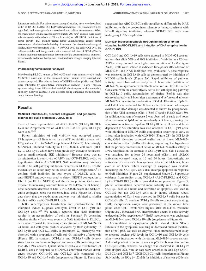
OCI-Ly10 and OCI-Ly19 cells were exposed to MLN4924 concen-trations that elicit 50% and 90% inhibition of viability in a 72-hourATPlite assay, as well as a higher concentration of 1�M (Figure2A-B). Cells were isolated at indicated time points after addition ofMLN4924, and NAE inhibition was evaluated. A dose-responsewas observed in OCI-Ly10 cells as demonstrated by inhibition ofNEDD8-cullin levels (Figure 2A). Rapid inhibition of pathwayactivity was observed as early as 1 hour after addition ofMLN4924, in agreement with effects observed in HCT-116 cells.3
Consistent with the constitutively active NF-�B signaling pathwayin OCI-Ly10 cells, accumulation of pI�B� (Ser32) was alsoobserved as early as 1 hour after treatment and before (and at lowerMLN4924 concentrations) elevation of Cdt-1. Elevation of pI�B�and Cdt-1 was sustained for 8 hours after treatment, whereuponevidence of DNA damage was detected as shown by phosphoryla-tion of the ATM substrate pChk-1 (Ser317) and p-H2AX (Ser 139).In addition, cleavage of caspase-3 was observed as early as 4 hoursafter treatment at 1�M and more robustly at 8 hours, showing thatapoptosis induction is rapid in OCI-Ly10 cells. Dose- and time-dependent NAE inhibition was also observed in OCI-Ly19 cells,with inhibition of NEDD8-cullin conjugation occurring as early as1 hour after incubation with MLN4924 (Figure 2B). In OCI-Ly19cells, Cdt-1 elevation occurred earlier and at lower MLN4924concentrations than pI�B� elevation, supporting the hypothesisthat the primary mechanism of action of MLN4924 in this setting isDNA rereplication. In contrast to OCI-Ly10 cells, Cdt-1 elevationwas sustained for at least 24 hours and pChk-1 and pH2AXactivation occurred later, at 16 and 24 hours. Interestingly, noactivation of caspase-3 cleavage was detected at 24 hours; how-ever, at 48 hours, robust cleavage of caspase-3 was evident,indicating that OCI-Ly19 cells also undergo apoptosis in responseto NAE inhibition (Figure 2B; supplemental Figure 2). Supportiveevidence from studies using OCI-Ly3 (ABC-DLBCL) and OCI-Ly7 (GCB-DLBCL) cells is provided in supplemental Figure 3;pI�B� accumulation occurred more robustly in OCI-Ly3 thanOCI-Ly7 cells at 4 hours and activation of apoptosis was seen inOCI-Ly3 but not OCI-Ly7 cells at 24 hours, whereas robustaccumulation of Cdt-1 was observed in OCI-Ly7 cells but not inOCI-Ly3 cells. To confirm OCI-Ly10 cells were not rereplicating,BrdU incorporation assays were performed at the 4-hour timepoint, when Cdt-1 levels were highest after MLN4924 treatment(Figure 2A). Increased BrdU incorporation occurs in cells activelyundergoing DNA rereplication.7,19 BrdU incorporation was unchangedin MLN4924-treated OCI-Ly10 cells (supplemental Figure 4).
Accumulation of cytoplasmic pI�B� should retain NF-�Bsubunits in the cytoplasm, resulting in decreased nuclear localiza-tion of p50-p65. We used an enzyme-linked immunosorbent assayto measure nuclear p65 levels in OCI-Ly10 and OCI-Ly19 cellsafter 4-hour incubation with increasing MLN4924 concentrations.A dose-dependent decrease in nuclear p65 levels was observed inOCI-Ly10 cells, whereas no change was observed in OCI-Ly19cells (Figure 2C). Similar data were observed in OCI-Ly3 (ABC-DLBCL) and OCI-Ly7 (GCB-DLBCL) cells (supplemental Figure5). Notably, the EC50 ( 25nM) for inhibition of nuclear p65 levels
ANTITUMOR ACTIVITY WITH MLN4924 IN DLBCL MODELS 1517BLOOD, 2 SEPTEMBER 2010 � VOLUME 116, NUMBER 9
For personal use only.on April 9, 2019. by guest www.bloodjournal.orgFrom
in OCI-Ly10 cells was similar to the EC50 (11nM) for inhibition ofviability (supplemental Table 2), suggesting an association betweenNF-�B pathway effects and cell viability for MLN4924.
TaqMan low-density arrays were used to measure the levels ofNF-�B target genes. We found that MLN4924 down-regulatedNF-�B target genes (eg, TNF�, IRF4, PIM1, I�B�, and CXCL10)in OCI-Ly10 cells in a dose- and time-dependent manner (Figure2D). At EC90 concentrations of MLN4924 that inhibit OCI-Ly10viability, a significant decrease in transcript levels of NF-�B targetgenes was noted as early as 3 to 6 hours after treatment. In contrast,minimal changes in NF-�B target gene expression were detected inOCI-Ly19 cells, consistent with the low level of NF-�B activityand lack of p65 redistribution.
These data demonstrate that MLN4924 inhibits NAE and leads tocell death in both OCI-Ly10 and OCI-Ly19 cells. In OCI-Ly10 cells, theprimary mechanism of action is probably the result of NF-�B signaling
inhibition, as evidenced by increased pI�B� levels, decreased nuclearp65, and inhibition of NF-�B target gene transcription. In OCI-Ly19cells, the primary mechanism of action is probably induction of DNArereplication, as evidenced by elevated Cdt-1 levels, accumulation ofcells in S-phase (with evidence of 4N DNAcontent), and no effect onNF-�B target gene transcription.
MLN4924 inhibits NAE in ABC- and GCB-DLBCL humanxenografts grown in mice
To determine the effects of NAE inhibition by MLN4924 in vivo,OCI-Ly10 and OCI-Ly19-luc cells were grown as subcutaneousxenografts in CB.17 SCID mice. OCI-Ly19-luc cells are a stablecell line generated after infection with a retrovirus expressing theluciferase transgene under the control of the cytomegaloviruspromoter. Mice received a single subcutaneous dose of 10, 30, or
Figure 1. MLN4924 is a potent inhibitor of NAE andinduces distinct cell-cycle profiles analyzed by flowcytometry in ABC- and GCB-DLBCL cell lines. OCI-Ly10, OCI-Ly3, OCI-Ly19, and OCI-Ly7 cells were treatedwith MLN4924 for 24 hours. (A) Inhibition of NAE wasassessed by immunoblot analysis of Ubc12-NEDD8thioester and cullin-NEDD8 levels, respectively. Thenonspecific band serves as the loading control. (B) DNAprofiles were analyzed by propidium iodide staining andflow cytometry.
1518 MILHOLLEN et al BLOOD, 2 SEPTEMBER 2010 � VOLUME 116, NUMBER 9
For personal use only.on April 9, 2019. by guest www.bloodjournal.orgFrom
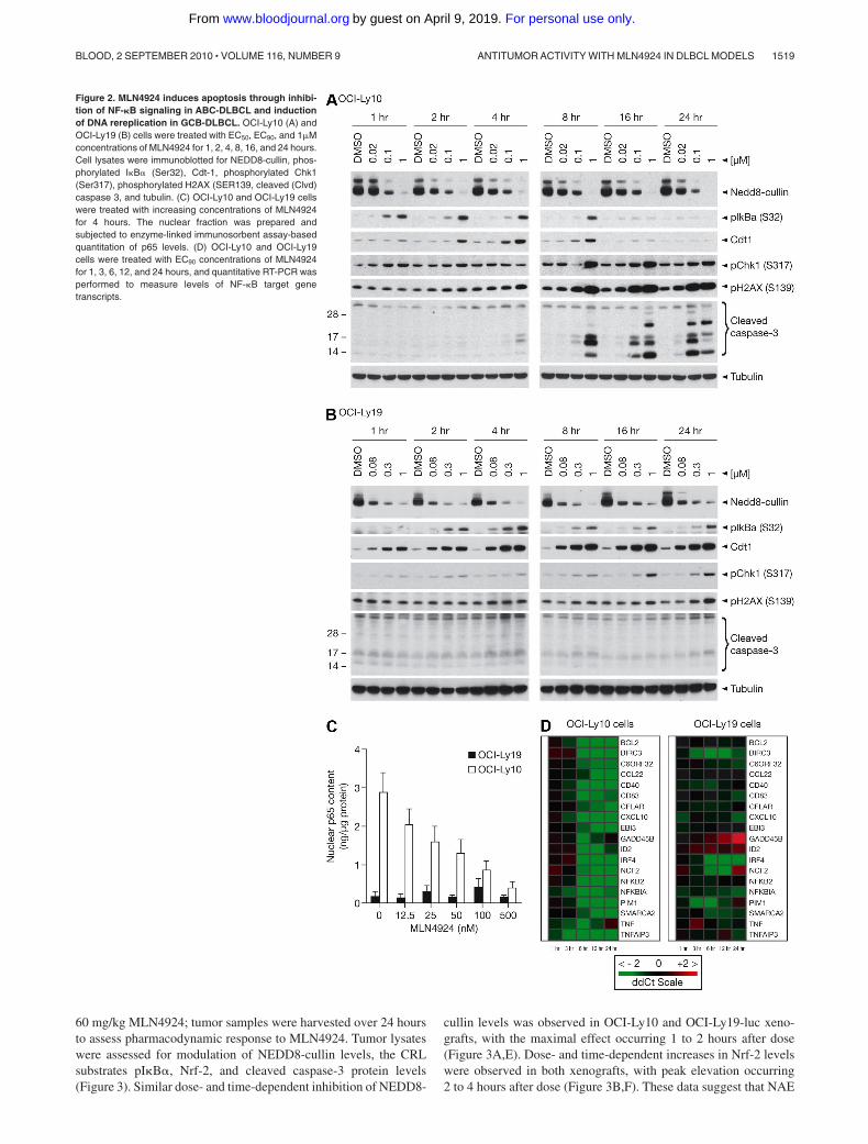
60 mg/kg MLN4924; tumor samples were harvested over 24 hoursto assess pharmacodynamic response to MLN4924. Tumor lysateswere assessed for modulation of NEDD8-cullin levels, the CRLsubstrates pI�B�, Nrf-2, and cleaved caspase-3 protein levels(Figure 3). Similar dose- and time-dependent inhibition of NEDD8-
cullin levels was observed in OCI-Ly10 and OCI-Ly19-luc xeno-grafts, with the maximal effect occurring 1 to 2 hours after dose(Figure 3A,E). Dose- and time-dependent increases in Nrf-2 levelswere observed in both xenografts, with peak elevation occurring2 to 4 hours after dose (Figure 3B,F). These data suggest that NAE
Figure 2. MLN4924 induces apoptosis through inhibi-tion of NF-�B signaling in ABC-DLBCL and inductionof DNA rereplication in GCB-DLBCL. OCI-Ly10 (A) andOCI-Ly19 (B) cells were treated with EC50, EC90, and 1�Mconcentrations of MLN4924 for 1, 2, 4, 8, 16, and 24 hours.Cell lysates were immunoblotted for NEDD8-cullin, phos-phorylated I�B� (Ser32), Cdt-1, phosphorylated Chk1(Ser317), phosphorylated H2AX (SER139, cleaved (Clvd)caspase 3, and tubulin. (C) OCI-Ly10 and OCI-Ly19 cellswere treated with increasing concentrations of MLN4924for 4 hours. The nuclear fraction was prepared andsubjected to enzyme-linked immunosorbent assay-basedquantitation of p65 levels. (D) OCI-Ly10 and OCI-Ly19cells were treated with EC90 concentrations of MLN4924for 1, 3, 6, 12, and 24 hours, and quantitative RT-PCR wasperformed to measure levels of NF-�B target genetranscripts.
ANTITUMOR ACTIVITY WITH MLN4924 IN DLBCL MODELS 1519BLOOD, 2 SEPTEMBER 2010 � VOLUME 116, NUMBER 9
For personal use only.on April 9, 2019. by guest www.bloodjournal.orgFrom
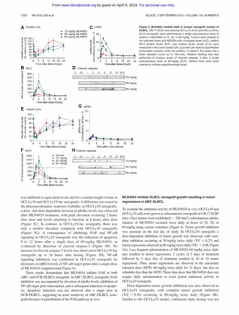
was inhibited to equivalent levels and for a similar length of time inOCI-Ly10 and OCI-Ly19-luc xenografts. A difference was noted inthe pharmacodynamic response of pI�B�; in OCI-Ly10 xenografts,a dose- and time-dependent increase in pI�B� levels was observedafter MLN4924 treatment, with peak elevation occurring 2 hoursafter dose and levels returning to baseline at 8 hours after dose(Figure 3C). In contrast, in OCI-Ly19-luc xenografts, there wasonly a modest elevation compared with OCI-Ly10 xenografts(Figure 3G). A consequence of inhibiting NAE and NF-�Bsignaling in OCI-Ly10 xenografts was the induction of apoptosis8 to 12 hours after a single dose of 60 mg/kg MLN4924, asevidenced by detection of cleaved caspase-3 (Figure 3D). Noincrease in cleaved caspase-3 levels was observed in OCI-Ly19-lucxenografts up to 24 hours after dosing (Figure 3H). NF-�Bsignaling inhibition was confirmed in OCI-Ly10 xenografts bydecreases in mRNA levels of NF-�B target genes after a single doseof MLN4924 (supplemental Figure 6).
These results demonstrate that MLN4924 inhibits NAE in bothABC- and GCB-DLBCLxenografts. InABC-DLBCLxenografts, NAEinhibition was accompanied by elevation of pI�B� levels, inhibition ofNF-�B target gene transcription, and a subsequent induction of apopto-sis. Apoptosis induction was not observed after a single dose inGCB-DLBCL, suggesting an acute sensitivity of ABC-DLBCL xeno-grafts because of perturbation of the NAE pathway in vivo.
MLN4924 inhibits DLBCL xenograft growth resulting in tumorregressions in ABC-DLBCL
To examine the antitumor activity of MLN4924 in vivo, OCI-Ly10 andOCI-Ly19 cells were grown as subcutaneous xenografts in CB.17 SCIDmice. Once tumors were established ( 200 mm3), subcutaneous admin-istration of MLN4924 occurred twice daily at doses of 10, 30, or60 mg/kg using various schedules (Figure 4). Tumor growth inhibitionwas assessed on the last day of study. In OCI-Ly10 xenografts adose-dependent inhibition of tumor growth was observed, with com-plete inhibition occurring at 30 mg/kg twice daily (T/C � 0.27) andtumor regressions observed at 60 mg/kg twice daily (T/C � 0.08, Figure4A). Less frequent administration of MLN4924 60 mg/kg twice dailyalso resulted in tumor regressions; 2 cycles of 5 days of treatmentfollowed by 5 days free of treatment resulted in 10 of 10 tumorregressions. Thus, tumor regressions are observed at the maximumtolerated dose (MTD, 60 mg/kg twice daily for 21 days), but also onschedules less than the MTD. These data show that MLN4924 does notrequire daily administration to exert potent antitumor activity inOCI-Ly10 xenografts.
Dose-dependent tumor growth inhibition was also observed inOCI-Ly19 xenografts, with complete tumor growth inhibition(T/C � 0.10) occurring at 60 mg/kg twice daily (Figure 4B).Similar to the OCI-Ly10 model, continuous daily dosing was not
Figure 3. MLN4924 inhibits NAE in human xenograft models ofDLBCL. CB.17 SCID mice bearing OCI-Ly10 (A-D) and OCI-Ly19-luc(E-H) xenografts were administered a single subcutaneous dose ofvehicle or MLN4924 at 10, 30, or 60 mg/kg. Tumors were excised atthe indicated times and NEDD8-cullin conjugate levels (A,E), relativeNrf-2 protein levels (B,F), and relative pI�B� levels (C,G) weremeasured in the tumor lysates (20 �g protein per lane) by quantitativeimmunoblot analysis. Units are arbitrary. In panel F, the assay has alinear standard curve up to 750 units. Western blotting was alsoperformed to analyze levels of cleaved caspase 3 after a singlesubcutaneous dose of 60 mg/kg (D,H). Vertical lines have beeninserted to indicate repositioned gel lanes.
1520 MILHOLLEN et al BLOOD, 2 SEPTEMBER 2010 � VOLUME 116, NUMBER 9
For personal use only.on April 9, 2019. by guest www.bloodjournal.orgFrom
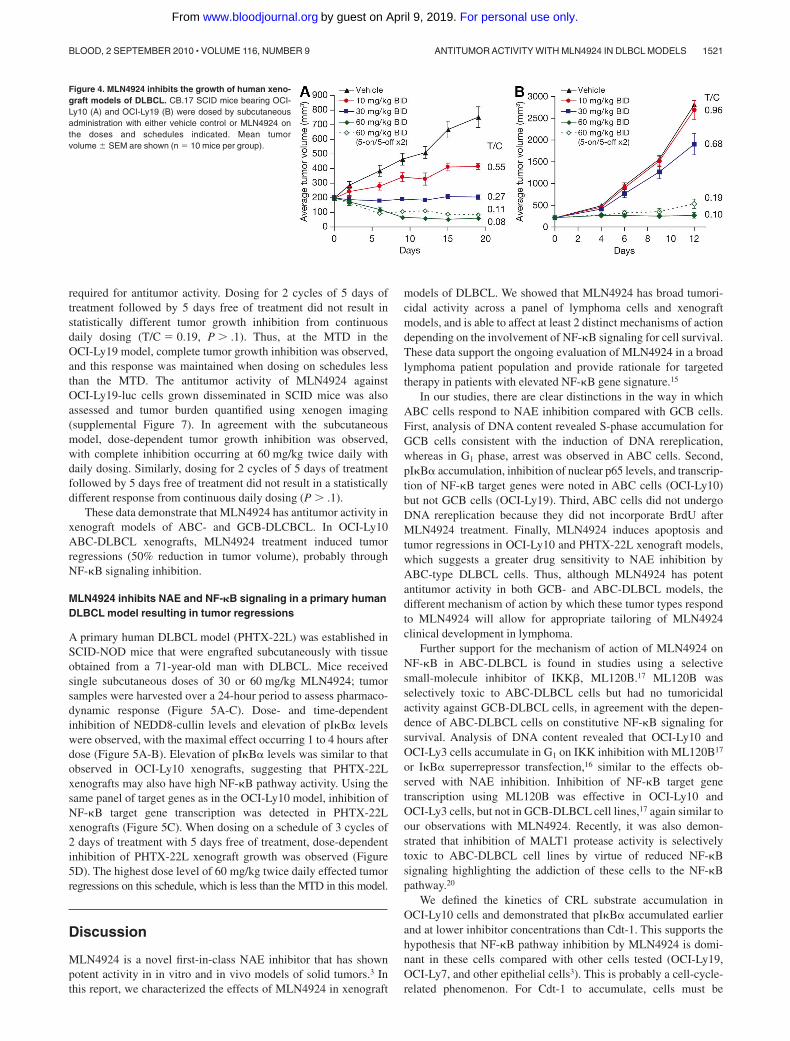
required for antitumor activity. Dosing for 2 cycles of 5 days oftreatment followed by 5 days free of treatment did not result instatistically different tumor growth inhibition from continuousdaily dosing (T/C � 0.19, P .1). Thus, at the MTD in theOCI-Ly19 model, complete tumor growth inhibition was observed,and this response was maintained when dosing on schedules lessthan the MTD. The antitumor activity of MLN4924 againstOCI-Ly19-luc cells grown disseminated in SCID mice was alsoassessed and tumor burden quantified using xenogen imaging(supplemental Figure 7). In agreement with the subcutaneousmodel, dose-dependent tumor growth inhibition was observed,with complete inhibition occurring at 60 mg/kg twice daily withdaily dosing. Similarly, dosing for 2 cycles of 5 days of treatmentfollowed by 5 days free of treatment did not result in a statisticallydifferent response from continuous daily dosing (P .1).
These data demonstrate that MLN4924 has antitumor activity inxenograft models of ABC- and GCB-DLCBCL. In OCI-Ly10ABC-DLBCL xenografts, MLN4924 treatment induced tumorregressions (50% reduction in tumor volume), probably throughNF-�B signaling inhibition.
MLN4924 inhibits NAE and NF-�B signaling in a primary humanDLBCL model resulting in tumor regressions
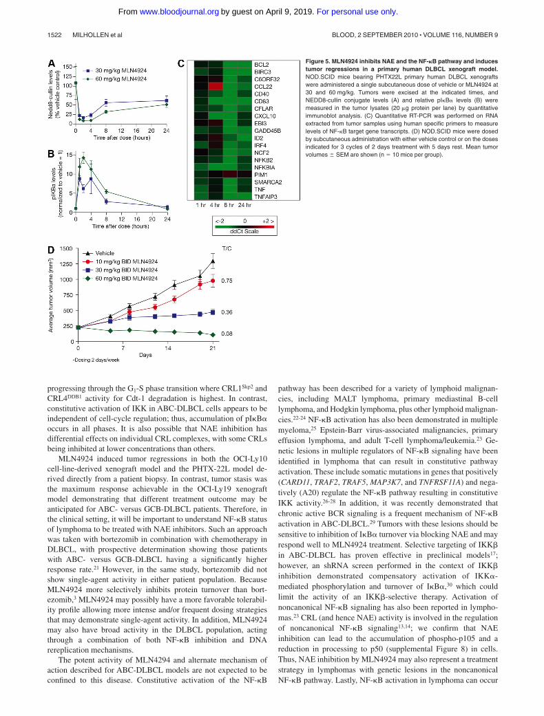
A primary human DLBCL model (PHTX-22L) was established inSCID-NOD mice that were engrafted subcutaneously with tissueobtained from a 71-year-old man with DLBCL. Mice receivedsingle subcutaneous doses of 30 or 60 mg/kg MLN4924; tumorsamples were harvested over a 24-hour period to assess pharmaco-dynamic response (Figure 5A-C). Dose- and time-dependentinhibition of NEDD8-cullin levels and elevation of pI�B� levelswere observed, with the maximal effect occurring 1 to 4 hours afterdose (Figure 5A-B). Elevation of pI�B� levels was similar to thatobserved in OCI-Ly10 xenografts, suggesting that PHTX-22Lxenografts may also have high NF-�B pathway activity. Using thesame panel of target genes as in the OCI-Ly10 model, inhibition ofNF-�B target gene transcription was detected in PHTX-22Lxenografts (Figure 5C). When dosing on a schedule of 3 cycles of2 days of treatment with 5 days free of treatment, dose-dependentinhibition of PHTX-22L xenograft growth was observed (Figure5D). The highest dose level of 60 mg/kg twice daily effected tumorregressions on this schedule, which is less than the MTD in this model.
Discussion
MLN4924 is a novel first-in-class NAE inhibitor that has shownpotent activity in in vitro and in vivo models of solid tumors.3 Inthis report, we characterized the effects of MLN4924 in xenograft
models of DLBCL. We showed that MLN4924 has broad tumori-cidal activity across a panel of lymphoma cells and xenograftmodels, and is able to affect at least 2 distinct mechanisms of actiondepending on the involvement of NF-�B signaling for cell survival.These data support the ongoing evaluation of MLN4924 in a broadlymphoma patient population and provide rationale for targetedtherapy in patients with elevated NF-�B gene signature.15
In our studies, there are clear distinctions in the way in whichABC cells respond to NAE inhibition compared with GCB cells.First, analysis of DNA content revealed S-phase accumulation forGCB cells consistent with the induction of DNA rereplication,whereas in G1 phase, arrest was observed in ABC cells. Second,pI�B� accumulation, inhibition of nuclear p65 levels, and transcrip-tion of NF-�B target genes were noted in ABC cells (OCI-Ly10)but not GCB cells (OCI-Ly19). Third, ABC cells did not undergoDNA rereplication because they did not incorporate BrdU afterMLN4924 treatment. Finally, MLN4924 induces apoptosis andtumor regressions in OCI-Ly10 and PHTX-22L xenograft models,which suggests a greater drug sensitivity to NAE inhibition byABC-type DLBCL cells. Thus, although MLN4924 has potentantitumor activity in both GCB- and ABC-DLBCL models, thedifferent mechanism of action by which these tumor types respondto MLN4924 will allow for appropriate tailoring of MLN4924clinical development in lymphoma.
Further support for the mechanism of action of MLN4924 onNF-�B in ABC-DLBCL is found in studies using a selectivesmall-molecule inhibitor of IKK�, ML120B.17 ML120B wasselectively toxic to ABC-DLBCL cells but had no tumoricidalactivity against GCB-DLBCL cells, in agreement with the depen-dence of ABC-DLBCL cells on constitutive NF-�B signaling forsurvival. Analysis of DNA content revealed that OCI-Ly10 andOCI-Ly3 cells accumulate in G1 on IKK inhibition with ML120B17
or I�B� superrepressor transfection,16 similar to the effects ob-served with NAE inhibition. Inhibition of NF-�B target genetranscription using ML120B was effective in OCI-Ly10 andOCI-Ly3 cells, but not in GCB-DLBCL cell lines,17 again similar toour observations with MLN4924. Recently, it was also demon-strated that inhibition of MALT1 protease activity is selectivelytoxic to ABC-DLBCL cell lines by virtue of reduced NF-�Bsignaling highlighting the addiction of these cells to the NF-�Bpathway.20
We defined the kinetics of CRL substrate accumulation inOCI-Ly10 cells and demonstrated that pI�B� accumulated earlierand at lower inhibitor concentrations than Cdt-1. This supports thehypothesis that NF-�B pathway inhibition by MLN4924 is domi-nant in these cells compared with other cells tested (OCI-Ly19,OCI-Ly7, and other epithelial cells3). This is probably a cell-cycle-related phenomenon. For Cdt-1 to accumulate, cells must be
Figure 4. MLN4924 inhibits the growth of human xeno-graft models of DLBCL. CB.17 SCID mice bearing OCI-Ly10 (A) and OCI-Ly19 (B) were dosed by subcutaneousadministration with either vehicle control or MLN4924 onthe doses and schedules indicated. Mean tumorvolume � SEM are shown (n � 10 mice per group).
ANTITUMOR ACTIVITY WITH MLN4924 IN DLBCL MODELS 1521BLOOD, 2 SEPTEMBER 2010 � VOLUME 116, NUMBER 9
For personal use only.on April 9, 2019. by guest www.bloodjournal.orgFrom
progressing through the G1-S phase transition where CRL1Skp2 andCRL4DDB1 activity for Cdt-1 degradation is highest. In contrast,constitutive activation of IKK in ABC-DLBCL cells appears to beindependent of cell-cycle regulation; thus, accumulation of pI�B�occurs in all phases. It is also possible that NAE inhibition hasdifferential effects on individual CRL complexes, with some CRLsbeing inhibited at lower concentrations than others.
MLN4924 induced tumor regressions in both the OCI-Ly10cell-line-derived xenograft model and the PHTX-22L model de-rived directly from a patient biopsy. In contrast, tumor stasis wasthe maximum response achievable in the OCI-Ly19 xenograftmodel demonstrating that different treatment outcome may beanticipated for ABC- versus GCB-DLBCL patients. Therefore, inthe clinical setting, it will be important to understand NF-�B statusof lymphoma to be treated with NAE inhibitors. Such an approachwas taken with bortezomib in combination with chemotherapy inDLBCL, with prospective determination showing those patientswith ABC- versus GCB-DLBCL having a significantly higherresponse rate.21 However, in the same study, bortezomib did notshow single-agent activity in either patient population. BecauseMLN4924 more selectively inhibits protein turnover than bort-ezomib,3 MLN4924 may possibly have a more favorable tolerabil-ity profile allowing more intense and/or frequent dosing strategiesthat may demonstrate single-agent activity. In addition, MLN4924may also have broad activity in the DLBCL population, actingthrough a combination of both NF-�B inhibition and DNArereplication mechanisms.
The potent activity of MLN4294 and alternate mechanism ofaction described for ABC-DLBCL models are not expected to beconfined to this disease. Constitutive activation of the NF-�B
pathway has been described for a variety of lymphoid malignan-cies, including MALT lymphoma, primary mediastinal B-celllymphoma, and Hodgkin lymphoma, plus other lymphoid malignan-cies.22-24 NF-�B activation has also been demonstrated in multiplemyeloma,25 Epstein-Barr virus-associated malignancies, primaryeffusion lymphoma, and adult T-cell lymphoma/leukemia.23 Ge-netic lesions in multiple regulators of NF-�B signaling have beenidentified in lymphoma that can result in constitutive pathwayactivation. These include somatic mutations in genes that positively(CARD11, TRAF2, TRAF5, MAP3K7, and TNFRSF11A) and nega-tively (A20) regulate the NF-�B pathway resulting in constitutiveIKK activity.26-28 In addition, it was recently demonstrated thatchronic active BCR signaling is a frequent mechanism of NF-�Bactivation in ABC-DLBCL.29 Tumors with these lesions should besensitive to inhibition of I�B� turnover via blocking NAE and mayrespond well to MLN4924 treatment. Selective targeting of IKK�in ABC-DLBCL has proven effective in preclinical models17;however, an shRNA screen performed in the context of IKK�inhibition demonstrated compensatory activation of IKK�-mediated phosphorylation and turnover of I�B�,30 which couldlimit the activity of an IKK�-selective therapy. Activation ofnoncanonical NF-�B signaling has also been reported in lympho-mas.23 CRL (and hence NAE) activity is involved in the regulationof noncanonical NF-�B signaling13,14; we confirm that NAEinhibition can lead to the accumulation of phospho-p105 and areduction in processing to p50 (supplemental Figure 8) in cells.Thus, NAE inhibition by MLN4924 may also represent a treatmentstrategy in lymphomas with genetic lesions in the noncanonicalNF-�B pathway. Lastly, NF-�B activation in lymphoma can occur
Figure 5. MLN4924 inhibits NAE and the NF-�B pathway and inducestumor regressions in a primary human DLBCL xenograft model.NOD.SCID mice bearing PHTX22L primary human DLBCL xenograftswere administered a single subcutaneous dose of vehicle or MLN4924 at30 and 60 mg/kg. Tumors were excised at the indicated times, andNEDD8-cullin conjugate levels (A) and relative pI�B� levels (B) weremeasured in the tumor lysates (20 �g protein per lane) by quantitativeimmunoblot analysis. (C) Quantitative RT-PCR was performed on RNAextracted from tumor samples using human specific primers to measurelevels of NF-�B target gene transcripts. (D) NOD.SCID mice were dosedby subcutaneous administration with either vehicle control or on the dosesindicated for 3 cycles of 2 days treatment with 5 days rest. Mean tumorvolumes � SEM are shown (n � 10 mice per group).
1522 MILHOLLEN et al BLOOD, 2 SEPTEMBER 2010 � VOLUME 116, NUMBER 9
For personal use only.on April 9, 2019. by guest www.bloodjournal.orgFrom
downstream of dysregulated CRL1�TRCP turnover of I�B�, includ-ing inactivating mutations in I�B�31,32 and amplifications inc-REL.23,33 Such genetic lesions would not be susceptible to NF-�Bpathway inhibition by MLN4924; experiments are ongoing todetermine whether these cells will undergo DNA rereplication.
These studies show that MLN4924 represents a promising agentfor the treatment of lymphoma, particularly for those with constitu-tively activated NF-�B. In addition, the pleiotropic effects ofinhibiting CRL-mediated protein turnover in cells may lead toidentification of additional subtypes of hematologic malignanciessensitive to NAE inhibition. Finally, because the NF-�B pathway isthought to play an important role in drug resistance to manychemotherapies,34 MLN4924 used in combination with standard ofcare agents has the potential to treat a diverse range of malignan-cies and patient populations.
Acknowledgment
The authors thank Bruce Dezube for critical reading of themanuscript.
Authorship
Contribution: P.G.S., M.A.M., A.J.B., T.A.S., and S.P.L. partici-pated in the conceptualization and analysis of studies; M.A.M.,J.A.-D., M.P.T., U.N., and E.K. performed in vitro cell cultureexperiments; T.T., J.Z., and M.P.T. performed in vivo antitumoractivity and pharmacodynamic experiments; J.Y., E.K., J.J.G., andM.P.T. performed pharmacodynamic analysis experiments; L.M.S.,L.D., L.R.D., M.R., M.M., and J.B.B participated in criticaldiscussions of data and reviewed and edited the paper; and P.G.Swrote the paper.
Conflict-of-interest disclosure: All authors (except L.M.S.)were employees of Millennium Pharmaceuticals at the time ofthese studies. There are no other competing financial interests.
The current affiliation for L.D. is Agios Pharmaceuticals,Cambridge, MA.
Correspondence: Peter G. Smith, Discovery, Millennium Phar-maceuticals Inc, 40 Landsdowne St, Cambridge, MA 02139;e-mail: [email protected].
References
1. Brownell JE, Sintchak MD, Gavin JM, et al. Sub-strate-assisted inhibition of ubiquitin-like proteinactivating enzymes: the NEDD8 E1 inhibitorMLN4924 forms a NEDD8-AMP mimetic in situ.Mol Cell. 2010;37(1):102-111.
2. Soucy TA, Smith PG, Rolfe M. Targeting NEDD8-activated cullin-RING ligases for the treatment ofcancer. Clin Cancer Res. 2009;15(12):3912-3916.
3. Soucy TA, Smith PG, Milhollen MA, et al. An in-hibitor of NEDD8-activating enzyme as a newapproach to treat cancer. Nature. 2009;458(7239):732-736.
4. Petroski MD, Deshaies RJ. Function and regula-tion of cullin-RING ubiquitin ligases. Nat Rev MolCell Biol. 2005;6(1):9-20.
5. Gong L, Yeh ET. Identification of the activatingand conjugating enzymes of the NEDD8 conjuga-tion pathway. J Biol Chem. 1999;274(17):12036-12042.
6. Fujita M. Cdt1 revisited: complex and tight regula-tion during the cell cycle and consequences ofderegulation in mammalian cells. Cell Div. 2006;1:22.
7. Vaziri C, Saxena S, Jeon Y, et al. A p53-dependent checkpoint pathway prevents rerepli-cation. Mol Cell. 2003;11(4):997-1008.
8. Karin M, Cao Y, Greten FR, Li ZW. NF-kappaB incancer: from innocent bystander to major culprit.Nat Rev Cancer. 2002;2(4):301-310.
9. Campbell KJ, Rocha S, Perkins ND. Active re-pression of antiapoptotic gene expression byRelA(p65) NF-kappa B. Mol Cell. 2004;13(6):853-865.
10. Ghosh S, Baltimore D. Activation in vitro of NF-kappa B by phosphorylation of its inhibitor I kappaB. Nature. 1990;344(6267):678-682.
11. Mercurio F, Zhu H, Murray BW, et al. IKK-1 andIKK-2: cytokine-activated IkappaB kinases essen-tial for NF-kappaB activation. Science. 1997;278(5339):860-866.
12. Read MA, Brownell JE, Gladysheva TB, et al.Nedd8 modification of cul-1 activatesSCF(beta(TrCP))-dependent ubiquitination ofIkappaBalpha. Mol Cell Biol. 2000;20(7):2326-2333.
13. Amir RE, Iwai K, Ciechanover A. The NEDD8pathway is essential for SCF(beta -TrCP)-medi-ated ubiquitination and processing of the NF-kappa B precursor p105. J Biol Chem. 2002;277(26):23253-23259.
14. Fong A, Sun SC. Genetic evidence for the essen-tial role of beta-transducin repeat-containing pro-tein in the inducible processing of NF-kappa B2/p100. J Biol Chem. 2002;277(25):22111-22114.
15. Alizadeh AA, Eisen MB, Davis RE, et al. Distincttypes of diffuse large B-cell lymphoma identifiedby gene expression profiling. Nature. 2000;403(6769):503-511.
16. Davis RE, Brown KD, Siebenlist U, Staudt LM.Constitutive nuclear factor kappaB activity is re-quired for survival of activated B cell-like diffuselarge B cell lymphoma cells. J Exp Med. 2001;194(12):1861-1874.
17. Lam LT, Davis RE, Pierce J, et al. Small moleculeinhibitors of IkappaB kinase are selectively toxicfor subgroups of diffuse large B-cell lymphomadefined by gene expression profiling. Clin CancerRes. 2005;11(1):28-40.
18. Ngo VN, Davis RE, Lamy L, et al. A loss-of-function RNA interference screen for moleculartargets in cancer. Nature. 2006;441(7089):106-110.
19. Melixetian M, Ballabeni A, Masiero L, et al. Lossof Geminin induces rereplication in the presenceof functional p53. J Cell Biol. 2004;165(4):473-482.
20. Ferch U, Kloo B, Gewies A, et al. Inhibition ofMALT1 protease activity is selectively toxic foractivated B cell-like diffuse large B cell lymphomacells. J Exp Med. 2009;206(11):2313-2320.
21. Dunleavy K, Pittaluga S, Czuczman MS, et al.Differential efficacy of bortezomib plus chemo-therapy within molecular subtypes of diffuse largeB-cell lymphoma. Blood. 2009;113(24):6069-6076.
22. Feuerhake F, Kutok JL, Monti S, et al. NFkappaBactivity, function, and target-gene signatures inprimary mediastinal large B-cell lymphoma anddiffuse large B-cell lymphoma subtypes. Blood.2005;106(4):1392-1399.
23. Jost PJ, Ruland J. Aberrant NF-kappaB signaling
in lymphoma: mechanisms, consequences, andtherapeutic implications. Blood. 2007;109(7):2700-2707.
24. Klein U, Dalla-Favera R. Germinal centres: role inB-cell physiology and malignancy. Nat Rev Immu-nol. 2008;8(1):22-33.
25. Ni H, Ergin M, Huang Q, et al. Analysis of expres-sion of nuclear factor kappa B (NF-kappa B) inmultiple myeloma: downregulation of NF-kappa Binduces apoptosis. Br J Haematol. 2001;115(2):279-286.
26. Compagno M, Lim WK, Grunn A, et al. Mutationsof multiple genes cause deregulation of NF-kappaB in diffuse large B-cell lymphoma. Nature.2009;459(7247):717-721.
27. Kato M, Sanada M, Kato I, et al. Frequent inacti-vation of A20 in B-cell lymphomas. Nature. 2009;459(7247):712-716.
28. Lenz G, Davis RE, Ngo VN, et al. OncogenicCARD11 mutations in human diffuse large B celllymphoma. Science. 2008;319(5870):1676-1679.
29. Davis RE, Ngo VN, Lenz G, et al. Chronic activeB-cell-receptor signalling in diffuse large B-celllymphoma. Nature. 2010;463(7277):88-92.
30. Lam LT, Davis RE, Ngo VN, et al. CompensatoryIKKalpha activation of classical NF-kappaB sig-naling during IKKbeta inhibition identified by anRNA interference sensitization screen. Proc NatlAcad Sci U S A. 2008;105(52):20798-20803.
31. Cabannes E, Khan G, Aillet F, Jarrett RF, HayRT. Mutations in the IkBa gene in Hodgkin’s dis-ease suggest a tumour suppressor role forIkappaBalpha. Oncogene. 1999;18(20):3063-3070.
32. Krappmann D, Emmerich F, Kordes U, et al. Mo-lecular mechanisms of constitutive NF-kappaB/Rel activation in Hodgkin/Reed-Sternberg cells.Oncogene. 1999;18(4):943-953.
33. Barth TF, Martin-Subero JI, Joos S, et al. Gains of2p involving the REL locus correlate with nuclearc-Rel protein accumulation in neoplastic cells ofclassical Hodgkin lymphoma. Blood. 2003;101(9):3681-3686.
34. Nakanishi C, Toi M. Nuclear factor-kappaB inhibi-tors as sensitizers to anticancer drugs. Nat RevCancer. 2005;5(4):297-309.
ANTITUMOR ACTIVITY WITH MLN4924 IN DLBCL MODELS 1523BLOOD, 2 SEPTEMBER 2010 � VOLUME 116, NUMBER 9
For personal use only.on April 9, 2019. by guest www.bloodjournal.orgFrom
online June 4, 2010 originally publisheddoi:10.1182/blood-2010-03-272567
2010 116: 1515-1523
Bolen and Peter G. SmithUsha Narayanan, Mark Rolfe, Louis M. Staudt, Teresa A. Soucy, Jie Yu, Julie Zhang, Joseph B.Lenny Dang, Lawrence R. Dick, James J. Garnsey, Erik Koenig, Steven P. Langston, Mark Manfredi, Michael A. Milhollen, Tary Traore, Jennifer Adams-Duffy, Michael P. Thomas, Allison J. Berger, lymphoma
dependent−BκB-cell lymphoma models: rationale for treatment of NF-MLN4924, a NEDD8-activating enzyme inhibitor, is active in diffuse large
http://www.bloodjournal.org/content/116/9/1515.full.htmlUpdated information and services can be found at:
(3019 articles)Lymphoid Neoplasia Articles on similar topics can be found in the following Blood collections
http://www.bloodjournal.org/site/misc/rights.xhtml#repub_requestsInformation about reproducing this article in parts or in its entirety may be found online at:
http://www.bloodjournal.org/site/misc/rights.xhtml#reprintsInformation about ordering reprints may be found online at:
http://www.bloodjournal.org/site/subscriptions/index.xhtmlInformation about subscriptions and ASH membership may be found online at:
Copyright 2011 by The American Society of Hematology; all rights reserved.of Hematology, 2021 L St, NW, Suite 900, Washington DC 20036.Blood (print ISSN 0006-4971, online ISSN 1528-0020), is published weekly by the American Society
For personal use only.on April 9, 2019. by guest www.bloodjournal.orgFrom

![ML3 Is a NEDD8- and Ubiquitin-Modi ed … Is a NEDD8- and Ubiquitin-Modified Protein1[C][W][OPEN] Jana P. Hakenjos, Sarosh Bejai, Quirin Ranftl, Carina Behringer, A. Corina Vlot,](https://img.pdfslide.net/doc/110x75/5ac5a9a47f8b9a57528dc479/ml3-is-a-nedd8-and-ubiquitin-modi-ed-is-a-nedd8-and-ubiquitin-modied-protein1cwopen.jpg)
![Research Paper Inhibition of CRL-NEDD8 pathway as a new … · 2018. 4. 2. · including acute myeloid leukemia[20]. To investigate effect of CRL -NEDD8 pathway in ATRAinduced differentiation](https://img.pdfslide.net/doc/110x75/6094d0db46df142e8230d376/research-paper-inhibition-of-crl-nedd8-pathway-as-a-new-2018-4-2-including.jpg)