Embed Size (px)
Citation preview

General Pathology
AmyloidosisFibrinoid, Hyalin
Institute of Pathology, 1st Faculty of Medicine, Charles University, Prague
Jaroslava Dušková

Amyloidosis, fibrinoid and hyalin –
disturbances of protein metabolism - contents
Amyloid
definition and history
morphology of amyloid
Amyloidoses – classification
systemic amyloidoses and
their complications
localized amyloidoses and
their complications
Diagnosis of amyloid –
clinical and morphological
Reversibility & therapy of
amyloid
Fibrinoid change of collagen
definiton
morphology of fibrinoid
change of collagen
complications, clinical
importance
Hyalin
definition
morphology
complications, clinical
importance
AmyloidosisDEF.:
disorder of protein
metabolism accompanied
with abnormal extracellular
deposition of proteinaceous
material - amyloid
Amyloid = starch likemisfolded PROTEIN with abundant
proteoglycans and glycosaminoglycans
Karl Freiherr von Rokitansky (1804-1878)
Rudolf Ludwig Karl Virchow (1821-1902).
Amyloid - nature - history
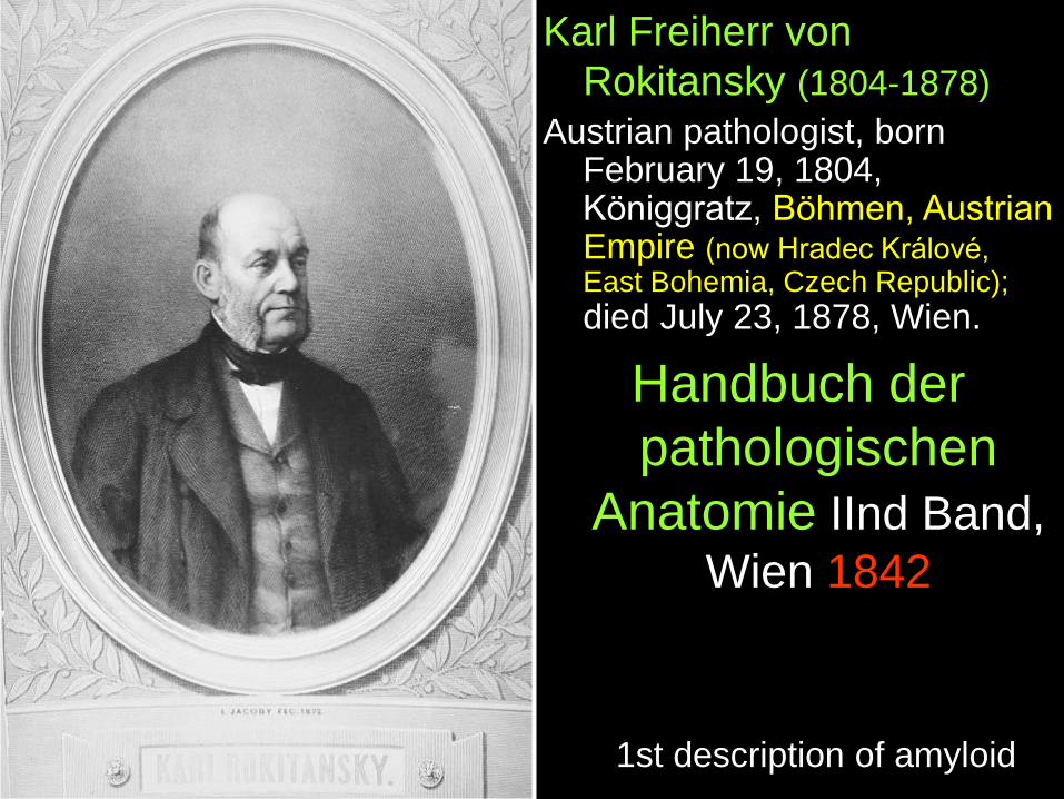
Karl Freiherr von
Rokitansky (1804-1878)
Austrian pathologist, born February 19, 1804, Königgratz, Böhmen, Austrian Empire (now Hradec Králové, East Bohemia, Czech Republic);
died July 23, 1878, Wien.
Handbuch der
pathologischen
Anatomie IInd Band,
Wien 1842
1st description of amyloid
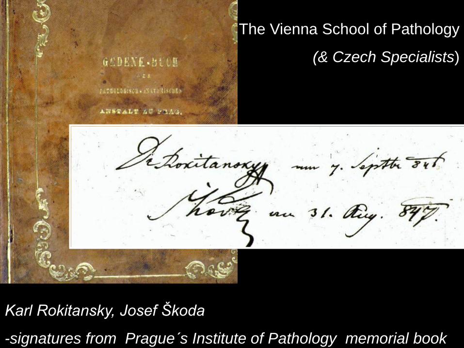
The Vienna School of Pathology
(& Czech Specialists)
Karl Rokitansky, Josef Škoda
-signatures from Prague´s Institute of Pathology memorial book

German anatomist and
pathologist,
Rudolf Ludwig Karl
Virchow (1821-1902).
Virchow´s macroscopy
reactions for amyloid

Amyloidosis – morphology
Macroscopy:
small amounts – invisible
larger deposits – enlarged,
firm, waxy organs
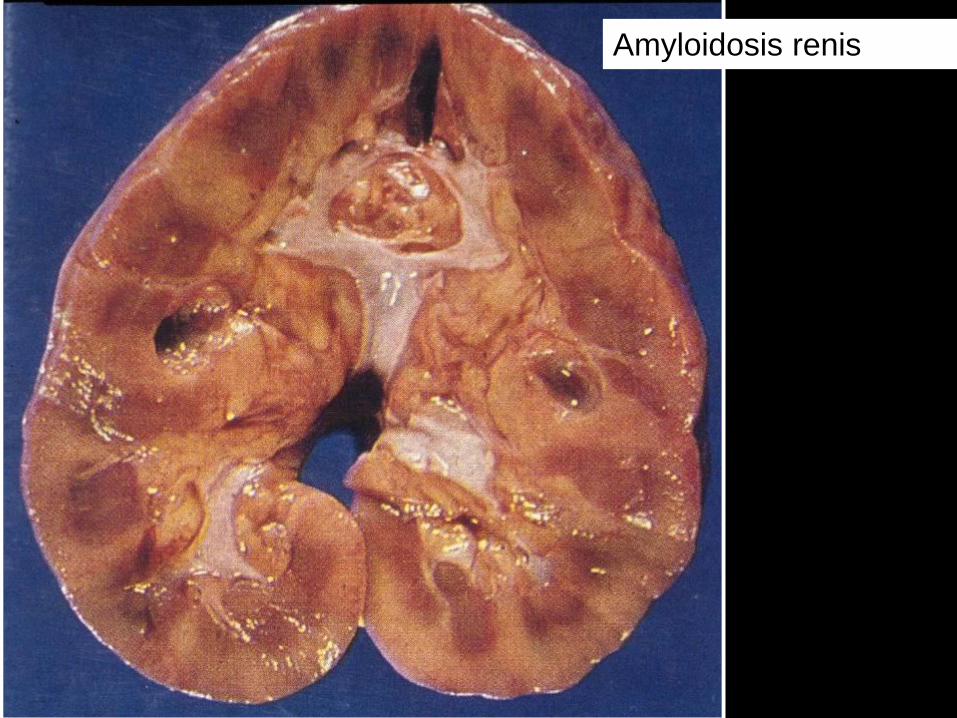
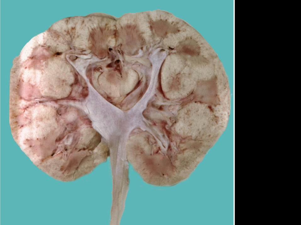
Amyloidosis renis
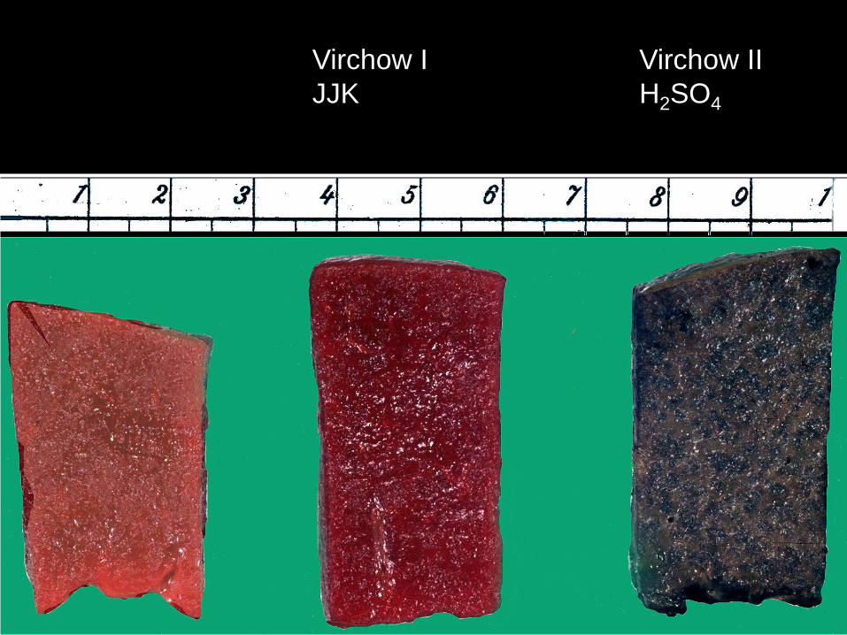
Virchow I
JJK
Virchow II
H2SO4
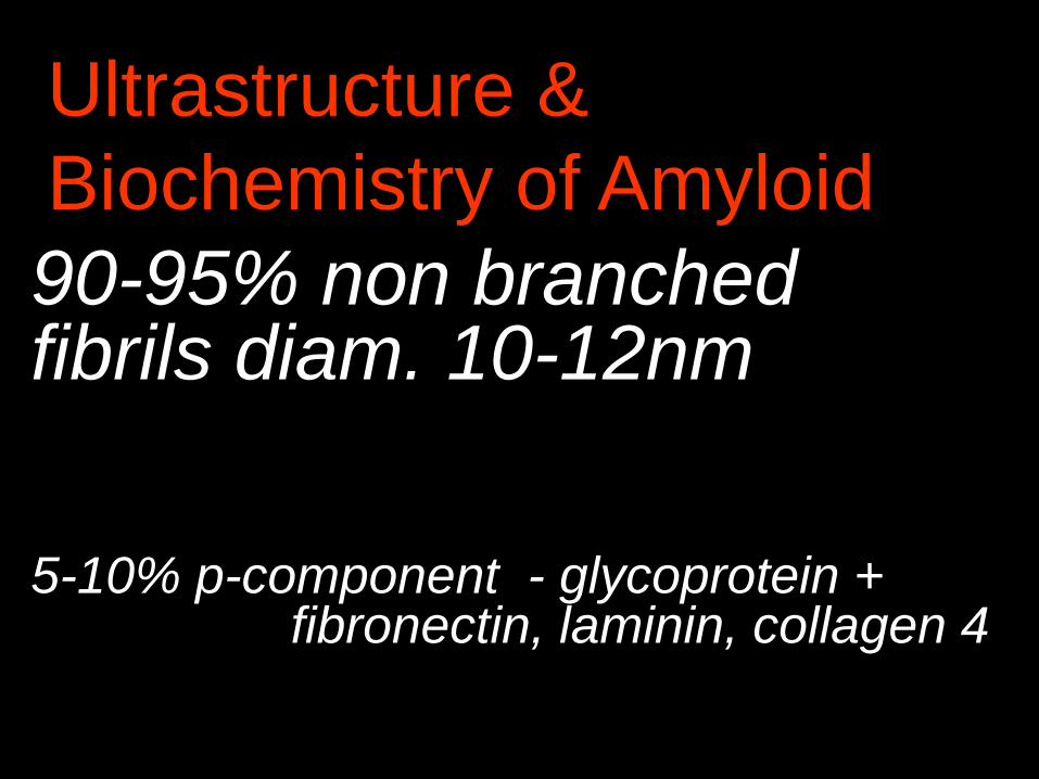
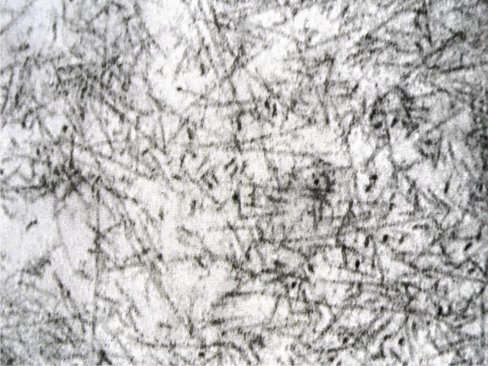
Ultrastructure &
Biochemistry of Amyloid
90-95% non branched fibrils diam. 10-12nm
5-10% p-component - glycoprotein + fibronectin, laminin, collagen 4
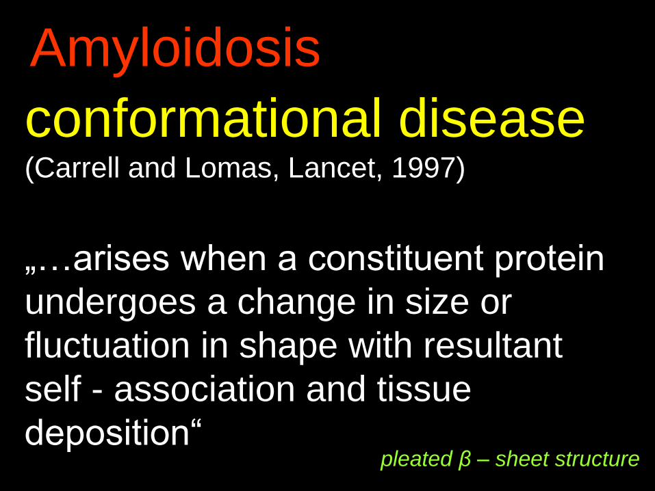
Amyloidosis
conformational disease(Carrell and Lomas, Lancet, 1997)
„…arises when a constituent protein
undergoes a change in size or
fluctuation in shape with resultant
self - association and tissue
deposition“ pleated β – sheet structure
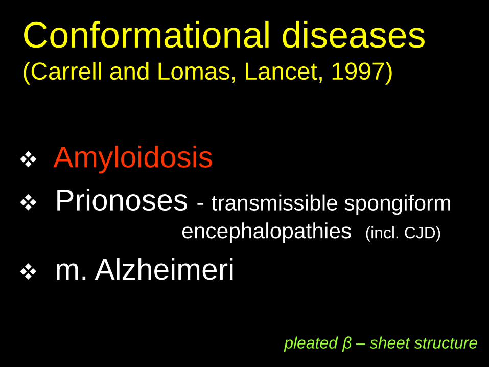
Conformational diseases (Carrell and Lomas, Lancet, 1997)
Amyloidosis
Prionoses - transmissible spongiform
encephalopathies (incl. CJD)
m. Alzheimeri
pleated β – sheet structure
Amyloidosis
Classification:
according to the source protein
(2015 - 31 different identified)
according to the distribution
systemic (generalised)
localised
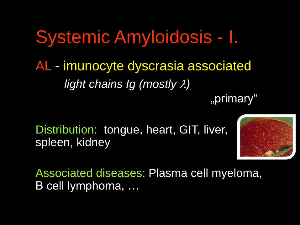
Systemic Amyloidosis - I.
AL - imunocyte dyscrasia associated
light chains Ig (mostly )
„primary“
Distribution: tongue, heart, GIT, liver, spleen, kidney
Associated diseases: Plasma cell myeloma, B cell lymphoma, …

Systemic Amyloidosis - II.
AA - reactive systemic amyloidosis
SAA = Serum Amyloid Associated
protein „secondary“
Distribution: liver, kidney, spleen, GIT, lymph nodes, bowel, adipose tissue
Associated diseases: rheumatoid arthritis, chronic
infections (tb, leprosy, bronchiectasiae,
osteomyelitis, IBD, neoplasms MLH , RCC…)

Systemic Amyloidosis - III.
Wild –type (senile) systemic/cardiac ATTR
25% people over the age of 80 years (!)
-normal (wild-type) transthyretin TTR (prealbumin)
-mostly heart & vessels involvement

Systemic Amyloidosis - IV.
A2 - hemodialysis associated
2 microglobulin
Hereditary
AA - Familial Mediterranean Fever
ATTR - Famil. polyneuropatia
transthyretin (mutated form)
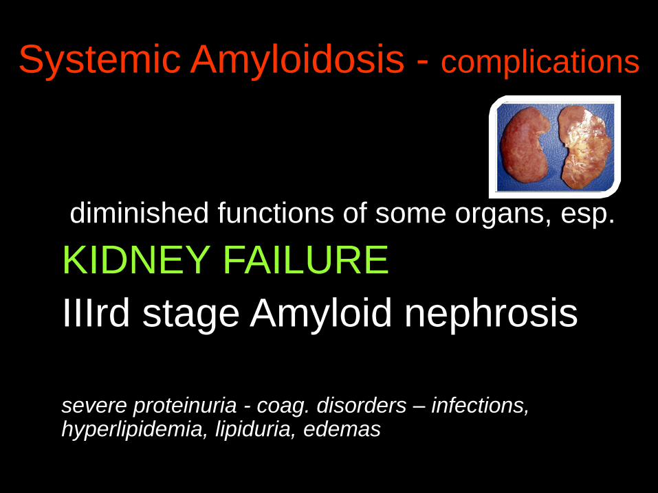
Systemic Amyloidosis - complications
diminished functions of some organs, esp.
KIDNEY FAILURE
IIIrd stage Amyloid nephrosis
severe proteinuria - coag. disorders – infections, hyperlipidemia, lipiduria, edemas
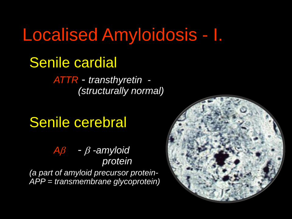
Localised Amyloidosis - I.
Senile cardialATTR - transthyretin -
(structurally normal)
Senile cerebral
A - -amyloid protein
(a part of amyloid precursor protein-APP = transmembrane glycoprotein)
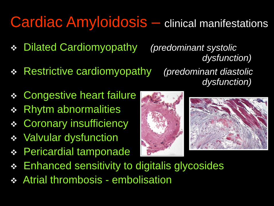
Cardiac Amyloidosis – clinical manifestations
Dilated Cardiomyopathy (predominant systolic dysfunction)
Restrictive cardiomyopathy (predominant diastolic dysfunction)
Congestive heart failure
Rhytm abnormalities
Coronary insufficiency
Valvular dysfunction
Pericardial tamponade
Enhanced sensitivity to digitalis glycosides
Atrial thrombosis - embolisation
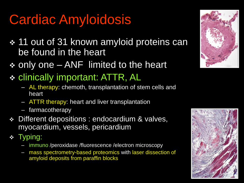
Cardiac Amyloidosis
11 out of 31 known amyloid proteins can be found in the heart
only one – ANF limited to the heart
clinically important: ATTR, AL– AL therapy: chemoth, transplantation of stem cells and
heart
– ATTR therapy: heart and liver transplantation
– farmacotherapy
Different depositions : endocardium & valves, myocardium, vessels, pericardium
Typing: – immuno /peroxidase /fluorescence /electron microscopy
– mass spectrometry-based proteomics with laser dissection of amyloid deposits from paraffin blocks

Localised Amyloidosis - II.
Endocrine
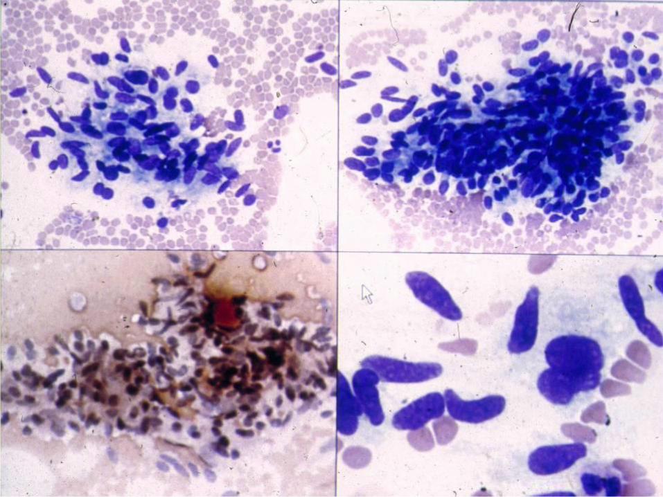
ACal - ca medullare gl. thyreoideae
AIAPP - islets of Langerhans associated
AANF - isolated atrial amyloidosis
atrial natriuretic polypeptide
Nodular tumoriform amyloid deposits(tongue, lung,larynx, skin, urinary bladder, orbita)

Clinical Symptoms of Amyloid
1. nephrotic proteinuria (edemas)
2. weekness, fatigue, loss of weight,
collapses, heart failure,
cardiomyopathy
3. hepatomegaly
4. idiopatic peripheral neuropathy
(parestesias)
5. diarrhoea, cachexia (GIT)
Clinical Diagnosis of Amyloid
Scintigraphy (in vivo)using human serum amyloid component
marked with 123J
Echocardiography (atrial amyloid)

Clinical Diagnosis of Amyloid
Biochemistry
sequening DNA -hered. forms
extraction of fibrils (from a biopsy
specimen)
spectrometry
sequening of the amyloid protein
Amyloidosis – morphology
Macroscopy:
small amounts – invisible
larger deposits – enlarged,
firm, waxy organs


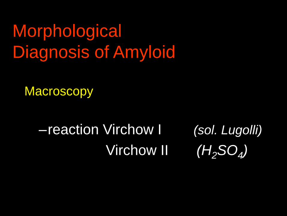
Morphological
Diagnosis of Amyloid
Macroscopy
–reaction Virchow I (sol. Lugolli)
Virchow II (H2SO4)

Virchow I
JJK
Virchow II
H2SO4
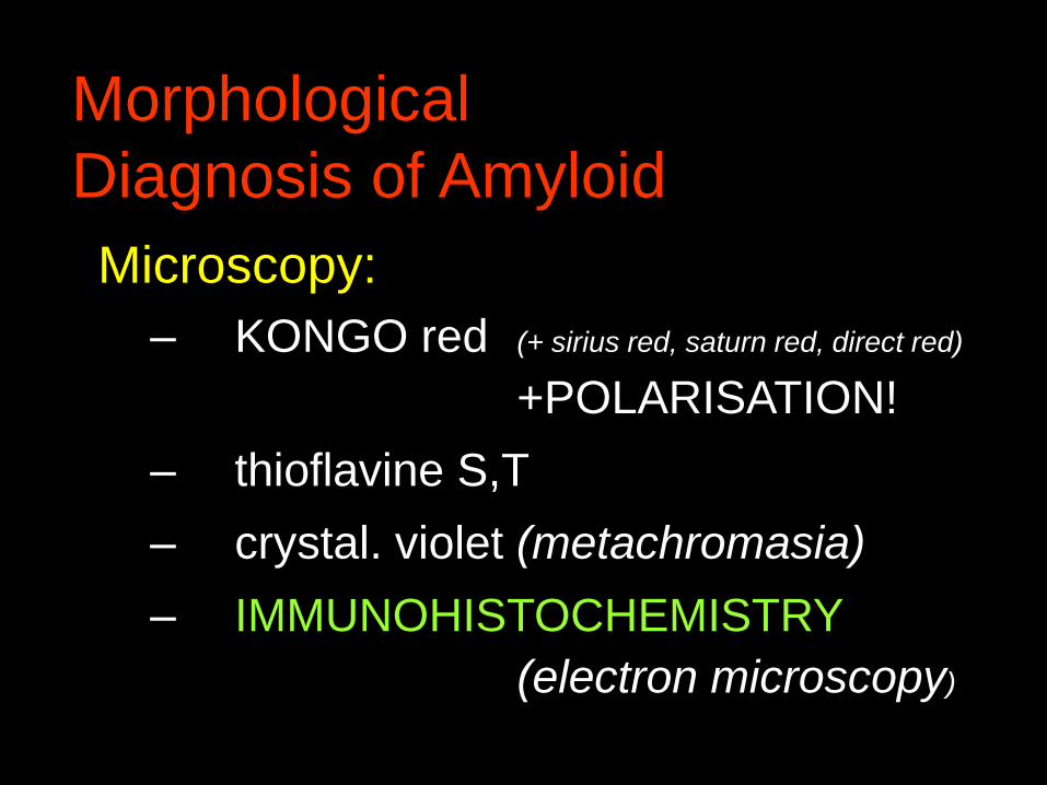
Morphological
Diagnosis of Amyloid
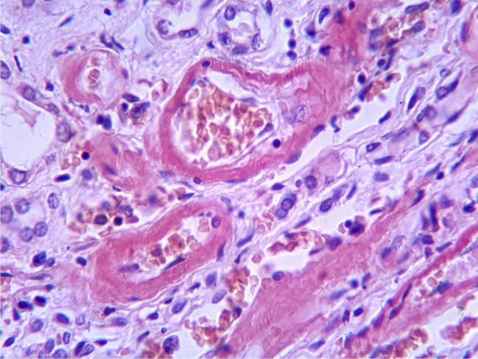
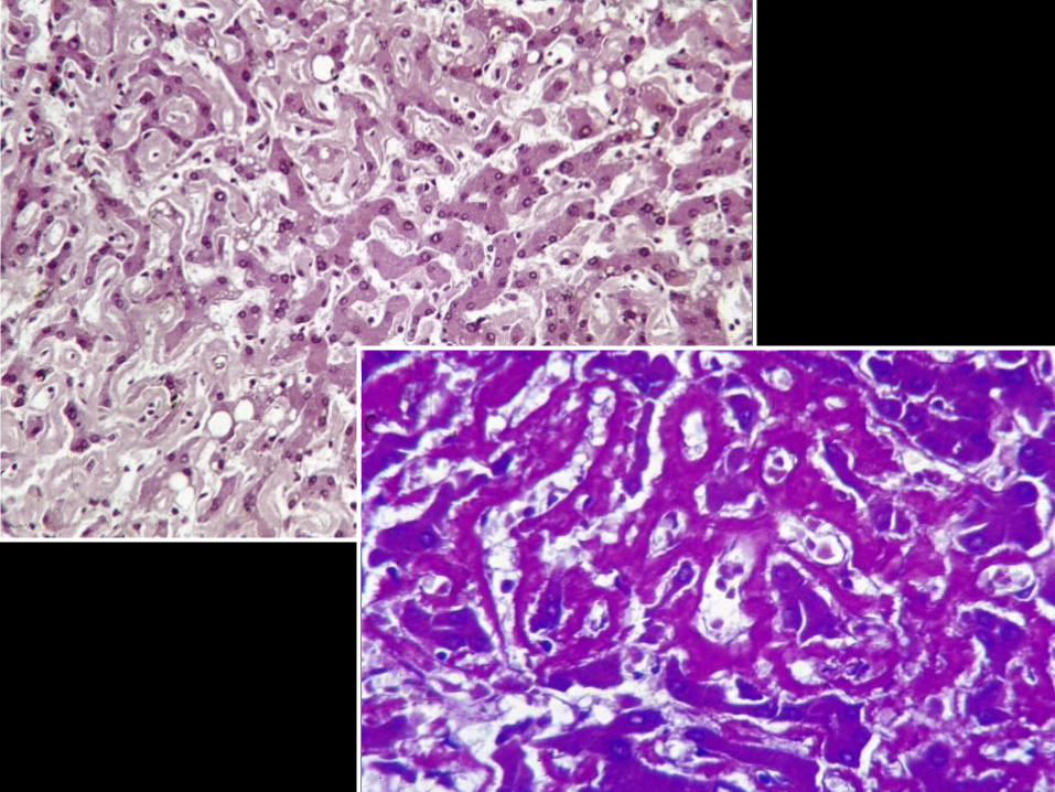
Microscopy:
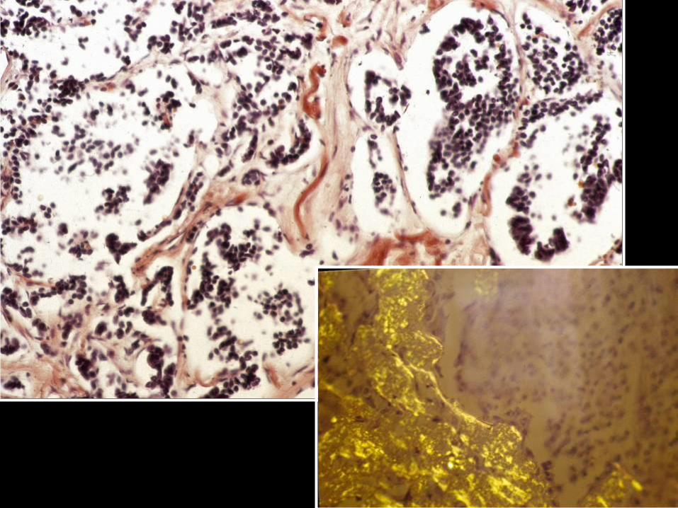
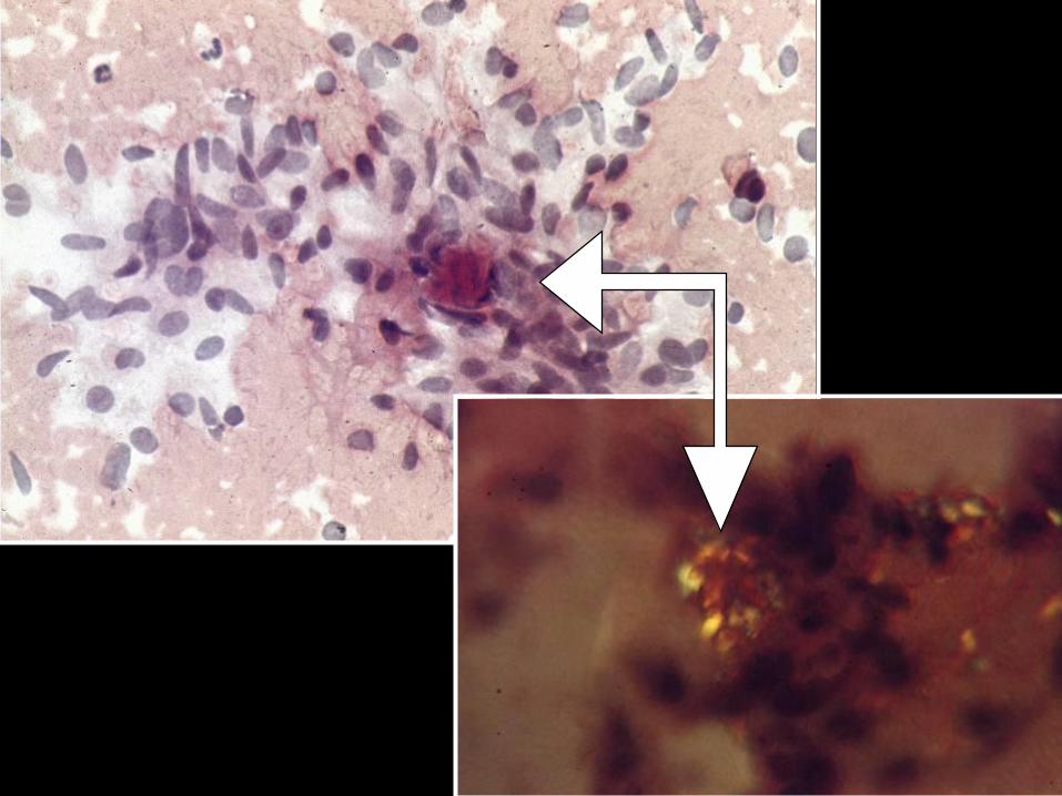
– KONGO red (+ sirius red, saturn red, direct red)
+POLARISATION!
– thioflavine S,T
– crystal. violet (metachromasia)
– IMMUNOHISTOCHEMISTRY
(electron microscopy)
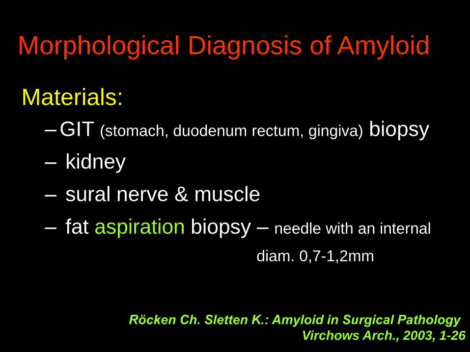
Morphological Diagnosis of Amyloid
Materials:
– GIT (stomach, duodenum rectum, gingiva) biopsy
– kidney
– sural nerve & muscle
– fat aspiration biopsy – needle with an internal
diam. 0,7-1,2mm
Röcken Ch. Sletten K.: Amyloid in Surgical Pathology
Virchows Arch., 2003, 1-26
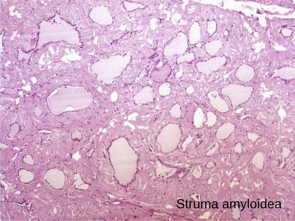
Struma amyloidea

Amyloidosis renis

CONGO Red synthesized by young chemist at Bayer comp. 1883 as
the first of economically lucrative direct (not needing a
mordant) textile dyes
patented by AGFA 1885
(AktienGeselschaft Für Anilinfarbenfabrikation)
3 weeks after the conclusion of the
West Africa Conference
to Europeans in 1885, the word Congo evoked exotic
images of far-off central Africa known as The Dark
Continent
the Congo red stain was named „Congo“ for marketing
purposes by a German textile dyestuff company in 1885
Steensma DP: „Congo“ Red. Out of Africa? Arch. Pathol.Lab.Med.,2001, 125, 250-2

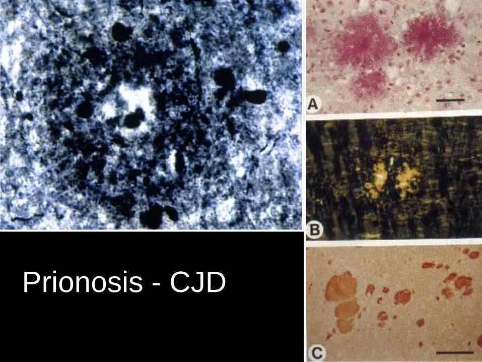
Prionosis - CJD


Reversibility of Amyloid
The deposits are NOT irreversible. e.g. Hrncic R. et al: Antibody mediated
resolution of light chain – associated amyloid deposits. Am.J. Pathol., 2000, 157,12369-46
Progression of generalised amyloidosis can be delayed or stopped by treatment of the underlying disease.
Röcken Ch. Shakespeare Ann: Pathology, diagnosis and pathogenesis of AA amyloidosis. Virchows Arch. , 2002, 440, 11-122
Prevention & Therapy of Amyloid
Prevention & treatment of the underlying diseases
Vaccination against β am. protein in mice diminished senile plaque formation and improved memory.
Nature Medicine, 2001, 7, 18th Jan.
A β –based experimental therapies based on degrading enzymes.
Zlokovic et al.: Neurovascular Pathways and Alzheimer
Amyloid β-peptide. Brain Pathol. , 2005, 15, 78-83

Solomon A., Murphy Ch.L., Westermark P.:
Unreliability of
Immunohistochemistry for Typing
Amyloid Deposits„Because the treatment as well as prognoses of patients with
amyloidosis is dependent on the amyloid type, it is crucial that
the nature of the fibrillar protein be established unequivocally
to avoid inappropriate and costly therapy that can have dire
and possible legal consequences.“
Archives of Pathology and Laboratory Medicine,2008, vol.
132, No. 1, pp. 14–14.

Amyloid Diagnosis Immunohistochemistry

Amyloid- DiagnosisImmunofluorescence most sensitive
fresh tissue sometimes needed
Pathogenesis of Amyloid
G. Joshi, M. E. Bekier, and Y.Wang
University of Michigan, Ann Arbor, MI, USA
Golgi fragmentation in Alzheimer's
disease. Front Neurosci. 2015; 9: 340.
The Golgi apparatus - post-translational modifications, sorting, and
trafficking of membrane and secretory proteins.
Proper functionality of the Golgi requires the formation of its unique
cisternal-stacking morphology.
Phosphorylation of the Golgi stacking protein GRASP65 disrupts its
function in Golgi structure formation, resulting in Golgi fragmentation.
Golgi defects may ultimately promote the development of AD.
Pathogenesis of Amyloid
Marin-Argany M et al: Mayo clinic, Rochester, USA
Mutations can cause light chains to be too stable or
too unstable to form amyloid fibrils.Protein Sci. 2015 Aug 24. doi: 10.1002/pro.2790. [Epub ahead of print]
Light chain (AL) amyloidosis is an incurable human
disease, where the amyloid precursor is a misfolding-prone
immunoglobulin light-chain.
Certain mutations either decrease (H32Y and H70D) or
increase (R65S and Q96Y) the protein thermal stability.
Interestingly, the most and the least stable mutants, Q96Y
and H32Y, do not form amyloid fibrils under physiological
conditions.
Within a thermal stability range, the most stable protein in
this study is the most amyloidogenic protein.

Pathogenesis of Amyloid
Penke B, Bogár F, Fülöp L. University of Szeged, Hungary
β-Amyloid and the Pathomechanisms of Alzheimer's Disease:
A Comprehensive View.
Molecules. 2017 Oct 10; 22(10). pii: E1692. doi: 10.3390/molecules22101692.
Protein dyshomeostasis is the common mechanism of neurodegenerative diseases such as
Alzheimer's disease (AD).
Aging is the key risk factor,
The extensive and complex network of proteostasis declines during aging and is not able to maintain
the balance between production and disposal of proteins.
Different cellular stress conditions result in the up-regulation of the neurotrophic, neuroprotective
amyloid precursor protein (APP).
Enzymatic processing of APP may result in formation of toxic Aβ aggregates (β-amyloids).
Chronic cerebral hypoperfusion causes dysfunction of the blood-brain barrier (BBB), and thus the Aβ-
clearance from brain-to-blood decreases.
Microglia-mediated clearance of Aβ also declines, Aβ accumulates in the brain and causes
neuroinflammation.
Protein folding is the basis of life and death.
Recognition of the above mentioned complex pathogenesis pathway resulted in novel drug targets in
AD research.
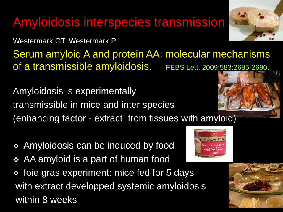
Amyloidosis interspecies transmission
Westermark GT, Westermark P.
Serum amyloid A and protein AA: molecular mechanisms
of a transmissible amyloidosis. FEBS Lett. 2009;583:2685-2690.
Amyloidosis is experimentally
transmissible in mice and inter species
(enhancing factor - extract from tissues with amyloid)
Amyloidosis can be induced by food
AA amyloid is a part of human food
foie gras experiment: mice fed for 5 days
with extract developped systemic amyloidosis
within 8 weeks
Fibrinoid & Hyalin
disorders of protein metabolism
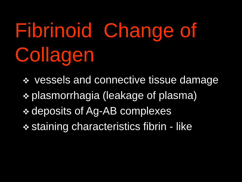
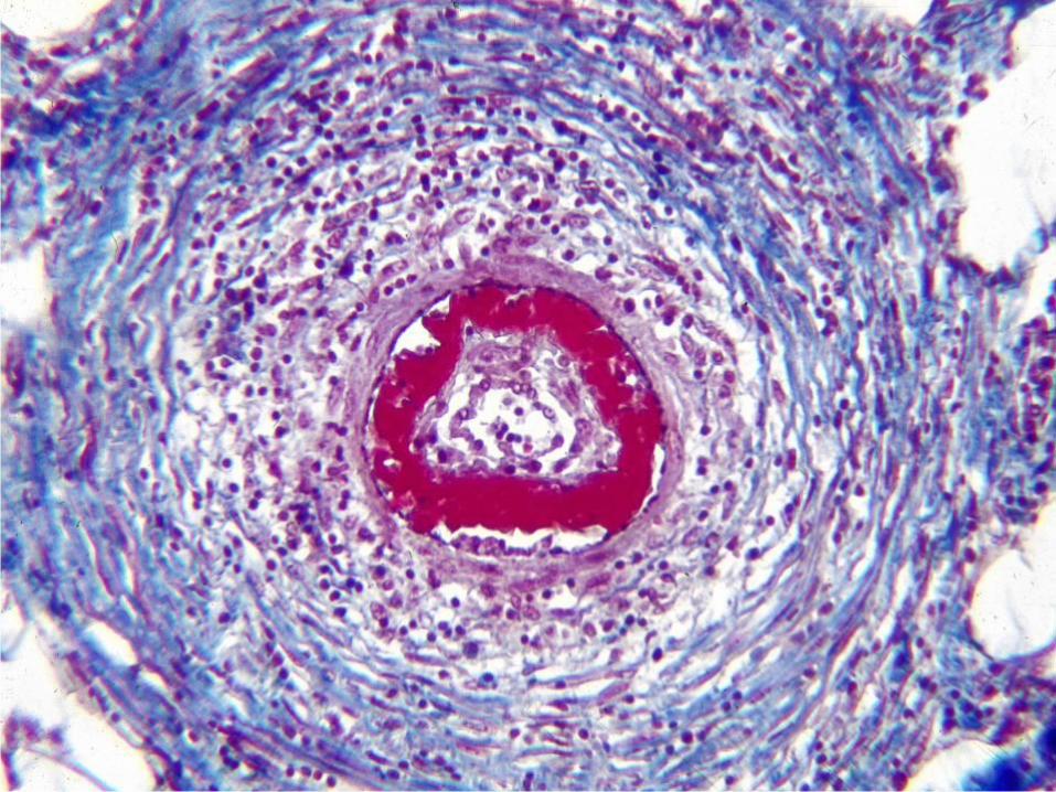
Fibrinoid Change of
Collagen vessels and connective tissue damage
plasmorrhagia (leakage of plasma)
deposits of Ag-AB complexes
staining characteristics fibrin - like
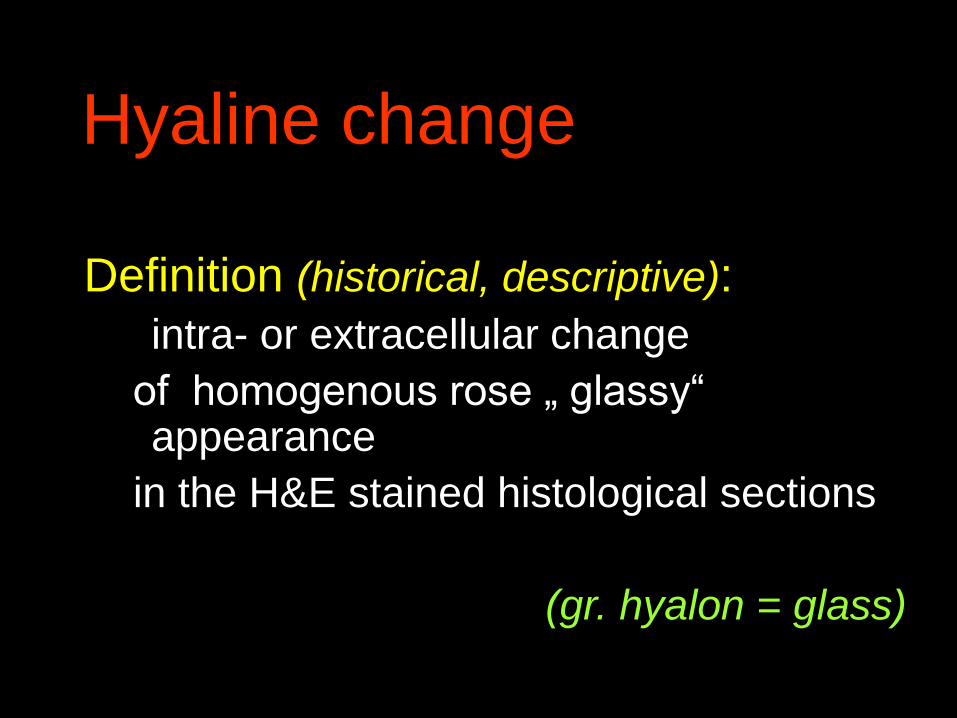
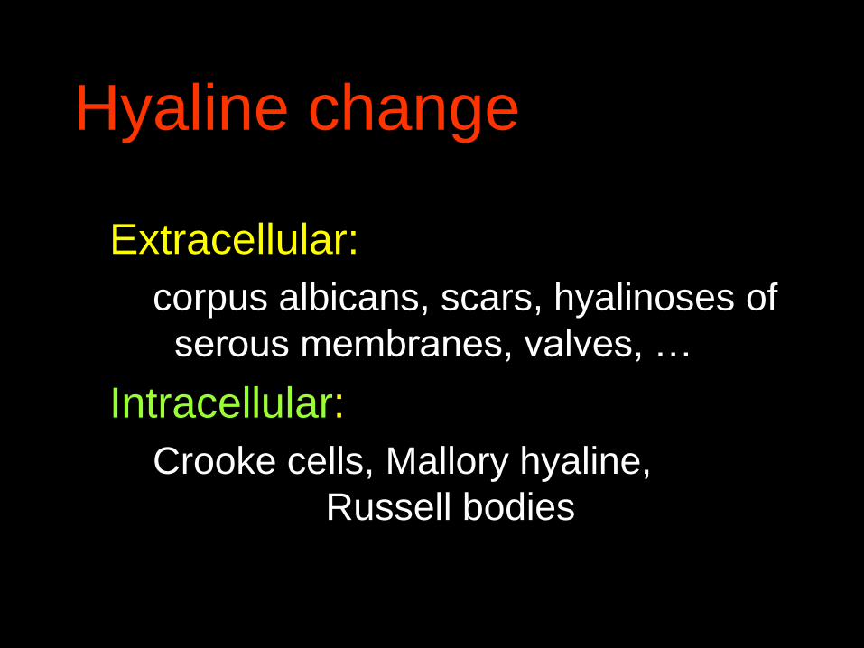
Hyaline change
Definition (historical, descriptive):
intra- or extracellular change
of homogenous rose „ glassy“ appearance
in the H&E stained histological sections
(gr. hyalon = glass)

Hyaline change
Extracellular:
corpus albicans, scars, hyalinoses of
serous membranes
Intracellular:
Crooke cells, Mallory hyaline,
Russell bodies
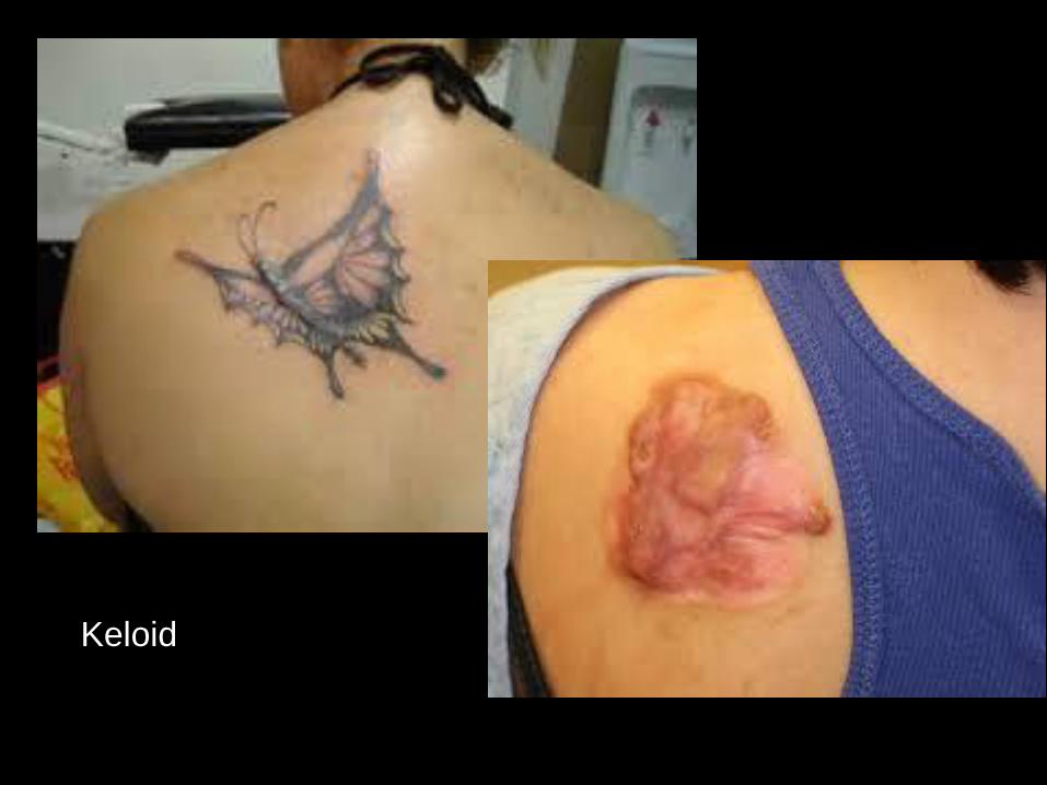
Keloid
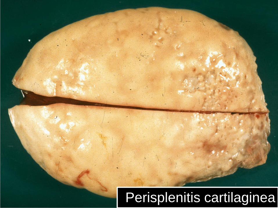
Perisplenitis cartilaginea
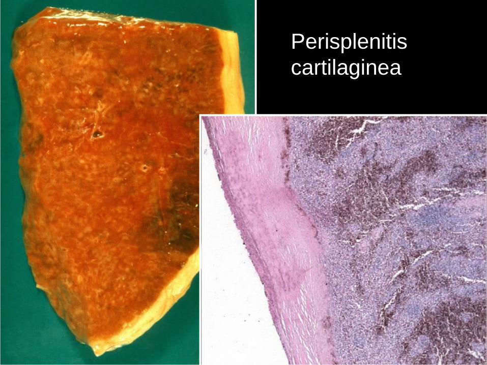
Perisplenitis
cartilaginea
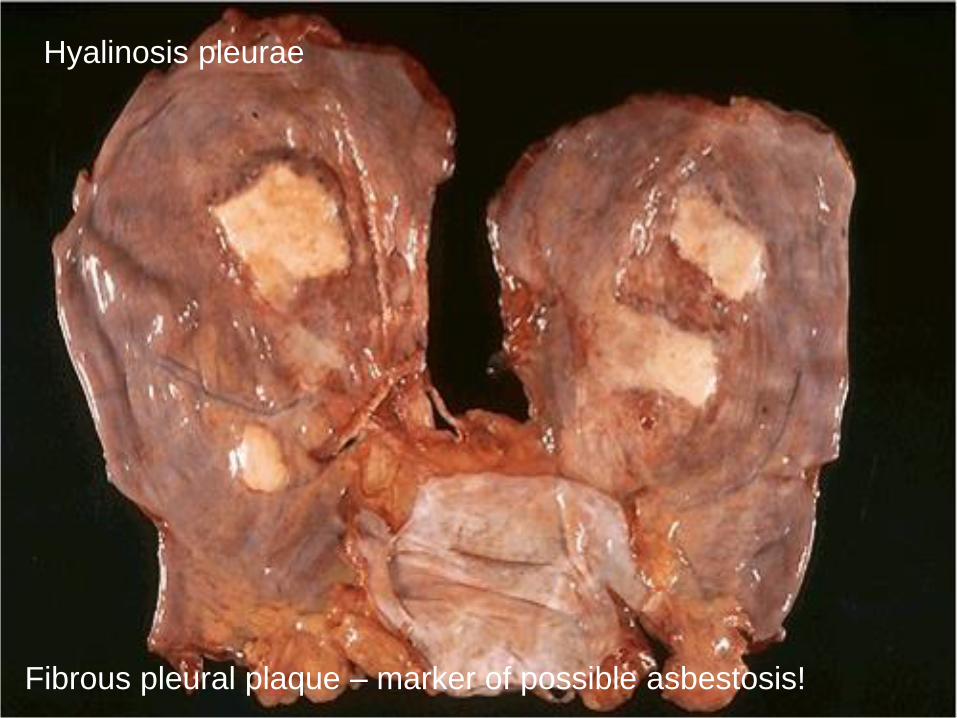
Fibrous pleural plaque – marker of possible asbestosis!
Hyalinosis pleurae
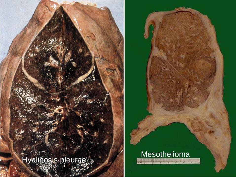
Hyalinosis pleuraeMesothelioma

Ca bronchogenesHyalinosis et metatases
carcinomatosae pleurae
parietalis
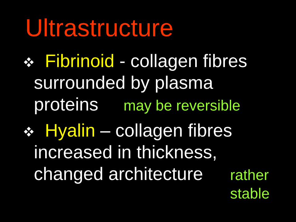
Ultrastructure
Fibrinoid - collagen fibres
surrounded by plasma
proteins may be reversible
Hyalin – collagen fibres
increased in thickness,
changed architecture rather
stable
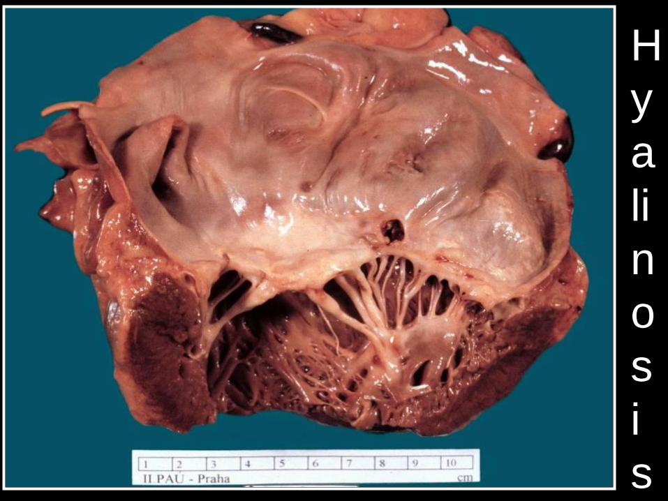
H
y
a
li
n
o
s
i
s
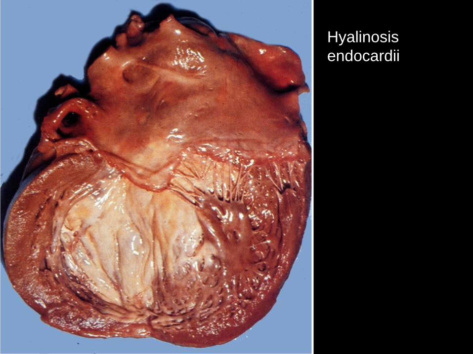
Hyalinosis
endocardii

Fibroelastosis
endocardii
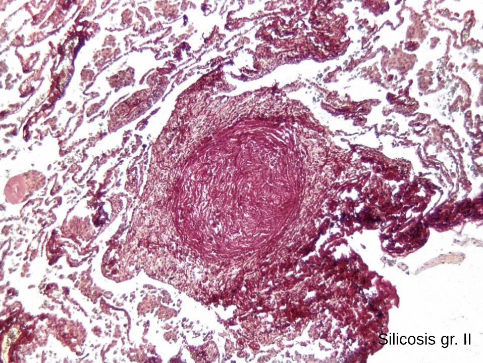
Silicosis gr. II
Hyaline change
Extracellular:
corpus albicans, scars, hyalinoses of
serous membranes, valves, …
Intracellular:
Crooke cells, Mallory hyaline,
Russell bodies
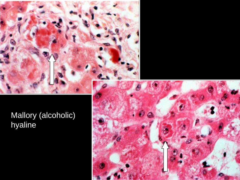
Mallory (alcoholic)
hyaline

Mallory (alcoholic) hyaline

Russel bodies
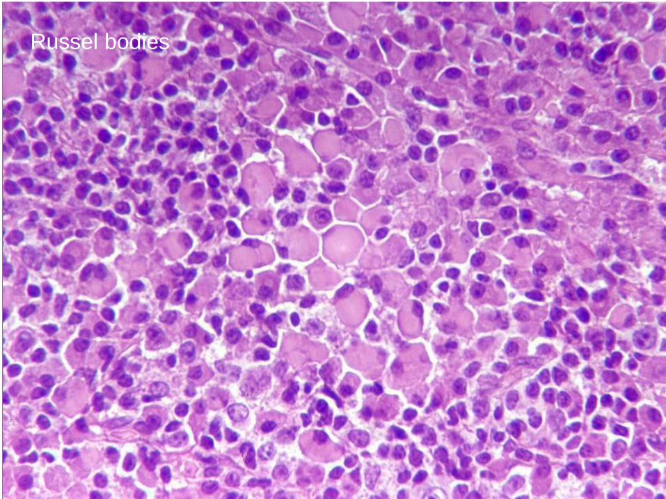
Russel bodies
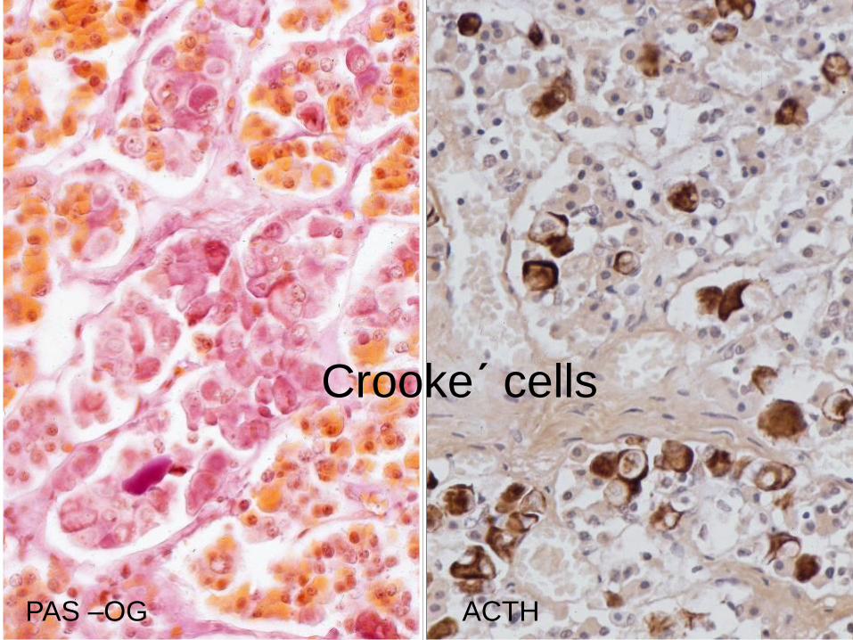
Crooke´ cells
PAS –OG ACTH


Significance of Fibrinoid
Change
diminished quality of the collagen ( firmness, permeability)
tendency to thrombosis in the
vessels, aneurysms formation
Significance of Hyalin
Change
diminished quality of the
collagen ( elasticity)
ischemia in organs with
thickened arterial walls
intracellular - function, death