Embed Size (px)
Citation preview
Proc. Natl. Acad. Sci. USAVol. 76, No. 11, pp. 5645-5649, November 1979Biophysics
Lateral mobility of an amphipathic apolipoprotein, ApoC-III, boundto phosphatidylcholine bilayers with and without cholesterol
(membrane lateral diffusion/fluorescence recovery after photobleaching)
WINCHIL L. C. VAZ*t, KENNETH JACOBSON**, EN-SHINN WU§, AND ZENON DERZKOT*Department of Experimental Pathology, Roswell Park Memorial Institute, Buffalo, New York 14263; §Department of Physics, University of Maryland,Baltimore, Maryland 21228; and IDepartment of Biophysical Sciences, State University of New York, Buffalo, New York 14226
Communicated by David Harker, July 9, 1979
ABSTRACT The technique of fluorescence recovery afterphotobleaching was used to investigate the lateral mobility ofa fluorescein-labeled amphipathic apolipoprotein, ApoC-II,bound to multibilayers prepared from dipalmitoyl phosphati-dylcholine, egg phosphatidylcholine, and a 1:1 (molar ratio)mixture of egg phosphatidylcholine and cholesterol. In dipal-mitoyl phosphatidylcholine bilayers the lateral diffusion coef-ficient (D) for the protein is about 2 X 10⁻⁹ cm2 sec1 at 200Cand about 9 X 10-s cm2 sec1 at 450C. Plots of D versus tem-perature in this system show a transition between about 30 and350C. Arrhenius activation energies for the diffusion in this casebetween 15 and 30'C and between 35 and 450C are 28.5 and 7.0kcal mol[1, respectively (1 calorie = 4.18 joules). In egg phos-phatidylcholine bilayers, D is about 3 X 10⁻⁸ cm2 sec1 at 20'Cand the Arrhenius activation energy for diffusion is 8.1 kcalmolh between 15 and 350C in this system. In bilayers preparedfrom an equimolar mixture of egg phosphatidylcholine andcholesterol D at 20'C is about 1.4 X 10- cm2 sec-1 and theArrhenius activation energy for the diffusion of the protein inthis system between 15 and 350C is 15.1 kcal mol'. Light-scattering and fluorescence-polarization results indicate thatbinding of this protein does not affect the gel-to-liquid crystal-line phase transition of bilayer membranes but does mediatea major, reversible aggregation of the vesicles at about 330C.These results lend support to the view that ApoC-III resides inthe head-group region of the bilayer and suggest that its lateraldiffusion coe ficient represents an upper bound for integralmembrane proteins.
The mobility of membrane components has drawn considerableattention in the recent literature (1, 2). Several reports on thelateral (3-7) and rotational diffusion (8, 9) of components in theplane of the membrane in model membrane systems have ap-peared. In model membranes, as opposed to biomembranes,it is possible in principle to examine simple membrane com-positions-for example, one protein and one lipid mem-brane-and subsequently to increase the complexity of thesystem at will. In this manner, it is possible to learn about theinfluence of a given membrane component on the diffusion ofother components in the membrane and also, by comparisonto natural membrane diffusion results, to learn about the rolethat nonmembrane components in the cell play in the regula-tion of membrane component mobility.
In the present communication we report the use of thetechnique of fluorescence recovery after photobleaching(FRAP) to study the lateral diffusion of an amphipathic apo-lipoprotein, ApoC-III, in bilayer membranes formed from di-palmitoyl phosphatidylcholine (Pam2PtdCho), egg-yolkphosphatidyl-choline (E-PtdCho), and an equimolar mixtureof E-PtdCho and cholesterol (Chol). The results have beencommunicated in a preliminary form (10). FRAP has been usedso far to study a wide range of natural membrane (11-21) and
model membrane systems (3-7). It offers a convenient methodfor studying lateral diffusion and is particularly attractive fromthe points of view of experimental and theoretical simplicity(19, 22).
ApoC-III is an amphipathic peptide having a molecularweight of about 9000 and a known amino acid sequence (23,24). Its interaction with lipid membranes has been extensivelystudied (24). It interacts with E-PtdCho (25) and Pam2PtdCho(26) bilayer vesicles without disrupting their structure, but itdisrupts the vesicular structure of dimyristoyl PtdCho unila-mellar vesicles (26). The association of ApoC-III with PtdChobilayer membranes is characterized by an association constantof about 2 X 10⁶ M (ref. 26; unpublished data) and a lipid/protein stoichiometry of about 50:1 (25). Its interaction withPtdCho bilayers involves a combination of electrostatic andhydrophobic interactions (27).
MATERIALS AND METHODSApoC-III was isolated according to the procedure of Brown etal. (28) and was labeled with fluorescein by reaction of theprotein (1 mg/ml) in 0.1 M sodium borate buffer at pH 9.0 witha 100-fold molar excess of fluorescein isothiocyanate (BaltimoreBiological Laboratory Division, Becton-Dickinson) for 1 hr at250C. After reaction, the labeled protein (dye/protein ratio =2) was separated from unreacted dye by filtration through acolumn of Sephadex G-25 using Dulbecco's Ca2+- andMg2+-free phosphate-buffered saline.The phospholipids used in this study were isolated or syn-
thesized by Tom Isac in the laboratory of D. Papahadjopoulos.Pam2PtdCho was synthesized as described by Robles and VanDen Berg (29) and E-PtdCho was isolated from egg yolks asdescribed by Papahadjopoulos and Miller (30). The phospho-lipids contained no detectable impurities as determined bythin-layer chromatography on silica gel H with chloroform/methanol/7 M ammonia, 230:90:15 (vol/vol), as solvent.
Slides for FRAP experiments were prepared by evenlyspreading a film of 2.5 mg of the lipid (in chloroform solution)over a 1-cm2 area of a cleaned glass slide. The solvent wasevaporated under reduced pressure. The dried lipid films werehydrated by dropping a coverslip with a hanging drop of 10 ,lof the fluorescein-labeled protein solution (50,gg/ml) over thefilm. This procedure was done at 250C for E-PtdCho, at 370Cfor E-PtdCho/Chol, and at 450C for Pam2PtdCho. After 15min for the lipid film to become hydrated, the slides were
Abbreviations: FRAP, fluorescence recovery after photobleaching;Pam2PtdCho, dipalmitoyl phosphatidylcholine; E-PtdCho, egg-yolkphosphatidylcholine; Chol, cholesterol; D, diffusion coefficient; 1/2,recovery halftime.t Present address: Max Planck-Institut fuer biophysikalische Chemie,Abteilung Spektroskopie, D-3400 Goettingen-Nikolausberg,G.F.R.
t To whom reprint requests should be addressed.
5645
The publication costs of this article were defrayed in part by pagecharge payment. This article must therefore be hereby marked "ad-vertisement" in accordance with 18 U. S. C. §1734 solely to indicatethis fact.
Proc. Natl. Acad. Sci. USA 76 (1979)
pressed with a heavy weight, with care taken to avoid lateralmotion of the coverslip over the hydrated lipid film as reported(3). The slides were then sealed with paraffin wax and incu-bated at 370C for 48 hr (these long incubations require use ofa pure paraffin to avoid contamination of the sample by fluo-rescent impurities). After the incubation, the slides were storedat room temperature. Slides older than 6 days after preparationwere not used for the FRAP experiments.The preparation of slides from Pam2PtdCho presents certain
problems leading to some degree of irreproducibility. Large,well-formed, multibilayer domains are only formed on theslides when all elements (reagents and equipment) in thepreparation, including the hydrating protein solution, are at450C (i.e., above the Pam2PtdCho phase-transition temperaturein its hydrated state). However, this high temperature is dele-terious to the protein stability. It was observed that often, andparticularly if the protein solution had been stored for longerthan about 15 min at 450C, protein binding to the lipid was
incomplete as identified by large amounts of the added fluo-rescent protein located in aqueous pockets within the specimen.When this was the case, it was found helpful to carefully washthe lipid bilayers on the slide with protein-free buffer bydrawing it under the coverslip with a filter-paper wick. Prep-aration of E-PtdCho and E-PtdCho/Chol systems did notpresent these difficulties. Finally, it was noted that fluores-cein-labeled ApoC-III changed character over prolongedstorage at 4VC as evidenced by less protein incorporation intothe multibilayer and slower diffusion rates.FRAP experiments were done as described (3), with a beam
diameter of t8 ,um. Photobleaching intensities were t10 mWfor E-PtdCho and Pam2PtdCho systems and -50 mW for E-PtdCho/Chol systems. Measuring beam intensities were at-tenuated by a factor of 105. During the measurement of theinitial or prebleach fluorescence (Ft) in the FRAP experiments(19), rapid fading of about 10% of the fluorescence often oc-
curred and then a stable level was achieved. The reason for thisbehavior is not understood. However, the maximal value of theprebleach fluorescence was selected as FI because fluorescencerecoveries back to this level were routinely obtained. Diffusioncoefficients were calculated as described by Axelrod et al. (22).Experimental fluorescence recovery curves showed good fitsto theoretical diffusion-limited recovery curves for one diffusingcomponent as described by equation 12 of Axelrod et al.(22).
Cholate dialysis was done as described by Kagawa andRacker (31) at 4VC for 1 week. The resulting liposomes with or
without protein are essentially cholate-free after this process(lipid/cholate, greater than 150:1). Unbound protein was sep-arated from protein-containing liposomes by centrifugation andwashing of the pellet. Cholate-dialyzed liposomes with or
without protein were labeled with 16-anthroyl palmitate, andfluorescence polarization measurements were done as describedby Cadenhead et al. (32).
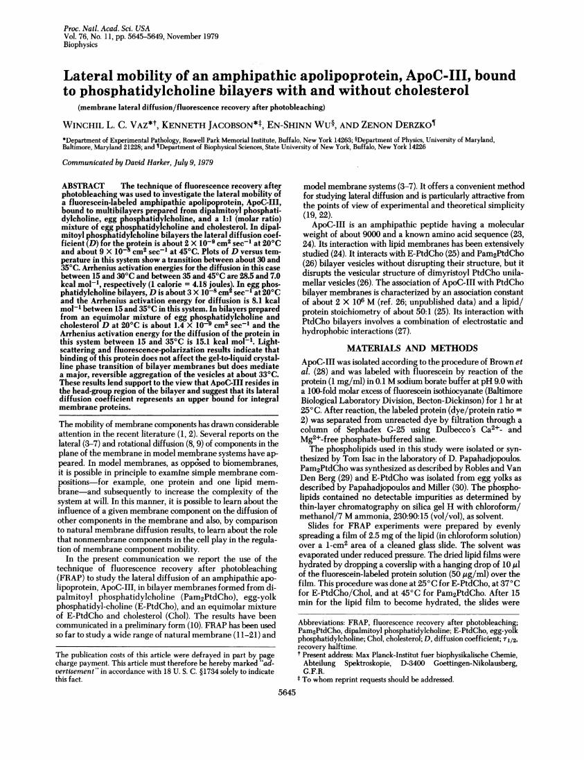
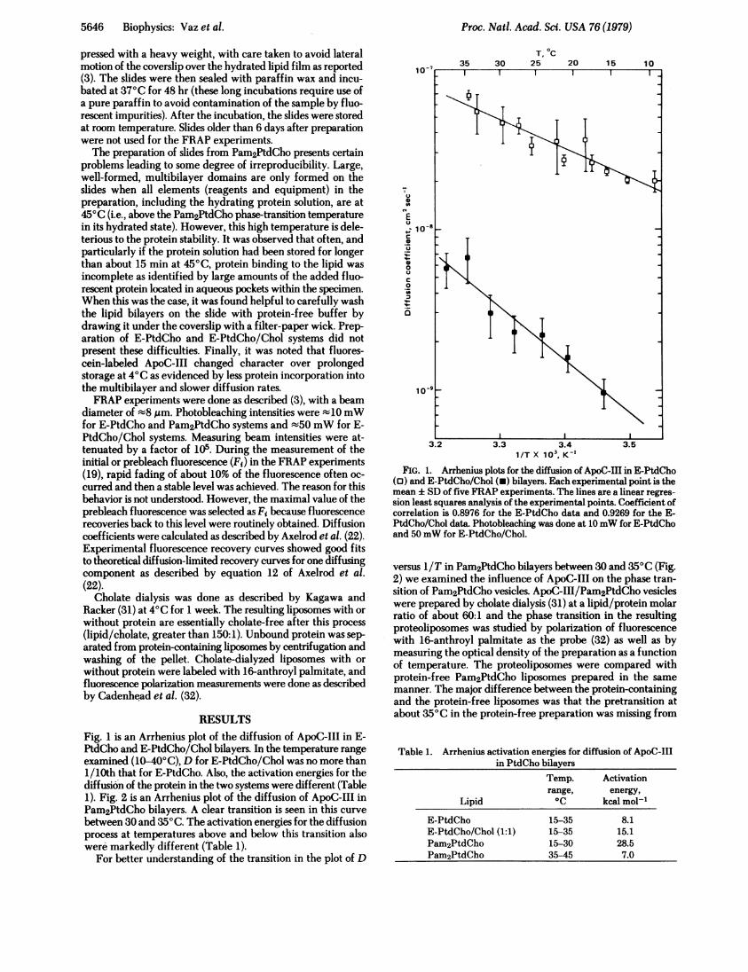
RESULTSFig. 1 is an Arrhenius plot of the diffusion of ApoC-III in E-PtdCho and E-PtdCho/Chol bilayers. In the temperature rangeexamined (10-40'C), D for E-PtdCho/Chol was no more than1/10th that for E-PtdCho. Also, the activation energies for thediffusion of the protein in the two systems were different (Table1). Fig. 2 is an Arrhenius plot of the diffusion of ApoC-III inPam2PtdCho bilayers. A clear transition is seen in this curve
between 30 and 350C. The activation energies for the diffusionprocess at temperatures above and below this transition alsowere markedly different (Table 1).
For better understanding of the transition in the plot of D
0
"E0
C
e
00
0
Cr
T, 0C25
3.2 3.3 3.4 3.51/T X 103, K-'
FIG. 1. Arrhenius plots for the diffusion of ApoC-IHl in E-PtdCho(0) and E-PtdCho/Chol (-) bilayers. Each experimental point is themean : SD of five FRAP experiments. The lines are a linear regres-
sion least squares analysis of the experimental points. Coefficient ofcorrelation is 0.8976 for the E-PtdCho data and 0.9269 for the E-PtdCho/Chol data. Photobleaching was done at 10mW for E-PtdChoand 50mW for E-PtdCho/Chol.
versus lIT in Pam2PtdCho bilayers between 30 and 350C (Fig.2) we examined the influence of ApoC-III on the phase tran-sition of Pam2PtdCho vesicles. ApoC-III/Pam2PtdCho vesicleswere prepared by cholate dialysis (31) at a lipid/protein molarratio of about 60:1 and the phase transition in the resultingproteoliposomes was studied by polarization of fluorescencewith 16-anthroyl palmitate as the probe (32) as well as bymeasuring the optical density of the preparation as a functionof temperature. The proteoliposomes were compared withprotein-free Pam2PtdCho liposomes prepared in the same
manner. The major difference between the protein-containingand the protein-free liposomes was that the pretransition atabout 350C in the protein-free preparation was missing from
Table 1. Arrhenius activation energies for diffusion of ApoC-IIIin PtdCho bilayers
Temp. Activationrange, energy,
Lipid 0C kcal mol1
E-PtdCho 15-35 8.1E-PtdCho/Chol (1:1) 15-35 15.1Pam2PtdCho 15-30 28.5Pam2PtdCho 35-45 7.0
5646 Biophysics: Vaz et al.
Proc. Natl. Acad. Sci. USA 76 (1979) 5647
T, C
n
E0C 1
0
._
._-
0._
1/T X 103, K_'
FIG. 2. Arrhenius plot for the diffusion of ApoC-III inPam2PtdCho bilayers. Each experimental point is the mean ± SD offive FRAP experiments. The points between 35 and 45°C and between15 and 30°C were fitted by a linear regression least squares analysis.Coefficient of correlation is 0.9784 between 35 and 45°C and 0.9908between 15 and 30°C. Photobleaching intensity was 10 mW.
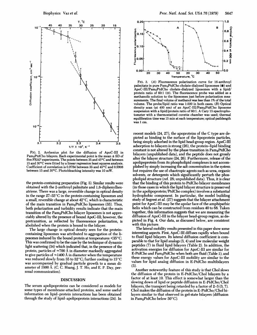
the protein-containing preparation (Fig. 3). Similar results wereobtained with the 2-anthroyl palmitate and 1,6-diphenylhex-atriene. There was a large, reversible change in optical densityin the range 27-35°C in the protein-containing liposomes anda small, reversible change at about 420C, which is characteristicof the main transition in Pam2PtdCho liposomes (33). Thus,both polarization and turbidity results indicate that the maintransition of the Pam2PtdCho bilayer liposomes is not appre-ciably altered by the presence of bound ApoC-III; however, thepretransition, as reflected by fluorescence polarization, isabolished when the protein is bound to the bilayer.The large change in optical density seen for the protein-
containing liposomes was attributed to aggregation of the li-posomes induced by the bound protein at temperatures <350C.This was confirmed to be the case by the technique of dynamiclight scattering (34) which indicated that, in the presence of theprotein, particles of -700 A in diameter markedly aggregatedto give particles of t1400 A in diameter when the temperaturewas reduced slowly from 35 to 320C; further cooling to 150Cwas accompanied by gradual particle growth to a mean di-ameter of 1900 A. (C. C. Hsang, J. T. Ho, and E. P. Day, per-sonal communication).
DISCUSSIONThe serum apolipoproteins can be considered as models forsome types of membrane-attached proteins, and some usefulinformation on lipid-protein interactions has been obtainedthrough the study of lipid-apolipoprotein interactions (24). In
20 30 40 50 20 30Temperature, "C
40 50
FIG. 3. (A) Fluorescence polarization curve for 16-anthroylpalmitate in pure Pam2PtdCho cholate-dialyzed liposomes (-) andApoC-III/Pam2PtdCho cholate-dialyzed liposomes with a lipid/protein ratio of 60:1 (o). The fluorescence probe was added as amethanolic solution to the liposomes just before polarization mea-surements. The final volume of methanol was less than 1% of the totalvolume. The probe/lipid ratio was 1:500 in both cases. (B) Opticaldensity scan (at 400 nm) of an ApoC-III/Pam2PtdCho liposomesuspension with a lipid/protein ratio of 60:1. A Cary 15 spectropho-tometer with a thermostatted cuvette chamber was used; thermalequilibration time was 15 min at each temperature; optical pathlengthwas 1 cm.
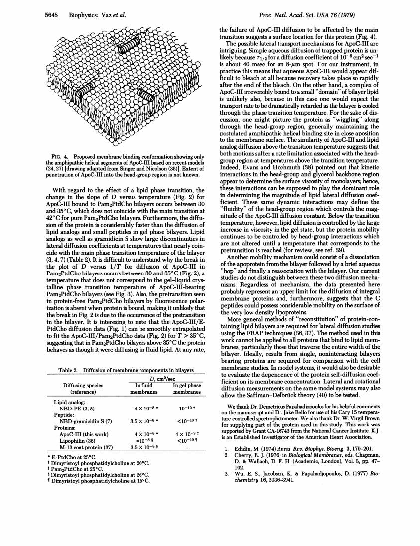
recent models (24, 27), the apoproteins of the C type are de-picted as binding to the surface of the lipoprotein particles,being simply adsorbed in the lipid head-group region. ApoC-IIIadsorption to bilayers is strong (26), the protein-lipid bindingconstant is not altered by the phase transition in Pam2PtdChobilayers (unpublished data), and the peptide does not grosslyalter the bilayer structure (24, 26). Furthermore, release of theapolipoprotein from its phospholipid complexes is not accom-plished by simply increasing the salt concentration in the systembut requires the use of chaotropic agents such as urea, organicsolvents, or detergents which significantly perturb the phos-pholipid structure (ref. 28; unpublished data). Thus, it is clearthat the binding of this protein to PtdCho bilayer membranes(in those cases in which the lipid bilayer structure is preservedin the apolipoprotein/PtdCho complex) involves a substantialhydrophobic component. In particular, the model buildingstudy of Segrest et al. (27) suggests that the bilayer attachmentpoint for ApoC-III may be the apolar face of the amphipathichelix which can be constructed from residues 40 to 68. Takentogether, this information suggests that we are measuring thediffusion of ApoC-III in the bilayer head-group region, as de-picted in Fig. 4. Our data, as discussed below, are consistentwith this picture.The lateral mobility results presented in this paper show some
interesting aspects. First, ApoC-III diffuses rapidly when boundto fluid lipid bilayers. Its lateral diffusion coefficient is com-parable to that for lipid analogs (3, 4) and low molecular weightpeptides (7) in fluid lipid bilayers (Table 2). In addition, theactivation energies for diffusion for ApoC-III are similar forE-PtdCho and Pam2PtdCho when both are fluid (Table 1); andthese energy values for ApoC-III mobility are similar to thevalues for lipid analog diffusion in E-PtdCho multibilayers(3).
Another noteworthy feature of this study is that Chol slowsthe diffusion of the protein in E-PtdCho/Chol bilayers by afactor of at least 10. This effect is somewhat larger than theslowing down of lipid or peptide diffusion in E-PtdCho/Cholbilayers, the transport being retarded by a factor of 2-3 (3, 7).Chol makes the diffusion of the protein in E-PtdCho/Chol bi-layers similar to that observed in gel-state bilayers (diffusionin Pam2PtdCho below 30°C).
Biophysics: Vaz et al.
Proc. Nati. Acad. Sci. USA 76 (1979)
FIG. 4. Proposed membrane binding conformation showing onlythe amphipathic helical segments of ApoC-III based on recent models(24, 27) [drawing adapted from Singer and Nicolson (35)]. Extent ofpenetration of ApoC-III into the head-group region is not known.
With regard to the effect of a lipid phase transition, thechange in the slope of D versus temperature (Fig. 2) forApoC-III bound to Pam2PtdCho bilayers occurs between 30and 350C, which does not coincide with the main transition at420C for pure Pam2PtdCho bilayers. Furthermore, the diffu-sion of the protein is considerably faster than the diffusion oflipid analogs and small peptides in gel phase bilayers. Lipidanalogs as well as gramidicin S show large discontinuities inlateral diffusion coefficients at temperatures that nearly coin-cide with the main phase transition temperature of the bilayer(3, 4, 7) (Table 2). It is difficult to understand why the break inthe plot of D versus 11T for diffusion of ApoC-III inPam2PtdCho bilayers occurs between 30 and 350C (Fig. 2), a
temperature that does not correspond to the gel-liquid crys-talline phase transition temperature of ApoC-III-bearingPam2PtdCho bilayers (see Fig. 3). Also, the pretransition seenin protein-free Pam2PtdCho bilayers by fluorescence polar-ization is absent when protein is bound, making it unlikely thatthe break in Fig. 2 is due to the occurrence of the pretransitionin the bilayer. It is interesing to note that the ApoC-III/E-PtdCho diffusion data (Fig. 1) can be smoothly extrapolatedto fit the ApoC-III/Pam2PtdCho data (Fig. 2) for T > 350C,suggesting that in Pam2PtdCho bilayers above 350C the proteinbehaves as though it were diffusing in fluid lipid. At any rate,
Table 2. Diffusion of membrane components in bilayersD, cm2/sec
Diffusing species In fluid In gel phase(reference) membranes membranes
Lipid analog:NBD-PE (3, 5) 4 X 10-8 * 10-10 t
Peptide:NBD-gramicidin S (7) 3.5 X 10⁻⁸ * <1010 t
Proteins:ApoC-III (this work) 4 X 10-8 * 4 X 10-9Lipophilin (36) t10-8 § <1o-loM-13 coat protein (37) 3.5 X 10⁻⁸ §
* E-PtdCho at 250C.t Dimyristoyl phosphatidylcholine at 200C.Pam2PtdCho at 250C.
§ Dimyristoyl phosphatidylcholine at 260C.Dimyristoyl phosphatidylcholine at 150C.
the failure of ApoC-III diffusion to be affected by the maintransition suggests a surface location for this protein (Fig. 4).The possible lateral transport mechanisms for ApoC-III are
intriguing. Simple aqueous diffusion of trapped protein is un-likely because T1/2 for a diffusion coefficient of 10-6 cm2 sec-1is about 40 msec for an 8-,gm spot. For our instrument, inpractice this means that aqueous ApoG-III would appear dif-ficult to bleach at all because recovery takes place so rapidlyafter the end of the bleach. On the other hand, a complex ofApoC-III irreversibly bound to a small "domain" of bilayer lipidis unlikely also, because in this case one would expect thetransport rate to be dramatically retarded as the bilayer is cooledthrough the phase transition temperature. For the sake of dis-cussion, one might picture the protein as "wiggling" alongthrough the head-group region, generally maintaining thepostulated amphipathic helical binding site in close apositionto the membrane surface. The similarity of ApoC-III and lipidanalog diffusion above the transition temperature suggests thatboth motions suffer a rate limitation associated with the head-group region at temperatures above the transition temperature.Indeed, Evans and Hochmuth (38) pointed out that kineticinteractions in the head-group and glycerol backbone regionappear to determine the surface viscosity of monolayers; hence,these interactions can be supposed to play the dominant rolein determining the magnitude of lipid lateral diffusion coef-ficient. These same dynamic interactions may define the"fluidity" of the head-group region which controls the mag-nitude of the ApoC-III diffusion constant. Below the transitiontemperature, however, lipid diffusion is controlled by the largeincrease in viscosity in the gel state, but the protein mobilitycontinues to be controlled by head-group interactions whichare not altered until a temperature that corresponds to thepretransition is reached (for review, see ref. 39).
Another mobility mechanism could consist of a dissociationof the apoprotein from the bilayer followed by a brief aqueous"hop" and finally a reassociation with the bilayer. Our currentstudies do not distinguish between these two diffusion mecha-nisms. Regardless of mechanism, the data presented hereprobably represent an upper limit for the diffusion of integralmembrane proteins and, furthermore, suggests that the Cpeptides could possess considerable mobility on the surface ofthe very low density lipoproteins.More general methods of "reconstitution" of protein-con-
taining lipid bilayers are required for lateral diffusion studiesusing the FRAP techniques (36, 37). The method used in thiswork cannot be applied to all proteins that bind to lipid mem-branes, particularly those that traverse the entire width of thebilayer. Ideally, results from single, noninteracting bilayersbearing proteins are required for comparison with the cellmembrane studies. In model systems, it would also be desirableto evaluate the dependence of the protein self-diffusion coef-ficient on its membrane concentration. Lateral and rotationaldiffusion measurements on the same model systems may alsoallow the Saffman-Delbruck theory (40) to be tested.
We thank Dr. Demetrious Papahadjopoulos for his helpful commentson the manuscript and Dr. Jake Bello for use of his Cary 15 tempera-ture-controlled spectrophotometer. We also thank Dr. W. Virgil Brownfor supplying part of the protein used in this study. This work wassupported by Grant CA-16743 from the National Cancer Institute. K.J.is an Established Investigator of the American Heart Association.
1. Edidin, M. (1974) Annu. Rev. Blophys. Bioeng. 3,179-201.2. Cherry, R. J. (1976) in Biological Membranes, eds. Chapman,
D. & Wallach, D. F. H. (Academic, London), Vol. 3, pp. 47-102.
3. Wu, E. S., Jacobson, K. & Papahadjopoulos, D. (1977) Bio-chemistry 16, 3936-3941.
5648 Biophysics: Vaz et al.
Proc. Natl. Acad. Sci. USA 76 (1979) 5649
4. Fahey, P. F. & Webb, W. W. (1978) Biochemistry 17, 3046-3053.
5. Smith, B. A. & McConnell, H. M. (1978) Proc. Natl Acddd. §.USA 75, 2759-2763.
6. Wolf, D. E., Schlessinger, J., Elson, E. L., Webb, W. W., Blu-menthal, R. & Henkart, P. (1977) Biochemistry 16, 3476-3483.
7. Wu, E. S., Jacobson, K., Szoka, F. & Portis, A. (1978) Biochemistry17,5543-5550.
8. Cherry, R. J., Mueller, U., Henderson, R. & Heyn, M. P. (1978)J. Mol. Biol. 121, 283-298.
9. Vaz, W. L. C., Austin, R. H. & Vogel, H. (1979) Biophys. J. 26,415-426.
10. Vaz, W. L. C., Wu, E. S. & Jacobson, K. (1979) Biophys. J. 25,178a.
11. Peters, R., Peters, J., Tews, K. H. & Bahr, W. (1974) Biochim.Biophys. Acta 367,282-294.
12. Edidin, M., Zagyansky, Y. & Lardner, T. J. (1976) Science 191,466-468.
13. Jacobson, K., Wu, E. S. & Poste, G. (1976) Biochim. Biophys. Acta433,215-222.
14. Zagyansky, Y. & Edidin, M. (1976) Biochim. Biophys. Acta 433,209-214.
15. Schlessinger, J., Koppel, D. E., Axelrod, D., Jacobson, K., Webb,W. W. & Elson, E. L. (1976) Proc. NatI. Acad. Sci. USA 73,2409-2413.
16. Schlessinger, J., Webb, W. W., Elson, E. L. & Metzger, H. (1976)Nature (London) 264,550-552.
17. Axelrod, D., Ravdin, P., Koppel, D. E., Schlessinger, J., Webb,W. W., Elson, E. L. & Podleski, T. R. (1976) Proc. NatI. Acad.Sci. USA 73,4594-4598.
18. Schlessinger, J., Axelrod, D., Koppel, D. E., Webb, W. W. &Elson, E. L. (1977) Science 195,307-309.
19. Jacobson, K., Derzko, Z., Wu, E. S., Hou, Y. & Poste, G. (1977)J. Supramol. Struct. 5,565-576.
20. Axelrod, D., Ravdin, P. M. & Podleski, T. R. (1978) Biochim.Biophys. Acta 511, 23-38.
21. Axelrod, D., Wight, A., Webb, W. W. & Horwitz, A. (1978)Biochemistry 17, 3604-3609.
22. Axelrod, D., Koppel, D. E., Schlessinger, J., Elson, E. L. & Webb,W. W. (1976) Biophys. J. 16, 1055-1069.
23. Brewer, H. B., Schulman, R., Herbert, P., Ronan, R. & Wehrly,K. (1972) Adv. Exp. Med. Biol. 26,280.
24. Morrisett, J. D., Jackson, R. L. & Gotto, A. M. (1977) Biochim.Biophys. Acta 472,93-133.
25. Morrisett, J. D., David, J. S. K., Pownall, H. J. & Gotto, A. M.(1973) Biochemistry 12, 1290-1299.
26. Trauble, H., Meddelhoff, G. & Brown, W. V. (1974) FEBS Lett.49,269-275.
27. Segrest, J. P., Jackson, R. L., Morrisett, J. D. & Gotto, A. M. (1974)FEBS Lett. 38, 247-253.
28. Brown, W. V., Levy, R. I. & Fredrickson, D. S. (1969) J. Biol.Chem. 244, 5687-5694.
29. Robles, C. & Van Den Berg, D. (1969) Biochim. Biophys. Acta187,520-526.
30. Papahadjopoulos, D. & Miller, N. (1967) Biochim. Biophys. Acta135,624-638.
31. Kagawa, Y. & Racker, E. (1971) J. Biol. Chem. 246, 5477-5487.
32. Cadenhead, D. A., Kellner, B. M. J., Jacobson, K. & Papahadjo-poulos, D. (1977) Biochemistry 16,5386-5392.
33. Yi, P. & MacDonald, R. (1973) Chem. Phys. Lipids 11, 114-134.
34. Day, E. P., Ho, J. T., Kunz, R. K. & Sun, S. T. (1977) Biochlm.Biophys. Acta 470,503-508.
35. Singer, S. J. & Nicolson, G. (1972) Science 175,920-931.36. Derzko, Z. & Jacobson, K. (1978) Biophys. J. 21, 204a.37. Smith, L. M., Smith, B. A. & McConnell, H. M. (1979) Bio-
chemistry 18, 2256-2259.38. Evans, E. & Hochmuth, R. (1978) Curr. Top. Membr. Transp.
10, 1-64.39. Seelig, J. (1978) Biochim. Biophys. Acta 515, 105-140.40. Saffman, P. G. & Delbruck, M. (1975) Proc. Natl. Acad. Sci. USA
72,3111-3113.
Biophysics: Vaz et al.
![16th APOC Goa, India - asiapacificoptometry.orgasiapacificoptometry.org/doc/16th-APOC-Goa-India.pdf16th APOC Goa, India - Come to India Education - Conference Programme [new] - Invited](https://img.pdfslide.net/doc/110x75/5b2738237f8b9a42708b4b3f/16th-apoc-goa-india-apoc-goa-india-come-to-india-education-conference-programme.jpg)