Embed Size (px)
Citation preview

Int.J.Curr.Microbiol.App.Sci (2014) 3(11) 501-520
501
Original Research Article
Mode of action of mosquitocidal protein in the larvae and pupae of Cx.quinquefasciatus and biochemical and physiological changes in the
mosquitoes exposed to protein
Usharani Brammacharry1*and Kummankottil Paily2
1Department of Genetics, Dr.A.L.M Post Graduate Institute for Basic Medical Sciences, University of Madras, Taramani, Chennai, Tamil Nadu, India
2Department of Microbiology, Vector Control Research Centre, Indira nagar, Puducherry, India *Corresponding author
A B S T R A C T
Introduction
The crystal proteins of Bacillus thuringiensis subsp. Israelensis when ingested by susceptible larvae dissolve in the alkaline environment of the larval midgut and release soluble proteins. Following their ingestion by susceptible insect larvae, these crystalline inclusions are solubilized in the highly alkaline midgut lumen and converted to active proteins by trypsin- like proteases. The activated proteins cross the peritrophic
membrane, bind to specific receptors on the
brush border apical membrane of midgut columnar cells, and insert into the membrane. Pore formation disrupts the ionic gradients and osmotic balance across the apical membrane and eventually causes the epithelial midgut cells to lyse. This leads to a massive disruption of the epithelium and ultimately to the death of the larvae by starvation or septicemia (Knowles et al., 1994). Binding of B. thuringiensis proteins
ISSN: 2319-7706 Volume 3 Number 11 (2014) pp. 501-520 http://www.ijcmas.com
K e y w o r d s
Pseudomonas fluorescens, Culex quinquefasciatus, cytochrome oxidase, Marker enzymes, Larvae, Pupae.
To study the biochemical and physiological changes in the larvae and pupae of Culex quinquefasciatus mosquitoes exposed to the proteins, the induction of marker enzymes, MTT assay, ex vivo toxicity assay, Cytochrome assay and cytotoxic assays were performed. The induction of the marker enzymes were observed in the protein treated midgut cells of larvae and pupae than the untreated larvae and pupae. The induction of cytochrome C oxidase activity in the treated Ae.albopictus (C6/36 cell lines) than the untreated Ae.albopictus cell lines concluded that the binding of mosquitocidal protein leads to induction of cytochrome oxidase in the treated larvae and pupae than the untreated larvae and pupae.Binding of Pseudomonas fluorescens proteins to specific receptors plays an important role in their mode of action. Itis concluded that the binding of mosquitocidal proteins to the midgutregion of treated larvae and pupae leads to considerable increase in the marker enzymes activity and cytochrome C oxidase activityin the treated Ae.albopictus cell lines.

Int.J.Curr.Microbiol.App.Sci (2014) 3(11) 501-520
502
to specific receptors plays an important role in their mode of action (Hofmann et al., 1988; Oddou et al., 1993). In various insect species (in particular those belonging to the lepidopteran class), amino peptidase N (APN) is one of the two receptor proteins that are considered to be involved in protein-receptor interactions.
The physiological effects of Bacillus sphaericus crystal protein have been poorly documented. Lakshmi and Gopinathan (1988) reported that oxygen uptake by mitochondria isolated from B. sphaericus-treated Culex pipiens, Culex quinquefmciutus larvae is inhibited, as itis the activity of larval choline acetyl transferase in the presence of protein. In addition, B. sphericus protein decreases oxygen uptake by mitochondria isolated from rat liver (Lakshmi and Gopinathan, 1988). As the differences in susceptibility between mosquito species do not seem related to the ability to solubilise and/or activate the binary protein, this variation presumably results from differences at the cellular level. Indeed, studies report the binding of fluorescently labeled protein to the gastric caecae and the posterior stomach only in very susceptible Culex species. The hypothesis that a specific receptor was involved in the protein binding was confirmed by in vitro binding assays using 125I-labelled activated crystal protein and midgut brush-border membrane fractions (BBMFs) isolated from either susceptible or nonsuceptible mosquito larvae (Oei et al., 1992).
The pathological effect of the B. thuringiensis Cry -endoproteins on susceptible insect larvae had extensive damage on the midgut epithelial cells. The cytolytic activity of a number of the Cry proteins has been demonstrated in vitro using a wide range of lepidopteron and
dipterans cell lines (Knowles et al., 1987; Thomas et al., 1983). The ex vivo cytotoxicity assay for assessing the insecticidal potency of the Cry4Bmosquito-larvicidal proteins, and demonstrated that the trypsin-activated Cry4B protein specifically exerted its cytolytic activity towards the isolated midguts from Aedes aegypti larvae. The availability of this ex vivo system will allow further investigation of the mechanism of action and the nature of the specific receptors on the Aedes midgut cell surface. In this study, the specific targets of the proteins in the mosquitoes exposed to the proteins were determined and the biochemical and physiological changes in the mosquitoes exposed to the proteins were elaborated.
Materials and Methods
Production of mosquitocidal protein from Pseudomonas fluorescens Migula
The culture supernatant of Pseudomonas fluorescens was collected after 72h of growth in GPS medium and the proteins were precipitated with 30-80%ammonium sulphate. The precipitated protein was dialyzed and fractionated by gel filtration using sephacryl S300 columns (Amersham-Pharmacia, Sweden) in an FPLC system. The column was eluted with Tris-HCl, containing sodium chloride buffer at a rate of 0.5ml/m. The thoroughly prewashed fractions displayed at 280nm a peak with a retention time of 17minutes. The fraction gave a single peak when checked by HPLC analysis, indicating homogeneity of the fraction. Pure protein was subjected to 10% Native page and single band was observed with a molecular weight of 90kDa. Thus the FPLC fractionated pure protein was found to be a monomer. Two prominent bands were observed on 10% SDS PAGE and their molecular weight was found to be 55kDa

Int.J.Curr.Microbiol.App.Sci (2014) 3(11) 501-520
503
and 35kDa (Usharani and Kummankottil, 2013).
-Galactosidase enzyme assay in the gut of larvae and pupae of Cx. quinquefasciatus
The 4th in star larvae and pupae of Cx.quinquefasciatus were treated with the mosquitocidal proteins and midgut were dissected outas described in earlier study Usharani and Kummankottil, 2014. The isolated midgut from the treated, untreated larvae and pupae were homogenized, centrifuged and supernatant was used for the assay. Z- Buffer (1ml) was allowed to react with 0.2ml of Ortho-nitrophenyl- -D-galactopyranoside (ONPG)(4mg/ml) in dark for 5minutes. Sample (100 l) was added and incubated in dark for 30minutes. The reaction was arrested by adding 500 l of sodium bicarbonate and absorbance was read at 410nm. The -Galactosidase enzyme was used as standard (Lycett et al., 2004).
Hydrogen peroxidase assay in the midgutof larvae and pupae of Cx. quinquefasciatus
The midgut of the mosquitocidal proteins treated 4th instar larvae and pupae of Cx.quinquefasciatus were dissected out as described in earlier report of Usharani and Kummankottil, 2014. The isolated midgutfrom the treated, untreated larvae and pupae were homogenized, centrifuged and supernatant was used for the assay.
Potassium phosphate buffer, 0.32ml of 0.1M, was added to 0.16ml of 30% Hydrogen peroxide and incubated in dark at room temperature for 10 minutes. To this, 100 l of sample and standard Hydrogen peroxidase enzyme was added and mixed well (Sanjeev et al., 2004). The reaction was arrested by the addition of 5% (w/v)
Pyrogallol solution and the absorbance was read at 420nm.
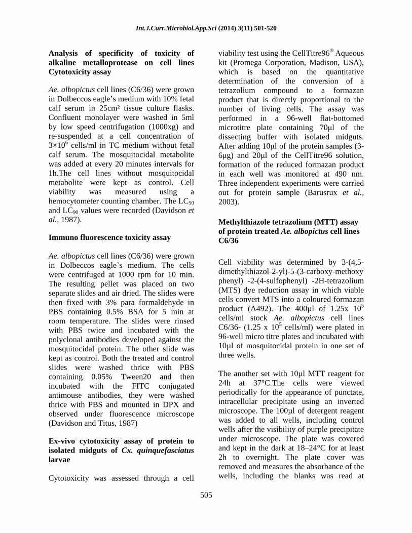
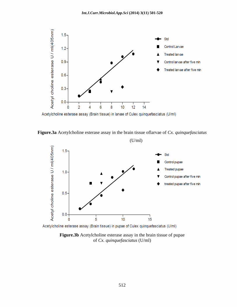
Acetylcholine esterase assay in the larvae and pupae of Cx. Quinquefasciatus
The brain tissue was isolated from the treated, untreated 4th instar larvae and pupae of Cx. quinquefasciatus by placing them on ice cooled microscope platform in PBS buffer (pH 7.5) and removing the outer most layer of head region with help of dissection needle. The isolated brain tissues were homogenized centrifuged and supernatant was used for the assay. Potassium phosphate buffer, 152 l of 0.1M, was taken in the control and activity wells of micro plate. To this, 1 l of 75mM concentration of substrate, acetylcholine iodide, was added. 5-thio-2-nitrobenzoate (DTNB) (5 l) was added to both the control and activity wells and incubated at room temperature for 10minutes. The absorbance was read at 405 nm and taken as Blank reading (Venkateswara et al., 2005). To this, 20 l of samples and standard acetylcholine esterase enzyme (1U/ml) was added, and the absorbance was read in the beginning and after five minutes at 405 nm.
Biochemical changes in the Ae.albopictus cell lines after treatment with the protein Cytochrome oxidase assay
Ae. albopictus cell lines (C6/36) were grown in Dolbeccos eagle s medium with 10% fetal calf serum in 25cm² tissue culture flasks. Confluent monolayer were washed in 5ml by low speed centrifugation (1000xg) and re-suspended at a cell concentration of 3×10⁶ cells/ml in Tissue Culture medium without fetal calf serum. The mosquitocidal protein was added to the cell lines at a concentration of 7.5µg/ml and incubated for 1h. The cell lines without the protein were maintained as control. The control and

Int.J.Curr.Microbiol.App.Sci (2014) 3(11) 501-520
504
treated cell lines were harvested by centrifuging at 600xg for 10 m. The pellets was recovered and washed with PBS and once again resuspended with five times the volume of solution A (0.25 M sucrose, 20 mM Hepes
KOH, 1 mM EDTA, 1 mM
DTT and 0.1mM PMSF). The cells were homogenized in the solution A and spun down at 750xg for 10 minutes. The supernatant was collected and centrifuged at 10000xg for 15 minutes. The supernatant was discarded and the crude mitochondrion recovered in the pellet was resuspended in the solution A.
The isolated intact mitochondria (40µl) were aliquoted in triplicate in 96 well plates. A volume of 150µl of reduced cytochrome C substrate was added to each well and the plate was assayed in a plate reader, reading at every 1m at 550nm for 10seconds. As 10x cytochrome oxidase oxidizes cyt C, the optical density of the solution declines at 550nm. The plate reader calculates the maximum slope of the plot of optical density (Timothy et al., 2003). The protein concentration for the mitochondrial homogenate was determined by the method of Lowry et al. (1951).
Western blot assay of membrane fractions of Ae. albopictus cell lines
The membrane fractions were prepared from the control and treated Ae. albopictus cell lines (1.25 x 10⁵ cells/ml) by washing the cells with TBS (10mM Tris HCl pH 7.5 and 150 mM NaCl). The washed cells were suspended in Tx 1000 and the homogenate was spun down at 4000xg for 30 minutes.
The supernatant was incubated at 37oC for 10m and spun down out at 60xg for 15 m. The pellets were suspended in TBS. The protein concentration was estimated by a
modification of the Lowry et al. (1951) method using bovine serum albumin as a standard. The cytoplasmic membrane fractions were subjected to 12% SDS-polyacrylamide gel electrophoresis (SDS-PAGE) and transferred onto a nitrocellulose membrane using a MiniTrans-Blot electrophoretic transfer cell (Bio-RAD) with transferring buffer (190 mM glycine, 20% methanol and 25 mM Tris-HCl, pH 8.3) at constant voltage for overnight (Laemmli 1970; Towbin et al., 1979). The membranes were washed thrice with phosphate buffered saline containing 0.05% Tween20 and treated with BSA for 2h to block non-specific binding sites.
After blocking, the proteins immobilized on the membrane were incubated for 5h with 1: 500 dilution of the polyclonal antibodies developed against the mosquitocidal protein. The membrane was washed thrice with PBS-0.05% Tween20 and the immunocomplexes were incubated with horseradish peroxidase-conjugated rabbit anti-mouse IgG (whole molecule) as the secondary antibody for 2h. Color was developed with TMB/H2O2 as the substrate and the reaction was stopped after 1h with water (Salas-Benito et al., 1997).
SDS-PAGE analysis of mitochondrial fraction to determine binding of protein
The mitochondrial fraction from the control, treated larvae and pupae of Cx. quinquefasciatus was suspended in 4x lamelli sample loading buffer [0.125 M tris/Hcl (pH 6.8), 4% (w/v) SDS, 20% (v/v) glycerol, 10% (v/v) 2-mercapto ethanol, 0.01% (w/v) bromophenol blue]. The samples were boiled for 5minutes, separated by 10% SDS PAGE to find out whether the binding of protein enhances the cytochrome oxidase IV complex subunit (Srikrishnaraj et al., 1995).
Int.J.Curr.Microbiol.App.Sci (2014) 3(11) 501-520
505
Analysis of specificity of toxicity of alkaline metalloprotease on cell lines Cytotoxicity assay
Ae. albopictus cell lines (C6/36) were grown in Dolbeccos eagle s medium with 10% fetal calf serum in 25cm² tissue culture flasks. Confluent monolayer were washed in 5ml by low speed centrifugation (1000xg) and re-suspended at a cell concentration of 3×10⁶ cells/ml in TC medium without fetal calf serum. The mosquitocidal metabolite was added at every 20 minutes intervals for 1h.The cell lines without mosquitocidal metabolite were kept as control. Cell viability was measured using a hemocytometer counting chamber. The LC50
and LC90 values were recorded (Davidson et al., 1987).
Immuno fluorescence toxicity assay
Ae. albopictus cell lines (C6/36) were grown in Dolbeccos eagle s medium. The cells were centrifuged at 1000 rpm for 10 min. The resulting pellet was placed on two separate slides and air dried. The slides were then fixed with 3% para formaldehyde in PBS containing 0.5% BSA for 5 min at room temperature. The slides were rinsed with PBS twice and incubated with the polyclonal antibodies developed against the mosquitocidal protein. The other slide was kept as control. Both the treated and control slides were washed thrice with PBS containing 0.05% Tween20 and then incubated with the FITC conjugated antimouse antibodies, they were washed thrice with PBS and mounted in DPX and observed under fluorescence microscope (Davidson and Titus, 1987)
Ex-vivo cytotoxicity assay of protein to isolated midguts of Cx. quinquefasciatus larvae
Cytotoxicity was assessed through a cell
viability test using the CellTitre96® Aqueous kit (Promega Corporation, Madison, USA), which is based on the quantitative determination of the conversion of a tetrazolium compound to a formazan product that is directly proportional to the number of living cells. The assay was performed in a 96-well flat-bottomed microtitre plate containing 70 l of the dissecting buffer with isolated midguts. After adding 10 l of the protein samples (3-6 g) and 20 l of the CellTitre96 solution, formation of the reduced formazan product in each well was monitored at 490 nm. Three independent experiments were carried out for protein sample (Barusrux et al., 2003).
Methylthiazole tetrazolium (MTT) assay of protein treated Ae. albopictus cell lines C6/36
Cell viability was determined by 3-(4,5-dimethylthiazol-2-yl)-5-(3-carboxy-methoxy phenyl) -2-(4-sulfophenyl) -2H-tetrazolium (MTS) dye reduction assay in which viable cells convert MTS into a coloured formazan product (A492). The 400µl of 1.25x 10⁵
cells/ml stock Ae. albopictus cell lines C6/36- (1.25 x 10⁵ cells/ml) were plated in 96-well micro titre plates and incubated with 10µl of mosquitocidal protein in one set of three wells.
The another set with 10µl MTT reagent for 24h at 37°C.The cells were viewed periodically for the appearance of punctate, intracellular precipitate using an inverted microscope. The 100µl of detergent reagent was added to all wells, including control wells after the visibility of purple precipitate under microscope. The plate was covered and kept in the dark at 18 24°C for at least 2h to overnight. The plate cover was removed and measures the absorbance of the wells, including the blanks was read at

Int.J.Curr.Microbiol.App.Sci (2014) 3(11) 501-520
506
570nm.Results presented are means of eight wells ±SD, and each experiment was repeated at least three times.
In vitro digestion of gut extracts of larvae and pupae of Cx. quinquefasciatus
The guts from 50nos of 4th instar control larvae and pupae were pulled out, and the peritrophic membranes were removed. The guts were then washed in 0.5ml of PBS and re-suspended in 0.2ml of PBS. The guts were sonicated for 60seconds in a 1.5ml Eppendorf tube and the protein concentration was determined by the method of Lowry et al. (1951). About 5µg of the crude gut extract was added to 10µg of the mosquitocidal protein. Trypsin digestion was carried out by adding 1µg of trypsin to 10µg of mosquitocidal protein. The mixtures were then incubated for 1h at 37ºC and the products were analyzed on a 10% SDS-PAGE. Bioassay was carried out in two tubes to determine the toxicity by introducing 5 pupae/ larvae in 2ml of water. To one tube 10mg/ml of the mosquitocidal protein and to the other tube trypsin treated protein was added and monitored for mortality for 1h (Thirumaran et al., 1992). Results and Discussion
The induction of the marker enzymes -galactosidase (Figure 1a,b,c), hydrogen peroxidise (Figure 2a,b,c), acetylcholine esterase (Figure 3a,b,c) were studied to find out the biochemical changes on the protein exposed 4th instar larvae and pupae of Cx. quinquefasciatus from the various tissues / organs. Since the P value of liner regression analysis is <0.0010 at 95% confidence intervals, the results of -Galactosidase, hydrogen peroxidase, acetylcholine esterase assay of larvae and pupae is statistically significant.
The target of the active mosquito cidal protein was identified using Ae. albopictus (C6/36 cell lines) exposed to the mosquito cidal metabolites and quantitative analyses of enzymes from intact mitocondria were studied. The results of cytochrome oxidase assay showed that there is an induction of cytochrome C oxidase activity in the treated Ae. albopictus (C6/36 cell lines) than the untreated Ae. albopictus cell lines (Figure 4a, b). The results of Western blot assay of membrane fractions of Ae. albopictus cell lines revealed that the binding of mosquitocidal protein to the 90kDa of Ae. albopictus cell lines (Figure 5). The results of qualitative binding assay of mosquitocidal protein to the cytochrome oxidase by SDS-PAGE analysis of mitochondrial fraction showed induction of ~18 kDa protein of cytochrome oxidase IV subunit in the treated larvae and pupae compared to that of untreated larvae and pupae (Figure 6).
The mechanism of action and receptor binding of a dual specificity of alkaline metalloprotease using insect cell line culture (C6/36) was studied. The mosquitocidal proteins incubated with C6/36 cell lines of Ae. albopictus caused rapid rounding of cells followed sequentially by swelling, bulging of membrane vesicles, vacoulation and finally lysis. Cell death was found to be induced by increasing the concentration of the purified protein (7µg/ml 15µg/ml), and showed the characteristics of apoptosis (Figure 7). The results of assay of toxicity through detection of immunofluorescence did not show fluorescence in control cell lines, where as in treated cell lines incubated with FITC conjugated anti-mouse antibodies showed bright fluorescence. The bulging of membrane vesicles and vacuolization of cells were observed in treated cell lines (Figure 8).

Int.J.Curr.Microbiol.App.Sci (2014) 3(11) 501-520
507
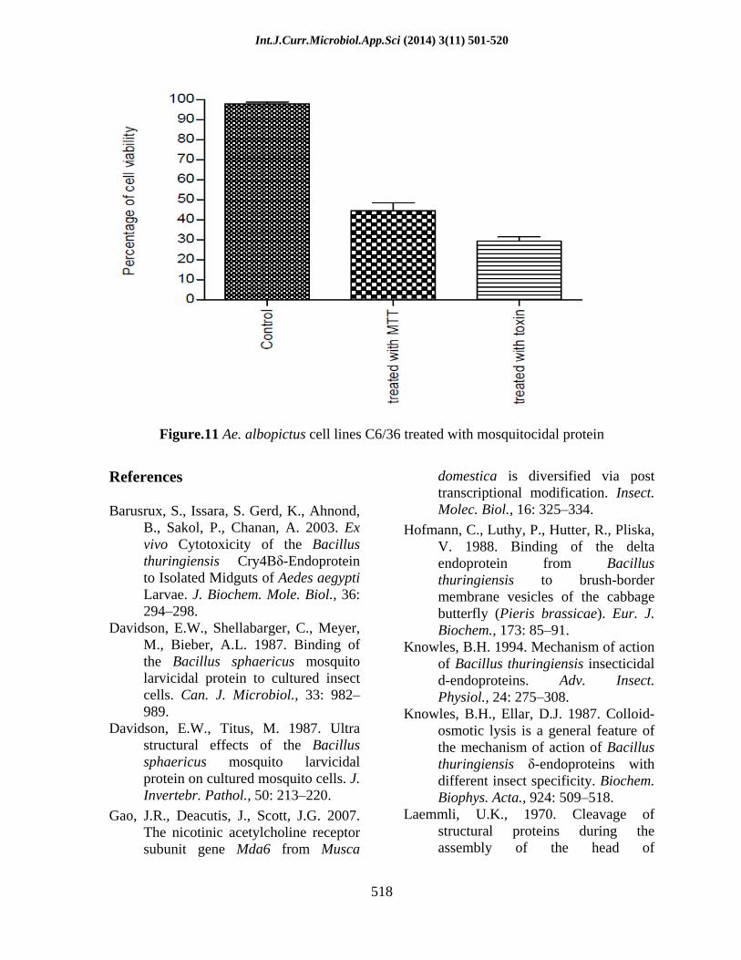
The trypsin digested gut extract of mosquitocidal protein exposed larvae and pupae were analysed on 10% SDS Poly acrylamide gel. The trypsin resistant bands were observed in the both larval and pupal gut extract. It showed that the trypsin did not cleave the 55kDa and 35kDa into smaller peptides (Figure 9). The midguts were isolated from larvae of Cx. quinquefasciatus for the assessment of mosquitocidal protein toxicity viaa cell viability assay. After incubating the dissected midguts with the protein samples, the number of viable epithelial cells was determined by a colorimetric method that monitors the quantity of water-soluble formazan products by photometric measurement at 490 nm. Changes in A490 were assumed to be directly proportional to the number of viable cells (Figure 10). The decline of A490 was due to the degradation of formazan that is possibly caused by oxidative agents from the dissected tissues. Each reaction curve was analyzed by nonlinear regression using Graph Pad PRISM version 2.0. A statistical analysis was carried out by using one-way analysis of variance (ANOVA) tests and the probabilities <0.05 were considered as significant. The MTT assay revealed 45% viability after 24 h after infection (Figure 11).Compared with uninfected control cells, there was a significant decrease in the viability of mosquitocidal protein exposed cell lines at 24h. Probabilities <0.05 versus untreated cell lines C6/36 were considered as being significant.
In the present investigation, specific target of the protein, biochemical and physiological changes in the protein exposed 4th instar larvae and pupae of Cx. quinquefasciatus were studied. The protein treated midgut cells of larvae and pupae showed induction of galactosidase activity. This may be due to the irreversible
binding of protein to the mid gut cells leading to constitutive expression of lacZ gene, which codes for galactosidase enzyme. In general, a wide range of carbohydrases are present in insects. The gut of insects like cockroach contains amylases
galactosidase and galactosidase (Sabine et al., 2006).
galactosidase utilizes lactose as a substrate by cleaving the anomeric carbon and glycosyl oxygen to form galactose and glucose. galactosidase is a reporter gene and regulated by lac Z under the control of Lac operon (Seeber et al., 1996). The protein treated midgut cells of larvae and pupae showed the induction of galactosidase activity and it could be due to the binding of protein to the midgut cells of treated larvae and pupae leading to increase in the galactosidase enzyme activity.
Similarly, hydrogen peroxidase activity in the midguts of treated larvae and pupae were studied after staining with TMB (Tetramethyl benzidine) peroxidase
specific chromopore and hydrogen peroxide as substrates. This histochemical study revealed that the treated midgut cells were stained very strongly than the control cells. This could be interpreted in such a way that the binding of mosquitocidal proteins to the mid gut epithelial cells leads to induced peroxidase enzyme activity, which subsequently leads the generation of hydrogen peroxide to promote apoptosis. It has been reported that the P. berghei invasion of An. stephensi midgut cells induces expression of nitric oxide synthase followed by increased peroxidase activity. To confirm the induction of peroxidase activity in response to malaria infection, the activity was determined in the mid gut homogenates by performing histochemical staining and spectrophotometric assay using TMB (Tetramethyl benzidine) peroxidase
specific chromopore and hydrogen peroxide as substrates. The parasite infection resulted

Int.J.Curr.Microbiol.App.Sci (2014) 3(11) 501-520
508
in a marked increase in the peroxidase activity when homogenates from mid guts that had been fixed with a mixture of glutaraldehyde and formaldehyde were used in the assay than the control midgut cells (Sanjeev et al., 2004). Similarly, in the present study also, mid guts of treated larvae and pupae were stained with TMB (Tetramethyl benzidine) peroxidase
specific chromopore and hydrogen peroxide as substrates.
The activity of marker enzyme acetylcholine esterase was increased significantly in the brain tissues of treated larvae and pupae than the midgut region of treated larvae and pupae. The enzyme activity was decreased considerably after five minutes interval. In general, acetylcholine receptors belong to the cys-loop super family of (GABA) gated chloride (cl-) channels consisting of five sub units. Each sub units possess a large N-terminal extra cellular domain that includes the components forming the acetyl choline binding site. The acetyl choline acts as a neurotransmitter and hydrolyzed by the enzyme acetylcholine esterase. The cuticular invasion of mosquitocidal protein to the central nervous system leads to increase in acetyl choline esterase activity in the treated larval and pupal brain tissues. This elucidates the biphasic effect of mosquitocidal proteins in such a way that the first phase of protein effect of increased levels of acetylcholine esterase is due to the inhibition of neuronal transmission while in second phase, the acetylcholine esterase activity was decreased; due to the neuronal degeneration of tissues. The acetylcholine esterase enzyme activity in the mid gut of treated larvae and pupae was decreased than the control larvae and pupae. This could be due to the fact that the mosquitocidal protein
acts as a potential inhibitor to the enzyme. The difference of acetylcholine esterase activity in the brain and mid gut region of treated larvae and pupae is depended on the function of the tissues involved (Zhen et al., 2004; Gao et al., 2007).
The induction of cytochrome C oxidase activity in the treated Ae. albopictus C6/36 cell lines than in the untreated cell lines indicates that the protein might have regulated mitochondrial ETC complex IV. The protein would have mediated the enhancement of mitochondrial biogenesis in the treated cell lines under the stress condition as reported by Luciakova et al. (1999). As a confirmation, the results showed induction of ~18 kDa protein of cytochrome oxidase IV subunit in the treated larvae and pupae as compared to that of untreated larvae and pupae. It has been reported that the proteases enhances the mitochondrial biogenesis (Nagy et al., 2004). The C6/36 cell lines incubated with the mosquitocidal proteins with caused rapid rounding of cells followed by swelling, bulging of membrane vesicles, vacoulation of cells and finally cell lysis. At lower concentrations, the protein caused no morphological change and the cells were viable. Cell death induced by increasing the concentration of purified protein had the characteristics of apoptosis. The fluorescence was not observed in control cell lines where as in treated cell lines, incubated with FITC conjugated antimouse antibodies showed bright fluorescence. The bulging of membrane vesicles and vacuolization of cells were observed in treated cell lines, indicating that the FITC conjugated antibody reacted with the protein which bound to the specific receptor on surface of the cell lines.

Int.J.Curr.Microbiol.App.Sci (2014) 3(11) 501-520
509
Figure.1a ß galactosidase enzyme assay in the larvae Cx.quinquefasciatus (U /ml)
Figure.1b ß galactosidase enzyme assay in the pupae of Cx.quinquefasciatus (U/ml)

Int.J.Curr.Microbiol.App.Sci (2014) 3(11) 501-520
510
Figure.1c Comparison of ß galactosidase assay in the control and protein treated larvae and pupae of Cx. quinquefasciatus
Figure.2a Peroxidase enzyme assay in the larvae of Cx. quinquefasciatus

Int.J.Curr.Microbiol.App.Sci (2014) 3(11) 501-520
511
Figure.2b Peroxidase enzyme assay in the pupae of Cx. quinquefasciatus
Figure.2c Comparison of peroxidase enzyme assay in the control and protein treated larvae and pupae of Cx. quinquefasciatus
Int.J.Curr.Microbiol.App.Sci (2014) 3(11) 501-520
512
Figure.3a Acetylcholine esterase assay in the brain tissue oflarvae of Cx. quinquefasciatus
(U/ml)
Figure.3b Acetylcholine esterase assay in the brain tissue of pupae of Cx. quinquefasciatus (U/ml)

Int.J.Curr.Microbiol.App.Sci (2014) 3(11) 501-520
513
Figure.3c Acetylcholine esterase assay in the brain tissue of larvae and pupae of Cx. quinquefasciatus
Figure.4a Cytochrome oxidase assay in mosquitocidal protein treated larvae

Int.J.Curr.Microbiol.App.Sci (2014) 3(11) 501-520
514
Figure.4b Cytochrome oxidase assay in mosquitocidal protein treated pupae
1 2
Lane 1: Mosquitocidalprotein treated membrane fractions, Lane 2: Control membrane
fractions
Figure.5 Western blot analysis of membrane fractions of Ae. aegypti cell lines treated with mosquitocidal proteins
Treated pupae
Control pupae
55 kDa
35 kDa

Int.J.Curr.Microbiol.App.Sci (2014) 3(11) 501-520
515
1 2 3
L 1: marker, L 2: treated membrane fractions, L 3: Control membrane fractions
Figure.6 Protein profile of mosquitocidalprotein treated membrane fractions of A. albopictuscell lines
Figure.7 Cytotoxicity assay a) Control cell line b) Initial vacoulation on Ae. albopictus C6/36 cell lines after treated with mosquitocidal metabolite

Int.J.Curr.Microbiol.App.Sci (2014) 3(11) 501-520
516
C) Vacoulation on Ae. albopictus C6/36 cell lines after treated with mosquitocidal protein
Figure.8 Immunofluorescence toxicity assay (a) Control cell line (b) Cell line treated with
FITC Conjugated anti-mouse antibodies

Int.J.Curr.Microbiol.App.Sci (2014) 3(11) 501-520
517
1 2 3 4 5 6 7
L 1: marker, L 2, 3: protein, L 4: treated pupal gut, L 5: treated larval gut L 6: larval gut , L 7 : pupal gut
Figure.9 In vitro tryptic digestion of midgut extracts of larvae and pupae of Cx. quinquefasciatus
Figure.10 Ex vivo cytotoxicity assay of mosquitocidal protein to isolated midguts of Cx. quinquefasciatus larvae
Int.J.Curr.Microbiol.App.Sci (2014) 3(11) 501-520
518
Figure.11 Ae. albopictus cell lines C6/36 treated with mosquitocidal protein
References
Barusrux, S., Issara, S. Gerd, K., Ahnond, B., Sakol, P., Chanan, A. 2003. Ex vivo Cytotoxicity of the Bacillus thuringiensis Cry4B -Endoprotein to Isolated Midguts of Aedes aegypti Larvae. J. Biochem. Mole. Biol., 36: 294 298.
Davidson, E.W., Shellabarger, C., Meyer, M., Bieber, A.L. 1987. Binding of the Bacillus sphaericus mosquito larvicidal protein to cultured insect cells. Can. J. Microbiol., 33: 982989.
Davidson, E.W., Titus, M. 1987. Ultra structural effects of the Bacillus sphaericus mosquito larvicidal protein on cultured mosquito cells. J. Invertebr. Pathol., 50: 213 220.
Gao, J.R., Deacutis, J., Scott, J.G. 2007. The nicotinic acetylcholine receptor subunit gene Mda6 from Musca
domestica is diversified via post transcriptional modification. Insect. Molec. Biol., 16: 325 334.
Hofmann, C., Luthy, P., Hutter, R., Pliska, V. 1988. Binding of the delta endoprotein from Bacillus thuringiensis to brush-border membrane vesicles of the cabbage butterfly (Pieris brassicae). Eur. J. Biochem., 173: 85 91.
Knowles, B.H. 1994. Mechanism of action of Bacillus thuringiensis insecticidal d-endoproteins. Adv. Insect. Physiol., 24: 275 308.
Knowles, B.H., Ellar, D.J. 1987. Colloid-osmotic lysis is a general feature of the mechanism of action of Bacillus thuringiensis -endoproteins with different insect specificity. Biochem. Biophys. Acta., 924: 509 518.
Laemmli, U.K., 1970. Cleavage of structural proteins during the assembly of the head of

Int.J.Curr.Microbiol.App.Sci (2014) 3(11) 501-520
519
bacteriophage T4. Nature, 227: 680685.
Lakshmi, N.M., Gopinathan, K.F. 1988. Effect of Bacillus sphaericus 1593 protein on choline acetyl transferase and mitochondrial oxidative activities of the mosquito larvae. Ind. J. Biochem. Biophy., 25: 253256.
Lowry, O.H., Rosen roughm, N.J., Randall, A.J. 1951. Protein measurement with the folin phenol reagent. J. Biol. Chem., 193: 265275.
Luciakova, K., Sokolikova, B., Chloupkova, M., Nelson, B.D. 1999. Enhanced mitochondrial biogenesis is associated with increased expression of the mitochondrial ATP-dependent Lon protease. FEBS Lett., 444: 186 188.
Lycett, J.G., Fotis, C.K., Thanasis, G.L. 2004. Conditional expression in the malaria mosquito Anopheles stephensi with Tet-On and Tet-Off Systems. Genetics, 167: 1781 1790.
Nagy, G., Maureen, B., Nick, G., Paul, E.P., Andras, P. 2004. Nitric oxide-dependent mitochondrial biogenesis generates Ca2 signaling pro le of lupus T cell. J. Immunol., 173: 3676 3683.
Oddou, P., Hartmann, H., Radecke, F., Geiser, M. 1993. Immunologically unrelated Heliothis sp. and Spodoptera sp. midgut-proteins bind Bacillus thuringiensis CryIA (b) d-endoprotein. Eur J Biochem., 212: 145 150.
Oei, C., Hindley, J., Berry, C. 1992. Binding of purified Bacillus sphaericus binary protein and its deletion derivatives to Culex quinquefasciatus gut: elucidation of
functional binding domains. J. Gen. Microbiol., 138: 1515 1526.
Sabine, E., Photini, S., Kai, M. 2006. Transgenic Plasmodium berghei sporozoites expressing -galactosidase for quantification of sporozoite transmission. Mol. Biochem. Parasitol., 146: 30 37.
Salas-Benito, J.S., Angel Rosa, M. 1997. Identification of two surface proteins from C6/36 cells that bind dengue type 4 virus. J. Virol., 71: 72467252.
Sanjeev, K., Lalita, G., Yeon Soo Han, Carolina, B.M. 2004. Inducible peroxidases mediate nitration of Anopheles midgut cells undergoing apoptosis in response to Plasmodium invasion. J. Bio. Chem., 279: 5347553482.
Seeber, F., Boothroyd, J.C. 1996. Escherichia coli -galactosidase as an in vitro and in vivo reporter enzyme and stable transfection marker in the intra-cellular protozoan parasite Toxoplasma gondii. Gene, 169: 39 45.
Srikrishnaraj, K.A., Ramasamy, R., Ramasamy, M.S.1995. Antibodies to Anopheles midgut reduces vector competence for Plasmodium vivax malaria. Med. Vet. Entomol., 9: 353357.
Thirumaran, T., John, H., Colin, B. 1992. Proteolytic processing of the mosquitocidal protein from Bacillus sphaericus SSII-1. J. Bacteriol., 174: 5051 5056.
Thomas, W.E., Ellar, D.J. 1983. Bacillus thuringiensis var.israelensis crystal -endoprotein: Effects on insect and
mammalian cells in vitro and in vivo. J. Cell. Sci., 60: 181 197.
Timothy, B., Sackton, A.H., Robert, David, M.R. 1997. Cytonuclear coadaptation in drosophila:

Int.J.Curr.Microbiol.App.Sci (2014) 3(11) 501-520
520
disruption of cytochrome c oxidase activity in backcross genotypes. Evolution., 57: 2315 2325.
Towbin, H., Staehelin, T., Gordon, J. 1979. Electrophoretic transfer of proteins from polyacrilamide gels to nitrocellulose sheets: procedure and some applications. PNAS USA, 76: 4350 4354.
Usharani, B, Kummankottil P. 2013. Chitinase like activity of metabolites of Pseudomonas fluorescens Migula on immature stages of the mosquito, Culex quinquefasciatus (Diptera: Culicidae). Afr. J. Microbiol. Res., 6(11): 2718 2726,.
Usharani, B, Kummankottil P. 2014. Gelatinase activity of metabolites of Pseudomonas fluorescens migula on larvae and pupae of Culex quinquefasciatus (Diptera: Culicidae). Int. J. Pharm. Bio. Sci., 5(3): (B) 234 245.
Venkateswara, R.J., Ghousia, B., Pallela, R., Usman, P.K., Nageswara, R.R.2005. Changes in behavior and brain acetylcholine esterase activity in mosquito fish, Gambusia affinis in response to the sub-lethal exposure to Chlorpyrifos. Int. J. Environ. Res. Public Health., 2: 478 483.
Zhen-Hua, T., Roger, J.W., Sue, L.C. 2004. Acetyl cholinesterase activity in organophosphorus and carbamate resistant and susceptible strains of the Culex pipiens complex. Pestic. Biochem. Physiol., 37: 192 199.


![INDEX []...INDEX Page 501-E01 CYLINDER, HEAD AND COVER 3 501-E02 PISTON/CRANKSHAFT 5 501-E03 INTAKE/ESHAUST 7 501-E04 WATER PUMP 11 501-E05 OIL PUMP 13 501-E06 OIL SYSTEM 15 501-E07](https://img.pdfslide.net/doc/110x75/5e9579482775034fef0cc642/index-index-page-501-e01-cylinder-head-and-cover-3-501-e02-pistoncrankshaft.jpg)

![ARTS OF FASHION @ La Cambre Mode[s] · Arts of Fashion @ La Cambre Mode[s] | Brussels Presentation The Arts of Fashion Foundation is a 501(c)(3), public, non-profit organization,](https://img.pdfslide.net/doc/110x75/5ecfa1126750746cf31bd7b2/arts-of-fashion-la-cambre-modes-arts-of-fashion-la-cambre-modes-brussels.jpg)