Embed Size (px)
DESCRIPTION
Our results support the hypothesis that alcohols (in particular10- to 14-C-atom alcohols) contribute to the stability of oleic acid vesicles under a wider range of experimental conditions. Moreover, studies of mixed oleic-acid-alkane and oleic-acid-alcohol systems using infrared spectroscopy and Langmuir troughmeasurements indicate that precisely those alcohols that increased vesicle stability also decreased the mobility of oleic acidpolar headgroups, as well as the area/molecule of lipid.
Citation preview

278 Biophysical Journal Volume 102 January 2012 278–286
Model Systems of Precursor Cellular Membranes: Long-Chain AlcoholsStabilize Spontaneously Formed Oleic Acid Vesicles
Adela Rendon,† David Gil Carton,‡ Jesus Sot,† Marcos Garcıa-Pacios,† L.-Ruth Montes,† Mikel Valle,‡
Jose-Luis R. Arrondo,† Felix M. Goni,†* and Kepa Ruiz-Mirazo†§*†Unidad de Biofısica (CSIC-UPV/EHU), and Departamento de Bioquımica, Universidad del Paıs Vasco, Bilbao, Spain; ‡Unidad de BiologıaEstructural, CIC-Biogune, Parque Tecnologico, Derio, Bizkaia, Spain; and §Departamento de Logica y Filosofıa de la Ciencia, Universidad delPaıs Vasco, Donostia-San Sebastian, Spain
ABSTRACT Oleic acid vesicles have been used as model systems to study the properties of membranes that could be theevolutionary precursors of more complex, stable, and impermeable phospholipid biomembranes. Pure fatty acid vesicles ingeneral show high sensitivity to ionic strength and pH variation, but there is growing evidence that this lack of stability can becounterbalanced through mixtures with other amphiphilic or surfactant compounds. Here, we present a systematic experimentalanalysis of the oleic acid system and explore the spontaneous formation of vesicles under different conditions, as well asthe effects that alcohols and alkanes may have in the process. Our results support the hypothesis that alcohols (in particular10- to 14-C-atom alcohols) contribute to the stability of oleic acid vesicles under a wider range of experimental conditions. More-over, studies of mixed oleic-acid-alkane and oleic-acid-alcohol systems using infrared spectroscopy and Langmuir troughmeasurements indicate that precisely those alcohols that increased vesicle stability also decreased the mobility of oleic acidpolar headgroups, as well as the area/molecule of lipid.
INTRODUCTION
Biomembranes consist of a wide variety of both lipidic andpeptidic components whose synthesis is metabolicallycontrolled and highly regulated, according to the specificrequirements of each living cell at a particular moment orstage in its development, which accounts for their complexdynamic behavior and their key role in the organization ofany biological organism. However, it is not easy to under-stand how those sophisticated supramolecular structurescould emerge from simpler membranes, or what thesesimpler membranes should look like. Recent investigationsin the fields of origins of life and synthetic-protocell biology(1) have focused on fatty acid vesicles as one of the mostplausible starting points in the evolution of biologically rele-vant lipid compartments (2–4). The work here reportedtakes the oleic acid system as an experimentally suitablemodel to study the general properties of this kind of simpli-fied membrane compartment and analyzes in particular howthe presence of alkanes and alcohols may influence some ofthe main biophysical features of the compartments.
Fatty acids, either unsaturated (e.g., oleic, myristoleic, orlinoleic acid) or short-chained, saturated (e.g., octanoic ordecanoic acid) can form vesicles in aqueous solution underdiverse experimental conditions (5–14). These supramolec-ular aggregates, in comparison with standard liposomes,have quite remarkable properties: they are more permeableand dynamic structures (whose growth and reproduction canbe achieved under laboratory conditions (15–19)), but theyare also less stable than phospholipid vesicles, since their
Submitted July 21, 2011, and accepted for publication December 9, 2011.
*Correspondence: [email protected] or [email protected]
Editor: Heiko Heerklotz.
� 2012 by the Biophysical Society
0006-3495/12/01/0278/9 $2.00
formation depends on a relatively narrow set of values ofexperimental parameters, e.g., pH and ionic strength.
According to Morigaki andWalde (20), fatty acid vesiclesare characterized by twomain features: 1), a critical aggrega-tion concentration (CAC) several orders of magnitude largerthan that of the usual phospholipids, as a result giving muchmore abundant and faster exchange processes betweenmonomers in solution and the actual molecules of the bilayeraggregate (aswell asmore frequent flip-flop processeswithinthe bilayer); and 2), the negative net charge of the aggregateas a whole, since these vesicles typically form through theconjunction of a fatty acid (in a neutral, nonionized state)and its respective soap (negatively charged) or conjugatedsalt (if the counterion is included). More specifically, whatdetermines whether closed bilayers form or not is the ratiobetween those two amphiphilic components, the neutral fattyacid and the soap, so the degree of protonation of the terminalcarbonyl group (and, therefore, the pH of the solution)becomes a critical variable in the system. Thus, althoughvesicle formation in principle would not be favored by themolecular properties of single-chain lipids with a conicalgeometry (i.e., spontaneous tendency to form micelles), thecreation of molecular pairs through hydrogen bonds betweenneutral fatty acid and anionic soap/salt (21), which wouldthen adopt a more cylindrical geometry, makes possible theirspontaneous assembly into bilayered closed structures.
As a result, if the pH of the system is maintained nearthe pKa of the fatty acid, favoring the balance between itsdeprotonated and protonated states (i.e., the stable unionof pairs) and, in addition, the monomer concentration isabove the minimum concentration threshold for molecularaggregation (CAC), then the formation of self-assembled
doi: 10.1016/j.bpj.2011.12.026

Oleic Acid Vesicles as Membrane Precursors 279
vesicular structures is observed. If pH varies and movesaway from the pKa value, the system will tend to other statesof aggregation: micelles (typically, in the alkaline region) orother, less regular supramolecular structures, like droplets(typically at lower pH).
However, the pH range in which the vesicular phase isstable can be extended in mixed systems, as Apel andcolleagues (22) have demonstrated, using short-chain fattyacids and alcohols. The presence of an alcohol whose chainlength is similar to that of the fatty acid (but with a pH-insensitive headgroup) is supposed to be an alternativeneutral component of the pair when the proportion ofuncharged fatty acid is low. This happens when pH isincreased significantly above the pKa, provoking excess offatty acid in deprotonated states. Moreover, since pure fattyacid vesicles are not very realistic membrane models,working with mixtures is becoming a research target itself(13,23–25), and this is actually a good strategy to overcomesome of the problems faced with the pure systems (26).
In that context, the primary target of this article is to inves-tigate the spontaneous formation of aggregates of a particularfatty acid (oleic acid) in aqueous solution, characterizing thedifferent aggregation phases as a function of pH and otherexperimental variables, like osmolarity, studying theirentrapment capacity and analyzing in particular the influencethat the presence of alcohols and alkanes of diverse chainlengths may have on the system. A long-chain (C18) fattyacid has been used to mimic more closely the thickness ofpresent-day membranes, even if this required the inclusionof a double bond to make the molecule amenable to vesicleformation at room temperature. Our studies are complemen-tary to a recently published report onmicelle-vesicle equilib-rium in the oleic/oleate system (27).
We therefore aim to gain further knowledge on how simpleamphiphilic molecules (certainly simpler than the glycero-phospholipids found in cell membranes) self-assemble intoclosed bilayers, and on the stability and permeability proper-ties of the latter. As mentioned above, this type of vesiclecould be a good model to study protocellular systems(6,7,28,29), namely possible precursors of present-daycellular membranes, without disregarding other potentialapplications they may have in the biotechnological domain.
MATERIALS AND METHODS
Materials
Oleic acid (C18:1) was from Merck (Darmstadt, Germany), and the various
salts were obtained by titration of the oleic acid with the corresponding
hydroxides. Methanol and chloroform were from Fisher (Suwanee, GA).
All other chemicals were purchased from Sigma-Aldrich (St. Louis, MO).
Preparation of vesicles
Oleic acid vesicles were prepared by injecting an aqueous suspension of
3.15 M oleic acid (OA) into 0.2 M sodium bicine buffer at different pH
values, typically 8.5. In experiments involving titration, the samples were
initially brought to higher pH by addition of NaOH (micellar phase), and
later adjusted by addition of HCl to the final pH values required. Final
OA concentrations were in the range 1–20 mM.
Mixed vesicles tested included OA with hexanol, octane, octanol, dec-
anol, dodecane, dodecanol, tetradecanol, hexadecane, hexadecanol, octade-
cane, and octadecanol (20:1,10:1, 5:1, and 2:1 molar ratios, 5 mM total
concentration of fatty acid). Mixed-vesicle preparations were achieved by
dissolving initially both fatty acid and alkane or alcohol in chloroform,
which was then evaporated under nitrogen flow. Later, the mixture was re-
suspended at 21�C in 0.2 mM bicine, pH 8.5, and incubated at room temper-
ature for 16 h before assay.
Buffer preparation
We prepared 0.2 M bicine by dissolving bicine in ultrapure water and ad-
justing the pH by addition of small amounts of 1 M HCl or 1 M NaOH
to pH 8.5 (unless otherwise specified).
Turbidity measurements
Turbidity was measured with a Cary 300 Bio UV/vis multicell spectropho-
tometer from Varian (Sydney, New South Wales, Australia), using quartz
cells with a path length of 1.0 cm, at l ¼ 400 nm.
Dynamic light scattering
Dynamic light scattering (DLS) measurements of particle sizes were carried
on a Malvern Zetasizer nano System. This instrument was equipped with
a 4 mW He-Ne LASER of 633 nm wavelength, and an Avalanche photo-
diode detector (quantum efficiency >50% at 633 nm) located at 173�
from the incident beam direction in a backscatter position. The temperature
of the sample holder was stabilized at 25�C through a Peltier thermostat.
Samples were introduced into plastic 50–2000-ml capacity disposable
cuvettes (UVette, Eppendorf, Hamburg, Germany).
Release/entrapment through fluorescencemeasurements
Vesicle leakage was estimated as an increase in absorbance at 500 nm,
measured in a spectrofluorometer FluoroMax-3 (Horiba, Kyoto, Japan).
Release of aqueous contents was assessed using the ANTS/DPX fluorescent
probe system (described in Ellens et al. (30)). For measurements of vesicle
leakage, lipids were hydrated in bicine 0.2 M, pH 8.5. Nonentrapped
ANTS/DPX was removed by gel filtration on Sephadex G-25 columns.
Fluorescence measurements were carried out in an LS-50B PerkinElmer
(Waltham, MA) spectrofluorometer, at room temperature (21 5 1�C) andwith continuous stirring. The osmolality of intra- and extravesicular solu-
tions was measured in a cryoscopic osmometer (Osmomat 030, Gonotec,
Berlin, Germany) and adjusted to 0.3 Osm/kg by adding NaCl. Fluores-
cence scales were calibrated as described previously (31). With pure buffer
in the cuvettes, 0% release was measured; 100% release was induced by
addition of the nonionic detergent Triton X-100, known to permeabilize
model and cell membranes, to a final 1 mM concentration. Excitation light
was adjusted to 490 nm. An interference filter (520 nm) was used to avoid
scattered excitation light.
Cryo-electron microscopy
Cryo-electron microscopy (cryo-TEM) is a suitable technique for direct
visualization of surfactant aggregates ranging in size from 5–10 nm to
1 mm, at the same time minimizing artifacts due to sample preparation/ fixa-
tion (through staining). The sample was placed in the controlled environ-
ment of the vitrification chamber at room temperature, where the relative
Biophysical Journal 102(2) 278–286

280 Rendon et al.
humidity was kept close to saturation to prevent water evaporation from the
sample. A 5-ml drop of the aqueous solution was placed on carbon-coated
holey film supported by a TEM copper grid. Most of the liquid was removed
by careful blotting with absorbent filter paper to create a thin liquid film.
The sample was then rapidly plunged into liquid ethane and cooled by
liquid nitrogen to its melting temperature to obtain a vitrified film. The vitri-
fied specimen was stored under liquid nitrogen and then transferred into the
electron microscope (Jeol JEM-2200FS) operating at 200 kVwith an under-
focus of 3–5 mm. The working temperature was kept below �175�C, andthe images were collected under low-dose conditions with a CCD camera
(UltraScan 4000, Gatan, Pleasanton, CA).
Preparation of giant unilamellar vesicles andfluorescence microscopy
Giant unilamellar vesicles (GUVs) were prepared by injecting an aqueous
solution of 3.15 M OA into sodium bicine buffer (0.2 M, pH 8.5). OA
(3.15 M in chloroform) was evaporated under nitrogen flow, and the lipid
was resuspended at 25�C in 0.2 mM bicine at pH 7.5, 8.5 or 10. The
hydrated sample was homogenized, passing it five times through a
25-mm, 23-gauge needle. A small aliquot (~3 ml) of aqueous solution
was deposited onto the platinum electrodes and allowed to dry in a vacuum
chamber for 30 min. Giant vesicles were obtained by using the electrofor-
mation method developed by Angelova et al. (32): a low-frequency AC field
(sinusoidal wave function with a frequency of 10 Hz and an amplitude of
2.5 V) was applied for 120 min at 75�C in the same, preheated, buffer.
Thin-layer chromatography revealed no lipid degradation under these
conditions. The vesicles were directly observed with an inverted confocal
microscope (TE2000 U, Nikon, Melville, NY). The excitation wavelength
for DiIC18 (bilayer concentration 0.4 mol %) was 488, and the fluorescence
signal was collected using a bandpass filter of 593 5 40 nm. The objective
used was a 40� oil immersion, NA 1.0 objective.
Infrared spectroscopy
For this set of measurements, OA (5 mM) was suspended in chloroform and
mixed with the corresponding alcohol/alkane (also in chloroform), when-
ever required, at either a 2:1 or a 10:1 mol ratio. We vacuum-dried 40 mL
of each sample overnight on a 56-mm-diameter CaF2 window (IR Select,
Spectroscopy Central, Warrington, UK). Before measuring, the films
were resuspended in 40 mL D2O (Merck), covered with another CaF2window interposing a 56-mm pathlength teflon spacer (Harrick Scientific,
Ossining, NY) and mounted on a Harrick cell. We acquired 360 scans
and took the average for each spectrum using an MCT detector in a Nexus
870 infrared (IR) spectrometer (Thermo Nicolet, Madison, WI).
Monolayer surface pressure measurements
In this case, samples were similarly prepared in chloroform and then mixed
with the alcohol/alkane at a 10:1 mol ratio. Monolayers were then spread
from 22 ml of chloroform solution onto a 235-cm2 Teflon trough filled
with 110 ml 10 mM Tris and 150 mM KCl, pH 8.5, at 22�C. The film
was relaxed for 30 min at 0 mN/m and later compressed to the collapse
phase at a speed of 0.15 nm2 mol�1 min�1. Surface pressure and film
area were measured in a Micro-Processor Interface IU4 (NIMA, Coventry,
UK). The reproducibility of experiments was within the maximum standard
error of 51 A2 for molecular areas.
FIGURE 1 Absorbance profiles as a function of pH for the pure OA
system. (A) [OA] ¼ 5 mM was chosen as the control condition. The pH
values that mark the two main phase transitions (pH1 and pH2) are indi-
cated. (B) Profiles for various concentrations of OA. (C) Close-up to
show more clearly the CAC threshold.
RESULTS
The OA-water system
To analyze the different aggregation phases of OA inaqueous solution (originally determined by others (5,8,9))
Biophysical Journal 102(2) 278–286
with our experimental setting and to study the conditionsunder which vesicles form spontaneously, we first usedturbidimetry (absorbance at 400 nm) and observed threeclearly differentiated regions as the pH of the solution wasmodified. As expected from previous work with this andother fatty acids, the slope of the absorbance profileundergoes two clear changes, marking the transition frommicelles to vesicles (around pH 9 in this case) and from vesi-cles to droplets (just below pH 8). This is shown in Fig. 1, Aand B (for different OA concentrations): the micellar phase(high pH) is characterized by an almost flat slope, whereasin the vesicular phase (pH around pKa 8.5 for OA), the slopebecomes moderate, and in the droplet phase (neutral pH orbelow), absorbance values increase markedly (steepersubset of points). These experiments rely on the fact thatthe formation and breakdown of fatty acid supramolecularaggregates, after pH variations, are relatively fast processes.

FIGURE 2 DLS size distributions of the population of aggregates at
different pH values. As pH decreases there is a progressive increase in
the mean size and heterogeneity (polydispersion index) of the population.
Oleic Acid Vesicles as Membrane Precursors 281
For the case of vesicle formation, the longest expected timesare in the range between 1 s and 1 min (33); for vesiclebreakdown and micellar processes, they are much shorter.
Two additional remarks should be made here: first, theobservation that titration curves change if the starting pointof the system is changed (in our experiments, since themicellar state is molecularly more ordered or homogeneousthan the droplets, we typically started from high pH values,to contribute to a smoother vesicle self-assembly). Forinstance, the turbidity of a sample directly prepared at pH8.5 is typically higher than the turbidity of the same sample,after bringing it up to the micellar phase (high pH) andtitrating it down, back to pH 8.5. This asymmetry or irre-versibility in the oleic system is not surprising, given theintrinsic dependence of this type of supramolecular self-assembly process on initial conditions—and on the prepara-tion procedure in general (34). Second, we confirmed theexistence of a CAC below which no vesicles are formed(see Fig. 1 C). Through the analysis of these turbidityprofiles for different OA concentrations, the 5 mM condi-tion, well above the CAC, was selected for subsequentexperiments.
With the aim of clarifying, as far as possible, the source ofthe aforementioned irreversibility, we carried out a seriesof experiments in which turbidity was measured underdifferent conditions of pH and osmolarity, using indepen-dently prepared samples (i.e., fixed values of pH and ionicstrength for each of them) and waiting for different timespans (24 and 48 h) after preparation. The results of theseexperiments are condensed in Fig. S1 in the SupportingMaterial, which essentially shows that although variationsin osmolarity do not seem to have a large effect at pH aroundthe pKa (50.3) of the fatty acid, moving away from the8–8.5 pH region, changes in the overall osmolite concentra-tion do have more radical consequences in the state of thesystem, as measured by variations in A400. There is, anyhow,an overall limit in terms of the osmotic strength that thesystem can withstand: the reported data at osmolarity values>0.8 are no longer reliable, since samples become nonho-mogenous, i.e., they are not opalescent any more but containmacroscopic aggregates, which precipitate in the absence ofstirring. Furthermore, we confirmed that the most orderlyand stable way to produce vesicles is when osmolarityvalues remain rather low (<0.4) and, optimally, when thesystem reaches the vesicle stability phase from the higherpH region (micellar phase).
DLS measurements consistently confirmed that the meansize of OA aggregates increases as the pH decreases (Fig. 2),and the average diameters obtained are compatible with thepredicted phase changes (from micelles to vesicles to drop-lets) as pH is decreased. This is in agreement with recentobservations by Dejanovic et al. (27). Morphology of theparticles in suspension was examined by cryo-TEM. Vesic-ular shapes were found mainly at pH between 7.8 and 9.Above and below those limits vesicles coexist with other
aggregation states of OA, like micelles or droplets, suggest-ing that pure phases may be separated from each other in thephase diagram by regions of phase coexistence. However,cryo-TEM images at different pH (see Fig. 3, A–C) demon-strate that vesicles constitute the statistically relevant (orspontaneously favored) aggregation state at pH ~ pKa,even if the size and lamellarity of the population is veryheterogeneous, as one would expect in nonextrudedsuspensions.
We collected an additional piece of evidence by means ofentrapment/release studies to verify that most of the aggre-gates formed under these conditions (pH ~ pKa) were stablevesicles, as compared to those found in other pH regions.According to the fluorimetric results shown in Table 1, ob-tained through the encapsulation and progressive release ofthe ANTS/DPX quenching pair, at different pH values, it isclear that only when pH is ~8.5 do OA aggregates showa significant capacity for solute entrapment.
Finally, we also checked that OA molecules can formGUVs (Fig. 3 D). However, in these cases, one cannot claimthat there is a spontaneous self-assembly process: rather,vesicle formation is induced by an electric (low-frequency)AC field applied on a platinum electrode in continuouscontact with the aqueous solution. Vesicle growth is, there-fore, electrodynamically directed by the progressive andordered accumulation of (both charged and uncharged)OA molecules on the electrode. Under these conditions,the formation of giant vesicles was achieved, even at pHvalues relatively removed from the pKa, although in those
Biophysical Journal 102(2) 278–286
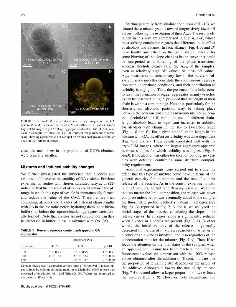
FIGURE 3 Cryo-TEM and confocal microscopy images of the OA
system (5 mM) in bicine buffer (0.2 M) at different pH values. (A–C)
Cryo-TEM images at pH 7.6 (large aggregates—droplets) (A), pH 8.4 (vesi-
cles) (B), and pH 9.7 (micelles) (C). (D) Confocal image (note the different
scale) showing a giant vesicle of OA (pH 8.5) with a background of smaller
ones, in the formation process.
282 Rendon et al.
cases the mean sizes in the population of GUVs obtainedwere typically smaller.
Mixtures and induced stability changes
We further investigated the influence that alcohols andalkanes could have on the stability of OA vesicles. Previousexperimental studies with shorter, saturated fatty acids (22)indicated that the presence of alcohols could enhance the pHrange in which this type of vesicle is spontaneously formedand reduce the value of the CAC. Therefore, we triedcombining alcohols and alkanes of different chain lengthswith OA in diverse ratios before hydrating them in the bicinebuffer (i.e., before the supramolecular aggregates were actu-ally formed). Note that alkanes are not soluble, nor can theybe dispersed in buffer unless in mixtures with OA (35).
TABLE 1 Percent aqueous content entrapped in OA
aggregates
Time (min)
Entrapment (%)
pH 7.5 pH 8.5 pH 10
0 5 5 0.73 75 5 0.95 14 5 0.97
60 1 5 1.82 56 5 1.34 13 5 0.24
180 <1 43 5 1.57 12 5 0.60
Content release was measured at various times after removal of nonentrap-
ped solutes by column chromatography (see Methods). 100% release was
measured after addition of 1 mM Triton X-100. Values are expressed as
the mean 5 SD (n ¼ 3).
Biophysical Journal 102(2) 278–286
Starting generally from alkaline conditions (pH ~10), wetitrated these mixed systems toward progressively lower pHvalues, following the evolution of their A400. The results ob-tained in this way are summarized in Fig. 4, A–F, whosemost striking conclusion regards the difference in the effectof alcohols and alkanes. In fact, alkanes (Fig. 4, A and D)have hardly any effect on the oleic system, except forsome blurring of the slope changes in the curve that couldbe interpreted as a softening of the phase transitions,whereas alcohols clearly raise the A400 of the samples,even at relatively high pH values. At these pH values,A400 measurements remain very low in the pure-control-system, since micelles constitute the spontaneous aggrega-tion state under those conditions, and their contribution toturbidity is negligible. Thus, the presence of alcohols seemsto favor the formation of bigger aggregates, mainly vesicles,as can be observed in Fig. 5, provided that the length of theirchain is within a certain range. Note that, particularly for theshorter-chain alcohols, partition may be taking placebetween the aqueous and lipidic environments. For an orig-inal alcohol/OA (1:10) ratio, the use of different-chain-length alcohols leads to significant increases in turbidityfor alcohols with chains in the 10- to 14-carbon range(Fig. 4, B and E). For a given alcohol chain length in themixture with OA, the effect on turbidity was dose-dependent(Fig. 4, C and F). These results correlated well with thecryo-TEM images, where the largest aggregates appearedin those samples for which turbidity was highest (Fig. 5,A–D). If the alcohol was either too short or too long, no vesi-cles were detected, confirming some structural compati-bility requirement.
Additional experiments were carried out to study theeffect that this type of mixture could have in terms of thegeneral capacity for entrapment and the rate of contentrelease of the vesicles. As in the control experiments withpure OAvesicles, the ANTS/DPX assay was used. We foundthat no matter the lipid composition, the release was nevercomplete unless Triton was eventually added to the sample:the fluorimetric profile reached a plateau in all cases (seeFig. 6). As reported in Fig. 7, A and B, we analyzed theinitial stages of the process, calculating the slope of therelease curves. In all cases, slope is significantly reducedwhen alkanes or alcohols are present (Fig. 7 A). In otherwords, the initial velocity of the release is generallydecreased by the use of mixtures, regardless of whether analcohol or an alkane is involved, and also regardless of theconcentration ratio for the mixture (Fig. 7 A). Then, if wefocus the attention on the final states of the samples, whenan apparent equilibrium has been reached, their relativefluorescence values (in comparison with the 100% releasevalues obtained after the addition of Triton), indicate thatthe proportion of remaining dye depends on the nature ofthe additive. Although it lowers the rate of dye release(Fig. 7 A), octanol allows a larger proportion of dye to leavethe vesicles (Fig. 7 B). However, both hexadecane and
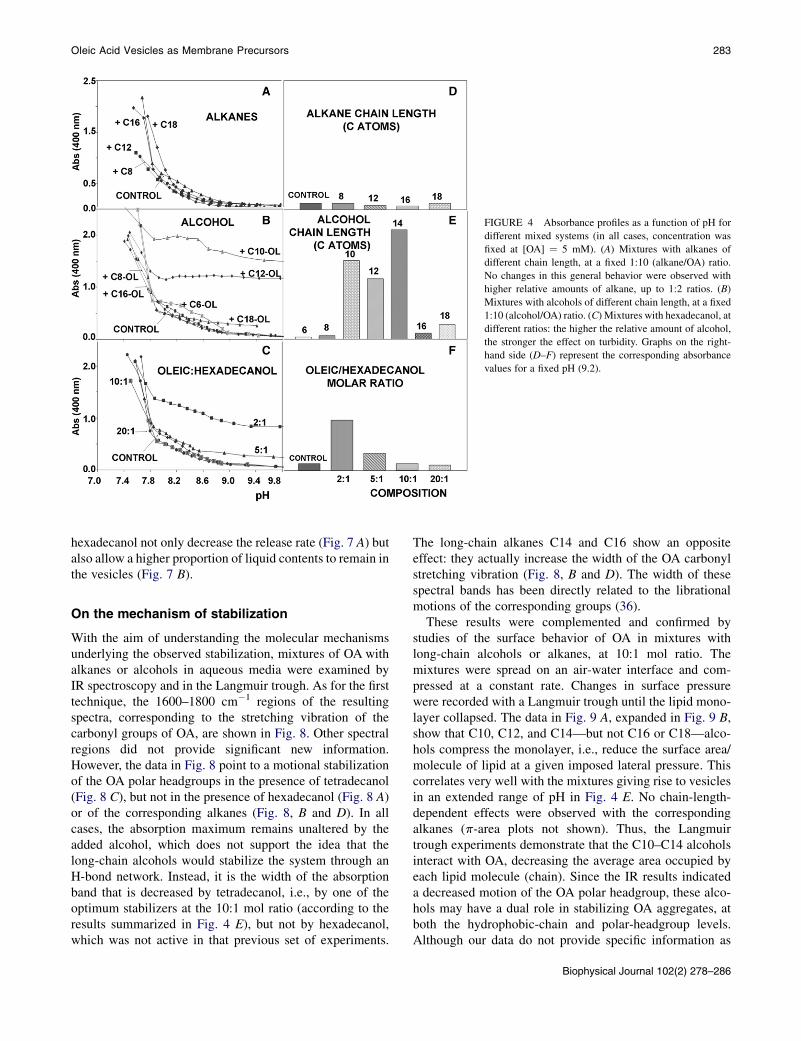
FIGURE 4 Absorbance profiles as a function of pH for
different mixed systems (in all cases, concentration was
fixed at [OA] ¼ 5 mM). (A) Mixtures with alkanes of
different chain length, at a fixed 1:10 (alkane/OA) ratio.
No changes in this general behavior were observed with
higher relative amounts of alkane, up to 1:2 ratios. (B)
Mixtures with alcohols of different chain length, at a fixed
1:10 (alcohol/OA) ratio. (C) Mixtures with hexadecanol, at
different ratios: the higher the relative amount of alcohol,
the stronger the effect on turbidity. Graphs on the right-
hand side (D–F) represent the corresponding absorbance
values for a fixed pH (9.2).
Oleic Acid Vesicles as Membrane Precursors 283
hexadecanol not only decrease the release rate (Fig. 7 A) butalso allow a higher proportion of liquid contents to remain inthe vesicles (Fig. 7 B).
On the mechanism of stabilization
With the aim of understanding the molecular mechanismsunderlying the observed stabilization, mixtures of OA withalkanes or alcohols in aqueous media were examined byIR spectroscopy and in the Langmuir trough. As for the firsttechnique, the 1600–1800 cm�1 regions of the resultingspectra, corresponding to the stretching vibration of thecarbonyl groups of OA, are shown in Fig. 8. Other spectralregions did not provide significant new information.However, the data in Fig. 8 point to a motional stabilizationof the OA polar headgroups in the presence of tetradecanol(Fig. 8 C), but not in the presence of hexadecanol (Fig. 8 A)or of the corresponding alkanes (Fig. 8, B and D). In allcases, the absorption maximum remains unaltered by theadded alcohol, which does not support the idea that thelong-chain alcohols would stabilize the system through anH-bond network. Instead, it is the width of the absorptionband that is decreased by tetradecanol, i.e., by one of theoptimum stabilizers at the 10:1 mol ratio (according to theresults summarized in Fig. 4 E), but not by hexadecanol,which was not active in that previous set of experiments.
The long-chain alkanes C14 and C16 show an oppositeeffect: they actually increase the width of the OA carbonylstretching vibration (Fig. 8, B and D). The width of thesespectral bands has been directly related to the librationalmotions of the corresponding groups (36).
These results were complemented and confirmed bystudies of the surface behavior of OA in mixtures withlong-chain alcohols or alkanes, at 10:1 mol ratio. Themixtures were spread on an air-water interface and com-pressed at a constant rate. Changes in surface pressurewere recorded with a Langmuir trough until the lipid mono-layer collapsed. The data in Fig. 9 A, expanded in Fig. 9 B,show that C10, C12, and C14—but not C16 or C18—alco-hols compress the monolayer, i.e., reduce the surface area/molecule of lipid at a given imposed lateral pressure. Thiscorrelates very well with the mixtures giving rise to vesiclesin an extended range of pH in Fig. 4 E. No chain-length-dependent effects were observed with the correspondingalkanes (p-area plots not shown). Thus, the Langmuirtrough experiments demonstrate that the C10–C14 alcoholsinteract with OA, decreasing the average area occupied byeach lipid molecule (chain). Since the IR results indicateda decreased motion of the OA polar headgroup, these alco-hols may have a dual role in stabilizing OA aggregates, atboth the hydrophobic-chain and polar-headgroup levels.Although our data do not provide specific information as
Biophysical Journal 102(2) 278–286

FIGURE 7 Effect of additives on the release of entrapped dyes. (A)
Changes in the intial slopes (rates). (B) Percent of nonreleased dye (taking
as the 100% reference the value obtained after Triton addition).FIGURE 5 Cryo-TEM images of various OA-alcohol mixtures ([OA] ¼5 mM) in bicine buffer (0.2 M) at high pH values. The images support the
absorbance profiles results shown in Fig. 4, demonstrating that vesicles and
aggregates of vesicles form in the strongly alkaline region (A–C) but not for
a short-chain alcohol, octanol (D).
284 Rendon et al.
to the location of alkanes/alcohols within the OA bilayer, theIR spectra shown in Fig. 8 clearly reflect that in most—if notall—cases, the lipid-water interface is perturbed by bothalcohols and alkanes, though perhaps in a different way.
DISCUSSION AND PERSPECTIVES
The experimental characterization of the OA system (Figs.1–3) reveals several of its most salient features. First, the
FIGURE 6 Release of entrapped water-soluble dyes. Release kinetics of
the ANTS/DPX pair, whose fluorimetric signal was followed for ~24 h,
after removal from the external milieu by a gel filtration column, showing
OA control (open circles), OA/octanol (20:1) (solid squares), OA/hexade-
cane (5:1) (solid triangles), and OA/hexadecanol (2:1) (solid circles). After
addition of Triton X-100, release measured 100%.
Biophysical Journal 102(2) 278–286
different aggregation states (micelles, vesicles, and drop-lets) depend on the pH of the solution. Second, osmolarityhas a critical influence on the stability of the aggregates,particularly the vesicles, as reported by Monnard andcolleagues (11) for decanoic acid vesicles. Third, thecapacity for entrapment of these spontaneously formedvesicle systems was also verified.
We have further demonstrated that mixtures with alcohols(but not with alkanes) have a strong effect on the aggrega-tion properties of the OA system, making possible the spon-taneous generation of vesicles for pH values well above thepKa of the acid. This confirmed similar experimental resultsobtained by Apel et al. (22) with shorter fatty acids. Therelevance of this finding can be better appreciated in thecontext of the appearance in the biosphere of the firstcompartments and their subsequent development throughincreasingly complex molecules, from fatty acids to thephospholipids that build up the present membranes. Har-greaves and Deamer (7) had already observed that disper-sion of dodecanoate gave rise to vesicles in a wider pHrange when dodecanol was also present. Monnard andDeamer (37) suggest that if alcohols manage to cometogether with fatty acids at some initial stage, for instance,by providing additional stability to the vesicles, as supportedalso by our results, the production of glycerol lipid deriva-tives would be facilitated, as an intermediate step towardamphiphilic compounds whose polar head included a phos-phate group. Several pieces of evidence (25,38) point inanalogous directions and highlight the importance ofstudying the prebiotically available compounds (e.g.,simple, single-stranded fatty acids) that only later, withthe advent of biosynthetic, protometabolic pathways, would

FIGURE 8 The baseline-corrected 1600–
1800 cm�1 region of the IR spectra of OA, either
pure or in mixtures with alkanes or alcohols. This
spectral region corresponds to the stretching vibra-
tion of the fatty acid carbonyl group. (A) OA 5
C16-alcohol (2:1). (B) OA 5 C16-alkane (2:1).
(C) OA 5 C14-alcohol (10:1). (D) OA 5 C14-
alkane (10:1). The spectrum of pure OA is drawn
with a dashed line.
Oleic Acid Vesicles as Membrane Precursors 285
develop into more complex (two-chain-stranded and largerpolar head) lipids (39).
Our results therefore confirm those by Apel andcolleagues (22) and extend them to long-chain fatty acids
FIGURE 9 Compression isotherms of pure OA and of mixtures with
alkanesor alcohols at a 10:1mol ratio.Measurementswere carriedout at 22�C.
and alcohols. In this context, it is not realistic to assume,as a premise, the presence of pure vesicles made of a singletype of compound, which is why we seek to combine OAwith other simple compounds, like alcohols and alkanes ofdiverse chain lengths, which surely coexist with fatty acidsin any prebiotic scenario. In addition to the analysis ofvesicle stability discussed above, the content-release studiesindicate other possible advantages of mixtures, namely,slower release profiles and a higher entrapment capacityof the vesicles. Moreover, the IR spectroscopic and Lang-muir trough experiments carried out provide solid evidencethat certain alcohols (C10, C12, and C14) compress laterallythe OA aliphatic chains and reduce the motility of theircarbonyl groups. These experiments thus help to builda coherent molecular explanation of the observed macro-scopic changes in lipid aggregation and vesicle stability.
The approach followed here could be extended in variousways. Mixtures with other compounds, not just alcohols andalkanes, should be tried (13,23). With the appropriatecombination of amphiphiles, many of the difficulties thatthis hypothetical scenario for the origin of cell membranesposes, like the apparent lack of stability of fatty acid vesi-cles, could be overcome. A growing number of laboratoriesare becoming aware of the relevance of using increasinglycomplex mixtures in the quest to elaborate more solid andrealistic membrane models, not only in the context of theorigins of cellularity, but also for a better understanding ofbiomembrane properties and behavior at large.
SUPPORTING MATERIAL
A figure is available at http://www.biophysj.org/biophysj/supplemental/
S0006-3495(11)05455-5.
Biophysical Journal 102(2) 278–286

286 Rendon et al.
The authors are grateful to Dr. P. Walde (Eidgenossische Technische Hoch-
schule, Zurich, Switzerland) for critically reading a previous version of the
manuscript.
This work was supported in part by grants from the Spanish Ministerio de
Ciencia e Innovacion (MICINN) to K.R.M. (FFI2008-06348-C02-01) and
F.M.G. (BFU2007-62062), and from the University of the Basque Country
to F.M.G. (GIV06/42). K.R.M. holds a Ramon y Cajal Research Fellowship
and also acknowledges support from COST Action CM0703.
REFERENCES
1. Sole, R. V., A. Munteanu, ., J. Macıa. 2007. Synthetic protocellbiology: from reproduction to computation. Philos. Trans. R. Soc.Lond. B Biol. Sci. 362:1727–1739.
2. Pohorille, A., and D. Deamer. 2009. Self-assembly and function ofprimitive cell membranes. Res. Microbiol. 160:449–456.
3. Mansy, S. S., and J. W. Szostak. 2009. Reconstructing the emergence ofcellular life through the synthesis of model protocells. Cold SpringHarb. Symp. Quant. Biol. 74:47–54.
4. Chen, I. A., and P. Walde. 2010. From self-assembled vesicles to pro-tocells. Cold Spring Harb. Perspect. Biol. 2:a002170.
5. Ekwald, P., and L. Mandell. 1969. Solutions of alkali soaps and waterin fatty acids. I. Region of existence of the solutions. Kolloid Z. Polym.233:938–944.
6. Gebicki, J. M., and M. Hicks. 1973. Ufasomes are stable particles sur-rounded by unsaturated fatty acid membranes. Nature. 243:232–234.
7. Hargreaves, W. R., and D. W. Deamer. 1978. Liposomes from ionic,single-chain amphiphiles. Biochemistry. 17:3759–3768.
8. Cistola, D. P., D. Atkinson,., D. M. Small. 1986. Phase behavior andbilayer properties of fatty acids: hydrated 1:1 acid-soaps. Biochemistry.25:2804–2812.
9. Cistola, D. P., J. A. Hamilton, ., D. M. Small. 1988. Ionization andphase behavior of fatty acids in water: application of the Gibbs phaserule. Biochemistry. 27:1881–1888.
10. Walde, P., R. Wick,., P. L. Luisi. 1994. Autopoietic self-reproductionof fatty acid vesicles. J. Am. Chem. Soc. 116:11649–11654.
11. Monnard, P. A., C. L. Apel,., D. W. Deamer. 2002. Influence of ionicinorganic solutes on self-assembly and polymerization processesrelated to early forms of life: implications for a prebiotic aqueousmedium. Astrobiology. 2:139–152.
12. Chen, I. A., R. W. Roberts, and J. W. Szostak. 2004. The emergence ofcompetition between model protocells. Science. 305:1474–1476.
13. Namani, T., and D. W. Deamer. 2008. Stability of model membranes inextreme environments. Orig. Life Evol. Biosph. 38:329–341.
14. Mansy, S. S. 2009. Model protocells from single-chain lipids. Int. J.Mol. Sci. 10:835–843.
15. Berclaz, N., M. Muller, ., P. L. Luisi. 2001. Growth and transforma-tion of vesicles studied by ferritin labeling and cryotransmission elec-tron microscopy. J. Phys. Chem. B. 105:1056–1064.
16. Hanczyc, M. M., S. M. Fujikawa, and J. W. Szostak. 2003. Experi-mental models of primitive cellular compartments: encapsulation,growth, and division. Science. 302:618–622.
17. Luisi, P. L., P. S. Rasi, and F. Mavelli. 2004. A possible route to prebi-otic vesicle reproduction. Artif. Life. 10:297–308.
18. Chen, I. A., and J. W. Szostak. 2004. A kinetic study of the growth offatty acid vesicles. Biophys. J. 87:988–998.
Biophysical Journal 102(2) 278–286
19. Stano, P., E. Wehrli, and P. L. Luisi. 2006. Insights into the self-repro-duction of oleate vesicles. J. Phys. Condens. Matter. 18:S2231–S2238.
20. Morigaki, K., and P. Walde. 2007. Fatty acid vesicles. Curr. Opin.Colloid Interface Sci. 12:75–80.
21. Haines, T. H. 1983. Anionic lipid headgroups as a proton-conductingpathway along the surface of membranes: a hypothesis. Proc. Natl.Acad. Sci. USA. 80:160–164.
22. Apel, C. L., D. W. Deamer, and M. N. Mautner. 2002. Self-assembledvesicles of monocarboxylic acids and alcohols: conditions for stabilityand for the encapsulation of biopolymers. Biochim. Biophys. Acta.1559:1–9.
23. Namani, T., and P. Walde. 2005. From decanoate micelles to decanoicacid/dodecylbenzenesulfonate vesicles. Langmuir. 21:6210–6219.
24. Mansy, S. S., J. P. Schrum, ., J. W. Szostak. 2008. Template-directedsynthesis of a genetic polymer in a model protocell. Nature. 454:122–125.
25. Maurer, S. E., D. W. Deamer, ., P. A. Monnard. 2009. Chemicalevolution of amphiphiles: glycerol monoacyl derivatives stabilize plau-sible prebiotic membranes. Astrobiology. 9:979–987.
26. Thomas, J. A., and F. R. Rana. 2007. The influence of environmentalconditions, lipid composition, and phase behavior on the origin ofcell membranes. Orig. Life Evol. Biosph. 37:267–285.
27. Dejanovi�c, B., V. Noethig-Laslo, ., P. Walde. 2011. On the surfaceproperties of oleate micelles and oleic acid/oleate vesicles studied byspin labeling. Chem. Phys. Lipids. 164:83–88.
28. Deamer, D. W., and J. P. Dworkin. 2005. Chemistry and physics ofprimitive membranes. Top. Curr. Chem. 259:1–27.
29. Walde, P. 2006. Surfactant assemblies and their various possible rolesfor the origin(s) of life. Orig. Life Evol. Biosph. 36:109–150.
30. Ellens, H., J. Bentz, and F. C. Szoka. 1985. Hþ- and Ca2þ-inducedfusion and destabilization of liposomes. Biochemistry. 24:3099–3106.
31. Nieva, J. L., F. M. Goni, and A. Alonso. 1989. Liposome fusion cata-lytically induced by phospholipase C. Biochemistry. 28:7364–7367.
32. Angelova, M. I., S. Soleau, ., P. Bothorel. 1992. Preparation of giantvesicles by external AC fields. Kinetics and application. Prog. ColloidPolym. Sci. 89:127–131.
33. Morigaki, K., P. Walde,., B. H. Robinson. 2003. Thermodynamic andkinetic stability. Properties of micelles and vesicles formed by the dec-anoic acid/decanoate system. Colloids Surf. A Physicochem. Eng. Asp.213:37–44.
34. Marques, E. F. 2000. Size and stability of catanionic vesicles: effects offormation path, sonication and aging. Langmuir. 16:4798–4807.
35. Urbina, P., A. Alonso,., J. Sot. 2006. Alkanes are not innocuous vehi-cles for hydrophobic reagents in membrane studies. Chem. Phys.Lipids. 139:107–114.
36. Castresana, J., J. M. Valpuesta,., F. M. Goni. 1991. An infrared spec-troscopic study of specifically deuterated fatty-acyl methyl groups inphosphatidylcholine liposomes. Biochim. Biophys. Acta. 1065:29–34.
37. Monnard, P.-A., and D. W. Deamer. 2002. Membrane self-assemblyprocesses: steps toward the first cellular life. Anat. Rec. 268:196–207.
38. Apel, C. L., and D. W. Deamer. 2005. The formation of glycerol mono-decanoate by a dehydration condensation reaction: increasing thechemical complexity of amphiphiles on the early Earth. Orig. LifeEvol. Biosph. 35:323–332.
39. Pereto, J., P. Lopez-Garcıa, and D. Moreira. 2004. Ancestral lipidbiosynthesis and early membrane evolution. Trends Biochem. Sci.29:469–477.