Embed Size (px)
Citation preview

Repository of the Max Delbrück Center for Molecular Medicine (MDC) Berlin (Germany) http://edoc.mdc-berlin.de/14021/
Models of chromosome structure
Nicodemi, M., Pombo, A. Published in final edited form as:
Current Opinion in Cell Biology. 2014 Jun ; 28: 90-95 | doi: 10.1016/j.ceb.2014.04.004 Elsevier ►

1
Title: Models of chromosome structure
Mario Nicodemi1,* and Ana Pombo2,*
1Universita' di Napoli "Federico II", Dipartimento di Fisica, INFN Sezione di Napoli, CNR-‐SPIN, Complesso Universitario di Monte S. Angelo, Via Cintia, 80126 Napoli, Italy. E-mail: [email protected] 2Berlin Institute for Medical Systems Biology, Max Delbrück Center for Molecular Medicine, Robert Rössle Strasse 10, 13125 Berlin-Buch, Germany. E-mail: [email protected] *To whom correspondence should be addressed:
Keywords: genome organization, long-‐range chromatin interactions, chromosome
conformation capture (3C), microscopy, fluorescence in situ hybridization, polymer
physics, computer simulations.

2
Abstract
Understanding the mechanisms that control chromosome folding in the nucleus of
eukaryotes and their contribution to gene regulation is a key open issue in
molecular biology. Microscopy and chromatin-‐capture techniques have shown that
chromatin has a complex organization, which dynamically changes across
organisms and cell types. The need to make sense of such a fascinating complexity
has prompted the development of quantitative models from physics, to find the
principles of chromosome folding, its origin and function. Here, we concisely review
recent advances in chromosome modeling, focusing on a recently proposed
framework, the Strings & Binders Switch (SBS) model, which recapitulates key
features of chromosome organization in space and time.
Introduction
Chromosomes have a complex organization across spatial scales within the cell
nucleus [1-‐3]. They occupy separate, yet interacting chromosomal territories [4, 1]
where long-‐range chromatin interactions are functionally important. Each territory
is partitioned in megabasepair-‐long domains enriched for internal contacts, known
as topological associated domains (TADs) [1, 5, 6], while long stretches of chromatin
interact with the nuclear lamina (the lamina-‐associated domains, LADs) [3], and
with a variety of other functional compartments [1]. Genes come together, for
instance, on specialized transcription factories, promoting proximity between
different transcription units according to their expression state [7-‐10]. Splicing
factors, the machinery that splices nascent transcripts into messenger RNA,
accumulate in splicing speckles, which are often associated with active genes, while
repressed chromatin associates with heterochromatic regions or Polycomb bodies
[1]. Early replication origins are also clustered in replication factories, which stably
reassemble in consecutive cell cycles [11].
The understanding of chromatin organization has inspired the development of
models for many years [12-‐15]. Early models were often a visual summary of
conformations identified by microscopy, aiming mostly to consolidate, in one image,

3
the many features observed (e.g. [1]). The increasing level of details exposed by
experimental advances has triggered the need to further develop quantitative
polymer models from physics. And here we focus especially on a few, more recent
developments.
Polymer models of chromatin organization
The simplest model of a free polymer, i.e., a polymer experiencing only self-‐
avoidance effects, is the Self-‐Avoiding Walk (SAW) model. A SAW polymer folds
spontaneously in a random conformation (Fig. 1) and, thus, in a mixture, entropy
induces full intermingling of polymers. Pioneer work by Kreth and Cremer (2004)
introduced a model where polymers also experience a strong self-‐attraction force
that can produce discrete chromosome territories [16].
Entropy might also play a role in favoring territorial separation by limiting the
intermingling between different chromosomes: models of free polymers folded into
clustered loops can hardly find the space to penetrate into each other, experiencing
an effective repulsion of entropic origin partially preventing overlap [17-‐19, 14].
These models help bridge the original view of chromosome territories fully
separated by channels devoid of DNA (the CT-‐IC view) [20, 1], and the discovery of
their intermingling (the ICN view) [4, 21]. Entropy-‐based models, however, cannot
explain the variety of specific, functional contacts occurring through chromatin
looping (such as enhancer-‐promoter interactions), its domain structure (LADs,
TADs) and, in particular, fail to describe the behavior of the contact probability
between genomic loci reported by Hi-‐C experiments [22, 23].
The development of ‘chromosome conformation capture’ (3C) methods [24], and
their genome-‐wide extensions such as Hi-‐C [25, 22, 26], has invigorated this field
with the prospect of semi-‐quantitative measures of chromosome folding. Hi-‐C
contact matrices have shown, for instance, that the average interaction probability
between pairs of genomic loci decreases with their genomic separation,
approximately with power law decay in the 0.5-‐7Mb range [22].

4
The contact probability and, in particular, its power law exponent, α, is different,
though, in different systems [23]. For instance, human embryonic stem cells (ESCs)
have α~1.6 [23], while α~1.1 in human interphase lymphoblastoid cells [22] and in
metaphase HeLa cells α~0.5 [27]. The exponent α reported in different species also
varies widely: in Drosophila α~0.7-‐0.8 [28], in yeast α~1.5 [29], in mouse ESCs the
Xist locus has α~0.7-‐0.9 [6, 30]. Even in a given cell system different chromosomes
can have different exponents [31, 23]; e.g. the gene-‐poor and gene-‐dense human
chromosomes 18 and 19 have α around 1.08 and 1.3, respectively, in human
lymphoblastoid cells [23]. These observations have contradicted the initial hope
that a single universal architecture, originally envisaged in the Fractal Globule (FG)
model [22] (where α=1), might describe chromosome folding.
The FG state [22] is a transient, highly unstable [32] conformation encountered, e.g.,
by a free polymer allowed to expand from an initial high-‐compaction, non-‐entangled
state. For the FG to be observable, the activity of topoisomerases (important nuclear
enzymes that resolve chromatin entanglement by cutting the DNA and passing it
through) must be negligible. The FG is knot free, which might allow for quick
unfolding events in the chromatin structure. However, the FG model also fails to
explain the formation of TADs or LADs, and microscopy data of inter-‐locus
distances, in particular the fact that the separation of distant regions on a
chromosome saturates at the territory size [23].
An alternative scenario to explain chromosome folding was proposed in the
Interchromatin Network (ICN) model [4], where folding derives from chromatin
interactions with complexes that promote looping (e.g. contacts between co-‐
expressed genes at transcription or replication factories) and associations with
nuclear landmarks, such as the lamina. The Strings & Binders Switch (SBS) model
[33, 34, 23] explored for the first time such a scenario on quantitative grounds using
polymer physics (Fig. 2a).

5
The SBS model, in its simplest version, investigates a polymer and its interactions
with diffusing binders that can crosslink the polymer specific binding sites. It can
also be expanded to consider many polymers, different binder species, and
interactions with nuclear landmarks [35, 36]. In the model, the position of the
binding sites along the polymer is assigned; the concentration and affinity of binders
can be changed [23], as resulting, for instance, from the up-‐regulation of a
corresponding gene or from chemical modifications of a chromosome sequence.
With simple and generic settings, the SBS model has been used to illustrate several
aspects of chromatin folding in quantitative terms: (a) three major folding classes
exist (open, closed and Θ-‐point; Fig. 2b), corresponding to stable emergent phases of
polymer physics (the Fractal Globule state is one of many possible transient states
of the SBS model); (b) conformational changes can be sharply regulated (switch-‐
like) by simple strategies, e.g., protein up-‐regulation or chromatin modifications,
which may help understand the mechanisms employed, e.g., to transduce (analog)
transcription factor levels into (digital) conformational switches; (c) that randomly
diffusing binding molecules can establish and dynamically change, by
thermodynamics mechanisms, architectural patterns, such as territory formation,
TADs or LADs, and the looping out of large stretches of chromatin from territories
(Fig. 2c); (d) that population and single cell microscopy and Hi-‐C data, such as
contact probabilities (Fig. 3) and spatial distances, can be rationalized in a single
framework [23]. The results of the SBS model are confirmed by similar findings in
the Dynamic Loop (DL) model, which has been used, in particular, to explore the
effects of entropy in mixtures of long, looped polymers [37, 14].
The picture that emerges from the SBS model is that chromatin exists inside nuclei
as a complex mixture of differently folded regions according to local specific factors,
which can self-‐organize across spatial scales by general physical mechanisms.
Specificity of binding at different loci or domains can be achieved, for example
through a combination of single molecular factors. In fact, more complex
architectures, with different, nested layers of organization can be constructed

6
within the SBS model. Beyond binding factors, other molecular mechanisms are
likely to contribute to chromatin folding into compact conformations, such as
supercoiling [30] and plectoneme formation [38].
Technical limitations
Irrespective of promising developments, it is helpful to appreciate the limitations of
the different methodologies. Imaging of nuclear architecture has improved to
exciting extents, but it is not fraught from artifacts (e.g. milder fixation and structure
loss) and currently suffers from low throughput options to screen broadly for
chromatin interactions, with exciting exceptions [39]. 3C-‐based mapping of
chromatin interactions has limitations too. Cells are also mildly fixed, broken up to
extract nuclei, DNA is cut by restriction enzymes and DNA free ends ligated, before
sequencing and alignment, all steps being fraught with unclear inefficiencies and
biases [40, 41]. Efforts to improve all these technologies are ongoing.
Similarly, we have to overcome limitations of physics models, such as their hidden
or oversimplifying assumptions. Ad-‐hoc prepared polymer structures have been
discussed, which might reproduce architectures identified experimentally without
adding a real new level of understanding of the underlying mechanisms. Due to
computer speed limitations, it is currently only possible to model comparatively
simple polymers, but as computer power grows soon whole chromosomes will be
modelled at high resolution. Yet, the discovery that polymer properties scale with
polymer size, awarded the Nobel Prize in Physics, has already demonstrated that
general and important features of chromosome folding can be derived using
relatively short and simple polymers [42].
Future perspectives
Many issues remain that require the development of more detailed models. On a
general ground, for instance, the effects of crowding have been only partially
considered as much as the constraints imposed by nuclear size and shape or by the
associations with nuclear bodies. Nevertheless, polymer models can be employed to

7
study specific genomic loci (e.g., the Xist locus) [36] to discriminate different
biological scenarios and thus helping to identify the determinants of folding.
Polymer models constrained by Hi-‐C data have been used to attempt
reconstructions of chromosome 3D shapes [43, 44]. These efforts suffer from the
limitation that population averaged Hi-‐C data return ‘average’ conformations
depending on the specific type of constraints and algorithms employed. To
circumvent such limitations, more realistic polymer models, such as the SBS, can be
used to reconstruct spatial conformations, and have the potential to predict
chromatin interaction sites that best explain Hi-‐C data.
From a biological point of view a number of key questions are still unresolved, such
as the origin of (a) specificity of interactions (e.g. why there is a preference for
pairing between specific chromosomes in specific cell types [45, 4] and why are
chromosome homologues often separated from each other [46]), (b) chromosomal
intermingling [4], or (c) the effects of other cellular processes, such as gene
expression or DNA replication, on chromosome organization.
In conclusion, as the complex 4D organization of chromatin remains still largely
mysterious, simple models of polymer physics, tested against real data, are trying to
provide a first picture of the basic principles and molecular mechanisms of folding.
In the near future, further developments in the models employed, exploiting
technical progresses and more detailed biological information, could push even
further our comprehension and could guide the design of targeted experiments to
resolve some of the many crucial open questions, hopefully advancing also our
understanding of related diseases.
Acknowledgements
A.P. thanks the Helmholtz Foundation for support.

8
References
[1] Lanctot C, Cheutin T, Cremer M, Cavalli G, Cremer T: Dynamic genome architecture in the nuclear space: Regulation of gene expression in three dimensions. Nature Rev Genet 2007, 8:104-‐115.
[2] Misteli T: Beyond the sequence: Cellular organization of genome function. Cell 2007, 128:787-‐800.
[3] Bickmore WA, van Steensel B: Genome architecture: Domain organization of interphase chromosomes. Cell 2013, 152:1270-‐1284.
[4] Branco MR, Pombo A: Intermingling of chromosome territories in interphase suggests role in translocations and transcription-‐dependent associations. PLoS Biol 2006, 4:e138.
[5] Dixon JR, Selvaraj S, Yue F, Kim A, Li Y, Shen Y, Hu M, Liu JS, Ren B: Topological domains in mammalian genomes identified by analysis of chromatin interactions. Nature 2012, 485:376-‐380.
[6] Nora EP, Lajoie BR, Schulz EG, Giorgetti L, Okamoto I, Servant N, Piolot T, van Berkum NL, Meisig J, Sedat J, et al.: Spatial partitioning of the regulatory landscape of the X-‐inactivation centre. Nature 2012, 485:381-‐385.
[7] Pombo A, Jackson DA, Hollinshead M, Wang Z, Roeder RG, Cook PR: Regional specialization in human nuclei: Visualization of discrete sites of transcription by RNA polymerase III. EMBO J 1999, 18:2241-‐2253.
[8] Osborne CS, Chakalova L, Mitchell JA, Horton A, Wood AL, Bolland DJ, Corcoran AE, Fraser P: Myc dynamically and preferentially relocates to a transcription factory occupied by igh. PLoS Biol 2007, 5:e192.
[9] Ferrai C, Xie SQ, Luraghi P, Munari D, Ramirez F, Branco MR, Pombo A, Crippa MP: Poised transcription factories prime silent upa gene prior to activation. PLoS Biol 2010, 8:e1000270.
[10] Schoenfelder S, Sexton T, Chakalova L, Cope NF, Horton A, Andrews S, Kurukuti S, Mitchell JA, Umlauf D, Dimitrova DS, et al.: Preferential associations between co-‐regulated genes reveal a transcriptional interactome in erythroid cells. Nat Genet 2010, 42:53-‐61.
[11] Jackson DA, Pombo A: Replicon clusters are stable units of chromosome structure: Evidence that nuclear organization contributes to the efficient activation and propagation of s phase in human cells. J Cell Biol 1998, 140:1285-‐1295.
[12] Langowski J: Polymer chain models of DNA and chromatin. Eur Phys J E Soft Matter 2006, 19:241-‐249.
[13] Emanuel M, Radja NH, Henriksson A, Schiessel H: The physics behind the larger scale organization of DNA in eukaryotes. Phys Biol 2009, 6:025008.
[14] Tark-‐Dame M, van Driel R, Heermann DW: Chromatin folding-‐-‐from biology to polymer models and back. J Cell Sci 2011, 124:839-‐845.
* This review discusses multiple roles of entropy in shaping chromosome conformations.
[15] Barbieri M, Scialdone A, Gamba A, Pombo A, Nicodemi M: Polymer physics, scaling and heterogeneity in the spatial organisation of chromosomes in the cell nucleus. Soft Matter 2013, 9:8631-‐8635.

9
[16] Kreth G, Finsterle J, von Hase J, Cremer M, Cremer C: Radial arrangement of chromosome territories in human cell nuclei: A computer model approach based on gene density indicates a probabilistic global positioning code. Biophys J 2004, 86:2803-‐2812.
[17] Marenduzzo D, Micheletti C, Cook PR: Entropy-‐driven genome organization. Biophys J 2006, 90:3712-‐3721.
*A discussion of the effects of entropy-‐based interactions on chromatin organization.
[18] Bohn M, Heermann DW, van Driel R: Random loop model for long polymers. Physical review. E, Statistical, nonlinear, and soft matter physics 2007, 76:051805.
[19] Rosa A, Everaers R: Structure and dynamics of interphase chromosomes. Plos Comp Biol 2008, 4:e1000153.
[20] Cremer T, Kreth G, Koester H, Fink RH, Heintzmann R, Cremer M, Solovei I, Zink D, Cremer C: Chromosome territories, interchromatin domain compartment, and nuclear matrix: An integrated view of the functional nuclear architecture. Crit Rev Eukaryot Gene Expr 2000, 10:179-‐212.
[21] Branco MR, Pombo A: Chromosome organization: New facts, new models. Trends Cell Biol 2007, 17:127-‐134.
[22] Lieberman-‐Aiden E, van Berkum NL, Williams L, Imakaev M, Ragoczy T, Telling A, Amit I, Lajoie BR, Sabo PJ, Dorschner MO, et al.: Comprehensive mapping of long-‐range interactions reveals folding principles of the human genome. Science 2009, 326:289-‐293.
**The Hi-‐C method was introduced, allowing genome-‐wide mapping of chromatin contacts, and measurement of the frequency of chromatin interactions. It also proposes the Fractal Globule model to explain properties of chromatin folding at 0.5-‐7Mb genomic scales.
[23] Barbieri M, Chotalia M, Fraser J, Lavitas LM, Dostie J, Pombo A, Nicodemi M: Complexity of chromatin folding is captured by the strings and binders switch model. PNAS 2012, 109:16173-‐16178.
**The SBS model is used here for the first time to explore single cell FISH data and population based HiC data. It explains many features of available experimental data, and proposes a scenario where chromatin is a mixture of differently folded regions.
[24] Dekker J, Rippe K, Dekker M, Kleckner N: Capturing chromosome conformation. Science 2002, 295:1306-‐1311.
[25] Tolhuis B, Palstra RJ, Splinter E, Grosveld F, de Laat W: Looping and interaction between hypersensitive sites in the active beta-‐globin locus. Mol Cell 2002, 10:1453-‐1465.
[26] Rodley CD, Bertels F, Jones B, O'Sullivan JM: Global identification of yeast chromosome interactions using genome conformation capture. Fungal Genet Biol 2009, 46:879-‐886.
[27] Naumova N, Imakaev M, Fudenberg G, Zhan Y, Lajoie BR, Mirny LA, Dekker J: Organization of the mitotic chromosome. Science 2013, 342:948-‐953.
*This paper shows that chromatin conformation depends on the cycle stage and that a compact folding state is observed at metaphase.

10
[28] Sexton T, Yaffe E, Kenigsberg E, Bantignies F, Leblanc B, Hoichman M, Parrinello H, Tanay A, Cavalli G: Three-‐dimensional folding and functional organization principles of the Drosophila genome. Cell 2012, 148:458-‐472.
[29] Duan Z, Andronescu M, Schutz K, McIlwain S, Kim YJ, Lee C, Shendure J, Fields S, Blau CA, Noble WS: A three-‐dimensional model of the yeast genome. Nature 2010, 465:363-‐367.
[30] Benedetti F, Dorier J, Burnier Y, Stasiak A: Models that include supercoiling of topological domains reproduce several known features of interphase chromosomes. Nucleic Acids Res 2013.
*This paper discusses the hypothesis that supercoiling can have an effect in the structure of Hi-‐C contact matrices.
[31] Kalhor R, Tjong H, Jayathilaka N, Alber F, Chen L: Genome architectures revealed by tethered chromosome conformation capture and population-‐based modeling. Nature Biotech 2011, 30:90-‐98.
**Introduces an alternative 3C-‐based method, TCC, and the concept of modelling ensemble of chromosome conformations from genome-‐wide chromatin conformation capture. It incorporates the concept of population conformation in the interpretation of HiC data.
[32] Schram RD, Barkema GT, Schiessel H: On the stability of fractal globules. J Chem Phys 2013, 138:224901.
*An exploration of the unstable properties of the Fractal Globule state and the difficulties in generating it.
[33] Nicodemi M, Panning B, Prisco A: A thermodynamic switch for chromosome colocalization. Genetics 2008, 179:717-‐721.
**Here the idea that chromosome conformations result from chromatin interactions and are controlled by basic thermodynamic mechanisms of polymer physics was first introduced.
[34] Nicodemi M, Prisco A: Thermodynamic pathways to genome spatial organization in the cell nucleus. Biophys J 2009, 96:2168-‐2177.
[35] Scialdone A, Nicodemi M: Diffusion-‐based DNA target colocalization by thermodynamic mechanisms. Development 2010, 137:3877-‐3885.
[36] Scialdone A, Cataudella I, Barbieri M, Prisco A, Nicodemi M: Conformation regulation of the X chromosome inactivation center: A model. PLoS Comput Biol 2011, 7:e1002229.
*This paper introduces the idea that symmetry breaking and polymer physics mechanisms can contribute to produce different conformations on different alleles.
[37] Bohn M, Heermann DW: Diffusion-‐driven looping provides a consistent framework for chromatin organization. PloS One 2010, 5:e12218.
**This is an extensive study of the effects polymer interactions on chromatin folding. [38] Le TB, Imakaev MV, Mirny LA, Laub MT: High-‐resolution mapping of the
spatial organization of a bacterial chromosome. Science 2013, 342:731-‐734. [39] Roukos V, Voss TC, Schmidt CK, Lee S, Wangsa D, Misteli T: Spatial dynamics
of chromosome translocations in living cells. Science 2013, 341:660-‐664. [40] O'Sullivan JM, Hendy MD, Pichugina T, Wake GC, Langowski J: The statistical-‐
mechanics of chromosome conformation capture. Nucleus 2013, 4:390-‐398.

11
**Important study exploring limitations of chromosome conformation capture methods, in particular the biases introduced by ligation of each DNA fragment to at most two other fragments.
[41] Belmont AS: Large-‐scale chromatin organization: The good, the surprising, and the still perplexing. Curr Opin Cell Biol 2014, 26C:69-‐78.
[42] de Gennes PG, Scaling concepts in polymer physics. 1979: Cornell University Press.
[43] Umbarger MA, Toro E, Wright MA, Porreca GJ, Bau D, Hong SH, Fero MJ, Zhu LJ, Marti-‐Renom MA, McAdams HH, et al.: The three-‐dimensional architecture of a bacterial genome and its alteration by genetic perturbation. Mol Cell 2011, 44:252-‐264.
[44] Nagano T, Lubling Y, Stevens TJ, Schoenfelder S, Yaffe E, Dean W, Laue ED, Tanay A, Fraser P: Single-‐cell Hi-‐C reveals cell-‐to-‐cell variability in chromosome structure. Nature 2013, 502:59-‐64.
[45] Parada LA, McQueen PG, Misteli T: Tissue-‐specific spatial organization of genomes. Genome Biol 2004, 5:R44.
[46] Khalil A, Grant JL, Caddle LB, Atzema E, Mills KD, Arneodo A: Chromosome territories have a highly nonspherical morphology and nonrandom positioning. Chromosome Res 2007, 15:899-‐916.

12
Figure Legends
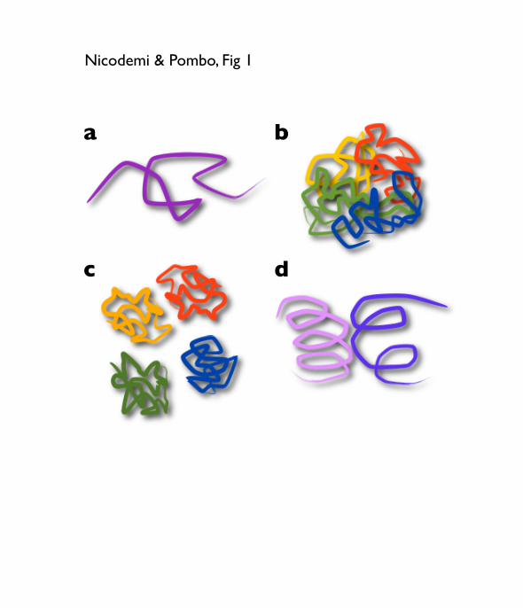
Fig. 1. Polymer models and conformations
a) The self-‐avoiding walk (SAW) model describes a free polymer, i.e., a polymer
having no interactions beyond excluded volume. A SAW polymer has a randomly
folded conformation.
b) A mixture of polymers would show substantial levels of intermingling because of
mixing entropy.
c) If each polymer experiences self-‐attraction (e.g. Kreth and Cremer model [16]),
polymers can confine into clearly separated territories.
d) If polymers have looped conformations, ‘rosette’-‐like, intermingling can be
reduced because loops interpenetrate each other with more difficulty and thus
experience an effective repulsion force (entropic force).
Fig. 2. The Strings & Binders Switch (SBS) model
a) In the SBS model [33, 23], chromatin is represented by a self-‐avoiding-‐walk
(SAW) chain, which has binding sites for diffusing binders. The basic model
parameters are the binder concentration, cm, and their affinity for the polymer
binding sites, EX. The interactions of polymer with molecular binders can produce
loops. The model stable emergent thermodynamic phases correspond to different
conformational classes.
b) The SBS model identifies a switch-‐like behavior in chromatin folding in response
to changes in binder affinity or concentration. The SBS phase diagram includes a
phase where the polymer folds in a random open conformation (in the universality
class of the free SAW) and a phase where it spontaneously folds into a compact
closed conformation. At the phase transition point, there is the Θ-‐point state.
Conformational changes are achieved by crossing the phase boundary, with no need
of parameter fine-‐tuning.
c) The formation of chromatin domains and chromatin looping can be modeled
within the SBS model, as result of the specialization of the polymer binding sites and
their binding molecules. The corresponding contact matrices have general features

13
similar to those found by Hi-‐C methods.
Fig. 3. Contact probabilities estimated from HI-‐C data are explained by the SBS
model.
a) The average contact probability, Pc(s), measures the extent of chromatin
interactions between pairs of loci separated by a given genomic distance.
b) In Hi-‐C data, the average contact probability, Pc(s), is found to decrease with their
genomic separation, s, with a power law decay at least within the 0.5-‐7Mb range,
Pc(s)~1/sα [22]. The exponent, α, of the power law is different in different systems
and in different chromosomes [23].
c) Within the SBS model, α is 2.1 in the open state, 1.5 in the Θ-‐point state, and 0 in
the compact state. Thus, mixtures of modelled SBS polymers including, for example,
a fraction, f, of open and a fraction, 1-‐f, of compact polymer regions can easily
explain the range of exponents found experimentally. This scenario simulates the
fact that different genomic regions assume open or closed chromatin conformations,
which need to be captured by polymer modelling approaches.
a b
d c
Nicodemi & Pombo, Fig 1

A schematic model
Strings&Binders Switch (SBS) model:System: a SAW polymer and a density, cm, of diffusing molecules with an affinity EX for its binding sites (a fraction f of the beads, e.g., 1/4).
Equilibrium/Dynamics: studied by Mean-Field theory & Monte Carlo simulations (on a lattice).
(Nicodemi&Prisco Biophys. J. ʻ09)
Questions: how are i) stable conformations (and their scaling properties) established? ii) architectural changes regulated?
b c Mean spatial distance
Contact Probability
Barbieri, Figure 1b,c
Gyration Radius
R
s
Rg
Rg is small
Rg
Rg is large
Polymer
�����������
���������� ���
Chromatin conformations, and patterns, can arise by interaction with randomly diffusing DNA-binding molecules
b c Mean spatial distance
Contact Probability
Barbieri, Figure 1b,c
Gyration Radius
R
s
Rg
Rg is small
Rg
Rg is large
Polymer
�����������
���������� ���
zoomSBS model Phase diagrama b
c
Nicodemi & Pombo, Fig 2

Hi-C data!
SBS model!
s!
Contact !probability, !
Pc!
Con
tact
pro
babi
lity
(Pc)
Con
tact
pro
babi
lity
(Pc)
Genomic distance, log(Mbp)
Genomic distance, log (s/s0)
Nicodemi & Pombo, Fig 3
![Compact and Discreet - yaesu.ru · Battery Life [5-5-90 duty with battery saver]MDC-1200® Features: - MDC-1200® ANI - MDC-1200® Call Alert - MDC-1200® Sel Call - MDC-1200® Radio](https://img.pdfslide.net/doc/110x75/5e80d3de6005c20fb639820d/compact-and-discreet-yaesuru-battery-life-5-5-90-duty-with-battery-savermdc-1200.jpg)