Embed Size (px)
Citation preview

Thomas Jefferson University Thomas Jefferson University
Jefferson Digital Commons Jefferson Digital Commons
Modern Surgery, 4th edition, by John Chalmers Da Costa Rare Medical Books
1903
Modern Surgery - Chapter 20. Diseases and Injuries of Muscles, Modern Surgery - Chapter 20. Diseases and Injuries of Muscles,
Tendons, and Bursae Tendons, and Bursae
John Chalmers Da Costa Jefferson Medical College
Follow this and additional works at: https://jdc.jefferson.edu/dacosta_modernsurgery
Part of the History of Science, Technology, and Medicine Commons
Let us know how access to this document benefits you
Recommended Citation Recommended Citation
Da Costa, John Chalmers, "Modern Surgery - Chapter 20. Diseases and Injuries of Muscles,
Tendons, and Bursae" (1903). Modern Surgery, 4th edition, by John Chalmers Da Costa. Paper
32.
https://jdc.jefferson.edu/dacosta_modernsurgery/32
This Article is brought to you for free and open access by the Jefferson Digital Commons. The Jefferson Digital Commons is a service of Thomas Jefferson University's Center for Teaching and Learning (CTL). The Commons is a showcase for Jefferson books and journals, peer-reviewed scholarly publications, unique historical collections from the University archives, and teaching tools. The Jefferson Digital Commons allows researchers and interested readers anywhere in the world to learn about and keep up to date with Jefferson scholarship. This article has been accepted for inclusion in Modern Surgery, 4th edition, by John Chalmers Da Costa by an authorized administrator of the Jefferson Digital Commons. For more information, please contact: [email protected].

Myalgia, or Muscular Rheumatism 553
XX. DISEASES AND INJURIES OF MUSCLES, TENDONS, AND BURSAE.
Myalgia, or muscular rheumatism, is a painful disorder of the voluntary muscles and of the fibrous and periosteal areas where they are attached. The term "muscular rheumatism" is not strictly correct. It is possible that in some cases the muscular structure is inflamed, but it is certain that in many cases the pain is distinctly neuralgic. Muscular rheumatism may he due to cold and wet, to over-exertion and strain, to acute infectious disorders, to syphilis, to chronic intoxications (lead, mercury, and alcohol), and to disturbances of the circulation. Gouty and rheumatic persons are especially predisposed, men being more liable to the disease than women. The disease is usually acute, but it may he chronic.
Symptoms.—Muscular rheumatism is apt to come on suddenly. The pain, which may be very acute and lancinating or may be dull and aching, is in some cases constantly present; in other cases it is awakened only by muscular contraction, and it is frequently relieved by pressure, though there is often some soreness. The skin above the muscle is sometimes tender to light pressure. The disease usually lasts for a few days, but it tends to recur. There is little, if any, fever.
Lumbago is myalgia of the muscles of the loins. Rheumatic iorticollis is myalgia of the muscles of the neck. Usually one side of the neck is attacked. The chin is turned from the affected side and the neck is stiff. Pleurodynia is myalgia of the intercostal muscles. The pain is very severe, is aggravated by deep respiration, by coughing, and by yawning, there may be tenderness, and the patient tries to limit chest-movement. In intercostal neuralgia the pain is limited, is not constant, but occurs in distinct paroxysms, and is linked with the presence of the tender spots of Valleix. Pleurodynia lacks the physical signs of pleurisy. Cephalodynia is myalgia of the muscles of the scalp. The muscles of the shoulder, upper dorsal region, abdomen, and extremities may also be attacked by myalgia. Myalgia must not be confused with the pains of locomotor ataxia.
Treatment.—Remove any obvious cause. Treat any existing diathesis, such as gout or rheumatism. Rest is of the first importance. For lumbago, put the person to bed. For pleurodynia, strap the side of the chest. A hypodermatic injection of morphin and atropin into the affected muscles at once allays the pain, and a deep injection of distilled water is sometimes curative. Relief may be afforded by painting the surface with 3o drops of a mixture of equal parts of guaiacol and glycerin and covering the painted area with cotton. The introduction of four or five aseptic needles into the muscles, and their retention for a few minutes, sometimes act most favor-ably. Ironing the skin above the painful muscles with a very warm iron, a piece of flannel being interposed, is a useful domestic remedy. Vigorous rubbing of the area with a piece of ice allays the pain. Hot poultices do good. If the pain is widely diffused, alters its seat, or is very obstinate, order hot baths or Turkish baths and administer diuretics. In chronic cases employ blisters or counter-irritation by the cautery, give iodid of potassium and nux vomica, and have the patient take a Turkish bath every week. The

554 Diseases and Injuries of Muscles, Tendons, and Bursx
constant electric current finds advocates. In an ordinary severe case order a hot bath, put the patient to bed with a hot-water bag over the part, and administer to grains of Dover's powder; the next morning order to be taken four times daily a capsule containing 5 grains of salol and 3 grains of phena. cetin, until the pain disappears. Citrate of potassium, citrate of lithium, chlorid of ammonium, or the salicylate of colchicin may be ordered instead of salol and phenacetin.
Infective myositis is a widespread inflammation of the voluntary muscles, due to an unknown infective cause. It is a disorder accompanied by pain and stitTness, by cutaneous edema, and by various paresthesiw. Myositis resembles trichinosis, and is distinguished from it only by spearing out a bit of muscle and examining it microscopically. Occasionally diffuse suppuration occurs.
Ordinary myositis arises from injuries, from syphilis, or from rheu-matism, and it presents the usual inflammatory symptoms. Contraction and adhesions may follow.
Treatment of Myositis.—lnfective myositis is treated by anodynes, stimu-lants, nutritious food, hot applications, and rest. If pus forms, it should be evacuated. Rheumatic myositis calls for the administration of the salicylates, the alkalies, or salol. Syphilitic myositis is treated with mercury and iodid of potassium. The remedies employed for myalgia are used in traumatic myositis.
Hypertrophy of the muscles may arise from their increased use. In pseudohypertrophic paralysis the bulk of the muscle is greatly augmented, but it contains less muscle-structure and more fat or connective tissue.
Atrophy of the muscles arises from want of use, from injury, from continuous pressure. from interference with the blood-supply, from disease of the nerves or their centers, or from lead-poisoning.
Degeneration of Muscles.—The muscles may undergo granular degeneration, waxy degeneration, .fatty degeneration, and calcareous de-generation, and may become pigmented.
Local Ossification and Myositis Ossificans.—It is not unusual for a small portion of bone to form in the periosteal insertion of a muscle which is subjected to frequent strain. In persons who ride many hours a day there not infrequently develops the "rider's hone," which is an area of ossification in the adductor muscles of the thigh. Myosilis ossificans, a widespread ossification of the muscles, is a rare disorder the cause of which is unknown, and which, if not congenital, begins at least in early life. In some cases a traumatic origin seems probable. It is seen more often among males than females. Columns of inflammatory swelling and induration slowly develop, each column running in the direction of the muscular fibers, and ossification of the indurated columns takes place. It is stated that the thumbs and great toes shorten (J. Jackson Clarke's "Orthopedic Surgery").
Tumors of the Muscles.—Primary tumors of the muscles are rare. Among those which may occur are sarcoma, fibroma, lipoma, osteoma, angioma, myxoma, and enchondroma. Most cases of supposed primary sarcoma of muscle are in reality cases of syphiloma (Esmarch).
Syphilis may cause inflammation. Gummata may form, or gum-matous infiltration may take place.

Ischemic Myositis, or Volkmann's Contracture 555
Trichinosis or trichiniasis is a disease due to the embryos of the trichina spiralis. The disease originates from eating insufficiently cooked meat which contains the trichina. These nematodes are carried into the intestine, there to develop and multiply. In from seven to nine days a horde of embryos develop in the bowel, and leave the alimentary canal by passing through the peritoneum or by means of the blood, and finally reach the connective tissue of the muscles. From the connective tissue the embryos migrate into the primitive muscle-fibers, where they dwell and enlarge. Myositis develops, and in the course of five or six weeks the parasites become encapsuled and develop no further. The cyst-walls may calcify and the worms may become calcified, or may live for years. The eating of infected meat is not inevitably followed by the disease, and a few embryos lodged in muscle may cause no symptoms.
Symptoms.—The symptoms of trichinosis often appear in a day or two after eating infected meat. The symptoms of acute gastro-intestinal catarrh or of cholera morhus are common, but in some cases no gastro-intestinal manifestations usher in the disease. In from seven to fourteen days after the infected meat is eaten the migration of the parasites develops obvious symptoms. A chill may be noted; there is usually fever; muscular pain, tenderness, swelling, and stiffness are complained of, This condition may he widespread. Involvement of the muscles of mastication interferes with chewing; of the larynx, with talking and respiration; of the intercostal~ and diaphragm, with respiration. Skin-edema and itching are marked, In some cases delirium exists. The writer saw in the Philadelphia Hospital one fatal case which was mistaken for erysipelas because of the high fever, the delirium. and the edematous redness of the face and neck. Dyspnea is frequent. Mild cases get well in a week or two; severe cases may last many weeks. The mortality varies in different epidemics from r to 3o per cent. (Osier). The diagnosis is made by spearing out a piece of muscle, which is then examined for trichina= under a microscope; or the worms may be detected in the feces by means of a pocket-lens. In a case under the care of the author, in St. Joseph's I Iospital, there was no record of any attack of gastro-intestinal disturbance and the first manifestation was enlargement of the calf of the left leg. In most cases of trichinosis there is eosinophilia, but in the author's case, previously referred to, eosinophilia was not present.
Treatment.—To treat trichinosis employ purgatives (senna and calomel) early in the case, and give glycerin, and also santonin or filix mas. When muscular invasion has taken place, sedatives, hypnotics, nourishing diet, and stimulants are indicated.
Ischemic Myositis, or Volkmann's Contracture.—It is occasionally noticed, particularly in children, that after prolonged fixation of the forearm, especially after prolonged fixation of the elbow-joint, by some appliance that impedes the freedom of circulation in the part, a contraction of the fingers occurs, or possibly a rigidity and contraction of the wrist. The same condition may come on after a severe injury in the neighborhood of the elbow-joint or may follow ligation of the main artery of a limb. This condi-tion is due to a muscular degeneration, infiltration, induration, and con-traction, the result of marked and prolonged ischemia; and it is frequently spoken .of as ischemic myositis (Dudgeon, " Lancet." Jan. 11, too2). In

556 Diseases and Injuries of Muscles, Tendons, and Bursae
some cases, distinct neuritis also exists. One characteristic of ischemic contracture is the rapidity with which it comes on. Dudgeon points out that in half a day, or even in less time in some cases, the symptoms appear, these symptoms being paralysis of the part with contracture. Pain is un-usual, unless the nerves are seriously involved. In some cases the fingers and hand swell and become discolored. The absence of pain frequently prevents the recognition of the condition; therefore, the causative splint or bandage pressure may be maintained for days after the trouble has become serious. When the splints and bandages are removed and the forearm is examined, there is almost always tenderness over the muscles and the nerve-trunks; and in the majority of cases in which a splint was the cause, a portion of the skin will have sloughed. Dudgeon points out the characteristic position of the deformity, as follows: When the wrist is extended, the metacarpo-phalangeal joints are also extended; but the interphalangeal joints of the fingers and the terminal joint of the thumb are so strongly bent that the tips of the fingers touch the palm, and this position cannot be corrected by any justifiable amount of force. As soon as the wrist-joint is bent to a right angle, the interphalangeal joints can readily be extended. In a very severe case the wrist itself will become markedly flexed, and it will be impossible to extend it. The forearm is usually semiflexed and the hand pronated. The ulceration or sloughing so frequently present is called a splint-sore. There is always marked induration about this splint-sore. The flexor muscles themselves are indurated and usually wasted. The condition of sensation depends upon the state of the nerves of the part. When neuritis is absent, it will be normal; but in accordance with the amount of neuritis and de-generation, there will be hyperesthesia, partial anesthesia, or complete anes-thesia. A curious feature of these cases that is dwelt upon by Dudgeon and commented upon by Turner is the fact that in young children there is a cessation of growth in the bone.
Treatment.—The old view of this condition was that it is practically hope-less. Anderson and Dudgeon, however, maintain that restoration may usually be obtained, the treatment consisting in regular, active motion, passive move-ment, massage, and electricity. Extension under ether is of no benefit what-ever. In a persistent and long-continued case an operation may be necessary. This operation may consist in dividing the flexor muscles in the forearm, as ad-vised by Davies Colley, and then, at a later period, dividing the flexor tendons. The objection to this procedure is that it destroys the capacity to flex the fingers for all time. Another suggestion has been to excise a piece from the radius and the ulna, and wire the fragments together. The best surgical treatment is probably tendon-lengthening, but this should not be done until all the improvement possible to secure by conservative treatment has been obtained by at least three months of effort.
Wounds and Contusions of the Muscles.—Wounds of muscles may be either open or subcutaneous. In a longitudinal wound the edges lie close together, and hence drainage must he provided for by the surgeon. In a transverse wound the edges separate widely, and catgut stitches must be inserted. Contusions of muscles, like contusions of other tissues, vary in extent and in severity. There are pain (which is increased by attempts to use the muscle), loss of function, swelling beneath the deep fascia, and dis-
Strains and Ruptures 557
coloration, which may appear at once because of superficial damage from the initial injury, or which may appear in dependent parts after many days by gravitation of the blood and the blood-stained serum. As a result of contusion, suppuration, inflammation, or atrophy may arise.
Treatment.— In a longitudinal wound, drain; in a transverse wound, suture the muscle. The further indications in wounds and contusions of muscles are to obtain rest by means of splints and to secure relaxation. Limitation of swelling is secured by bandaging. Inflammation is combated first by cold and lead-water and laudanum; later by indin, blue ointment, ichthyol, and intermittent heat. To prevent loss of function, employ, as soon as the acute symptoms subside, massage, passive motion, and stimulat-ing liniments, and, later in the case, electricity (galvanism if the reactions of degeneration exist; farad ism, if absent).
Strains and Ruptures.—A strain is a stretching of a muscle with a small amount of rupture. The muscle is swollen, tender, stiff, weak, and sore, and attempts at motion produce sharp pain. Strains are common in the deltoid, the hamstring muscles, the back, the calf, the biceps, and the great pectoral. Strain of the psoas muscle causes pain on flexing the thigh, and is associated with tenderness in the iliac fossa. Strain of the right psoas may be mistaken for appendicitis, but it lacks the intense local tenderness, the abdominal rigidity, and the constitutional symptoms. "Lawn-tennis arm" is a strain of the pronator radii teres muscle. "Riders' leg" is a strain of the adductor muscles of the thigh. A strain may be the only injury, or may be associated with some other condition (fracture of bone, dislocation, sprain, contusion, etc.). A strain may be followed by periostitis at the point of insertion of the muscle.
The muscle is often rigid, is tender, and pains greatly when an attempt is made to use it. The skin over it, especially over its point of insertion, is usually tender.
A strain of the back is a very common accident which is often associated with sprains of the vertebral articulations. There is great pain when the patient voluntarily straightens up. If the vertebral ligaments are not damaged, the patient can he straightened by passive motion without pain. The skin is tender in certain areas. The muscles are often rigid. There may be unilateral rigidity. In a back injury make a careful examination to he sure no damage has been inflicted upon the vertebra or cord.
Treatment.—Relaxation by suitable position;. rest by the use of splints or by putting the patient to bed; bandages for compression; hot fomentations or a hot-water hag, or ichthyol. As soon as acute symptoms subside employ frictions and massage. If there is much pain after a strain, administer Dover's powder, or even morphin.
Rupture of a muscle is announced by a sudden and violent pain and by loss of function arising during powerful muscular contraction or strong traction on a muscle. The rupture may be announced by a clearly audible snap (A. Pearce Gould). A distinct gap is felt between the ends; great pain develops on movement; there are tenderness, loss of power, and swelling. Strains and ruptures may be followed by atrophy, as are contusions. Among the muscles which occasionally rupture we may mention the quadriceps, biceps, triceps, deltoid, plantaris, etc. Rupture of the plantaris 'muscle

558 Diseases and Injuries of Muscles, Tendons, and Bursa!
(coup de jouel ; lawn-tennis leg) is an injury which is frequently not diag-nosticated. It occurs during exercise (walking, bicycling, jumping, playing tennis) or is first complained of after exercise. It produces sudden pain in the middle of the calf, inability to walk except with a rigid ankle and everted toes, swelling, and often ecchymosis.
Treatment.—In limited rupture treat as a severe strain. In treating extensive rupture of an important muscle, when the ends are widely separated, expose by aseptic incision, unite the divided ends with sutures of chromic catgut, and sew up the skin with silkworm-gut. Treat the part in any case by rest and relaxation, and combat inflammation by appropriate means. Passive motion and massage are employed as soon as union is firm. In rupture of the quadriceps extensor femoris, operation should be undertaken, because mechanical treatment gives frequently a bad result and confines the patient to bed for weeks. Rupture of the plantaris is treated at first by rest and compression and later by massage and the use of an elastic bandage.
Hernia of Muscles.—When a tear takes place in a muscular sheath a portion of the muscle protrudes. The treatment is incision and suturing of the sheath.
Contractions of muscles may result from injury, from joint-disease, from malposition of parts (as in old dislocation or torticollis), or from diseases of the nervous system. The treatment in some cases is sudden extension, in other cases gradual extension, tenotomy, or myotomy. Macewen recom-mends the making of a number of V-shaped incisions in the muscle. In some cases of spasmodic contraction nerve-stretching is of value.
Dislocation of Muscles and Tend ons.—The long head of the biceps is oftenest displaced. The flexor carpi ulnaris, the peroneus brevis, the peroneus longus, the tibialis posticus, the sartorius, the plantaris, the quad-riceps extensor femoris, and the extensors back of the wrist may be dislocated. What is known as dislocation of the latissimus dorsi, a condition in which that muscle no longer lies upon the angle of the scapula, is not a dislocation, but a paralysis. Most of these accidents are associated with chronic joint-disease or with fracture, but displacement may exist as' a solitary injury. Dislocation of the long head of the biceps may occur tolerably early in the progress of rheumatoid arthritis of the shoulder-joint, and the displaced tendon may be absorbed.
Symptoms.—After dislocation of a tendon the muscle of the tendon can still contract, but it acts at a disadvantage; thus the corresponding joint exhibits partial loss of function. The displaced tendon can be felt, and a hollow exists where it normally resides.
When the muscle contracts the tendon is felt to slip from its groove. When the tendon of the biceps is dislocated the head of the bone passes forward (so-called subluxation of the humerus).
Treatment.—In tendon-dislocation reduction is easy, but the displace-ment is apt to recur because of laceration of the sheath. The treatment usually advised is to effect reduction by relaxation of the limb and manipula-tion of the tendon, to place the part upon a splint so that the muscle belonging to the tendon will be relaxed, and to apply pressure over the point of injury. This treatment generally fails, and if the tendon does not become firmly anchored in its proper situation in four weeks we should operate. In some

Palmar Abscess
559
tendons it is enough to incise, freshen the edges of the torn sheath, and sew up with kangaroo-tendon or chromic catgut. In a tendon lying in a long groove, make a halter for the tendon by incising the periosteum and suturing it over the tendon.* Passive movements are begun at the end of the first week. Even if the tendon will not remain reduced, a useful joint will he obtained. Wood, of New York, advised in obstinate cases tenotomy and immobilization.
Wounds of Tendons.—Subcutaneous wounds of tendons are usually inflicted by the surgeon, and they heal well. Open wounds require rigid antisepsis and suturing of the tendon. In wounds of the wrist especially always suture the tendons (Fig. 282), and be sure to bring the proper ends into apposition.
Rupture of Tendons.—A violent muscular effort may rupture a tendon, and as the accident occurs a snap may often be heard. The symptoms are sudden pain and loss of power, fulness of the associated muscle from re-traction, and absolute inability to bring the tendon into action. A gap may often he felt in the tendon.
Treatment.—The best procedure in treating rupture of a tendon is exposure by incision and the introduction of sutures. Some surgeons relax the parts and apply splints.
Thecitis, or tenosynovitis, is inflammation of the sheath of a tendon. Acute thecitis may arise from a contusion, from a wound, from repeated
overaction in working, or while engaged in some sport, from rheumatism, from gonorrhea, from influenza, from the continued fevers, or from syphilis. In early syphilis certain tendon-sheaths may rapidly develop effusion because of hyperemia of the sheaths (Taylor).
Symptoms.— In non-su ralive cases of thecitis the symptoms are pain, swelling, tenderness, and moist crepitus along the tendon-sheath, due to inflammatory roughening. The crepitus disappears as the swelling increases, but it reappears as the swelling diminishes. In suppurative cases the symp-toms are great swelling, pulsatile pain, dusky discoloration, inflammation spreading up the tendon-sheaths, and •often the constitutional symptoms of sepsis.
Treatmen1.—In treating non-suppurative thecitis, employ splints and apply locally iodin, blue ointment, or ichthvol, and administer suitable remedies to combat any causative constitutional disease. In the suppurative form make free incisions, irrigate, drain, and dress with hot antiseptic fomentations.
Palmar Abscess.—A thecal abscess in a flexor tendon of a finger travels rapidly upward and may produce a palmar abscess. A thecal abscess of either the index, ring, or middle finger is usually arrested at the lower end of the palm, but suppurative thecitis of the thumb or the little finger may diffuse pus over a large surface of the palm and also up the arm (Fig. 278). Palmar abscess is a most serious affection. The pus may dissect up all the structures of the palm, may reach the dorsum, or may pass beneath the anterior annular ligament into the connective-tissue planes of the forearm.
Treatment.—A palmar abscess demands free incision and drainage at
*Walshatn's (1144: of dislocation of the pentneus longns, Brit. Med. Jour., Nov. 2, 1895.
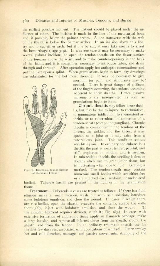
Fig. 278.—Diagram of tendon-sheaths of the hood (Tillauxi.
56o Diseases and Injuries of Muscles, Tendons, and Burns
the earliest possible moment. The patient should be placed under the in-fluence of ether. The incision is made in the line of the metacarpal' bone and, if possible, below the palmar arches. A line transverse with the web of the thumb is below the palmar arches. In an incision above this line, try not to cut either arch; but if one he cut, at once take means to arrest the hemorrhage (page 319). In a severe case it may be necessary to make several palmar incisions, to open the tendon-sheaths on the flexor surface of the forearm above the wrist, and to make counter-openings in the back of the hand, and it is sometimes necessary to introduce tubes, and drain through and through. After operation apply hot antiseptic fomentations and put the part upon a splint. When granulations begin to form, dry dressings are substituted for the hot moist dressing. It may be necessary to give
morphin for pain, and stimulants may be' needed. There is great danger of stiffness of the fingers occurring, the tendons becoming adherent to their sheaths. Hence, passive movements are inaugurated as soon as granulations begin to form.
Chronic thecitis may follow acute theci-tis, but may be due to injury, to rheumatism, to gummatous infiltration, to rheumatoid ar-thritis, or to tuberculous inflammation of a tendon-sheath (compound ganglion). Chronic thecitis is commonest in the tendons of the fingers, the ankles, and the knees; it may spread to a joint or it may arise from a tuberculous joint. This condition causes very little pain. In ordinary non-tuberculous thecitis the part is weak, tender, painful, and stiff, crepitates on motion, and is swollen. In tuberculous thecitis the swelling is firm or doughy when due to granulation-tissue, but is fluctuating when due to fluid. Grating is marked. The tendon-sheath may contain numerous small bodies which are either free or are attached (rice, riziform, or melon-seed
bodies). Tubercle bacilli are present in the fluid or in the granulation tissue.
Treatment.—Tuberculous cases are treated as follows: If there is a fluid effusion make a small incision, wash out with salt solution, introduce some iodoform emulsion, and close the wound. In cases in which there are rice-bodies, open the sheath, evacuate the contents, scrape the walls thoroughly, inject with iodoform emulsion, and close the wound. (If the annular ligament requires division, stitch it; Fig. 285.) In cases with extensive formation of embryonic tissue apply an Esmarch bandage, make a large incision, and remove all infected tissue from the sheath, around the sheath, and from the tendon. In an ordinary traumatic thecitis use for the first few days rest associated with applications of ichthyol. Later employ hot and cold douches, massage, and passive movements, strapping of the

Felon, or Whitlow 561
part, inunctions of ichthyol, and the hot-air bath. If effusion is persistent or rice-bodies exist, make an incision and scrape the interior of the tendon-sheath. In rheumatic cases give antirheumatic remedies and employ the hot-air bath. In syphilitic cases administer mercury and iodid of potassium.
Ganglia.—In connection with tendon-sheaths simple ganglia may de-velop. They are small, tense, round swellings, which arc firm, grow pro-gressively though slowly, are painless when uninflamed, and contain a fluid of the appearance and consistence of glycerin-jelly (Bowlby). Ganglia are commonest upon the dorsum of the wrist, and they occur especially in those who constantly use the NvriA-muscles. Paget states that a .cifnpic ganglion is due to cystic degeneration of a synovial fringe inside a tendon-sheath,, and that the fluid of the ganglion does not communicate with the fluid of the tendon-sheath. Other pathologists believe a simple ganglion to he a hernia of synoyial membrane through a rent in a tendon-sheath, all com-munication between the herniated part and the tendon-sheath being soon obliterated. Compound ganglion is an old name for tuberculous thecitis.
Treatment.—A ganglion is treated by aseptic puncture with a tenotome, evacuation, :,earilicatiort of the culls, antiseptic dressing, and pressure. An ohl-time method of treatment ww, subcutaneous rupture brought about by striking with a heavy book. Dupla). treats a ganglion by injecting a few drops of iodin through a hypodermatic needle. The cyst is not evacuated before injection. The parts are dressed antiseptically, ;Intl cure is obtained in one week. Recurrent ganglia, very large ganglia, and ganglia with very thick contents should be dissected out.
Felon, or whitlow, is a violent, rapidly spreading pyogenic inflamma-tion of a finger or in toe which resembles cellulitis, and which is sometimes followed by gangrene or by necrosis of bone. As a rule, an injury precedes the whitlow—an abrasion of the surface which admit- 1ms-organisms or a contusion which creates a point of least resistance. The commonest seat of a felon is the last digit of the finger or thumb. An abrasion of the surface at this point absorbs pus-organisms and the superficial lymphatics carry the bacteria directly inward, lodging them, it may be, in the skin, in the subcutaneous !issues, in the tendon-sheath, or beneath the periosteum.
Felons are very rare in infants, but may occur in children. Women are more liable to them than are men. The fingers are much more prone to infection than are the toes, because they are more exposed to injury. Several fingers may be attacked at once or successively in persons of dilapidated constitution. Whitlow is most apt to occur and is most severe in persons broken down by disease, alcoholism, overwork, or worry. In certain cases of neuritis painless suppuration may arise. In syringomyelia painless felons are common and they are apt to be associated with necrosis of bone. Pain-less and destructive whitlows constitute a characteristic part of Morvan's disease.
There are two forms of felons, the superficial and the deep. If the infection is in the skin, the point of infection becomes dark red,
swollen, painful, and tender. The epidermis is lifted up by the pus which forms, and a considerable area may be attacked before the spread of the process is arrested. If the subcutaneous tissues only are involved, the symp- toms are those of an ordinary cellulitis. Paronychia a cellulitis starting
36

562 Diseases and Injuries of Muscles, Tendons, and Bursm
at the end or side of the digit, and involving the parts around and below the nail. The pus-organisms obtain entrance by means of an abrasion, a puncture, or an ulcerated "step-mother." The pain is throbbing and violent; is increased by motion, pressure, or a dependent position; the skin is dusky red, but the swelling is slight. In about forty-eight hours pus forms in the superficial parts, the epidermis being lifted into pustules or blebs, and pus may also form under the nail. A portion of the nail, or the entire nail, may be lost.
If the tendon-sheath is involved as well as the subcutaneous tissue, the symptoms are those of suppurative thecitis, with more marked discoloration of the skin.
Deep felon, or bone-felon, involves most of the structures of the finger (periosteum, hone, tendon, tendon-sheath, and cellular tissue), and may destroy the digit or the finger. It arises in the same manner as paronychia,
but the organisms are lodged in the deeper parts. The pain is agonizing, entirely preventing sleep, pulsatile in character, associated with excruciat-ing tenderness, greatly aggravated by motion or a dependent position, and often extending up the hand and forearm. The skin is dusky red and edematous, and the part is enormously swollen. Pus forms quickly; diffuse cellulitis may arise; thecal suppuration may occur; sloughing of the tendon and subcutaneous tissue may take place; necrosis of one or more bones may ensue, and in some cases gangrene of the finger follows.
In deep whitlow lymphangitis of the forearm and arm is not unusual, adenitis of the axillary glands is common, and almost always there
Fig. 27.4.--1, 2, and ,Incisions is fever. In superficial felon constitutional
3 for felon of finger and for ordinary symptoms are slight or absent, and lymph- suppu rat Ion ; 4, palmr incision. angitis and adenitis arise in a minority of
cases. A felon may be followed by a palmar abscess, and is particularly apt to be if the disease arises in the thumb or little finger.
Treatment.—Even a superficial felon demands instant incision in all cases, and the parts must be irrigated, dressed with hot antiseptic fomenta-tions, and the hand must be placed upon a splint. A bone-felon requires prompt incision to the bone alongside the tendon. Fig. 279 shows the proper lines of incision in the fingers and palm. Do not wait for pus to form, but allay tension and prevent pus-formation by early incision. Do not waste time with poultices; to wait means agonizing pain, sleepless nights, consti-tutional involvement, and, perhaps, sloughing of tendons or death of bone. Incision and drainage constitute the treatment, followed by irrigation, anti-septic fomentations, and splinting of the extremity. If the patient cannot sleep, give morphin. See that the bowels are moved once a day. Give quinin, iron, and milk punch. Opening a felon is exquisitely painful; hence ether should be given to obtain the first stage of anesthesia, nitrous oxid

Bursitis 563
should he administered, or the superficial parts should be frozen by a spray of chlorid of ethyl.
Bursitis is inflammation of a bursa. Acute bursitis arises from strain or from traumatism. The symptoms of acute bursitis are pain, limited swelling, moist crepitus, fluctuation, and discoloration in the anatomical position of a bursa. Bursitis of the retrocalcaneal bursa (Albert's disease) is a painful affection which is often overlooked. Walking causes great pain in the heel. Raising up on the toes is excessively painful. It is usually associated with flat-foot. In these cases osteophytes often form within the bursa. There are numerous bursa about the hip. Some anatomists count twenty-one.* The two most important bursa, and the ones usually affected, are the iliac and the deep bursa over the great trochanter.f Inflammation of the iliac bursa produces swelling below Poupart's ligament, which swelling is tense, but exhibits fluctuation on careful examination. In some cases the sac can he emptied by pressure, the fluid passing into an adjacent bursa or into the joint. The enlargement often presses on the anterior crural nerve and causes pain throughout the nerve's trajectory. The limb, according to Zuelzer, is usually slightly flexed, abducted and rotated outward, and movement in an opposite direction causes pain. Inflammation of the bursae about the hip may produce symptoms resembling those of incipient coxalgia, but in bursitis the symptoms do not remit as in hip-disease. In inflamma-tion of the gluteal bursae there is moderate pain back of the thigh and knee which disappears when the patient is at rest; there is a marked limp, limita-tion of motion, and an area of deep fluctuation in the buttock (Brackett). In inflammation of the iliac bursa flexion is not so marked as in coxalgia, and the trochanter is never above Islelaton's line. In inflammation of the deep trochanteric bursa the position is the same as in iliac bursitis, and re-sembles that of coxalgia. In coxalgia, however, there is pain on pressure upon the front of the joint or directly on the trochanter or on tapping the sole of the foot. These manipulations do not cause pain in bursitis (Zuelzer).
It is difficult to differentiate between inflammation of a deep bursa and synovitis; indeed, in bursitis the joint is apt to be secondarily affected. This difficulty is especially vexatious in distinguishing between joint-injury and injury of the bursa beneath the deltoid. Suppuration may take place in a bursa. Direct force may rupture a bursa. The bursa beneath the deltoid is frequently ruptured. When this accident happens there are pain, marked swelling, a large area of moist crepitus, and later extensive discoloration from blood. Chronic bursitis may follow acute bursitis, or the disease may be chronic from the start. Its symptom is swelling with little or no pain unless acute inflammation arises. Chronic bursitis of the subhyoid bursa is known as Boyer's cyst.
Treatment.—Acute bursitis is treated by rest, pressure, and the appli-cation of iodin, blue ointment, or ichthyol. If the swelling persists, aspirate and apply pressure, or incise the sac and remove it partly or completely. If pus forms, incise, paint the interior of the sac with pure carbolic acid, and pack with iodoform gauze. Chronic bursitis may be cured by the use of pressure and the application of blue ointment, and with treatment of any causative diathesis; but most cases require incision and packing. A
* Synnestvedt, of Sweden. f. ?Atelzer, in Zeit. f. Chir., vol. t.

Pr'
,564 Diseases and Injuries of Muscles, Tendons, and Bursae
ruptured bursa is treated as an acute bursitis. Some cases of retrocalcaneal bursitis get well from rest, but others demand incision and drainage. If osteophytic formation takes place in Albert's disease, remove the bony sta-lactites with a rongeur forceps or a gouge.
Housemaids' knee is thickening and enlargement of the prepatellar bursa, due to intermittent pressure (Fig. 28o). In effusion into the knee-joint the fluid is behind the patella and the bone floats up; in housemaids' knee the fluid is above the bone and the osseous surface can be felt be-neath it.
" Miners' elbow," which is a condition similar to housemaids' knee, affects the olecranon bursa,
" Weavers' bottom " is enlargement of the bursa over the tuberosity
Fig. 043.—Housemaids' knee.
of the ischium. A bursa which is simply thickened and enlarged rarely gives rise to annoyance; but when it inflames, as it is apt to do, it causes the ordinary symptoms of bursitis.
Treatment.—Some few cases of housemaids' knee may be cured by.rest and blistering, but in most cases it is necessary to incise and pack with iodo-form gauze. In enlargement of the bursa beneath the ligamentum patella, if rest and blistering fail to cure, aspirate or incise. In enlargement of the bursa beneath the tendon of the semimembranosus and also in "weavers' bottom" and in " miners' elbow," incise and pack.
Bunion. —A bunion is a bursa due to pressure, and it is most commonly situated above the metatarsophalangeal articulation of the great toe, but is occasionally seen over the joint of another toe. When the big toe is pushed

Fig. stp pa. Talus !or bunions.
Subcutaneous Tenotomy of the Tendo Achillis 565
inward by ill-fitting boots, a bunion forms. When a bunion is not inflamed, it may cause but little trouble; but when it inflames, the bursa enlarges and the parts become hot, tender. and excessively painful. Suppuration may occur and pus may invade the joint, and the hone not unusually becomes diseased.
Treatment.- - In treating a bunion the patient most wear shoes that are not pointed, that have the inner borders straight, and that have rounded toes (Jacobson). For a mild case a bunion-plaster gives comfort. Sayre advises the use of a linen glove over the digits, the phalanges being drawn inward by a piece of elastic webbing, one end of which is fastened to the glove and the other end to a piece of strapping from the heel. A special apparatus may be worn (Fig. 281). In many cases osteotomy of the first phalanx or of the first metatarsal bone is required; in some cases excision of the joint is necessary; in others amputation must be performed. When the bursa is not inflamed, but only thickened, blisters should he em-ployed over it, or there should be applied tincture of iodin, ichthyol, or mercurial ointment. When the bursa inflames, ichthyol ointment is applied, and intermittent heat by foot-baths gives relief. Suppuration demands immediate incision and antiseptic dressing. If an ulcerated bunion does not heal by antiseptic dressing, stimulate it with nit rate of silver and dress it with unguent. hydrarg. nitrat. (t part to 7 of cosmulin). Jacobson recommends skin-grafting for some cases.
OPERATIONS UPON MUSCLES AND TENDONS.
Tenotomy is the cutting of a tendon. It may be open or subcutaneous, the open operation being preferred in dangerous regions.
Open Division of the Sternocleidomastoid Muscle for Wry-neck.—Subcutaneous tenotomy for wry-neck has been largely abandoned. It is not only more unsafe than the open operation, but it never completely divides all of the contracted band.
The instruments required consist of a scalpel, dissecting forceps, hemo-static forceps, scissors, needles, ligatures, etc. The patient is placed recum-bent, the chin being drawn more toward the opposite side.
A transverse incision is made over the muscle about one-fourth of an inch above the clavicle. The superficial parts are divided, the muscle is exposed and sectioned, bleeding is arrested, and the skin is sutured. Avoid the anterior jugular vein, which is underneath the muscle, and also the ex-ternal jugular, which is close to the outer edge of the muscle. Mikulicz advocates the removal of almost the entire muscle, leaving, however, the upper and posterior portion where the spinal accessory nerve passes. After operation for wry-neck support the head with sand-bags or a plaster-of-Paris dressing until healing occurs, and then inaugurate motions, active and passive.
Subcutaneous Tenotomy of the Tendo Achillis.—This operation is performed for club-foot, in which the heel is raised. The tendon is cut
566 Diseases and Injuries of Muscles, Tendons, and Bursm
about one inch above its point of insertion. The instrument used for the first puncture is a sharp tenotome. The patient lies upon his back "with his body rolled a little toward the affected side" (Treves), the foot being placed upon its outer side on a sand pillow. The surgeon stands to the outer side. The tendon is rendered moderately rigid, and the sharp tenotome, with its blade turned upward, is inserted along the anterior border of the tendon until the surgeon's finger feels the knife approaching the outer side. The sharp-pointed instrument is withdrawn and a blunt-pointed tenotome is inserted in its place. The tendon is drawn into rigidity, and the surgeon turns the blade of his knife toward the tendon, places his finger over the skin, and saws toward his finger. The tendon gives way with a snap. Treves states that a beginner is apt not to push the knife far enough toward the outside, or he may in the first puncture push the knife through the tendon; in either case the tendon is not completely cut. The little wound, which is covered with a hit of gauze, will he entirely closed in forty-eight hours. In club-foot cases after tenotomy some surgeons at once correct the deformity and immobilize the limb in plaster; some partially correct the deformity and apply plaster for one week, at which time they remove the plaster, correct the deformity further, reapply the plaster, and so on; other surgeons do not attempt correction of the deformity until the cut tendon has begun to unite, when they gradually stretch the new material.
Subcutaneous Tenotomy of the Tendon of the Tibialis Anticus Muscle.—The tendon is divided about one and a half inches above its point of insertion. It can be made tense by extending and abducting the foot. The sharp-pointed tenotome is entered upon the outside of the tendon, and is passed well around it. The blunt-pointed tenotome is used to cut the tense tendon.
Subcutaneous Tenotomy of the Tendons of the Peroneus Lon-gus and Brevis Muscles.—These two tendons are cut together back of the external malleolus, and one and a half inches above the tip of the malleolus, so as to avoid the synovial sheath (Treves). The patient lies upon the sound side, the outer aspect of the deformed foot being upward and the inner aspect of the ankle of the deformed side resting upon a sand pillow. A sharp tenotome is introduced close to the fibula, and is carried around the loose tendons. A blunt-pointed tenotome is now introduced, its edge is turned toward the tendons, and these structures are cut as they arc made tense.
Subcutaneous Tenotomy of the Tendon of the Tibialis Posticus Muscle.—This tendon is sectioned above the point where its synovial sheath begins; that is, above the internal annular ligament (Treves). The tendon is made tense and the pointed knife is entered above the base of the inner malleolus. The knife is entered just back of the inner edge of the tibia, and is carried around the muscle while it is kept close to the bone. The tendon is sectioned with a blunt knife.
Subcutaneous pasciotomy of the Plantar Fascia.—The con- tracted bands are discovered by motions which render them tense, and they are divided just in front of the attachments to the os calcis. The sharp knife passes between the skin and fascia at the inner side of the sole of the foot. The fascia is cut from without inward by the blunt-pointed tenotome. It is usually necessary to section the fascia at more than one point.

A B
Tendon-suture and Tendon-lengthening 567
Tendon-suture and Tendon-lengthening.—The instruments re-quired in these operations are an Esmarch apparatus; curved needles, and needle-holder; chromicized gut, kangaroo-tendon, or silk for an ordinary case, silver wire for a suppurating wound. In performing tendon-suture make the part aseptic and bloodless. It is wise to apply a rubber bandage on the prox-imal side, the bandage being applied centrifugally, forcing the proximal end of the tendon into view (Haegler). If searching for the proximal end of a flexor of the finger, flex the injured finger, and hyper-extend the adjoining fingers (Filiget). If this expedient fails, enlarge the incision, or, what is
Fig. 2R2.—Tendon-sutures : t, of Le I on ; 2, of Fig. 283.—Anderson'a method of tendon- Le Mann ; 3, of Le)ars. lengthening.
better, make a large flap in the skin. After finding the ends approximate them, being sure the proper ends are brought into contact; stitch them to-gether with a continuous suture or with one of the sutures shown in Fig. 282, 1, 2, and 3. In a suppurating wound suture by silver wire should he tried, though it usually fails. After suturing, remove the Esmarch apparatus, arrest bleeding, close the wound and dress it antiseptically, relax the parts, and place the limb on a splint. If, after suturing, there is much tension, s itch the cut tendon above the sutures to an adjacent tendon, and apply a splint, the finger which was injured being flexed, the others being extended.
Fig. 28.$.—Czerny's method of tendon-limigtli- Fig. 285.—Method of suturing the annular ening, ligament of the wrist.
If only the distal end of the tendon can be found, graft it upon the nearest tendon with a like anatomical course and function. When a tendon has been sutured begin gentle massage in two weeks. Positive passive motion is begun in three or four weeks. In old injuries, when the ends cannot be brought into apposition, lengthen one end or both ends, either by the method of Anderson (Fig. 283) or by the method of Czerny (Fig. 284). Dr. J. Neely Rhoads (" Med. News," Nov. 28, 1891) suggested that slight lengthening could be accomplished by "cutting half through the tendon at different levels and from opposite sides, leaving some longitudinal fibers to slip on

568 Diseases and Injuries of Muscles, Tendons, and Bursm
each other, thus gaining slight elongation" (H. Augustus Wilson, in " Inter-national Clinics," vol. i, 4th series). Poncet makes several zigzag incisions on each side of the tendon, and when the tendon is pulled upon it elongates decidedly. One of these methods of lengthening may he used if there is deformity from tendon-contraction. If the tendon cannot be lengthened sufficiently, make a bridge of catgut from one end of it to the other, or graft in another tendon from one of the lower animals, or graft the distal end to a tendon of like function.
The annular ligament is sutured as shown in Fig. 285. Tendon-transplantation.—In some cases in which a muscle has been
paralyzed, Nicoladoni and others have divided the tendon of the paralyzed muscle and have united its distal end with the tendon of a normal muscle, the normal tendon being split to receive it. It has also been pointed out that when a muscle or the tendon of a muscle is sutured to a paralyzed an-tagonistic muscle, the transplanted structure will actually execute the func-tions of the paralyzed muscle. For instance, a flexor, when so transplanted, may become an extensor and act under the mental impulse of extension. These principles have been utilized when some or many of the muscles of a limb have been paralyzed, the tendon of an unparalyzed muscle or the tendons of an unparalyzed group of muscles being fastened to the tendons of the paralyzed muscle. It has been shown that the success of this procedure depends upon the accuracy of diagnosis, the division of secondary contrac-tures, the correction of existing deformities, and careful after-treatment. (See the article by Dr. J. Hilton Watterman, in " Med. News," July 12, 1902.) In a paralysis of the lower extremity, as Goldthwait points out, the sartorius usually retains power, and it may be advisable in such a case to divide the sartorius and suture its upper end to the quadriceps above the patella.
CAA