Embed Size (px)
DESCRIPTION
Our mission is to enhance your ability to practice equine medicine by providing the latest info you need.
Citation preview

Equine VetThe Modern
Vol 2 Issue 5 2013www.modernequinevet.com
Managing neonatal colicDetermining risk of navicular disease
Tying up:Should you order genetic test?

2 Issue 5/2013 | ModernEquineVet.com
TABLE OF CONTENTS
ORTHOPEDICS
Determining risk of navicular disease ........................................... 7Presence of distal border fragments might be key COLIC
Managing the neonate with colic ................................................... 8Good outcome depends on many factors
NEWS
Standing MRI eliminates need for anesthesia ......................10Looking for a few good broodmares .........................................10EIA confirmed in Nebraska ...........................................................11Patent issued for beneficial animal "candy" ..........................13Adequan limited ...............................................................................13Penn works to improve stem cell's cartilage formation ....17
TECHNICIAN UPDATE
Good medical records decrease risk ...............................................................................15Deborah Reeder talks about this important technician responsibility
TO CONTACT US, EMAIL MARIE ROSENTHAL
LEGAL DISCLAIMER: The content in this digital issue is for general informational purposes only. PercyBo Publishing Media LLC makes no representations or warranties of any kind about the completeness, accuracy, timeliness, reliability or suitability of any of the information, including content or advertisements, contained in any of its digital content and expressly disclaims liability of any errors or omissions that may be presented within its content. PercyBo Publishing Media LLC reserves the right to alter or correct any content without any obligations. Furthermore, PercyBo disclaims any and all liability for any direct, indirect, or other damages arising from the use or misuse of the information presented in its digital content. The views expressed in its digital content are those of sources and authors and do not necessarily reflect the opinion or policy of PercyBo. The content is for veterinary professionals. ALL RIGHTS RESERVED. Reproduction in whole or in part without permission is prohibited.
Genetic testing for horse that is tying up?
COVER STORY: 4
Cover photo by photographer Bob Langrish http://www.boblangrish.com
Equine VetThe Modern
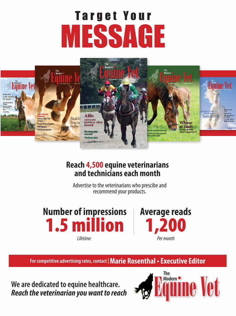
We are dedicated to equine healthcare. Reach the veterinarian you want to reach
For competitive advertising rates, contact | Marie Rosenthal • Executive Editor
MESSAGET a r g e t Y o u r
Reach 4,500 equine veterinarians and technicians each month
Advertise to the veterinarians who prescibe and recommend your products.
Number of impressions 1.5 million
Average reads 1,200
Lifetime Per month

4 Issue 5/2013 | ModernEquineVet.com
Tie me up,
Exertional rhabdomyolysis can have many causes and some of these are related to an underly-ing genetic susceptibility to muscle damage with exercise.
There are two genetic tests avail-able for forms of exertional rhab-domyolysis, type 1 polysaccharide storage myopathy (PSSM1) and malignant hyperthermia (MH).
PSSM1 is characterized by chronic bouts of stiffness, muscle
pain, cramping, and cell damage during exercise. A dominant ge-netic mutation in the glycogen synthase (GYS1) gene is responsi-ble for PSSM1. MH can lie hidden in horses and periodically give rise to severe episodes of muscle damage with exercise or with general anesthesia. Horses with signs of MH have a severe meta-bolic imbalance and high body temperature, and this can be fatal.
COVER STORY
B y M a r i e R o s e n t h a l , M S
Should vets order genetic tests for tying up? It depends.
ties me down
Who’s at risk?PSSM1 is known to affect more
than 20 different breeds but to date the GYS1 genetic mutation has not been found in purebred light breeds, such as Standardbred, Thorough-bred and Arabians. If these light breeds have exertional rhabdomy- olysis, the likelihood that it is due to the GYS1 mutation is very low.
That doesn’t mean the horse isn’t tying up, it just means the cause is not due to the GYS1 mutation, ac-cording to Stephanie Valberg, DVM, PhD, DACVIM, ACVSMR, professor at the University of Min-nesota and director of its equine center, and one of the patent owners of the PSSM1 genetic test.
In general, less than 10% of Paints, Appaloosas and Morgans will test positive for the genetic mu-tation.
About 10% of quarter horses will test positive, but there are variations within the Quarter horse breed. Knowing which performance types are most affected can help veteri-narians decide when to perform ge-netic testing.
For example, about 28% of hal-ter horses will test positive. “If you have a halter horse with exertional rhabdomyolysis then testing for the PSSM1 genetic mutation is a good place to start when searching for the cause,” Valberg said. “On the other side of the spectrum, less than 2% of racing Quarter horses have the gene for PSSM1, so it may not be the place to start.”
If the genetic test is performed, owners should be told that further diagnostics will be needed if it is negative to find the cause of this horse’s exertional rhabdomyolysis.
The prevalence of PSSM1 is very high in certain Draft breeds. More than 30% of Belgians and 50% of Percherons have the PSSM1 genetic mutation, but this mutation is rare in Shires and Clydesdales, she said. “This is important to take into ac-count because no matter what dis-ease you are looking at you have a
Phot
os c
ourte
sy of
Dr. S
teph
anie
Valbe
rg

ModernEquineVet.com | Issue 5/2013 5
50% chance in Percherons that it will come back positive,” she said.
“Clinical judgment is needed to interpret the positive result. Veteri-narians need to ensure that the clin-ical signs in a Percheron horse are consistent with the consequences of the GYS1 mutation.”
In contrast, MH is only known to exist in Quarter horses and paints and has a very low prevalence. Less than 3% of quarter horses are affect-ed, so a positive genetic test is highly significant in horses with exertional rhabdomyolysis.
Some Quarter horses have both MH and PSSM1 that makes their clinical signs worse and harder to manage.
What does a positive genetic test mean?
“There are many things that impact whether a horses with the GYS1 mutation have clinical dis-ease or not: turn out, pasture, diet and exercise regime,” said Valberg.
In addition, a horse has about 30,000 genes, and the unique com-position of these genes in an indi-vidual influence whether or not one genetic mutation will result in mild or severe clinical signs.
Valberg likes to use a poker analogy when discussing this with clients. Having the GYS1 gene mu-tation is like being dealt the Ace of Clubs. If the hand doesn’t have any aces or clubs, an ace of clubs has less impact than if the hand has two aces. If the hand has the King, Queen, Jack and 10 of clubs, the ace of clubs becomes very significant.
When recommending a genetic test, veterinarians must explain the odds of a positive test and the im-plications of both a positive and a negative test, Valberg said.
If the odds are low due to the breed, clients need to be prepared for the next steps, such as a muscle biopsy, if the genetic test is negative.
“For example, less than 20% of Warmblood horses with exertional rhabdomyolysis have the GYS1 mu-
tation, the genetic test is a pain-free place to start exploring the cause of tying up but the odds are it will be negative and a muscle biopsy might be needed,” Valberg said.
Owners, especially breeders, also need to be prepared if a test is positive. A positive test can occur in some horses that have not had clini-cal signs of exertional rhabdomy-olysis. This can be a nasty surprise for breeders.
In cases where the test is posi-tive but horses are symptom-free, horses may have had lots of exer-cise throughout their life, been on the right diet, large sparse pastures or they may have luckily had the right combination of other genes to prevent the disease from being expressed.
Owners should consider what they will do with a positive test re-sult before they test.
Managing PSSM1The product of GSY1, glycogen
synthase, is involved in the produc-tion of glycogen, which provides energy to the muscles during exer-tion. In normal horses, there is a set point for how much glycogen is made and stored. The mutation in GSY1 in PSSM1 horses disrupts that set point and they keep synthe-sizing glycogen even when enough glycogen is stored. The enzyme ac-tivity is greatly enhanced by insulin, so a high-grain diet will further en-hance glycogen synthesis.
“For there not to be any confu-sion about whether a muscle is go-ing to synthesize or use glycogen, there is reciprocal activation and inactivation of glycogen synthase and phosphorylase, which tells the muscle when to break down glyco-gen,” explained Valberg.
In horses with PSSM1, the mus-cle signal to make glycogen is usu-ally greater than the signal to break down glycogen, so the body has difficulty metabolizing glycogen to supply energy for aerobic exercise. The muscle cannot supply energy
Taking a biopsy sample for testing.Source: Dr. Valberg
Phot
os c
ourte
sy of
Dr. S
teph
anie
Valbe
rg
6 Issue 5/2013 | ModernEquineVet.com
COVER STORY
for muscle contraction, and the horse experiences the clinical signs that stop its activity.
“Most horses develop tying up at around 15 minutes of exercise and that is a time when they are reliant on glycogen metabolism for energy and before they have circulating free fatty acids as an energy source,” she said.
A healthy dietIf a horse has the GSY1 muta-
tion, veterinarians can offer practi-cal advice to improve the horse’s function by helping it to start ac-tivating glycogen breakdown and find another energy source.
Changing the diet and keep-ing the horse active are the keys to helping the horse.
“Get rid of insulin stimulation of the glycogen synthase enzyme by modulating dietary starch and sugar. That is the whole basis for wanting to ensure that the hay is low in nonstructural carbohy-drates, that the grass is limited if it is lush and high in nonstruc-tural carbohydrates, and that we eliminate the grain that these horses are being fed because it will decrease stimulation of this enzyme,” she said.
Make sure there is enough oxi-dative capacity to use other sub-strates and that means exercising these horses. “We can’t let them stand still. We can’t let them go without exercise because we want to increase the mitochondria in their muscle and increase their capacity to use an alternative energy source and that energy source can come in the form of fat. By bypassing their inability to metabolize glycogen early in exercise, clinical signs can be re-duced.
Watch the calories when switch-
ing the diet, she warned.“Owners sometimes read on
the internet, ‘feed them a pound of fat a day.’ I don’t think that is the place to start,” she said.
Instead, determine what the daily caloric intake of that horse should be and make sure that the nonstructural carbohydrates are low and that the right balance of energy is supplied as fats for that individual. A veterinary nutri-tionist can be very helpful in rec-ommending an appropriate diet.
If the horse is already over-weight or obese, consider a man-agement technique of horse own-ers in the late 1800s; take their feed away before exercising so they have a negative energy balance.
“If the owner of an overweight horse rides in the morning, I usu-ally recommend throw it their flake of hay at night, don’t give them their morning feed and then ride the horse. And if you do that, it will have a much higher amount of cir-culating free fatty acids concentra-tion and you get the same benefit as feeding them fat. But you also get the added benefit of taking off weight,” she said.
If the horse is in good body condition and receives about 45 minutes of exercise a day, feed it a hay that is about 12% nonstructural carbohydrate. Staying below 12% will prevent the release of insulin.
Supplement with oil. The cheap-est method is to moisten hay cubes and put oil on top. If that is too messy, rice bran is a great choice. Make sure that fat supplies about 15% of the energy in the total diet and carbohydrate provides less than 15% of the total energy.
If the horse is exercising in-tensively and needs more feed to maintain weight, high-fat, low-starch concentrates can be used.
In general, to get the right amount of fat this often means at least 4 lbs a day of some of these concen-trates. Some owners who think the diet isn’t working are feeding a high-fat, low-starch concentrate, but they don’t feed enough to get an adequate amount of fat. Make sure the horse is getting about 10-15% of the calories a day in the form of fat, Valberg suggested.
The diet is only effective if the horse receives an appropriate amount of daily exercise, but the exercise does not have to be too long or too strenuous to be effec-tive, according to Valberg.
“The thing that is important for owners to know is that it doesn’t take much exercise. In our trials, those horses could not do more than 20 minutes of exercise, but we could with 20 minutes of exercise a day and very little in the way of turn out dramatically improve their lifestyle and im-proved their rhabdomyolysis and the pain they experienced.
“So, even if they can’t ride, get-ting the horse out is important. Even exercising on a lunge line for 10 minutes a day makes a huge difference for these horses.”
Turnout is an excellent activ-ity. Try to put the horse out with other horses that will encourage activity and make sure they have to forage for food. Use a grazing muzzle if the grass is lush, she suggested.
“When you adjust the diet and give them some exercise, the mus-cles feel better and their perfor-mance is better,” she said, warning that veterinarians should watch out for previous undetected lameness when the horse becomes active again. It often goes unnoticed be-cause everyone is focused on the tying up. MeV
For more information:
Dr. Valberg is one of the owners of the patent for the PSSM genetic test and receives sales income from its use. Her financial interest has been reviewed and managed by the University of Minnesota in accordance with its conflict of interest policies.

ModernEquineVet.com | Issue 5/2013 7
The presence of distal border fragments of the navicular bone might indicate that a horse is at risk for navicular disease.
Therefore, during the purchase examinations and when selecting breeding stallions, veterinarians should look for horses with more favorable navicular border shapes (straight or convex), which may re-duce the prevalence of distal border fragments and likely the risk for de-veloping navicular disease, suggest-ed Sarah Claerhoudt, DVM, PhD, of Ghent University in Belgium.
Researchers have determined that the shape of the proximal ar-ticular border is hereditary and the distribution of biomechanical forces exerted on the navicular bone de-pend on that shape. There is also a shape-grade association, in which concave and undulating shapes are associated with the highest risk for developing navicular disease. The fragments may arise from a fracture at the insertion of the distal sesa-moid (impar) ligament (avulsion fracture), mineralization in the liga-ment or from a separate ossification center.
Claerhoudt and her colleagues reviewed radiographs from 325 Belgian Warmbloods and found that concave and undulating shapes were associated with the highest risk
of having distal border fragments. “These fragments were significantly more prevalent in bones with a con-cave or undulating shape,” she said. “We hypothesized that distal border fragments result from unfavorable loading of the navicular region.
“In our study, all of the navicular bones with fragments had a corre-sponding defect in the bone, which may strengthen our hypothesis that distal border fragments arise from a fracture due to abnormal strain at the attachment of the impar liga-ment, [which holds the navicular bone in the hoof capsule],” she said.
The clinical significance of these distal border fragments remains un-clear. However, today, if Belgian vet-erinarians find a distal border frag-ment, they typically will recommend against selecting that breeding stal-lion, and therefore, this defect has an important financial consequence.
Finding fragmentsDistal border fragments are sel-
dom prominent, so they can be difficult to see on a radiograph. To evaluate the foot for distal border fragments, Claerhoudt takes three standard radiographic projec-tions (lateromedial, dorso55°- and dorso65°proximal-palmarodistal oblique projections). Two dorsopal-mar views are taken from different
angles with the horizontal, which results in a better study of the distal border. If a distal border fragment is not obvious, additional oblique pro-jections should be made.
Magnetic resonance imaging (MRI) and computed tomography (CT) scans may help overcome ra-diography’s limitations. MRI is used for diagnosing the exact location of the fragment and the presence of na-vicular bone edema, representing an inflammation or degenerative bone change, while CT is good for de-tailed imaging of normal bone and bony disorders.
“CT and MRI are complementa-ry, but since the introduction of the standing MRI, MRI is the technique of choice for evaluating distal foot pain,” Claerhoudt said.
Although the clinical significance of these fragments remains unclear, the hereditary of shape, the shape-grade and shape-fragment associa-tions described, one may assume a possible relationship between these fragments and navicular disease, she added. MeV
Risk of
Presence of distal border fragments might be key
A radiograph of a navicular bone with a distal border fragment at the medial and lateral aspect.Source: Dr. Claerhoudt
For more information:
Claerhoudt S, Pile F, Vanderperren K, et al. Association between navicular bone fragmentation and shape in Belgian warmblood horses. Vet Comp Orthop Traumatol. 2011;24(2):132-6. doi: 10.3415/VCOT-10-03-0037. Epub 2011 Jan 11. http://www.ncbi.nlm.nih.gov/pubmed/21225084
ORTHOPEDICS
naviculardisease
B y M a r i e R o s e n t h a l , M S

8 Issue 5/2013 | ModernEquineVet.com
COLIC
Most cases of neonatal colic can be managed medically with good outcomes, according to Michelle Harris, VMD, DACVIM, a lecturer
in emergency and critical care at New Bolton Center, University of Pennsylvania.
“The vast majority of neonates
presenting to a referral hospital for colic signs can be managed medi-cally. The need for surgical interven-tion is less common than it is for
Good outcome depends on many factors, but most neonates fair well
B y M a r i e R o s e n t h a l , M S
neonate with colicManaging the

adult horses,” Harris said recently at the 58th Annual Convention of the American Association of Equine Practitioners.
Harris discussed a study lead by Melissa MacKinnon, DVM, DACVS, while she was a surgi-cal resident at New Bolton Center. MacKinnon now practices at Milton Equine Hospital in Campvellville, Ontario, Canada.
In this retrospective study, the re-searchers reviewed the medical re-cords of 137 neonates younger than 30 days of age with signs of colic that were treated at the George Widner Hospital for Large Animals between January 2000 and August 2010. Most were Thoroughbreds (72) with
Standardbred (28) being the second largest group represented. Seventy-six were colts and 61 were fillies.
They obtained information about signalment, history, physical exam, laboratory results, ancillary diagnostic tests, details of treatment and primary diagnosis.
The primary diagnosis was en-terocolitis, followed by necrotizing enterocolitis (NEC), meconium-associated colic, and small intestinal strangulating obstruction (SISO).
“The majority or 89% of neo-nates were managed medically,”
Harris said. Nineteen had surgical lesions, and 11 underwent surgery.
The researchers found that the overall short-term survival was good to excellent. Neonates that were less likely to survive to dis-charge were diagnosed with severe NEC or SISO.
Overall short-term survival was 75% and was not significantly different between surgically and medically managed cases, accord-ing to Harris.
Four neonates with severe NEC that underwent surgery were euthanized under general anesthesia due to grave prognosis. “All of the foals that were treated surgically and were allowed to
recover from general anesthesia survived to discharge,” she said.
Concurrent diseases in neonates with colic are common. In this study, 87 neonates had a comorbid-ity, which included sepsis, neonatal encephalopathy, neonatal nephrop-athy, failure of passive transfer and umbilical remnant infection.
“It is important to keep in mind the potential presence of a concur-rent disease, when assessing and managing a neonate with potential colic. The impact of the severity of the concurrent disease should be
considered carefully,” she said. In the neonates diagnosed
with transient medical colic, four were euthanized due to concur-rent diseases.
Long-term survivalThe veterinarians were able to
provide 12-month follow up for 69% of the neonates; 93% of those survived to 12 months of age.
“None of these horses died or were euthanized as a result of their colic as a neonate. Colic after dis-charge was uncommon,” she said.
“If the neonate survived to matu-rity, the neonate had a good chance of being used as intended at an ex-pected age. If they failed to be used
as intended or at the expected age, the reason was unrelated to colic,” she added.
Don’t discourage owners from consenting to an exploratory celi-otomy if it is indicated, Harris said.
“Early treatment, close moni-toring and prompt surgical inter-vention if necessary are recom-mended.”
MacKinnon worked with Jon Palmer, VMD, DACVIM, a neo-natologist, and Louise Southwood, BVSc, PhD, DACVS, DACVECC, an emergency clinician, at New Bolton Center. MeV
ModernEquineVet.com | Issue 5/2013 9
neonate with colicFoals with colic. Thanks to Dr. Michelle Abraham for permission to use them.
Concurrent Diseases Clinical Features
87 neonates with concurrent disease• Sepsis• Neonatal encephalopathy• Neonatal nephropathy• Failure of passive transfer• Umbilical remnant infection
Associated with survival• Plasma lactate• Total plasma protein• Oral mucous membrane color• Intestinal borborgmi• Primary colic diagnosis• Concurrent disease

10 Issue 5/2013 | ModernEquineVet.com
NEWS NOTES
Hallmarq Veterinary Imaging continues to increase the number of clinics throughout North America using their standing equine MRI machine.
Because the standing MRI eliminates the need for general anesthesia, equine clinics can now obtain scans of the foot and lower limbs with virtually no risk to the horse.
“The standing MRI machine allows us the abil-
ity to clearly determine what type of injury the horse has without the use of general anesthesia. It’s more convenient for me and my staff, and it is much safer for the horse,” said Wesley Sutter, DVM, MS, DACVS, Lexington Equine Surgery & Sports Medicine.
Sutter, a specialist in equine orthopedic surgery and sports medicine, says his clinic plans to install and offer the standing equine MRI machine to pro-vide clients a better option for MRI.
“We are an orthopedic surgery and lameness referral clinic that sits about one mile from the Ken-tucky Horse Park here in Lexington, so as you can imagine, we specialize in premier equine athletes,” Sutter said. “Conducting an MRI will be a routine procedure at our clinic, so being able to do that in the least invasive way and still get the information we need is not only important to our business, but to the client experience as well.”
The Hallmarq standing equine MRI machine al-lows veterinarians to get clear, high-resolution scans of the soft tissue in the horse’s foot or leg to allow for a more precise diagnosis. The system is unique, as the horse is simply walked in for the scan, making it less labor-intensive for staff. MeVhttp://www.hallmarq.net/equine
Standing MRI eliminates need for general anesthesia
The University of California, Davis William R. Pritchard Veterinary Medical Teaching Hospital is asking for dona-tions of young, healthy mares for its teaching herd. The horses will serve as embryo recipients for hospital clients with mares participating in the embryo transfer program.
UC Davis will give the donated horses a quality home during their time in the program. If a mare successfully re-ceives an embryo, she will be sold to the client whose mare provided the embryo. That client will care for the recipient mare until the foal is born and is weaned. The client can sell the mare back to the program afterward.
To be accepted into the recipient herd, mares must: • Be at least 15 hands and (ideally) 1,100 lbs • Be between 3 and 10 years of age• Be (preferably) Warmbloods, Standardbreds, Drafts, Quarter Horses or Thoroughbreds• Be "broodmare sound" (sound in a pasture situation, not necessarily sound enough to ride or perform)• Be halter broken • Be free of any history of reproductive problems or subfertility • Can be a maiden or foaled mare “These mares will play a critical role as we expand our student teaching and resident training programs, and fulfill
our teaching and service visions,” explained Bruce Christensen, DVM, MS, DACT, chief of the equine reproduction service at VMTH.
To discuss a donation, please contact Christensen or through the VMTH Large Animal Clinic at (530) 752-0290. MeV
Looking for a few good brood mares
Phot
o cou
rtesy
of H
allm
arq V
eter
inary
Imag
ing

ModernEquineVet.com | Issue 5/2013 11
Nebraska Department of Agriculture (NDA) officials worked quickly to contain an outbreak of equine infectious anemia (EIA) in one horse herd located in Northwestern Nebraska.
EIA is a bloodborne disease and is typically transmitted by biting insects (such as horseflies and deerflies), but also can be transmitted from horse to horse through infected needles. There are no treatment options for infected horses, according to State Veterinarian Dr. Dennis Hughes. Symptoms include fever, depression, weight loss, swelling and anemia.
Horse owners are encouraged to take biosecurity precautions to reduce the risk of infection in their herds, including:
• Implement control measures, including husbandry practices that reduce biting insects, such as horseflies and deerflies;
• Follow the rule of one horse-one needle; and
• Additions to herds should have a negative Cog-
gins test before being allowed to intermingle with other equids.
For more information about protecting horses, direct clients to www.nda.nebraska.gov.
Hughes reminded those who are importing horses into Nebraska for show/exhibition or other reasons to follow Nebraska’s horse import regula-tions, which includes the requirement of a nega-tive Coggins test. Producers with questions about import regulations should contact NDA at (402) 471-2351. MeV
EIA confirmed and contained in Nebraska
Equine infectious anemia (EIA) is a potentially fatal viral disease of equids. No vaccine or treatment exists for the disease. It can be difficult to differ-entiate from other fever-producing diseases, including anthrax, influenza, and equine encephalitis.
Degrees of InfectiousnessAcute — When horses are exposed to EIAV, they may develop severe, acute signs of disease and die within 2 to 3 weeks. This type is the most damag-ing and the most difficult to diagnose because the signs appear rapidly, and often only a fever is noted. One-fifth of a teaspoon of blood from a horse with acute EIA contains enough virus to infect 1 million horses.
The clinical signs of the acute form of EIA are nonspecific. In mild cases, the initial fever may be short lived (often less than 24 hours). As a result,
horse owners and veterinarians may not recognize this initial sign as EIAV. These infected horses often recover and continue to move freely among the herd. The first indication that a horse was exposed to, and infected with, EIAV may well be a positive result on a routine Coggins test.Chronic — If the horse survives this first acute bout, it may develop a recur-ring clinical disease with these signs:
• Fever—An infected horse’s temperature may rise suddenly to about 105° F or, rarely, as high as 108° F. Then it may drop back to normal for an indeterminate period until the onset of another episode.
• Petechial hemorrhages• Depression• Weight loss• Dependent edema• Anemia.
The horse with chronic EIA is the classic “swamper” who has lost condi-tion, is lethargic and anorexic, has a low hematocrit, and demonstrates a persistent decrease in the number of blood platelets, especially coincident with fever induced by EIAV.Inapparent — Most horses are latent carriers without overt clinical signs. They survive as reservoirs of the infec-tion for extended periods. Carriers have dramatically lower concentrations of EIAV in their blood than horses with active clinical signs of the disease. Only 1 horsefly out of 6 million is likely to pick up and transmit EIAV from this horse.
All horses infected with EIAV are thought to be lifelong carriers. The in-apparent form may become chronic or acute due to severe stress, hard work, or the presence of other diseases. MeVSource: USDA
acute EIAOne-fifth of a teaspoon
of blood from a horse withcontains enough virus to
infect 1 million horses.
Equine Infectious Anemia

12 Issue 5/2013 | ModernEquineVet.com
INFECTION CONTROL
Methicillin-resistant Staphylo-coccus aureus (MRSA) infections in horses are difficult to treat because few antibiotics are effective against these resistant bacteria.
But veterinarians can reduce the spread of resistant bacteria by improving hygiene in the equine hospital, according to Karin Berg-ström, of the Swedish National Vet-erinary Institute, who defended her dissertation on the topic this month.
“An infection-control program requires continuous work with au-dits, training and monitoring. Hos-pital leaders need to give their sup-port by allocating resources and by their active engagement. The intro-duction of infection-prevention and -control measures is a self-evident responsibility of horse hospitals, as MRSA involves both patient safety and the working environment,” said Bergström, who is the assistant state veterinarian.
In the summer of 2008, MRSA was found at an equine hospital in Sweden. Bergström studied the out-break and the biosecurity measures
that were implemented for her dis-sertation. Most of the horses were suffering from superficial wounds that healed without the use of anti-biotics, which she felt was a key ob-servation.
The bacteria in the outbreak belonged to a type of MRSA called CC398 associated with food-pro-ducing animals, but it has also been found in horses in Europe. However, this was the first time this type of bacteria had caused equine infec-tions in Sweden.
Of a total of nine horses that could be monitored after the in-fection, all but one showed nega-tive samples within two to seven months, and the nasal passages proved to be the most reliable sam-pling site for obtaining MRSA.
Collaboration among the hospi-tal where the infection had spread, experts in human infection control and public authorities contributed to the development of a program for infection control. But the cost to the hospital was steep — almost $200,000.
The horse hospital presents chal-lenges that are not seen in human hospitals, and further studies are needed to find better ways to con-trol equine infections, she said. For example, the development of surface materials that are suited to horses, but are easy to disinfect facilitated infection control in these hospitals.
Environmental sampling showed that MRSA was prevalent in places accessible only to people, which means that hand hygiene is key to stopping the spread of this bacteria.
MRSA was found on furnish-ings that are difficult to clean. Therefore, mangers and water cups were replaced by buckets that could be disinfected. Observations at three horse hospitals showed that biosecurity routines regard-ing work clothing and the like were exemplary, but workers were less likely to comply with routines for hand hygiene and disposable glove use. Reasons for lack of com-pliance were practical difficulties, insufficient knowledge and high workloads. MeV
Prevent MRSA
CDC/
Janic
e Han
ey Ca
rr/ Je
ff Ha
gem
an, M
.H.S.
i n e q u i n e h o s p i t a l s
Due to renovations at a New York factory, Luit-pold Pharmaceuticals Inc. announced that supplies of polysulfated glycosaminoglycan (PSGA, Ad-equan) will be limited over the coming months.
"Our factor in New York has undergone a signifi-cant renovation to meet enhanced quality standards and address observations of the FDA. This has resulted in depletion of our existing inventories," the company said in a press release.
Although the company has tried to manage supplies as well as possible, it expects to be out of the product for a short time and to resume shipments of the 5 mL size in early July followed by the 50 mL size in August.
Since approved for equine use by the FDA in
1984, Ad-
equan is the only PSGAG labeled to treat non-infectious degenerative and/or traumatic joint dysfunction and associated lame-ness of the carpal and hock joints in horses. There is no generic product labeled for use anywhere in the word, the company said.
For the most recent updates on expected release dates, visit www.adequan.com. MeV
ModernEquineVet.com | Issue 5/2013 13
Supplies of Adequan Limited
NEWSNOTES
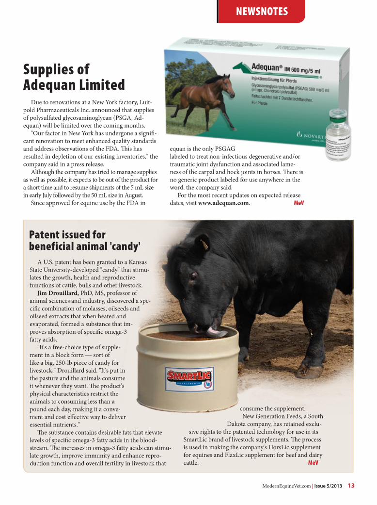
Patent issued for beneficial animal 'candy'
A U.S. patent has been granted to a Kansas State University-developed "candy" that stimu-lates the growth, health and reproductive functions of cattle, bulls and other livestock.
Jim Drouillard, PhD, MS, professor of animal sciences and industry, discovered a spe-cific combination of molasses, oilseeds and oilseed extracts that when heated and evaporated, formed a substance that im-proves absorption of specific omega-3 fatty acids.
"It's a free-choice type of supple-ment in a block form — sort of like a big, 250-lb piece of candy for livestock," Drouillard said. "It's put in the pasture and the animals consume it whenever they want. The product's physical characteristics restrict the animals to consuming less than a pound each day, making it a conve-nient and cost effective way to deliver essential nutrients."
The substance contains desirable fats that elevate levels of specific omega-3 fatty acids in the blood-stream. The increases in omega-3 fatty acids can stimu-late growth, improve immunity and enhance repro-duction function and overall fertility in livestock that
consume the supplement. New Generation Feeds, a South
Dakota company, has retained exclu-sive rights to the patented technology for use in its
SmartLic brand of livestock supplements. The process is used in making the company's HorsLic supplement for equines and FlaxLic supplement for beef and dairy cattle. MeV

AAEVT MembershipAAEVT* membership is open to US and international equine veterinary technicians, assistants, practice managers, and support staff employed in the veterinary industry. It is also open to students of AVMA/CVMA accredited programs
AAEVT MembershipBi-Annual NewsletterWeekly “HoofBeats” email NEwsblastFull access to www.aaevt.org, including the Career Center and the LibraryUp-to-date information on the AAEVTDiscounted registration for AAEVT Regional Meetings and the annual AAEP/AAEVT ConventionNTRA, Working Advantage and Platinum Performance BenefitsThe opportunity to participate in the AAEVT Online Certification Program or to become a member of the AEVNT Academy-Specialty in Equine Veterinary Nursing Scholarship opportunities. AAEVT’s Equine Manual for Veterinary Technicians (Blackwell Publishing 20% discount on purchase price)Subscription to THE HORSE Magazine, compliments of Intervet Schering/Plough Opportunity to attend Purina’s Annual Equine Veterinary Technician Conference - All Expenses paid!
••••••••
••
•
AAEVT ObjectivesProvide opportunities for CE, training, communication, and networkingEducate the equine veterinary community and the public about our professionInform Members of issues affecting our professionAssist in providing the best medical care to improve the health and welfare of the horse
••••
AAEVT Online Equine Certification Program
For more information visit www.aaevt.org*American Association of Equine Veterinary Technicians and Assistants
AAEVT Mission Statement: To promote the health and welfare of the horse through the education and professional enrichment of the equine veterinary technician and assistant.
A three course, 10 module, equine-only online program offered through ACTGeared toward Credentialed Veterinary Technicians, Assistants, Support staff, & StudentsAreas of study include: equine medical terminology, anatomy and physiology, parasitology, laboratory, diagnostics, equine basics (breeds, wellness, husbandry,) diagnostic procedures, emergency medicine, restraint, pharmacology, surgical assistance and anesthesia, equine office proceduresA certificate of completion is awarded to those who: Successfully complete required courses Complete the list of required skills (per a supervising DVM who is an AAEP member) Attend an AAEVT regional CE symposium and participate in the we labsThose individuals who successfully complete the programs will be recognized as AAEVT Certified Equine Veterinary Technicians / AAEVT Certified Equine Veterinary Assistants depending on their current designation. The certificate is recognized by the AAEVT and the AAEP but does not grant the credentialed status by the AVMAFor more information go to www.aaevt.4act.com or call 800-357-3182
•••
•
•
•

ModernEquineVet.com | Issue 5/2013 15
By Deborah Reeder, RVT, VTS-EVNAAEVT Executive Director
Accurate medical records, completed in a timely fashion are a legal necessity and the only protection against suc-cessful malpractice litigation.
There is little doubt that equine practitioners face some unique challenges in their practices when com-pared with companion animal veterinarians. For in-stance, the critical issue of obtaining the consent to treatment from the owner becomes blurred when the equine clinician regularly accepts instructions from a trainer or stable manager.
It is important for the proper management of the equine practice to make sure that the actual owner of the horse is both identified and provides specific instructions that the trainer is authorized to act on the owner’s behalf. Too often the equine practice is compromised when the owner refuses to pay for ser-vices because the instructions to provide service were never actually authorized by the owner. During the initial engagement the veterinarian should ensure that authorization can be provided by the trainer or other designated party. This is particularly true when a syndicate owns the horse.
Talk to me!Communication is the key in the success of any
service business. Veterinary medicine is no excep-tion, and the clients’ interactions with the practice can be the most important factor in the success of a practice. On a daily basis there may be com-munications with the backyard owner of a single horse, multiple owners of million dollar racing Thoroughbreds, buyers, sellers, agents, referring veterinarians, barn managers, trainers, riders, in-surance companies, an export agent, a show office, the USDA or a farrier. All of these communica-tions need to be documented.
Timely, complete and correct messages de-livered to the appropriate person, with prompt follow-up, will go a long way toward reassuring clients that their requests are being well handled. Confidentiality when dealing with client informa-tion is an absolute necessity, which, if not strictly adhered to, can result in the loss of a client or prac-tice reputation at the minimum or a lawsuit attack-ing profession ethics. Consistent and timely com-
munication among staff is a challenge, which must be met if the practice is to run smoothly.
Medical Records are the practice communica-tion between staff and to the client. Creating and maintaining correct, accurate and timely medical records is the single most important responsibility of the equine technician in a practice.
Who owns whatThe challenges associated with obtaining and
recording this information must be met. Ethically the information within veterinary medical records is considered privileged and confidential. It must not be released except by court order or consent of the owner of the patient. This is especially true when horses change owners or are presented for a pre-purchase examination, as the absolute con-fidentiality of information must be maintained and prior medical history cannot be included in the medical record or communicated to a second owner or potential buyer without the express writ-ten permission of the original owner.
It is important to note that the physical images (radiographs, ultrasound images or bone scan im-ages for instance) are the property of the practice, which created them. The information contained in these images is the property of the client who paid for them to be made.
Release of this information requires a signed release from that client. Absolute adherence to this law is essential. The time that patient records, including imaging, must be maintained by a prac-tice, regardless of the status of the patient, varies from state to state; however a minimum of 10 years provides a good rule of thumb.
Medical Records should contain the minimum standards of information collected and docu-mented. A complete and thorough description of the horse, it’s age and weight, it’s use, identifiable
Medical records reduce risk in equine hospitals
TECHNICIAN UPDATE
Creating and maintaining correct,
accurate and timely medical records is
the single most important responsibility
of the equine technician in practice.

16 Issue 5/2013 | ModernEquineVet.com
TECHNICIAN UPDATE
markings or tattoos, breeding history if relevant, and any medical history, including medications, feed and supplements. Complete client information should be documented, and information collected that assures the practice has done due diligence in assuring ownership/ agency authorization of the horse. The physical examination should be recorded and should include notations on normal findings as well as any abnormalities. In addition, the following should always be included in any medical record for any visit or examination and noted as to day, time and person performing the procedure:
• Presenting complaint(s)• Diagnostic recommendations• Treatment provided• Prognosis, if indicated• Written and oral client communication• Authorizations to treat as well as declining of
any recommended procedures• Notation of any Educational materials given
to clients
All communication with clients must be noted in the medical record, whether they are made in person, by telephone, email, fax, text or a voice-mail message. Note the time and day the message was delivered, as well as the initials of the person involved in the communication.
The saying that often comes up in a court case is “If it is not in the medical record ... it did not happen.”
The records should be consistent in the infor-mation collected and format and they should be legible. If they cannot be easily read or deciphered, they will be thrown out in any court or state board case. Do not include any editorial comments and make sure that any corrections done are noted that they are a correction and why.
Equine practitioners must do better, and the more information collected, the better. Have your practice perform a records audit at least twice a year and discuss ways to reduce your risk through better management of your medical records. MeV
The AAEVT is looking for veterinary technicians to submit case study papers to be presented at its annual AAEVT conference held in conjunction with AAEP Annual Convention. Technicians who are already planning on attending AAEP in Nashville, TN (December 7-11, 2013) are invited to submit an interesting case in which they participated for a panel discussion during the scientific sessions.
case study papers:Call for
Case studies will be evaluated and two cases per category will be selected to present during our scientific session in a panel discussion and appear in our annual AAEVT proceedings
The categories for submission are:• Anesthesia/Surgery • Diagnostic Imaging • Critical Care
Guidelines:Submit a 1-2 page, originally written case study in which you participated in the care of an equine patient.
Include the following information:• History, PE findings, lab values, diagnostics, treatment and outcome.• Any appropriate references
Information for submission:• Please submit your case study in a word document, via e-mail to
Jessie Loberg on or before July 1st, 2013.• Authors will be notified by August 1, 2013 of the decision to include
your case study in the 2013 Annual AAEVT scientific sessions.

ModernEquineVet.com | Issue 5/2013 17
NEWSNOTES
Bioengineers are interested in finding innovative ways to grow new cartilage from a human patient’s own stem cells, and, thanks to a new study from the University of Pennsylvania, such a treatment is a step closer to reality.
Jason Burdick and his colleagues at the Univer-sity of Pennsylvania have been studying mesenchy-mal stem cells, a kind of adult stem cell found in bone marrow that is capable of turning into bone, fat or cartilage cells. His group has been particularly interested in deducing the microenvironmental sig-nals that tell these cells which way to differentiate. The group recently investigated conditions that can preferentially coax these stem cells into becoming either fat-like or bone-like cells while encapsulated in hydrogels, polymer networks that simulate some of the environmental conditions in which stem cells naturally grow.
The first step in growing new cartilage is initiat-ing chondrogenesis, or convincing the mesenchymal stem cells to differentiate into chondrocytes, which in turn generate the spongy matrix of collagen and sug-ars that cushions joints. One challenge in prompting this differentiation is that, despite the low density of adult chondrocytes in tissues, the actual formation of cartilage begins with cells in close proximity.
“In typical hydrogels used in cartilage tissue en-gineering,” Burdick said, “we’re spacing cells apart, so they’re losing that initial signal and interaction. That’s when we started thinking about cadherins, which are molecules that these cells use to interact with each other, particularly at the point they first become chondrocytes.”
To simulate that environment, the researchers used a peptide sequence that mimics these cadherin interactions, which they bound to the hydrogels used to encapsulate the mesenchymal stem cells, which “tricks” them into making cartilage.
To test the efficacy of their cadherin-mimicking peptide, the researchers encapsulated mesenchymal stem cells in several other kinds of gels: a regular hy-drogel with no peptide; one with a non-functional, scrambled version of the peptide; and one with the peptide as well as an antibody that blocked cadherin interactions.
After a week, cells within gels containing the cad-
herin peptide exhibited more genetic markers of chondrogenesis than any of the controls.
A second experiment involved growing gels for four weeks, long enough for them to start develop-ing cartilage matrix. This allowed the researchers to conduct functional tests, such as subjecting them to mechanical loads. They found the peptide-contain-ing gels performed more like natural cartilage than the other gels.
The researchers also sectioned the gels and stained them for type-II collagen and chondroitin sulfate, molecules that are part of the cartilage matrix. Once again, the peptide-containing gels produced more of these markers of matrix formation than the controls.
“All together,” Burdick said, “these experiments provide a thorough demonstration that this cadherin signal can improve the chondrogenesis response when presented from a synthetic hydrogel.” MeV
Penn works to improve stem cell’s cartilage formation
Phot
o: M
egan
Farre
ll
For more information:
L. Bian M, et al. Hydrogels that mimic developmentally relevant matrix and N-cadherin interactions enhance MSC chondrogenesis. Proceedings of the National Academy of Sciences, 2013; DOI: 10.1073/pnas.1214100110. http://www.pnas.org/content/110/25/10117.long
Fluorescently labeled mesenchymal stem cells in a hyaluronic acid hydrogel.

Reach your veterinarians where ever they are, whenever they want.
FOR ADVERTISING RATES AND INFORMATION, EMAILMarie Rosenthal, MS
Equine VetThe Modern