Embed Size (px)
Citation preview
Modified DNA Aptamer Inhibitors of IL-6 Signaling
1
Chemically-Modified DNA Aptamers Bind Interleukin-6 with High Affinity and Inhibit Signaling by Blocking its Interaction with Interleukin-6 Receptor
Shashi Gupta1, Masao Hirota2, Sheela M. Waugh1, Ikuo Murakami2, Tomoki Suzuki2, Masahiro Muraguchi2, Masafumi Shibamori2, Yuichi Ishikawa2, Thale C. Jarvis1, Jeffrey D. Carter1, Chi
Zhang1, Bharat Gawande1, Michael Vrkljan1, Nebojsa Janjic1 and Daniel J. Schneider1
1SomaLogic, Inc., 2945 Wilderness Place, Boulder, CO, 80301
2Otsuka Pharmaceutical Co., Ltd., 463-10 Kagasuno, Kawauchi-cho, Tokushima 771-0192, Japan
Running title: Modified DNA Aptamer Inhibitors of IL-6 Signaling
To whom correspondence should be addressed: Daniel J. Schneider, SomaLogic, Inc., 2945 Wilderness Place, Boulder, CO, 80301, USA, Tel.: (303) 625-2089; E-mail: [email protected] Keywords: Aptamers; Interleukin; SELEX; SOMAmer; Nucleic acid chemistry; Protein DNA- interaction; Cell signaling; Nuclease stability; Molecular evolution; Drug discovery Background: IL-6 signaling is a key component of inflammatory diseases. Results: Modified DNA aptamers that inhibit IL-6 signaling were discovered and optimized. Conclusion: Modified aptamers are stable in serum and block the interaction of IL-6 with its receptor IL-6Rα. Significance: Modified aptamers are a new class of antagonist with properties potentially suitable for clinical treatment of inflammation. ABSTRACT Interleukin-6 (IL-6) is a pleiotropic cytokine that regulates immune and inflammatory responses and its overproduction is a hallmark of inflammatory diseases. Inhibition of IL-6 signaling with the anti-IL-6 receptor antibody tocilizumab has provided some clinical benefit to patients; however, direct cytokine inhibition may be a more effective option. We used the SELEX process to discover SOMAmers (Slow Off-rate Modified Aptamers) with hydrophobic base modifications that inhibit IL-6 signaling in vitro. Two classes of IL-6 SOMAmers were isolated from modified DNA libraries containing 40 random positions and either 5-(N-benzylcarboxamide)-2'-deoxyuridine (Bn-dU) or 5-[N-(1-naphthylmethyl)carboxamide]-2'-deoxyuridine (Nap-dU) replacing dT. These modifications facilitate the high affinity binding
interaction with IL-6 and provide resistance against degradation by serum endonucleases. Post-SELEX optimization of one Bn-dU and one Nap-dU SOMAmer led to improvements in IL-6 binding (10-fold) and inhibition activity (greater than 20-fold), resulting in lead SOMAmers with sub-nanomolar affinity (Kd = 0.2 nM) and potency (IC50 = 0.2 nM). Although similar in inhibition properties, the two SOMAmers have unique sequences and different ortholog specificities. Furthermore, these SOMAmers were stable in human serum in vitro for more than 48 hours. Both SOMAmers prevented IL-6 signaling by blocking the interaction of IL-6 with its receptor, and inhibited the proliferation of tumor cells in vitro as effectively as tocilizumab. This new class of IL-6 inhibitor may be an effective therapeutic alternative for patients suffering from inflammatory diseases. IL-6 is a member of the cytokine family of immunomodulating proteins, characterized by a long chain four-helix bundle (1-3). IL-6 is produced by B cells, T cells, monocytes, fibroblasts and other cell types and exhibits both pro- and anti-inflammatory properties (4,5). IL-6 activates cells by binding to its specific non-signaling IL-6 receptor (IL-6Rα, gp80, CD126) present on the cell membrane. This ligand-receptor complex then binds to the signal transducing protein gp130 (CD130) and activates
http://www.jbc.org/cgi/doi/10.1074/jbc.M113.532580The latest version is at JBC Papers in Press. Published on January 12, 2014 as Manuscript M113.532580
Copyright 2014 by The American Society for Biochemistry and Molecular Biology, Inc.
by guest on April 14, 2019
http://ww
w.jbc.org/
Dow
nloaded from
Modified DNA Aptamer Inhibitors of IL-6 Signaling
2
the JAK-STAT3 signaling pathway (1). IL-6Rα is expressed as a membrane-bound protein in only a few cell types, whereas gp130 is expressed ubiquitously in all cell types and acts as a signaling protein for other members of the IL-6 cytokine family. IL-6 signaling through membrane-bound IL-6Rα is known as the classical signaling pathway, or cis-signaling. In addition to the membrane-bound IL-6Rα, a soluble form of IL-6Rα (sIL-6Rα) is present in high concentration in blood and other body fluids (6,7) and has an affinity for IL-6 that is similar to the membrane bound receptor. Upon interaction with IL-6, sIL-6Rα does not act as an antagonist; instead it increases the circulating half-life of IL-6 and activates the signaling pathway in cells where the membrane bound form of IL-6Rα is not expressed. This is also known as the trans-signaling pathway (8,9). The ubiquitous expression of gp130 suggests that the IL-6 trans-signaling pathway can activate all or most of the cell types in the body. A soluble form of gp130 is also expressed in cells and acts as an antagonist for the IL-6 signaling pathway. Thus, the different forms of IL-6Rα and gp130 play a role in regulating IL-6 mediated pathways in different cell types. IL-6 is a pleiotropic regulator of a wide range of biological activities including host immune defense mechanisms and hematopoiesis. It is also involved in the proliferation and differentiation of various tumor cells (10). Under some acute inflammatory conditions, IL-6 concentrations in plasma can dramatically increase from pg/mL to μg/mL (11). The role of cytokines and their receptors in various inflammatory diseases has been elucidated in preclinical studies and some have become major therapeutic targets (12,13). There are now several available anti-TNF-α agents (such as infliximab, adalimumab, etanercept, golimumab and centolizumab pegol) that are broadly used to reduce inflammation. Since these drugs are not effective in all patients, there is a need to explore other cytokines as targets for therapeutic intervention in inflammation, such as IL-6. Anti-IL-6Rα antibody tocilizumab was the first antagonist of the IL-6 signaling pathway to receive regulatory approval and is currently used for treating rheumatoid arthritis (14-16). Tocilizumab has also been tested in clinical trials for various other diseases (17) and several other antagonists of the IL-6 pathway are in
development, including direct inhibitors of IL-6 (12). Although treatment options for inflammatory diseases have improved over the last several decades, there is still a need for alternative interventions for patients that don't respond to current therapies. We report the discovery of novel aptamer-based antagonists of IL-6. Aptamers are oligonucleotides that bind their targets with high affinity and specificity and are selected by the process of Systematic Evolution of Ligands by EXponential Enrichment (SELEX)3 (18,19). Aptamers have been used for a wide range of both in vitro and in vivo applications including affinity chromatography, image microscopy and biomarker identification (20-22). With one approved drug, pegaptanib (Macugen) (23,24), and several in late-stage clinical development (such as REG1 (25), E10030 (Fovista) (26) and ARC1905 (27)), aptamers are of increasing interest as therapeutic agents. Aptamers have relatively small size (6-12 kDa) and therefore good diffusibility, low immunogenicity, and tunable binding and pharmacokinetic properties (28,29), and may represent a superior treatment option for certain indications. We recently described a new class of aptamers called SOMAmers (Slow Off-rate Modified Aptamers) containing modified nucleotides with functional groups absent in natural DNA (21,30). In addition to the polar and charge-charge contacts typical of conventional aptamer-target interactions, these novel base modifications mediate hydrophobic interactions between SOMAmers and their targets, leading to significant improvements in binding affinity and slower off-rates. The modified nucleotides also provide convenient handles for targeted post-SELEX modification of SOMAmers aimed at further improving their binding affinity, functional activity and metabolic stability. We set out to identify SOMAmers that bind to human IL-6 with high affinity and specificity and inhibit the first and essential step in the IL-6 signaling pathway, binding of IL-6 to its cell surface receptors IL-6Rα and gp130. Herein we describe the discovery and characterization of two SOMAmers, each possessing a different hydrophobic modification. Both display high affinity binding to human IL-6 and neutralizing activity in functional cell-based assays, but differ in species cross-reactivity.
by guest on April 14, 2019
http://ww
w.jbc.org/
Dow
nloaded from
Modified DNA Aptamer Inhibitors of IL-6 Signaling
3
These SOMAmers have the potential to be effective inhibitors of IL-6 mediated signaling in vivo, offering an alternative treatment option for inflammatory diseases. EXPERIMENTAL PROCEDURES Proteins - Recombinant human IL-6 was purchased from PeproTech (Rocky Hill, NJ, #200-06) for SELEX and binding assays, and from R&D Systems (Minneapolis, MN, #206-IL-050/CF) or EMD Millipore (Billerica, MA, #IL006) for the luciferase gene reporter assay. Recombinant rat IL-6 (#506-RL-050/CF) and mouse IL-6 (#406-ML-005/CF) were purchased from R&D Systems. Soluble human IL-6 receptor was purchased from Sigma-Aldrich (St Louis, MO, #I5771). Glycosylated human IL-6 for was purchased from GenWay Biotech (San Diego, CA, #10-006-22054). Cynomolgus monkey IL-6 was prepared in-house as follows. The cynomolgus monkey IL-6 gene (Macaca fascicularis, GenBank accession no. AB000554) with 6 repetitive histidine codons (CATCATCATCATCATCAT) was cloned into pcDNA5/FRT (Life Technologies, Carlsbad, CA, #V6010-20) and co-transfected with pOG-44 (Life Technologies, #V6005-20) into Flp-InTM CHO cells (Life Technologies, #R758-07) to establish a stable cell line. Expressed monkey IL-6 was purified from supernatants of the cell culture using Ni-NTA His-Bind Resin and Buffer Kit (EMD Millipore, #70666 and #70899) according to the manufacturer's instructions. Protein concentration was determined by ELISA (R&D Systems, #D6050). SOMAmer Synthesis - SOMAmers were prepared by solid phase synthesis using the phosphoramidite method (31) with some adjustments to the protocol to account for unique base modifications. Modified nucleoside phosphoramidite and triphosphate monomers were synthesized according to protocols described previously (30,32). Biotin was added to SL1032 as a biotin serinol phosphoramidite, and to SL1025 as a photo-cleavable biotin phosphoramidite, along with a Cy3 phosphoramidite. All phosphoramidites were purchased from Glen Research, Sterling, VA. SOMAmers with 5'-PEG modifications were prepared via PEG-NHS ester conjugation to hexylamine-modified SOMAmers using standard methods.
SOMAmer Discovery - SOMAmers were discovered using the SELEX process described in Gold et al. (21), from a modified DNA library with 40 random positions containing either Bn-dU or Nap-dU in place of dT. The 40 random positions (N40) were flanked by PCR priming regions with the following sequence: 5'- GATGTGAGTGTGTGACGAG-N40-CACAGAGAAGAAACAAGACC-3'. Recombinant human IL-6 was biotinylated by covalent coupling of NHS-PEO4-biotin (Thermo Scientific, Pittsburgh, PA, #21329) to lysine residues according to the manufacturer's protocol. Protein (300 pmoles in 50 µL) was exchanged into SB17T buffer (40 mM HEPES, pH 7.5, 102 mM NaCl, 5 mM KCl, 5 mM MgCl2, 1 mM EDTA, 0.05% TWEEN-20) with a Sephadex G-25 microspin column. NHS-PEO4-biotin was added to 30 μM and the reaction was incubated at 4°C for 16 hours. Unreacted NHS-PEO4-biotin was removed with a Sephadex G-25 microspin column. Biotinylated IL-6 was equilibrated with DNA library in SB17T and complexes were captured via target protein biotins using MyOne-streptavidin paramagnetic beads (Life Technologies, #65001)). A kinetic challenge was applied to preferentially select sequences with slow complex dissociation rates. This was accomplished in rounds 2-5 by diluting the pre-equilibrated protein:SOMAmer complexes 20-fold in SB17T 15 minutes prior to capture, in rounds 6-7 by diluting 400-fold in SB17T 60 minutes prior to capture, and in round 8 by diluting 400-fold in SB17T containing 10 mM dextran sulfate 60 minutes prior to capture. After eight rounds of the SELEX process, the converged pools were cloned and sequenced. Pool Sequencing and Analysis - Sequences for 48 clones from the enriched Bn-dU and Nap-dU pools were obtained using the Sanger method and analyzed using custom software that determines sequence counts/copy number and identifies common convergence patterns using a local-alignment algorithm. Sixteen of these clones bound the MyOne-streptavidin beads in the absence of IL-6 protein, indicating they were streptavidin binders. Of the remaining clones, sequences with highest representation (copy number) in the pool and sequences that shared common binding motifs were chosen for affinity screening. SOMAmers and their truncated
by guest on April 14, 2019
http://ww
w.jbc.org/
Dow
nloaded from

Modified DNA Aptamer Inhibitors of IL-6 Signaling
4
variants were prepared synthetically as described previously (32). Determination of Consensus Sequence - The enriched Bn-dU and Nap-dU pools were also sequenced using 454 pyrosequencing technology to acquire a larger number of sequences. For each pool, the DNA was amplified with 454 primers and the PCR product was purified and normalized using a SequalPrep normalization plate (Life Technologies, #A10510-01). The eluate was run on a gel to confirm the size and purity of each amplicon. The purified PCR product was sequenced at the 454 pyrosequencing facility at the University of Colorado Health Science Center (Aurora, CO). 14,404 Bn-dU and 7,758 Nap-dU sequences were acquired and analyzed using the algorithm described above. Solution Measurement of Equilibrium Binding Constants (Kd) - Equilibrium binding constants of SOMAmers were measured in SB17T at 37°C as described in Gold et al. (21). Briefly, radiolabeled SOMAmer was equilibrated with various concentrations of IL-6 protein, and IL-6:SOMAmer complexes were captured with ZORBAX PSM-300 resin (Agilent Technologies, Santa Clara, CA) and quantified with a phosphorimager. The fraction of SOMAmer captured was plotted as a function of IL-6 concentration and data were fit to a 3-parameter sigmoid dose response model to determine the Kd value. Surface Plasmon Resonance Measurement of Interaction Kinetics - Kinetic analysis of SOMAmer binding to IL-6 was performed using a 404pi biosensor (BiOptix, Boulder, CO). Biotin-labeled SOMAmer was immobilized on a streptavidin-coated sensor surface by injection of a 300 nM solution in running buffer (SB17T) for 17.5 minutes at a flow rate of 20 μL/min. Binding was initiated by injection of recombinant human IL-6 in running buffer for 3.5 minutes at 100 μL/min (association phase), followed by injection of running buffer alone for 60 minutes at 100 μL/min (dissociation phase). Data were collected at 0, 4, 8, 16, 32 and 64 nM IL-6 with regeneration between runs using 10 mM NaOH. All data were collected at 37°C and each curve was referenced to a paired streptavidin-coated surface without SOMAmer. Sensorgrams were generated by plotting response units (RU) as a function of time for each IL-6 concentration after subtraction of
control data without IL-6. Sensorgram data for all IL-6 concentrations were globally fit to determine the binding model parameters, namely, the association rate constant (𝑘𝑜𝑛 ), the dissociation rate constant ( 𝑘𝑜𝑓𝑓 ), and the maximum SPR response (𝑅𝑚𝑎𝑥 ). Model input includes total protein concentration (𝑃𝑡 ), response units as a function of time (𝑅(𝑡)), and the time at which the dissociation phase begins (𝑡𝑑). If a reasonable 3-parameter fit ( 𝑘𝑜𝑓𝑓,1,𝑘𝑜𝑛,1,𝑅𝑚𝑎𝑥,1 ) was not achieved with a one-site binding model (𝑛 = 1), a two-site binding model (𝑛 = 2) was applied using a 6-parameter fit (𝑘𝑜𝑓𝑓,1,𝑘𝑜𝑛,1,𝑅𝑚𝑎𝑥,1 , 𝑘𝑜𝑓𝑓,2,𝑘𝑜𝑛,2,𝑅𝑚𝑎𝑥,2 ). The equations governing the one- and two-site binding models are, 𝑅(𝑡) = ∑ 𝑅𝑖𝑛
𝑖=1 (𝑡) (1) for 𝑡 ≤ 𝑡𝑑 𝑅𝑖(𝑡) = 𝑅𝑚𝑎𝑥,𝑖(�
𝑃𝑡𝑃𝑡+𝐾𝑑,𝑖
�)(1 − 𝑒−�𝑃𝑡𝑘𝑜𝑛,𝑖+𝑘𝑜𝑓𝑓,𝑖�𝑡) (2) where 𝐾𝑑,𝑖 =
𝑘𝑜𝑓𝑓,𝑖
𝑘𝑜𝑛,𝑖 (3)
for 𝑡 > 𝑡𝑑 𝑅𝑖(𝑡) = 𝑅𝑑,𝑖𝑒−𝑘𝑜𝑓𝑓,𝑖(𝑡−𝑡𝑑) (4) where 𝑅𝑑,𝑖 = 𝑅𝑖(𝑡𝑑) (5) and 𝑅𝑖(𝑡𝑑) is defined by Eq(2). Luciferase Gene Reporter Assay - The STAT responsive sequence (5’-TGTTGCTCAATCGACTTCCCAAGAACAGGCTGTTGCTCAATCGACTTCCCAAGAACAGGCTGTTGCTCAATCGACTTCCCAAGAACAGGCTGTTGCTCAATCGACTTCCCAAGAACAG-3’), which contains the STAT consensus sequence 5'-TT(N4-6)AA-3', was cloned into the pGL3-promoter vector (Promega, Madison, WI, #E1761) (33). This construct and pWL-neo plasmid (Stratagene, La Jolla, CA, #200285-85) were co-transfected into HeLa cells and a stable cell line was established (L4 cells). L4 cells were plated in DMEM containing 10% FBS at 5 x 10⁴ cells per well in a 96-well white plate (Corning, Corning, NY, #3903) and cultured for 1 day at 37°C in a CO2 incubator. Recombinant human IL-6 (10 ng/mL) was incubated with or without IL-6 SOMAmer, and was added into supernatants. Cells were cultured for 1 day at 37°C in a CO2 incubator. After culture,
by guest on April 14, 2019
http://ww
w.jbc.org/
Dow
nloaded from
Modified DNA Aptamer Inhibitors of IL-6 Signaling
5
luciferase substrate reagent (TOYO B-Net, Tokyo, Japan, #302-16163) was added to the cells for 30 minutes at ambient temperature after discarding supernatants. Luminescence was measured with a Wallac 1420 ARVO Light (PerkinElmer, Waltham, MA). Cell Proliferation Assays - U266B1 cells (human myeloma, ATCC #TIB-196) were suspended with SOMAmer (1, 10 or 100 μg/mL) or tocilizumab (ACTEMRA 200 mg, Genentech, Inc., South San Francisco, CA; 1, 10 or 100 μg/mL) in RPMI 1640 medium containing 10% FBS at 104 cells per well and cultured for 30 minutes at 37°C in a 5% CO2 incubator. Human recombinant IL-6 (R&D Systems, #206-IL; 100 ng/mL) was added and cells were incubated for 2 days at 37°C. alamarBlue (Bio-Rad, Hercules, CA, #BUF012A) was added and cells were incubated an additional 2-3 hours at 37°C. Fluorescence (excitation at 560 nm, emission at 590 nm) was measured with a luminometer (Wallac 1420 ARVO Light, Perkin Elmer). HepG2 cells (human hepatoma, ECACC #EC85011430) or U87MG cells (human glioma, ECACC #EC89081402) were plated in DMEM medium containing 10% FBS at 104 cells per well in a 96 well plate and cultured for 1 day at 37°C in a 5% CO2 incubator. SOMAmer (0.83 or 8.3 μM) or tocilizumab (1 or 10 μM) was added and cells were incubated for 7 days at 37°C. Proliferation was measured with alamar blue as described above. Serum Stability Assay - SOMAmer (0.5 μM) was incubated with 90% human serum (Innovative Research, Novi, MI, #IPLA-SER) in SB17T buffer containing 0.01% TWEEN-20. Samples were incubated at 37°C and aliquots were drawn at various time points from 0-48 hours. A control DNA of different length was added to each sample for normalization. Aliquots were extracted once with phenol and once with chloroform, and concentrated with a YM-10 molecular weight cut-off filter (EMD Millipore, #MRCPRT010). Samples were analyzed by denaturing PAGE using a 10% polyacrylamide/urea gel and SOMAmer was stained with SYBR Gold Nucleic Acid Gel Stain (Life Technologies, #S-11494). Stained DNA was imaged with a FluorChem Q Fluorescent Image Analyzer (Alpha Innotech, San Leandro, CA) and quantified using the AlphaView Q software package. The fraction of intact
SOMAmers was normalized to the control DNA sample at each time point. IL-6 Receptor Binding Assay - Soluble IL-6Rα expressed in Sf21 insect cells was coupled to the surface of a microtiter plate (Nunc, Roslilde, Denmark, #468667) by passive adsorption. Biotinylated IL-6 (50 ng/mL) was mixed with different concentrations of SOMAmer in assay buffer (PBS with 1% BSA, 0.05% TWEEN-20, 5 mM MgCl2) and added to the plate. After incubating for 120 minutes at 25°C with shaking at 200 RPM, unbound IL-6 was removed by washing with PBST Buffer (PBS with 0.05% TWEEN-20) and the amount of remaining biotinylated IL-6 was measured with streptavidin horseradish peroxidase (Thermo Scientific, #21130) according to standard procedures. The percent of biotinylated IL-6 bound to soluble IL-6Rα (relative to the no-competitor control) was plotted as a function of SOMAmer concentration. RESULTS Discovery of High Affinity IL-6 SOMAmers - IL-6 SOMAmers were isolated using the SELEX process described in Gold et al. (21) with modified DNA libraries containing 40 random positions and either Bn-dU or Nap-dU replacing dT (Fig. 1A). Selections included a kinetic challenge (addition of a large molar excess of the non-specific competitor dextran sulfate after equilibration of IL-6 and the DNA library) to favor sequences with slow dissociation rates. Following 8 rounds of selection and amplification, affinity-enriched libraries were sequenced and three of the most abundant sequences from each enriched library representing different sequence patterns were synthesized and screened for IL-6 binding. High affinity binding to human recombinant IL-6 protein was observed for all six SOMAmers, with Kd values ranging from 1- 5 nM (data not shown). IL-6 SOMAmers Act as Antagonists in Cell Assays - The six SOMAmers described above were evaluated for their ability to inhibit IL-6 mediated activation of cellular responses using a bioluminescent gene reporter assay. Treatment of L4 cells with IL-6 resulted in an IL-6-dependent increase in luciferase activity. Percent luciferase activity in the presence of SOMAmer (relative to the control with no SOMAmer) is plotted in Figure 1B. All three of the Bn-dU and 2 of the 3 Nap-dU SOMAmers inhibited IL-6 mediated luciferase
by guest on April 14, 2019
http://ww
w.jbc.org/
Dow
nloaded from
Modified DNA Aptamer Inhibitors of IL-6 Signaling
6
activity under these conditions. SOMAmer inhibition activity was confirmed in a cell proliferation assay (data not shown). The Bn-dU SOMAmer 3 (SL1022) and the Nap-dU SOMAmer 4 (SL1029) were chosen for further evaluation. Dose-dependent inhibition of IL-6 by SL1022 and SL1029 was demonstrated in the luciferase gene reporter assay. Luciferase activity was plotted as a function of SOMAmer concentration (Fig. 1C) and data were fit to a 4-parameter sigmoidal dose-response model to determine half-maximal inhibitory concentration (IC50) values. The inhibition profiles for Bn-dU and Nap-dU SOMAmers were markedly different. Bn-dU SOMAmer SL1022 exhibited an apparent IC50 value of about 4 nM, but achieved only 60% inhibition at the highest SOMAmer concentration tested (256 nM), whereas Nap-dU SOMAmer SL1029 achieved nearly 100% inhibition, but with a higher IC50 value of about 30 nM. Additional optimization of SL1022 and SL1029 was initiated to further enhance inhibitory potency and achieve more complete inhibition of IL-6. Deep Sequence Analysis and Consensus Identification - The two lead SOMAmers, SL1022 and SL1029, were initially identified from an analysis of 48 sequences from their respective affinity-enriched pools. A larger number of sequences was acquired by re-sequencing the Bn-dU and Nap-dU pools using Next Generation Sequencing technology. Pattern search analysis of 14,404 Bn-dU and 7758 Nap-dU sequences led to the identification of consensus motifs in SL1022 and SL1029 within a family of related sequences that may be required for SOMAmer binding to IL-6 (Fig. 2). Bn-dU SOMAmer SL1022 was the most abundant sequence in the Bn-dU pool (922 copies) and contained the consensus motif GGZZZGG (Z represents Bn-dU), whereas Nap-dU SOMAmer SL1029 contained two circularly permuted consensus motifs, CGPAAGGCGGP and PPAPGPAG (P represents Nap-dU), with intervening sequence of variable length. SOMAmer Truncation - The minimal binding domains of SL1022 and SL1029 were determined empirically by synthesizing variants with 5' and 3' truncations and screening for binding activity (data not shown). This iterative process resulted in the reduction of Bn-dU SOMAmer SL1022 to a 32 nucleotide sequence
(SL1023) with equivalent affinity as its full-length parent SOMAmer (Fig. 3A). Truncation of the Nap-dU SOMAmer SL1029 led to a 39 nucleotide sequence (SL1030) (Fig. 3C), and to a 30 nucleotide sequence (SL1031) after further rounds of optimization. A list of all SOMAmers described in this report can be found in Table 1. Post-SELEX SOMAmer Optimization - The affinity, specificity, and nuclease stability of aptamers can be improved significantly with modifications to the phosphodiester backbone, ribose or deoxyribose sugar, and base components (28). Modification of the 2' sugar position with a methoxy group (2'-OMe) provides very effective resistance to DNase and RNase activity in vivo, but the added methoxy groups have the potential to interfere with target protein binding. To assess the tolerance of SOMAmers SL1023 and SL1030 to 2'-OMe substitutions, each dA, dC and dG nucleotide was substituted individually with the corresponding 2'-OMe derivative, and the affinities of the 21 synthetic variants of SL1023 and 31 variants of SL1030 were measured. A similar scan was performed with a C3-spacer (3-carbon alkyl linker in place of the nucleoside while preserving the spacing) to determine the contribution of specific nucleosides to SOMAmer binding. Results are illustrated in Figure 3. Affinity ratios (Kd
variant/Kdparent) are reported and
shaded with a color gradient from blue (affinity enhancement) to red (affinity loss), where the color intensity represents the magnitude of the affinity ratio. Both 2'-OMe and C3-spacer substitutions had no effect on the binding affinity of Bn-dU SOMAmer SL1023 in the region of A16-C20 and C18-G21, respectively, suggesting that this region of the SOMAmer is not involved in IL-6 binding (Fig. 3A). A slight enhancement in binding affinity was observed with 2’-OMe substitutions at positions C3, G6 and C28, and an additional C3-spacer substitution was tolerated at C28. A significant (> 5-fold) affinity loss was observed with a 2'-OMe or C3-spacer substitution at any of the other positions, indicating their role in maintaining the SOMAmer structure for IL-6 binding. The interaction surface between SL1023 and IL-6 was probed using alternative hydrophobic dU modifications at Bn-dU positions to identify contact points and opportunities for
by guest on April 14, 2019
http://ww
w.jbc.org/
Dow
nloaded from

Modified DNA Aptamer Inhibitors of IL-6 Signaling
7
affinity enhancement. Forty singly-substituted variants of SL1023 were synthesized with each Bn-dU replaced by 5-[N-(phenyl-2-ethyl)carboxamide]-2'-deoxyuridine (Pe-dU, possessing one additional alkyl carbon in the linker), 5-[N-(phenyl-3-propyl)carboxamide]-2'-deoxyuridine (Pp-dU, possessing two additional alkyl carbons in the linker), Nap-dU or dT. Variants were tested for binding and the results are summarized in Figure 3B. Substitution of Bn-dU at position 8 with any other dU modification resulted in loss of binding to IL-6, suggesting that this residue plays a critical role in the SOMAmer interaction with IL-6. Bn-dU at position 14 in SL1023 tolerated an additional benzyl ring but not increased linker length, whereas at other Bn-dU positions (27 and 30), changing the linker length had no deleterious effect but an additional ring was not tolerated. Substitutions with any dU modification (Nap-dU, Pe-dU and Pp-dU) were tolerated at Bn-dU positions 7, 9, 12, 15, 22 and 23. All but one Bn-dU (at position 27) were sensitive to dT replacement, with complete loss of affinity observed from substitutions at positions 8, 12 or 14, and partial loss of affinity with substitutions at positions 7, 9, 15, 22 or 23. All the binding data for these substitutions were supported by corresponding differences in inhibitory potency, as measured in the luciferase gene reporter assay (data not shown). A similar series of substitution scans was done with the Nap-dU SOMAmer SL1030. Single 2'-OMe substitutions had no deleterious effect between G12-G16, and C3-spacer substitutions were tolerated between C13-G16 (Fig. 3C). This region is between the two consensus motifs of the Nap-dU SOMAmer. Both 2'-OMe and C3-spacer substitutions were tolerated at nearly all positions within the 3’end of the SOMAmer suggesting that this region is not required for interaction with the IL-6 protein. Consistent with these observations, we were able to remove positions 31 through 39 in a subsequent round of truncation variants without compromising the activity of the Nap-dU SOMAmer. Almost all the residues forming the conserved motifs did not tolerate any substitutions. Affinity was significantly reduced by dT substitutions at all Nap-dU positions except 5 and 17. Of the seven Nap-dU nucleotides present in SL1030, position 17 is the only one that is not part of a consensus motif (Fig. 3D). This implies that
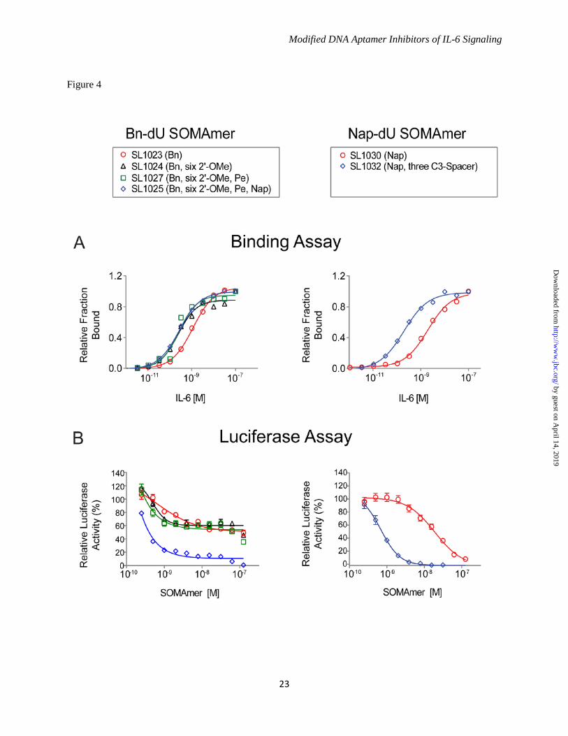
hydrophobic modifications at all other positions contribute to the SOMAmer structure and/or interaction with IL-6. From the single substitution data summarized in Figure 3, more than 100 combinations of tolerated 2'-OMe and C3-spacer substitutions and beneficial 5-dU substitutions were synthesized for each SOMAmer and evaluated for IL-6 binding and inhibition activity (data not shown). Many combinations with up to a 10-fold affinity improvement were identified for both the Bn-dU and Nap-dU SOMAmers. The most favorable combination of substitutions in the Bn-dU SOMAmer was observed in variant SL1025, which contained six 2'-OMe groups (at positions C3, G6, A16, A19, C20 and C28) and two 5-dU mutations (Bn-dU9 Pe-dU9 and Bn-dU12 Nap-dU12). The affinity of SL1025 for IL-6 (Kd = 0.2 nM) was approximately 5-fold greater than that of its precursor SL1023 (Kd = 1 nM). The Nap-dU SOMAmer tolerated fewer substitutions; however, compared to its precursor SL1030, a 10-fold affinity improvement was observed for variant SL1032 (Kd = 0.2 nM), with three C3-spacer substitutions at positions G1, G14 and A15, and the 3' terminal 9 nucleotides removed (see Fig. 4A). These two optimized variants were chosen as leads for further analysis in functional activity assays. Single Bn-dU to Nap-dU in SOMAmer SL1025 Leads to Complete Inhibition of IL-6 - The inhibitory activities of the lead Bn-dU and Nap-dU SOMAmers were compared in the luciferase gene reporter assay before and after optimization. Optimized Nap-dU SOMAmer SL1032 (IC50 = 0.9 nM) was approximately 30-fold more potent than its truncated un-optimized parent SL1030 (IC50 = 30 nM) and its full-length un-optimized parent SL1029 (Fig. 4B and Fig. 1C). Optimized Bn-dU SOMAmer SL1025 also increased in potency compared to its full-length un-optimized parent SL1022, as evident from the twenty-fold shift in IC50 (0.2 nM vs. 4 nM) (Fig. 4B and Fig. 1C). Surprisingly, inhibition of IL-6 by SL1023 increased from 60% to nearly 100% after optimization to SL1025. This observed increase was attributable to the single substitution of Bn-dU to Nap-dU at position 12. A direct comparison of inhibition dose response curves between SL1025 (with Nap-dU12) and SL1027 (SL1025 with Bn-dU12) revealed that this single
by guest on April 14, 2019
http://ww
w.jbc.org/
Dow
nloaded from
Modified DNA Aptamer Inhibitors of IL-6 Signaling
8
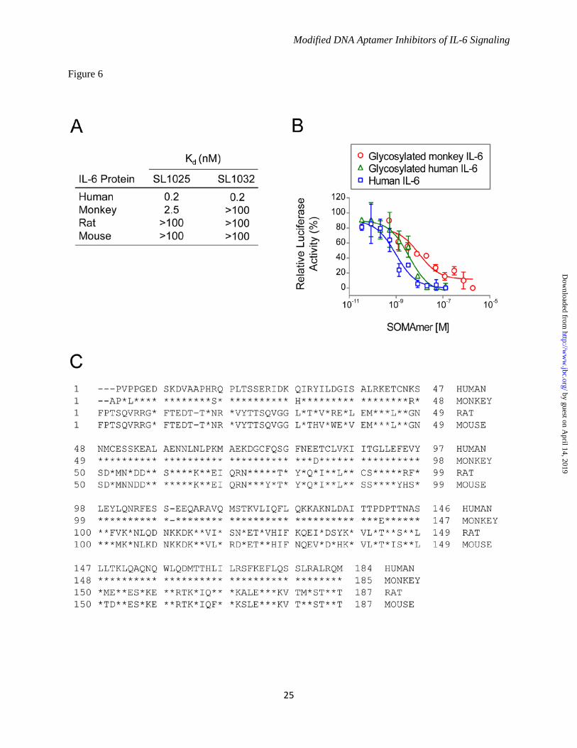
substitution was responsible for the observed increase in percent inhibition (Fig. 4B). It is important to note that the IC50 values are limited by the concentration of IL-6 used in this assay (0.5 nM), and therefore the true inhibitory potency of the lead SOMAmers may be greater than those reflected in the IC50 values. The combined effects of SOMAmer truncation and optimization on IL-6 binding and inhibition are summarized in Table 2. Kinetic Analysis of SOMAmer Binding to IL-6 - A kinetic evaluation of SL1025 and SL1032 binding to IL-6 was performed at 37°C using surface plasmon resonance (SPR). Biotin-labeled SOMAmer was immobilized on a streptavidin-coated surface and IL-6 was injected for 3.5 minutes (association phase), followed by buffer without IL-6 for 60 minutes (dissociation phase). Response units (RU) were plotted as a function of time for all five IL-6 concentrations (Fig. 5). A global fit of the SL1025 data was performed with a one-site binding model, and association and dissociation rate constants were determined (kon = 1.2 x 10⁵ M-1s-1, koff = 2.8 x 10⁻⁵ s-1). The equilibrium binding constant calculated as the ratio of koff/kon (Kd = 2.3 x 10⁻¹⁰ M) was consistent with solution measurements. Rate constants for SL1032 were determined in a similar manner using a two-site binding model (kon,1 = 7.9 x 10⁴ M-1s-1, koff,1 = 6.9 x 10⁻⁶ s-1, kon,2 = 1.4 x 10⁶ M-1s-1, koff,2 = 2.2 x 10⁻³ s-1). The equilibrium binding constant for the high affinity ligand interaction (Kd,1 = koff,1/kon,1 = 8.7 x 10⁻¹¹ M) was also consistent with solution measurements. Cross Species Reactivity and Effect of Protein Glycosylation on SOMAmer Activity - The binding properties of SL1025 and SL1032 to IL-6 from different species including rat, mouse and monkey were profiled. Neither SOMAmer showed measurable binding to any of these orthologs, with the exception of SL1025, which bound monkey IL-6, although with a 10-fold reduction in affinity (Kd = 2.5 nM)(Fig. 6A). The monkey IL-6 used for this study was expressed in eukaryotic cells and was therefore glycosylated, while the human IL-6 used for SELEX was expressed in E. coli and was non-glycosylated. The effect of target glycosylation on SL1025 activity was determined by comparing inhibition of glycosylated and non-glycosylated human IL-6 in the gene reporter assay along with glycosylated monkey IL-6 (Fig. 6B). SL1025 inhibited all three
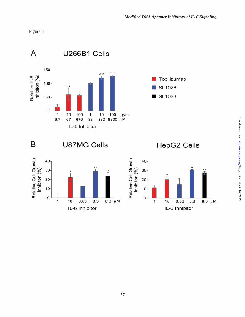
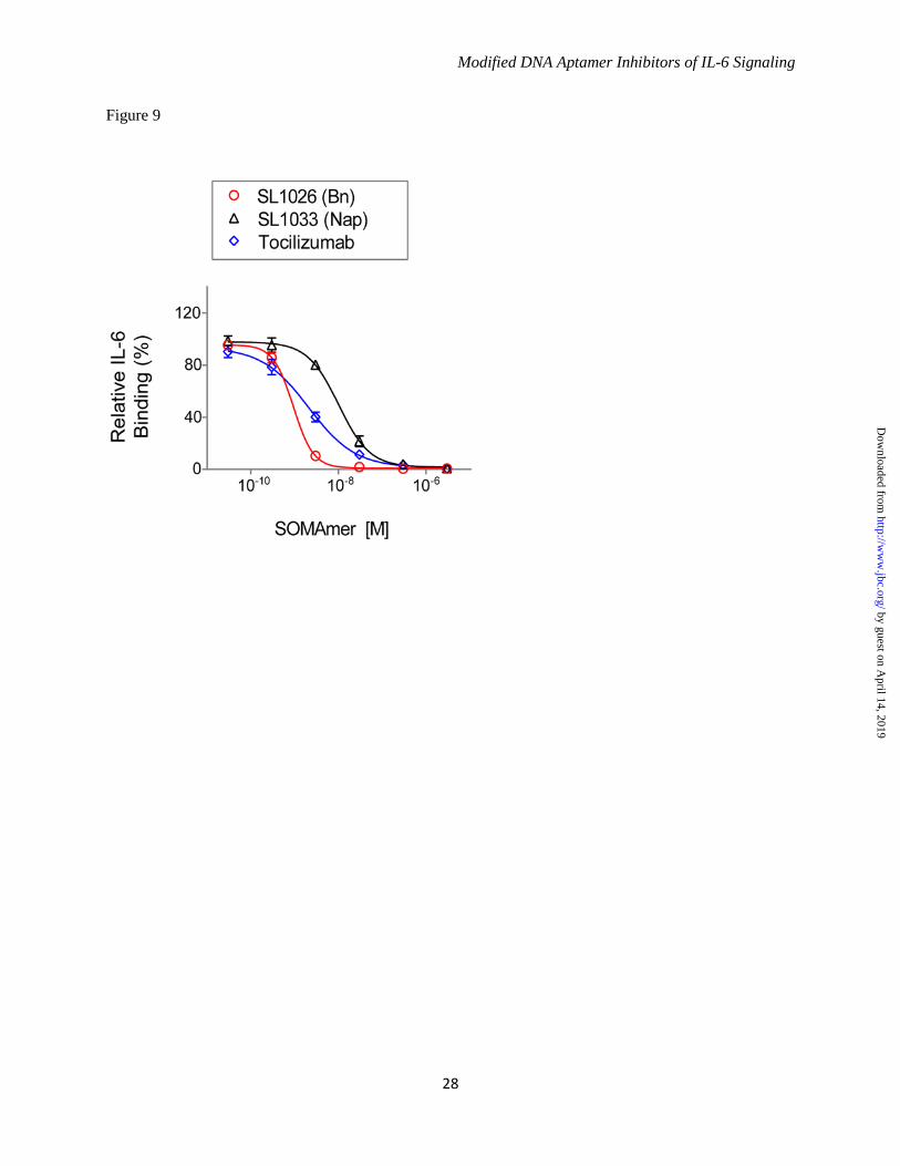
forms of IL-6, but a reduction in potency (4-fold) was observed with glycosylated human IL-6, and a further reduction (3-fold) was observed with glycosylated monkey IL-6. Modified Nucleotides Impart Resistance to Nuclease Attack - The sensitivity of optimized SOMAmers SL1025 and SL1032 to serum nucleases was measured in an in vitro nuclease stability assay. Active SOMAmers were compared to un-optimized versions SL1023 and SL1031, and inactive analogs in which all modified dU residues were replaced with natural dT residues (SL1023dT and SL1031dT). All SOMAmers tested contained a 3' inverted dT (3'-idT) group to block 3' to 5' exonuclease activity (34). SOMAmers were incubated with 90% human serum at 37°C for up to 48 hours, and samples were analyzed at different time points by denaturing PAGE (Fig. 7A). Percent intact SOMAmer was plotted as a function of time, and fit to a one-phase exponential decay model to determine half-life (Fig. 7B). As expected, the dT controls were rapidly cleaved (t1/2 = 5.5 hours for SL1023dT, and t1/2 = 8.5 hours for SL1031dT). The un-optimized SOMAmers, however, were significantly more stable (t1/2 = 50 hours for SL1023, and t1/2 = 77 hours for SL1031). Nevertheless, some degradation of SL1023 and SL1031 was observed, (along with a discreet metabolite of SL1023), indicating certain positions within the SOMAmers remained sensitive to nuclease cleavage. Further stability enhancement was achieved with the addition of 2'-OMe and C3-spacer substitutions, as very little cleavage occurred in 48 hours with the optimized SOMAmers SL1025 and SL1032. Comparison of IL-6 SOMAmer Inhibition Activity with Tocilizumab - To prevent renal elimination in future in vivo studies, a branched 40 kDa PEG was conjugated to SOMAmers SL1025 and SL1032 to create SL1026 and SL1033, respectively (35,36). Addition of the PEG moiety did not affect the inhibitory activity of the SOMAmers in the luciferase gene reporter assay (data not shown). We then compared the activity of Bn-dU SOMAmer SL1026 with tocilizumab in the U266B1 human myeloma cell proliferation assay as described in methods. SL1026 achieved complete inhibition of IL-6 at 1 μg/mL (83 nM), while tocilizumab achieved 60% inhibition at a roughly equivalent molar concentration (67 nM)
by guest on April 14, 2019
http://ww
w.jbc.org/
Dow
nloaded from
Modified DNA Aptamer Inhibitors of IL-6 Signaling
9
(Fig. 8A). Inhibition of cell proliferation by SL1026 and SL1033 was also measured for two human tumor cell lines, HepG2 (hepatoma) and U87MG (glioma), and compared to tocilizumab. Percent proliferation was quantified relative to a no-inhibitor control and plotted in Figure 8B. Both SL1026 and SL1033 suppressed proliferation of U87MG and HepG2 cells to a greater extent than tocilizumab at similar molar concentrations. SL1026 and SL1033 Block Binding of IL-6 to IL-6 Receptor - To further understand the mechanism of inhibition, we developed a plate-based sandwich assay to determine if SOMAmers block IL-6 binding to IL-6Rα, the first step in the IL-6 signaling pathway. We tested sIL-6Rα binding to biotinylated IL-6 pre-incubated with different concentrations of SOMAmer or tocilizumab. The percent of IL-6 bound to sIL-6Rα (relative to the no-competitor control) was plotted as a function of inhibitor concentration (Fig. 9). As the concentration of IL-6 inhibitor increased, the amount of bound IL-6 decreased, indicating that SL1026, SL1033 and tocilizumab block the binding of IL-6 to its receptor. DISCUSSION Aptamers are an established technology for inhibiting protein function in vitro and in vivo (28,29,37). Aptamers bind their molecular targets with high affinity and specificity by virtue of surface shape and charge complementarities. SOMAmers are a new class of aptamers with enhanced functionality offering more favorable properties for target inhibition, including exquisite affinity (typical Kd < 1 nM) and slow dissociation rate (typical t1/2 > 30 minutes). These properties are enabled by hydrophobic adducts at the 5 position of uridine that facilitate the formation of unique intramolecular structures and direct interactions with hydrophobic amino acids on the surface of the target protein (32). We performed the SELEX process against human IL-6 with two random libraries, one with benzyl modifications on the 5 position of dUTP (Bn-dU) and the other with naphthyl modifications (Nap-dU), and identified two SOMAmers with similar binding affinity to IL-6 (Kd = 2-3 nM) but different inhibitory properties in vitro. Optimization efforts resulted in improvements in affinity and nuclease stability, and more potent and complete inhibition of IL-6 activity.
Different primary sequence motifs were identified for SL1022 (Bn-dU) and SL1029 (Nap-dU). Although both modifications are planar, hydrophobic and aromatic, and the SELEX process leading to the discovery of these two SOMAmers was identical, the size and chemical properties of the two modifications were sufficiently different to yield unique SOMAmer sequence solutions (and, by inference, structure solutions) for IL-6 binding. In other words, an apparently subtle change in the functional group of the side chain was sufficient to cause different sequences to be favored during affinity selections. This has been observed previously with modifications at the 2’ position of ribose: for example, SELEX aimed at the same target but accomplished with different starting libraries (including unmodified RNA, DNA, 2’-aminopyrimidine and 2’-fuoropyrimidine RNA) has invariably led to completely different primary sequence solutions to high affinity binding (38-40). Our results extend this observation to highly related modifications at the 5 position of uridine, and provide further support for the notion that nucleic acid ligands represent precisely assembled structures in which individual nucleotides make aggregate contributions to scaffold assembly and presentation of key functional groups to their binding partner to enable the formation of high affinity complexes. Post-SELEX optimization is commonly performed to shorten an aptamer and enhance affinity and nuclease resistance, which often leads to improved potency. For conventional aptamers, backbone protection has been necessary to improve the metabolic stability and achieve adequate plasma residence time for systemic use. RNA aptamers are typically selected with fluorine modifications on the 2' ribose position of pyrimidines, and only purine positions require stabilization, most commonly with 2'-methoxy groups (2'-OMe). DNA SOMAmers are selected with no sugar modifications and therefore all backbone positions are susceptible to endonuclease attack. Because addition of a methoxy group at any 2' position might interfere with protein binding, we surveyed by chemical synthesis each dA, dG and dC position of SL1023 individually to identify those that tolerated a 2'-OMe, and screened combinations to find six positions in SL1025 that could be substituted
by guest on April 14, 2019
http://ww
w.jbc.org/
Dow
nloaded from
Modified DNA Aptamer Inhibitors of IL-6 Signaling
10
without sacrificing binding activity. Unlike conventional RNA aptamers reported in the literature that are broadly substituted with 2'-OMe purines without loss of activity, a smaller number of 2'-OMe substitutions were tolerated in SL1025. This may be attributed in part to a conformational preference in DNA for the 2’-endo sugar pucker and its likely perturbation to the 3’-endo pucker upon 2’-OMe substitution. The 2'-endo conformation is energetically favorable in natural deoxyribose, while the 3'-endo conformation is preferred in ribose (RNA), 2'-OMe ribose and 2’-fluoro ribose (41). The same preference for the 3’-endo conformation in the latter is likely responsible for the high degree of tolerance of the 2’-flouropyrimidine, 2’-ribopurine nucleic acid libraries (which are often used in SELEX) toward 2’-OMe substitution. In contrast, the switch from 2'- to 3'-endo sugar pucker in DNA-based ligands means that such substitutions can be tolerated at positions where ribose conformation is not important or where it can be compensated for by other changes in the molecule. Empirically, at many positions such substitutions perturb the DNA-based SOMAmer structure to reduce the binding affinity to IL-6. Another substitution that is resistant to endonuclease attack is a C3-spacer. While this 3-carbon methylene linker preserves the inter-nucleotide spacing, it lacks a sugar and base and is therefore a more drastic change at any single position of the SOMAmer than a 2'-OMe substitution. The C3-spacer unlocks the conformational restriction imposed by the cyclic deoxyribose thus providing a large degree of additional conformational flexibility through fully rotatable bonds in the inter-nucleotide linkage. We also surveyed each dA, dC and dG position of SL1023 with a C3-spacer substitution and identified a region spanning positions C18-G21 that tolerated this substitution. This region is likely to be spatially distinct from the core binding elements, and not in direct contact with IL-6. SOMAmers are selected from libraries containing a single dU modification. These modifications are essential components of the intermolecular interaction with the target protein, and while they are acceptable solutions, they may not be ideally complementary to the interaction surface. Additional or fewer linker carbons or planar hydrophobic rings might allow one or more
benzyl or naphthyl adducts to adopt a more favorable orientation resulting in improved affinity (32). With this goal in mind, we surveyed each dU position of SL1023 with alternative modifications or no modification (dT), but observed only modest affinity improvements at only a few positions. However, when we tested combinations, we found the Pe-dU and Nap-dU substitutions at positions 9 and 12, paired with the six 2'-OMe additions in SL1025, resulted in improved inhibition activity and serum stability in vitro. One serendipitous outcome of this approach was an increase in percent inhibition achieved with the substitution of Nap-dU at position 12 of SL1025. It is possible that the additional ring of the naphthyl group (compared to the benzyl group) helps to form a more extensive contact surface with IL-6 thereby further stabilizing the complex. Optimization of Nap-dU SOMAmer SL1030 was performed with a similar strategy. Combinations of C3-spacer substitutions resulted in SL1032, with three C3-spacers in the truncated 30-mer providing a 30-fold improvement in inhibition activity. Perhaps removal of these nucleotides relaxed a structural constraint or facilitated a new interaction with IL-6. Combinations of the three C3-spacer substitutions in SL1032 with 2'-OMe substitutions at tolerated positions have not been extensively evaluated, but may offer additional nuclease protection. The optimization process led to improvements in the binding affinities of SL1025 and SL1032 for human IL-6. SPR analysis revealed that the high affinities can be attributed at least in part to exceptionally slow complex dissociation rates, an intrinsic property of SOMAmers. SL1025 exhibited simple monophasic kinetics with a dissociation half-life of 6.9 hours, while SL1032 showed biphasic kinetics with a fast-dissociating species (5 minute half-life) and a slow-dissociating species (28 hour half-life). This biphasic behavior may be due to the existence of two kinetically-trapped conformations of SL1032, one with tight IL-6 binding and one with weaker binding. To study ortholog specificity of the lead SOMAmers the binding properties of SL1025 and SL1032 to rat, mouse and monkey IL-6 were profiled. Human and monkey IL-6 have about 97% sequence identity as compared to only 40% with rat and mouse IL-6 (Fig. 6C). Neither
by guest on April 14, 2019
http://ww
w.jbc.org/
Dow
nloaded from
Modified DNA Aptamer Inhibitors of IL-6 Signaling
11
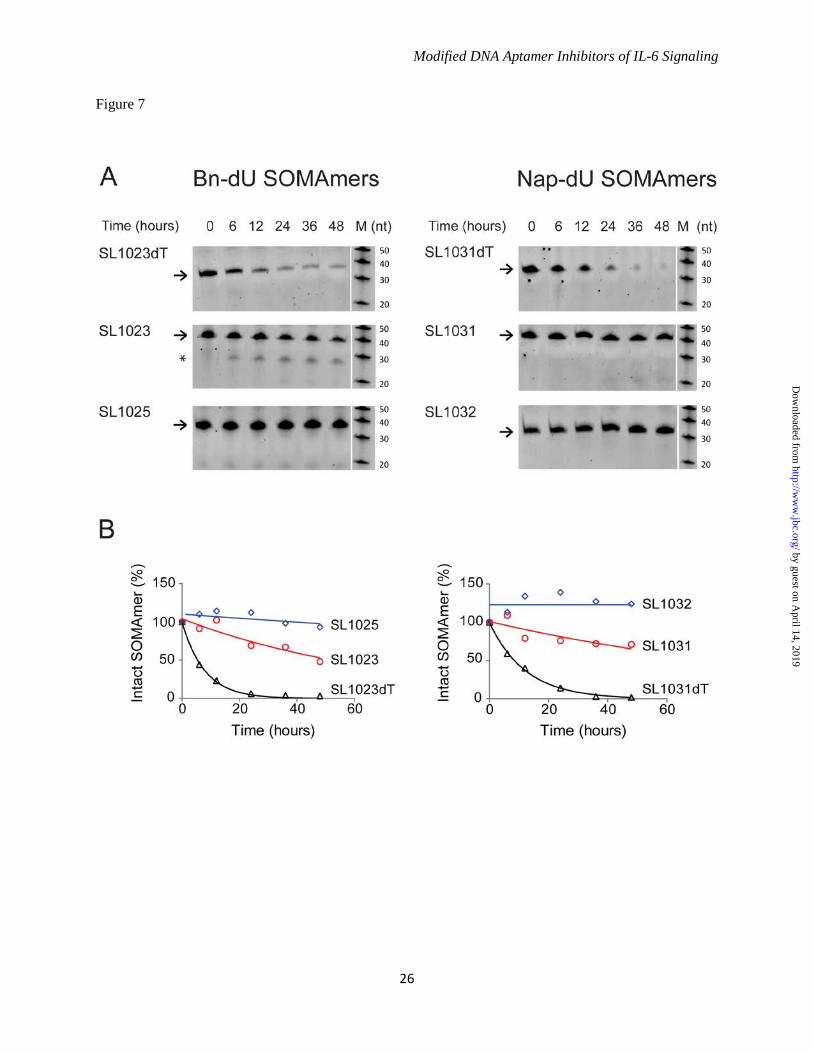
SOMAmer bound rodent IL-6, which was predicted based on the low amino acid sequence conservation. The binding properties of SL1025 and SL1032 were quite different with regards to monkey IL-6 protein. The binding affinity of SL1025 for monkey IL-6 was 10-fold lower than for the human ortholog, while SL1032 showed no binding to monkey IL-6 protein. Because the two SOMAmers compete with each other for IL-6 binding (data not shown) and block IL-6 binding to IL-6Rα (Fig. 9), they likely bind IL-6 protein at overlapping but non-identical sites and make contact with different residues. There are six amino acid differences between human and monkey IL-6 (P3L, R16S, Q28H, K46R, E81D, D140E), and one or more of these may be involved in the interaction of SL1032 with IL-6 (Fig. 6C). Differences in glycosylation may also contribute to the reduction in affinity for monkey IL-6, as the monkey IL-6 used in these experiments was glycosylated while the human IL-6 was not. This effect was observed in inhibition studies where the IC50 of SL1025 was higher for glycosylated human IL-6 than its non-glycosylated form, and even higher for glycosylated monkey IL-6 (Fig. 6B). While inhibiting either IL-6Rα or gp130 binding will interfere with the IL-6 signaling pathway, as we show in the accompanying manuscript (Gelinas et al., 2014), blocking both receptor binding sites may lead to more effective inhibition of IL-6 signaling in vivo. Unmodified DNA and RNA aptamers are rapidly degraded by nucleases present in blood and tissues, resulting in half-lives as short as 2 minutes (42). RNA aptamers for clinical use are heavily modified at the 2' position of ribose with fluorine (2'-F), methoxy (2'-OMe) or amine (2'-NH2) groups, or in the phosphodiester backbone with phosphorothioate (PS) groups to achieve suitable in vivo stability (28). To determine the effect of the 5-dU modifications and 2'-OMe and C3-spacer substitutions on metabolic stability, we tested the SOMAmers in an in vitro serum stability assay. This assay served as a surrogate for an in vivo pharmacokinetic evaluation to assess the sensitivity of SOMAmers to endonucleases present in plasma in the absence of other clearance mechanisms. As expected, unmodified DNA control sequences SL1023dT and SL1031dT were rapidly degraded. The benzyl and naphthyl modifications present in SL1023 and SL1031
increased half-life by about 10-fold, indicating that these adducts interfere with substrate recognition by serum nucleases and alone offer a degree of resistance not present in unmodified aptamers. It is also possible that SL1023dT and SL1031dT adopt alternate conformations, exposing endonuclease sites not present on SL1023 and SL1031. Another substantial increase in stability was achieved by the addition of 2'-OMe and C3-spacer modifications in SL1025 and SL1032, indicating our optimization efforts were successful. In addition to its role in inflammatory disease, IL-6 signaling is also a key component of tumor cell proliferation (43). IL-6 receptor is over-expressed in some cancers including brain, prostate, and kidney, and elevated IL-6 ligand and receptor expression are associated with poor patient survival. Inhibition of IL-6 signaling may suppress growth, survival, and/or metastatic potential of tumor cells. Inhibition of tumor cell proliferation by SL1026 and SL1033 was demonstrated in U266B1 myeloma, HepG2 hepatoma and U87MG glioma cells, and the potency of SOMAmer inhibition of IL-6 was equal to or greater than tocilizumab inhibition of IL-6Rα in all cases (Fig. 8). The role of constitutive activation of the IL-6 signaling pathway is well established in inflammation and an increase in IL-6 and soluble IL-6Rα in the synovial fluid of joints in rheumatoid arthritis patients was shown to correlate with disease progression (44). The humanized anti-IL-6 receptor antibody tocilizumab has afforded some benefit to patients with rheumatoid arthritis, Castleman’s disease and juvenile idiopathic arthritis (45). However, side effects of both classical and trans-signaling inhibition of IL-6Rα by tocilizumab have been reported, including increased cholesterol and triglyceride levels accompanied by weight gain (45-47). Also, an increase in IL-6 protein is seen in patients treated with tocilizumab (46). Direct inhibition of IL-6 may offer some advantages over inhibition of IL-6Rα because of the known role of soluble IL-6Rα in facilitating clearance of IL-6 from the circulation (46). Inhibition of IL-6 with the anti-IL-6 antibody clazakizumab was recently shown to be effective at controlling the symptoms of rheumatoid arthritis in a Phase IIb clinical study (48). Clazakizumab was also shown to be more
by guest on April 14, 2019
http://ww
w.jbc.org/
Dow
nloaded from
Modified DNA Aptamer Inhibitors of IL-6 Signaling
12
potent than tocilizumab at blocking IL-6 induced cell functions in vitro (49). The IL-6 SOMAmers described in this report have properties well-suited for this therapeutic challenge - high affinity, slow complex dissociation, endonuclease resistance, and potent
inhibition of IL-6 signaling. While these SOMAmers antagonize IL-6 activity in vitro, an in vivo evaluation in an inflammation model is required to understand the true therapeutic potential of this new class of IL-6 inhibitors.
by guest on April 14, 2019
http://ww
w.jbc.org/
Dow
nloaded from
Modified DNA Aptamer Inhibitors of IL-6 Signaling
13
REFERENCES 1. Heinrich, P. C., Behrmann, I., Muller-Newen, G., Schaper, F., and Graeve, L. (1998) Interleukin-
6-type cytokine signalling through the gp130/Jak/STAT pathway. Biochem. J. 334, 297-314 2. Hirano, T., Yasukawa, K., Harada, H., Tata, T., Watanabe, Y., Matsuda, T., Kashiwamura, S.,
Nakajima, K., Koyoma, K., Iwamatsu, A., Tsunasawa, S., Sakiyama, F., Matsui, H., Takahara, Y., Taniguchi, T., and Kishimoto, T. (1986) Complementary DNA for a novel human interleukin (BSF-2) that induces B lymphocytes to produce immunoglobulin. Nature 324, 73-76
3. Somers, W., Stahl, S., and Seehra, J. S. (1997) 1.9 Å crystal structure of interleukin 6: implications for a novel mode of receptor dimerization and signaling. The EMBO J. 16, 989-997
4. Kishimoto, T. (2005) Interleukin-6: from basic science to medicine--40 years in immunology. Annu. Rev. Immunol. 23, 1-21
5. Scheller, J., Chalaris, A., Schmidt-Arras, D., and Rose-John, S. (2011) The pro- and anti-inflammatory properties of the cytokine interleukin-6. Biochim. Biophys. Acta 1813, 878-888
6. Honda, M., Yamamoto, S., Cheng, M., Yasukawa, K., Suzuki, H., Saito, T., Osugi, Y., Tokunaga, T., and Kishimoto, T. (1992) Human soluble IL-6 receptor: its detection and enhanced release by HIV infection. J. Immunol. 148, 2175-2180
7. Novick, D., Engelmann, H., Wallach, D., and Rubinstein, M. (1989) Soluble cytokine receptors are present in normal human urine. J. Exp. Med. 170, 1409-1414
8. Waetzig, G. H., and Rose-John, S. (2012) Hitting a complex target: an update on interleukin-6 trans-signalling. Expert Opin. Ther. Targets 16, 225-236
9. Rose-John, S. (2012) IL-6 trans-signaling via the soluble IL-6 receptor: importance for the pro-inflammatory activities of IL-6. Int. J. Biol. Sci. 8, 1237-1247
10. Guo, Y., Xu, F., Lu, T., Duan, Z., and Zhang, Z. (2012) Interleukin-6 signaling pathway in targeted therapy for cancer. Cancer Treat. Rev. 38, 904-910
11. Waage, A., Brandtzaeg, P., Halstensen, A. S., Kierulf, P., and Terje Espevik, T. (1989) The complex pattern of cytokines in serum from patients with meningococcal septic shock. J. Exp. Med. 169, 333-338
12. Kopf, M., Bachmann, M. F., and Marsland, B. J. (2010) Averting inflammation by targeting the cytokine environment. Nat. Rev. Drug Discov. 9, 703-718
13. Dayer, J. M., and Choy, E. (2010) Therapeutic targets in rheumatoid arthritis: the interleukin-6 receptor. Rheumatology (Oxford) 49, 15-24
14. Tanaka, T., Narazaki, M., and Kishimoto, T. (2012) Therapeutic targeting of the interleukin-6 receptor. Annu. Rev. Pharmacol. Toxicol. 52, 199-219
15. Nishimoto, N., Kishimoto, T., and Yoshizaki, K. (2000) Anti-interleukin 6 receptor antibody treatment in rheumatic disease. Ann. Rheum. Dis. 59 Suppl 1, i21-27
16. Smolen, J. S., and Maini, R. N. (2006) Interleukin-6: a new therapeutic target. Arthritis. Res. Ther. 8 Suppl 2, S5
17. Tanaka, T., and Kishimoto, T. (2012) Targeting interleukin-6: all the way to treat autoimmune and inflammatory diseases. Int. J. Biol. Sci. 8, 1227-1236
18. Ellington, A. D., and Szostak, J. W. (1990) In vitro selection of RNA molecules that bind specific ligands. Nature 346, 818-822
19. Tuerk, C., and Gold, L. (1990) Systematic evolution of ligands by exponential enrichment: RNA ligands to bacteriophage T4 DNA polymerase. Science 249, 505-510
20. Gupta, S., Thirstrup, D., Jarvis, T. C., Schneider, D. J., Wilcox, S. K., Carter, J., Zhang, C., Gelinas, A., Weiss, A., Janjic, N., and Baird, G. S. (2011) Rapid histochemistry using slow off-rate modified aptamers with anionic competition. Appl. Immunohistochem. Mol. Morphol. 19, 273-278
21. Gold, L., Ayers, D., Bertino, J., Bock, C., Bock, A., Brody, E. N., Carter, J., Dalby, A. B., Eaton, B. E., Fitzwater, T., Flather, D., Forbes, A., Foreman, T., Fowler, C., Gawande, B., Goss, M., Gunn, M., Gupta, S., Halladay, D., Heil, J., Heilig, J., Hicke, B., Husar, G., Janjic, N., Jarvis, T.,
by guest on April 14, 2019
http://ww
w.jbc.org/
Dow
nloaded from

Modified DNA Aptamer Inhibitors of IL-6 Signaling
14
Jennings, S., Katilius, E., Keeney, T. R., Kim, N., Koch, T. H., Kraemer, S., Kroiss, L., Le, N., Levine, D., Lindsey, W., Lollo, B., Mayfield, W., Mehan, M., Mehler, R., Nelson, S. K., Nelson, M., Nieuwlandt, D., Nikrad, M., Ochsner, U., Ostroff, R. M., Otis, M., Parker, T., Pietrasiewicz, S., Resnicow, D. I., Rohloff, J., Sanders, G., Sattin, S., Schneider, D., Singer, B., Stanton, M., Sterkel, A., Stewart, A., Stratford, S., Vaught, J. D., Vrkljan, M., Walker, J. J., Watrobka, M., Waugh, S., Weiss, A., Wilcox, S. K., Wolfson, A., Wolk, S. K., Zhang, C., and Zichi, D. (2010) Aptamer-based multiplexed proteomic technology for biomarker discovery. PloS one 5, e15004
22. Mascini, M., and Tombelli, S. (2008) Biosensors for biomarkers in medical diagnostics. Biomarkers 13, 637-657
23. Ruckman, J., Green, L. S., Beeson, J., Waugh, S., Gillette, W. L., Henninger, D. D., Claesson-Welsh, L., and Janjic, N. (1998) 2'-Fluoropyrimidine RNA-based aptamers to the 165-amino acid form of vascular endothelial growth factor (VEGF165). Inhibition of receptor binding and VEGF-induced vascular permeability through interactions requiring the exon 7-encoded domain. J. Biol. Chem. 273, 20556-20567
24. Gragoudas, E. S., Adamis, A. P., Cunningham, E. T., Feinsod, M., and Guyer, D. R. (2004) Pegaptanib for Neovascular Age-Related Macular Degeneration. New Engl. J. Med. 351, 2805-2816
25. Chan, M. Y., Rusconi, C. P., Alexander, J. H., Tonkens, R. M., Harrington, R. A., and Becker, R. C. (2008) A randomized, repeat-dose, pharmacodynamic and safety study of an antidote-controlled factor IXa inhibitor. Journal of thrombosis and haemostasis : JTH 6, 789-796
26. Ophthotech website press release. (2013) Ophthotech Enrolls First Patient in Phase 3 Clinical Trial of Fovista™ Anti-PDGF Therapy in Combination with Anti-VEGF Therapy for Wet AMD. (http://www.ophthotech.com/news).
27. Ophthotech website. (2012) ARC1905 Anti-C5 Aptamer Clinical Development. (http://www.ophthotech.com/product-candidates/arc1905/)
28. Keefe, A. D., Pai, S., and Ellington, A. (2010) Aptamers as therapeutics. Nat. Rev. Drug Discov. 9, 537-550
29. Bouchard, P. R., Hutabarat, R. M., and Thompson, K. M. (2010) Discovery and development of therapeutic aptamers. Annu. Rev. Pharmacol. Toxicol. 50, 237-257
30. Vaught, J. D., Bock, C., Carter, J., Fitzwater, T., Otis, M., Schneider, D., Rolando, J., Waugh, S., Wilcox, S. K., and Eaton, B. E. (2010) Expanding the Chemistry of DNA for in Vitro Selection. J. Am. Chem. Soc. 132, 4141-4151
31. Beaucage, S., and Caruthers, M. (1981) Deoxynucleoside phosphoramidites - A new class of key intermediates for deoxypolynucleotide synthesis. Tetrahedron Lett. 22, 1859-1862
32. Davies, D. R., Gelinas, A. D., Zhang, C., Rohloff, J. C., Carter, J. D., O'Connell, D., Waugh, S. M., Wolk, S. K., Mayfield, W. S., Burgin, A. B., Edwards, T. E., Stewart, L. J., Gold, L., Janjic, N., and Jarvis, T. C. (2012) Unique motifs and hydrophobic interactions shape the binding of modified DNA ligands to protein targets. Proc. Natl. Acad. Sci. U S A 109, 19971-19976
33. Aaronson, D. S., and Horvath, C. M. (2002) A road map for those who don't know JAK-STAT Science 296, 1653-1655
34. Beigelman, L., McSwiggen, J. A., Draper, K. G., Gonzalez, C., Jensen, K., Karpeisky, A. M., Moday, A. S., Matulic-Adamic, J., DiRenzo, A. B., Haeberli, P., Sweedlere, D., Tracz, D., Grimm, S., Wincott, F., Thackray, V., and Usman, N. (1995) Chemical Modification of Hammerhead Ribozymes. J. Biol. Chem. 270, 25702-25708
35. Kawaguchi, T., Asakawa, H., Tashiro, Y., Juni, K., and Sueishi, T. (1995) Stability, specific binding activity, and plasma concentration in mice of an oligodeoxynucleotide modified at 5'-terminal with poly(ethylene glycol). Biol. Pharm. Bulletin 18, 474-476
36. Watson, S. R., Chang, Y. F., O'Connell, D., Weigand, L., Ringquist, S., and Parma, D. H. (2000) Anti-L-selectin aptamers: binding characteristics, pharmacokinetic parameters, and activity against an intravascular target in vivo. Antisense Nucleic Acid Drug Dev. 10, 63-75
by guest on April 14, 2019
http://ww
w.jbc.org/
Dow
nloaded from
Modified DNA Aptamer Inhibitors of IL-6 Signaling
15
37. Burnett, J. C., and Rossi, J. J. (2012) RNA-based therapeutics: current progress and future prospects. Chem. Biol. 19, 60-71
38. Eaton, B. E., Gold, L., Hick, B. J., Janjic, N., Jucker, F. M., Sebesta, D. P., Tarasow, T. M., WIllis, M. C., and Zichi, D. A. (1997) Post-SELEX combinatorial optimization of aptamers. Bioorg. Med. Chem. 5, 1087-1096
39. Green, L. S., Jellinek, D., Bell, C., Beebe, L. A., Feistner, B. D., Gill, S. C., Jucker, F. M., and Janjic, N. (1995) Nuclease-resistant nucleic acid ligands to vascular permeability factor/vascular endothelial growth factor. Chem. Biol. 2, 683-695
40. Jellinek, D., Green, L. S., Bell, C., and Janjic, N. (1994) Inhibition of receptor binding by high-affinity RNA ligands to vascular endothelial growth factor. Biochemistry 33, 10450-10456
41. Guschlbauer, W., and Jankowski, K. (1980) Nucleoside conformation is determined by the electronegativity of the sugar substituent. Nucleic Acids Res. 8, 1421-1433
42. Griffin, L., Tidmarsh, G., Bock, L., Toole, J., and Leung, L. (1993) In Vivo Anticoagulant Properties of a Novel Nucleotide-Based Thrombin Inhibitor and Demonstration of Regional Anticoagulation in Extracorporeal Circuits. Blood 81, 3271-3276
43. Ara, T., and Declerck, Y. A. (2010) Interleukin-6 in Bone Metastasis and Cancer Progression. Eur. J. Cancer 46, 1223-1231
44. Kishimoto, T. (2010) IL-6: from its discovery to clinical applications. Int. Immunol. 22, 347-352 45. Jones, S. A., Scheller, J., and Rose-John, S. (2011) Therapeutic strategies for the clinical
blockade of IL-6/gp130 signaling. J. Clin. Invest. 121, 3375-3383 46. Nishimoto, N., Terao, K., Mima, T., Nakahara, H., Takagi, N., and Kakehi, T. (2008)
Mechanisms and pathologic significances in increase in serum interleukin-6 (IL-6) and soluble IL-6 receptor after administration of an anti-IL-6 receptor antibody, tocilizumab, in patients with rheumatoid arthritis and Castleman disease. Blood 112, 3959-3964
47. Singh, J. A., Beg, S., and Lopez-Olivo, M. A. (2011) Tocilizumab for rheumatoid arthritis: a Cochrane systematic review. J. Rheumatol. 38, 10-20
48. Bristol-Myers Squibb press release. (2013) Promising Phase IIb Data On Clazakizumab In Patients With Moderate-To-Severe Rheumatoid Arthritis To Be Presented At The 2013 Annual Meeting Of The American College Of Rheumatology. (www.bms.com/news/press_releases)
49. Zhao, Q., Pang, J., Shuster, D., Hung, C., Baglino, S., Dodge, R., Sun, H., Trigona, W., and Salter-Cid, L. (2013) Anti-IL-6 Antibody Clazakizumab Is More Potent Than Tocilizumab In Blocking In Vitro and Ex Vivo IL-6-Induced Functions. Abstract: #2385. Session title: Rheumatoid Arthritis Treatment - Small Molecules, Biologics and Gene Therapy III. ACR/ARHP Annual Meeting 13
by guest on April 14, 2019
http://ww
w.jbc.org/
Dow
nloaded from

Modified DNA Aptamer Inhibitors of IL-6 Signaling
16
Acknowledgements - We gratefully acknowledge the contributions of Dominic Zichi for the alignment of sequences and pattern identification, SPR data analysis, and advice on statistical analyses; Steve Wolk for his assistance with the SPR experiments; Dan Drolet for advice on statistical analyses and critical review of the manuscript; Nancy Kim for graphic artist services; and BiOptix of Boulder, Colorado for expert technical assistance and use of the 404pi instrument for all SPR experiments. SOMAmer is a registered trademark of SomaLogic, Inc. FOOTNOTES 1To whom correspondence may be addressed: SomaLogic, Inc., 2945 Wilderness Place, Boulder, CO, USA, Tel.: (303) 625-2089; E-mail: [email protected] 2Otsuka Pharmaceutical Co., Ltd., 463-10 Kagasuno, Kawauchi-cho, Tokushima 771-0192, Japan 3The abbreviations used are: SELEX, Systematic Evolution of Ligands by EXponential enrichment; SOMAmer, Slow Off-rate Modified Aptamer; Bn-dU, 5-(N-benzylcarboxamide)-2'-deoxyuridine; Nap-dU, 5-[N-(1-naphthylmethyl)carboxamide]-2'-deoxyuridine; Pe-dU, 5-[N-(phenyl-2-ethyl)carboxamide]-2'-deoxyuridine; Pp-dU, 5-[N-(phenyl-3-propyl)carboxamide]-2'-deoxyuridine; 2'-OMe, 2'-methoxy; SPR, surface plasmon resonance FIGURE LEGENDS FIGURE 1. Properties of SOMAmers discovered by the SELEX process. A. Structure of 5-(N-benzylcarboxamide)-2'-deoxyuridine (Bn-dU) and 5-[N-(1-naphthylmethyl)carboxamide]-2'-deoxyuridine (Nap-dU). B. IL-6 inhibition activity of six high affinity SOMAmers (3 Bn-dU and 3 Nap-dU) in the luciferase gene reporter assay. Percent luciferase activity was plotted relative to a no-SOMAmer control sample. Each bar represents the mean +/- SEM with 2 replicates in three independent experiments. C. Dose-dependent inhibition of IL-6 by lead SOMAmers SL1022 and SL1029 in the luciferase gene reporter assay. Percent IL-6 activity (relative to a no-SOMAmer control sample) was plotted as a function of SOMAmer concentration. Each data point represents the mean +/- SEM with 4 replicates for each condition. The data were fit to a 4-parameter sigmoid dose response model and IC50 values were determined. The reported IC50 value for SL1022 is an apparent IC50 because incomplete inhibition (<100%) was observed at the highest SOMAmer concentration tested. FIGURE 2. Conserved Sequence Patterns in Lead SOMAmers. A. Alignment of SL1022 (indicated with *) with additional sequences from the Bn-dU SELEX pool containing the consensus motif GGZZZGG. The number of copies of each sequence in a pool of 14,404 is shown. B. Alignment of SL1029 (indicated with *) with additional sequences from the Nap-dU SELEX pool containing the consensus motifs CGPAAGGCGGP and PPAPGPAA. The number of copies of each sequence in a pool of 7,758 is shown. FIGURE 3. Post-SELEX optimization of truncated IL-6 SOMAmers SL1023 and SL1030. For all panels, affinity ratios (Kd
variant/Kdparent) are reported and shaded with a color gradient from blue (affinity
enhancement) to red (affinity loss), where the color intensity represents the magnitude of the affinity ratio. No affinity data was acquired at positions marked with "−". A. 2'-OMe and C3-spacer substitution scans at A, C and G positions of SL1023. The sequence of lead optimized truncate SL1025 is shown with modifications indicated (Bn = Bn-dU, Pe = Pe-dU, Nap = Nap-dU, mC = 2'-OMe C, mG = 2'-OMe G, mA = 2'-OMe A). B. 5-position modification scans at Bn-dU positions of SL1023. Structures of hydrophobic modifications Bn-dU, Pe-dU, Pp-dU and Nap-dU are illustrated. C. 2'-OMe and C3-spacer substitution scans at A, C and G positions of SL1030. The sequence of lead optimized truncate SL1032 is shown with modifications indicated (L = C3-spacer). D. 5-position modification scans at Nap-dU positions of SL1030.
by guest on April 14, 2019
http://ww
w.jbc.org/
Dow
nloaded from
Modified DNA Aptamer Inhibitors of IL-6 Signaling
17
FIGURE 4. Improvements in IL-6 binding and inhibition activity after SOMAmer optimization. A. IL-6 dose-dependent binding of SL1023, SL1030 and optimized variants. Equilibrium binding constants of truncated SOMAmers SL1023 and SL1030 and optimized variants were measured using a solution affinity assay. The fraction of complexed SOMAmer was plotted as a function of IL-6 concentration and equilibrium binding constants (Kd) were determined by fitting the data to a 4-parameter sigmoid dose-response model. B. Dose-dependent inhibition of IL-6 by SL1023, SL1030 and optimized variants. Functional inhibitory activity (IC50) values of truncated SOMAmers SL1023, SL1030 and optimized variants were measured in the luciferase gene reporter assay as described in Figure 1C. Each data point represents the mean +/- SEM with 4 replicates for each condition. FIGURE 5. Kinetic evaluation of SL1025 and SL1032 binding to IL-6. Kinetic constants (kon, koff) for the interaction between IL-6 and SOMAmer were determined by SPR analysis with a BiOptix 404pi biosensor. Raw data are shown in gray with curve fits overlaid in black and IL-6 concentrations indicated to the right of each curve. A. SL1025 data were fit globally to a one-site binding model to determine kinetic constants. B. SL1032 data were fit globally to a two-site binding model to determine kinetic constants. FIGURE 6. Cross reactivity of SOMAmers with IL-6 from different species. A. Equilibrium binding constants (Kd) of SL1025 and SL1032 to human, cynomolgus monkey, rat and mouse IL-6 protein. Kd values were determined as described in Figure 4A. B. Dose-dependent inhibition of human, glycosylated human and glycosylated cynomolgus monkey IL-6 by SL1026 in the luciferase gene reporter assay. IC50 values were determined as described in Figure 1C. C. Amino acid sequence alignment of mature human, cynomolgus monkey, rat and mouse IL-6 proteins. Sequences of IL-6 proteins with Swiss-Prot accession numbers P05231 (human), P79341 (cynomolgus monkey), P20607 (rat) and P08505 (mouse) were aligned with UniProt after removing the signaling peptide sequence at the N-terminus. Amino acid sequences are presented in single letter code and residues that are identical with the sequence of human IL-6 protein are indicated with a star. FIGURE 7. Stability of SOMAmers in human serum in vitro. A. Gel images of truncated SOMAmers SL1023 and SL1031, dT controls SL1023dT and SL1031dT, and optimized SOMAmers SL1025 and SL1032 after serum exposure for the indicated amount of time. DNA markers (M) were run on a different part of the gel. Intact SOMAmers are indicated with arrows, and a stable metabolite of SL1023 is marked with an asterisk. B. Graphical representation of PAGE results. The fraction of intact SOMAmer (relative to the zero time point control sample) was plotted as a function of time. The data were fit to a one-phase exponential decay model to determine half-life values. FIGURE 8. Inhibition of tumor cell proliferation by SL1026, SL1033 and tocilizumab. A. Inhibition of U266B1 (human myeloma) cell proliferation. Cell proliferation was measured with alamarBlue and percent IL-6 inhibition (relative to a no-inhibitor control sample) was plotted. Each bar represents the mean +/- SEM of 3 experiments. The data were analyzed by one way ANOVA (Dunnet’s two –tailed) and each data point was compared with control data without IL-6 inhibitor. Statistically significant differences are denoted as *(p-value < 0.05), **(p-value < 0.01), ****(p-value < 0.0001). B. Inhibition of U87MG (human glioma) and HepG2 (human hepatoma) cell proliferation. Percent inhibition of cell growth (relative to a no-inhibitor control sample) was plotted. FIGURE 9. SL1026 and SL1033 compete with soluble IL-6 receptor for IL-6 binding. IL-6 binding to immobilized IL-6Rα was measured in the presence of SOMAmer or tocilizumab. Percent IL-6 binding (relative to a no-inhibitor control sample) was plotted as a function of inhibitor concentration. Each data point represents the mean +/- SEM with 2 replicates for each condition. The data were fit to a 4-parameter sigmoid dose-response model.
by guest on April 14, 2019
http://ww
w.jbc.org/
Dow
nloaded from

Modified DNA Aptamer Inhibitors of IL-6 Signaling
18
Table 1
Key to SOMAmers described in this report
SOMAmer Version Length (nt) Composition 5' Terminal Group
SL1022 parent 78 Bn OHSL1023 truncate 32 Bn OHSL1023dT unmodified truncate 32 dT OHSL1024 optimized truncate 32 Bn, 2'-OMe OHSL1025 lead optimized truncate 32 Bn, 2'-OMe, Pe, Nap OHSL1026 lead optimized truncate 32 Bn, 2'-OMe, Pe, Nap PEGSL1027 optimized truncate 32 Bn, 2'-OMe, Pe, Nap OH
SL1029 parent 79 Nap OHSL1030 truncate 39 Nap OHSL1031 truncate 30 Nap OHSL1031dT unmodified truncate 30 dT OHSL1032 lead optimized truncate 30 Nap, L OHSL1033 lead optimized truncate 30 Nap, L PEG
Modifications are denoted as follows: Bn (Bn-dU), Nap (Nap-dU), Pe (Pe-dU), 2'-OMe (2'-methoxy), L (C3-spacer), OH (hydroxyl), PEG (polyethylene glycol, 40 kDa)
by guest on April 14, 2019
http://ww
w.jbc.org/
Dow
nloaded from

Modified DNA Aptamer Inhibitors of IL-6 Signaling
19
Table 2
Properties of SOMAmers before and after post-SELEX optimization
SOMAmer Modification Version Length (nt) Kd ,nM IC50 ,nM Percent Inhibition*
SL1022 Bn-dU parent 78 3.4 (2.0-4.9) 4.0 (2.8-5.7)** 62 (57-68)SL1025 Bn-dU lead optimized truncate 32 0.16 (0.11-0.20) 0.20 (0.15-0.27) 90 (81-102)
SL1029 Nap-dU parent 79 2.1 (1.9-2.3) 30 (17-77) 100 (78-106)SL1032 Nap-dU lead optimized truncate 30 0.19 (0.17-0.22) 0.87 (0.78-0.98) 102 (101-103)
95% confidence intervals of curve fit values are reported in parenthases after Kd, IC50 and Percent Inhibition values* Percent inhibition observed at highest SOMAmer concentration tested** Apparent IC50
by guest on April 14, 2019
http://ww
w.jbc.org/
Dow
nloaded from
Modified DNA Aptamer Inhibitors of IL-6 Signaling
20
Figure 1
by guest on April 14, 2019
http://ww
w.jbc.org/
Dow
nloaded from

Modified DNA Aptamer Inhibitors of IL-6 Signaling
21
Figure 2
by guest on April 14, 2019
http://ww
w.jbc.org/
Dow
nloaded from
Modified DNA Aptamer Inhibitors of IL-6 Signaling
22
Figure 3
by guest on April 14, 2019
http://ww
w.jbc.org/
Dow
nloaded from
Modified DNA Aptamer Inhibitors of IL-6 Signaling
23
Figure 4
by guest on April 14, 2019
http://ww
w.jbc.org/
Dow
nloaded from

Modified DNA Aptamer Inhibitors of IL-6 Signaling
24
Figure 5
by guest on April 14, 2019
http://ww
w.jbc.org/
Dow
nloaded from
Modified DNA Aptamer Inhibitors of IL-6 Signaling
25
Figure 6
by guest on April 14, 2019
http://ww
w.jbc.org/
Dow
nloaded from
Modified DNA Aptamer Inhibitors of IL-6 Signaling
26
Figure 7
by guest on April 14, 2019
http://ww
w.jbc.org/
Dow
nloaded from
Modified DNA Aptamer Inhibitors of IL-6 Signaling
27
Figure 8
by guest on April 14, 2019
http://ww
w.jbc.org/
Dow
nloaded from
Modified DNA Aptamer Inhibitors of IL-6 Signaling
28
Figure 9
by guest on April 14, 2019
http://ww
w.jbc.org/
Dow
nloaded from
Zhang, Bharat Gawande, Michael Vrkljan, Nebojsa Janjic and Daniel J. SchneiderMuraguchi, Masafumi Shibamori, Yuichi Ishikawa, Thale C. Jarvis, Jeffrey D. Carter, Chi
Shashi Gupta, Masao Hirota, Sheela M. Waugh, Ikuo Murakami, Tomoki Suzuki, MasahiroInhibit Signaling by Blocking its Interaction with Interleukin-6 Receptor
Chemically-Modified DNA Aptamers Bind Interleukin-6 with High Affinity and
published online January 12, 2014J. Biol. Chem.
10.1074/jbc.M113.532580Access the most updated version of this article at doi:
Alerts:
When a correction for this article is posted•
When this article is cited•
to choose from all of JBC's e-mail alertsClick here
by guest on April 14, 2019
http://ww
w.jbc.org/
Dow
nloaded from