Embed Size (px)
Citation preview
Instructions for use
Title Modified "Dredging Method" for complicated solid/multicystic ameloblastoma in the mandible: Report of a case treatedby fractionated enucleation
Author(s) Ohiro, Yoichi; Yamada, Tamaki; Kakuguchi, Wataru; Kobayashi, Ichizo; Kitamura, Tetsuya; Tei, Kanchu
Citation Journal of oral and maxillofacial surgery, medicine, and pathology, 31(2), 121-125https://doi.org/10.1016/j.ajoms.2018.11.001
Issue Date 2019-03
Doc URL http://hdl.handle.net/2115/76828
Rights © [2019]. This manuscript version is made available under the CC-BY-NC-ND 4.0 licensehttp://creativecommons.org/licenses/by-nc-nd/4.0/
Rights(URL) http://creativecommons.org/licenses/by-nc-nd/4.0/
Type article (author version)
File Information Ohiro_31 (2)_121_201903.pdf
Hokkaido University Collection of Scholarly and Academic Papers : HUSCAP
Title:
Modified “Dredging Method” for complicated solid/multicystic ameloblastoma in the
mandible: Report of a case treated by fractionated enucleation
Author names and affiliations:
Yoichi Ohiroa*, Tamaki Yamadaa, Wataru Kakuguchi a, Ichizo Kobayashib, Tetsuya Kitamurac
and Kanchu Teia
a Department of Oral and Maxillofacial Surgery, c Department of Oral Pathology and Biology,
Faculty of Dental Medicine and Graduate School of Dental Medicine, Hokkaido University
Kita13 Nishi7, Kita-ku, Sapporo 060-8586, Japan
b Department of Oral Surgery, JR Sapporo Hospital
Kita3 Higashi1, Chuou-ku, Sapporo, 060-0033, Japan
*Corresponding author:
Tel.: +81 11 706 4283; Fax: +81 11 706 4283.
E-mail address: [email protected] (Y. Ohiro)
Abstract
The purpose of this paper is to describe the treatment procedures for a solid/multicystic
ameloblastoma which was treated with the “Dredging Method” modified by fractionated
enucleation adopted to avoid the pathologic fracture because of the complicated expansive
nature in the mandible. Ameloblastoma is defined as a benign epithelial odontogenic tumor with
progressive growth potential. Currently employed surgical treatment as curative treatment is
resection with adequate margins because of the characteristics of high recurrence rates with
conservative treatment. In this article, a 38-year-old male with swelling on the right mandible
was referred to our hospital. Image analysis showed an expansile partially honeycombed
multilocular radiolucent lesion from the body to the ascending ramus of the mandible. A
follicular ameloblastoma was diagnosed by the biopsy. The paper also details the management
of the ameloblastoma with the “Dredging Method” to remove the tumor completely and
maintain the form and function of the mandible and the 13-year follow-up post treatment.
Keywords
ameloblastoma, Dredging method, conservative treatment, pathologic fracture
1. Introduction
The prognosis of ameloblastoma depends on the kind of treatment and conservative treatments
result in a high recurrence rates [1-3]. Current recommended surgery is resection with adequate
margins [4-9], however the treatment of a characteristic slow and painlessly growing
ameloblastoma involves extensive loss of the mandible. The loss of jaw bone support induces
many adverse effects with loss of oral function and deformities even after reconstruction. To
improve on these complications and further to remove the tumor completely, the “Dredging
Method” was established as an alternative conservative treatment in 1973 [10]. We report a case
of a partially honeycombed multilocular ameloblastoma. Following the standard “Dredging
Method”, pathologic fracture was anticipated during the operation, but here the authors
successfully treated the patient with the “Dredging Method” modified by fractionated
enucleation.
2. Case Report
A 38-year-old male patient was referred to the Department of Oral and maxillofacial Surgery,
Hokkaido University in January 2003. The chief complaint of the patient was swelling in the
right mandibular body. Hypoesthesia of the mental nerve was not determined. Intraoral
palpation showed a swelling with poorly defined boundaries from the right first premolar to the
ascending ramus. On the body of the mandible, the obvious fluctuation was palpated. A
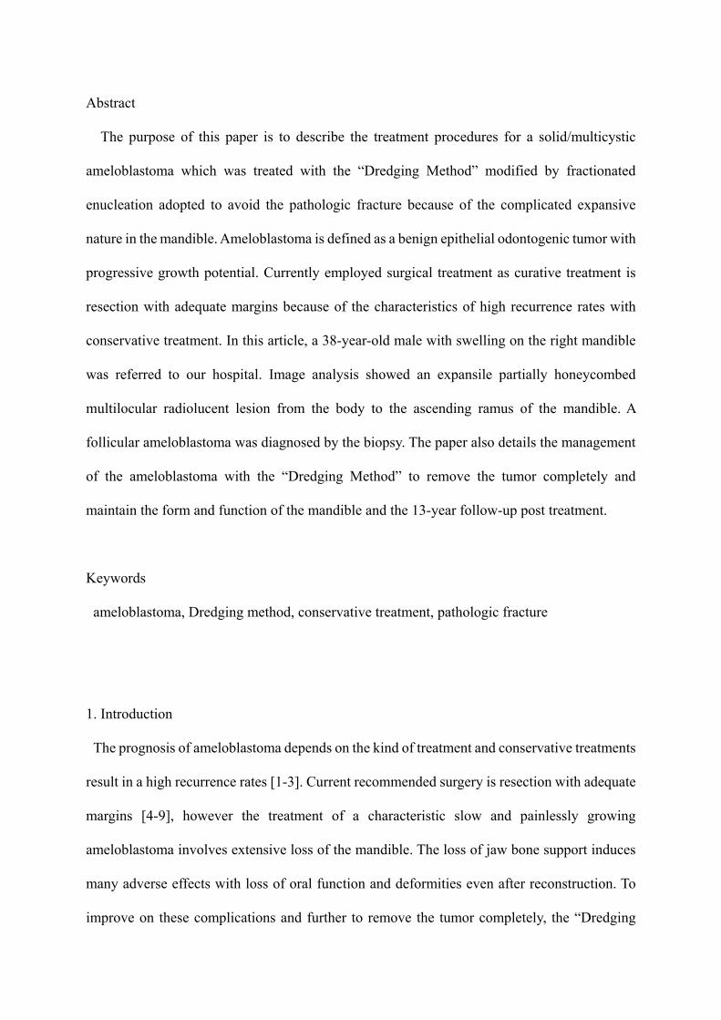
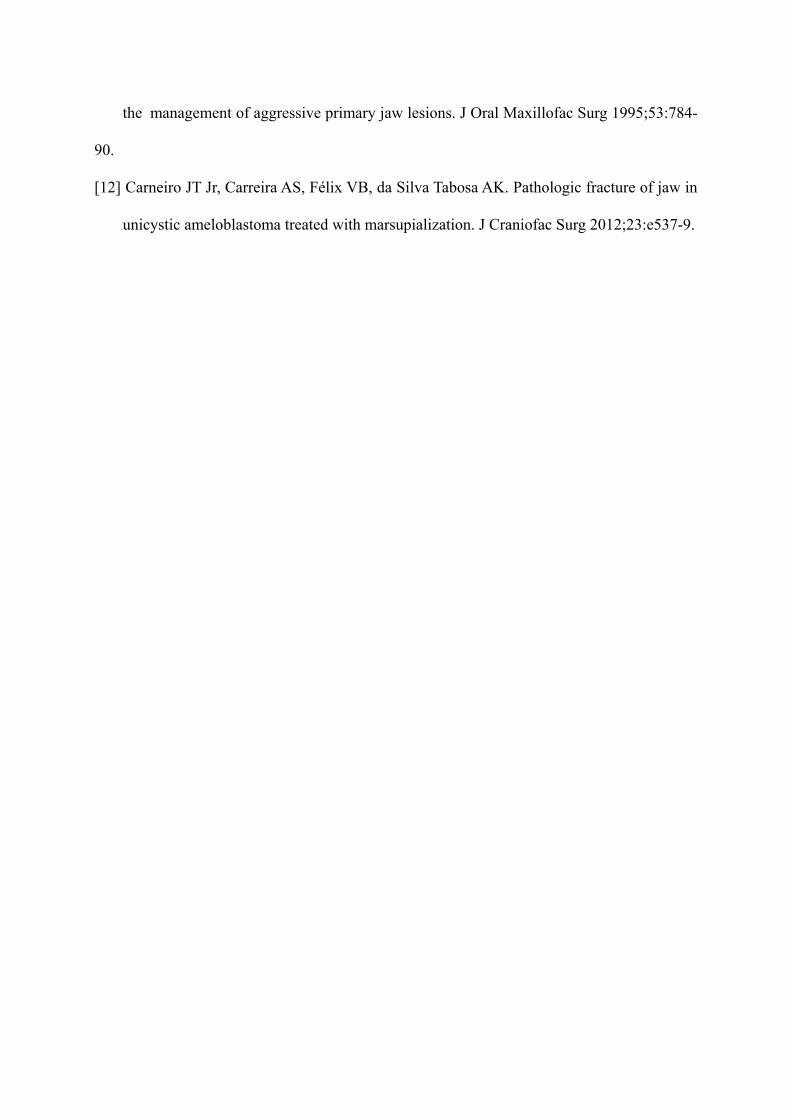
panoramic radiograph and computed tomography (CT) image showed a large multilocular
radiolucent lesion with well circumscribed borders in the body of the mandible and a slightly
obscure honeycombed lesion at an angle to the ascending ramus of the mandible (Fig. 1A and
1B). The wisdom tooth was involved in the lesion. The clinical diagnosis was a benign tumor
of the mandible. A biopsy and deflation was performed simultaneously under local anesthesia.
A follicular ameloblastoma was diagnosed from the biopsy specimens (Fig. 1C and 1D).
Hemimandibulectomy and reconstruction with a fibular flap was determined to need to be
performed as a radical treatment. The patient did not accept this treatment plan, because of the
decline of the oral function and facial deformity due to the loss of occlusion and continuity of
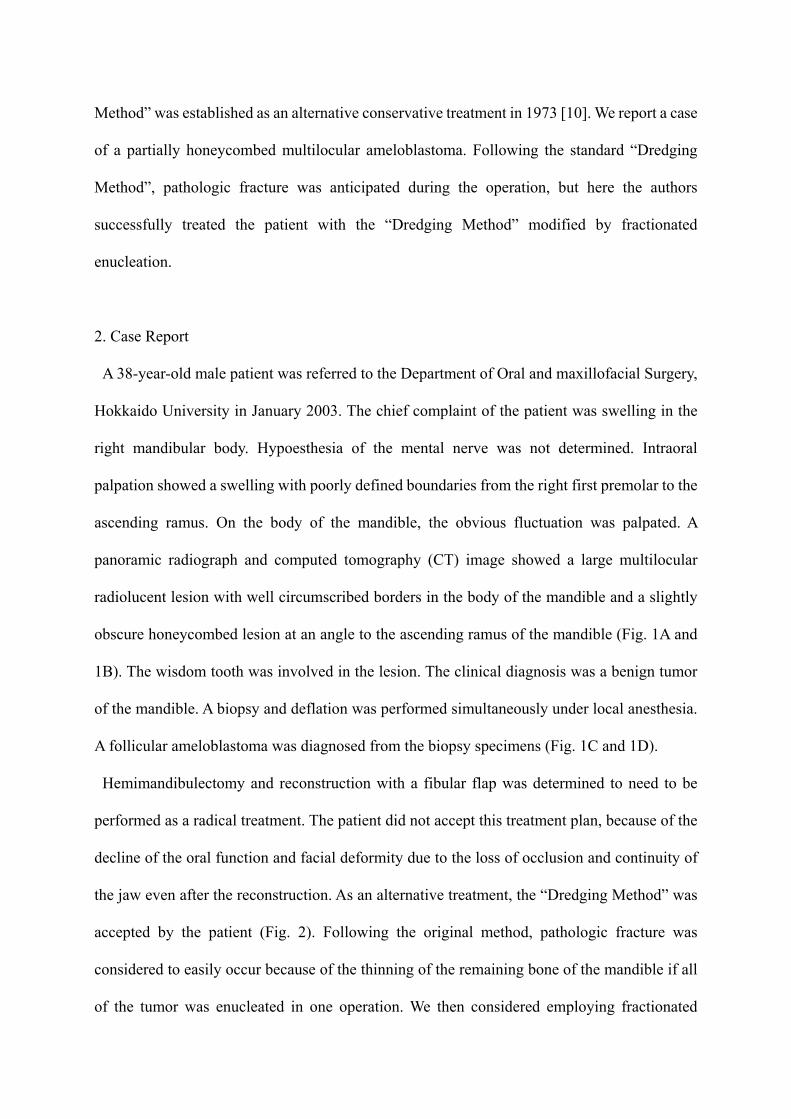
the jaw even after the reconstruction. As an alternative treatment, the “Dredging Method” was
accepted by the patient (Fig. 2). Following the original method, pathologic fracture was
considered to easily occur because of the thinning of the remaining bone of the mandible if all
of the tumor was enucleated in one operation. We then considered employing fractionated
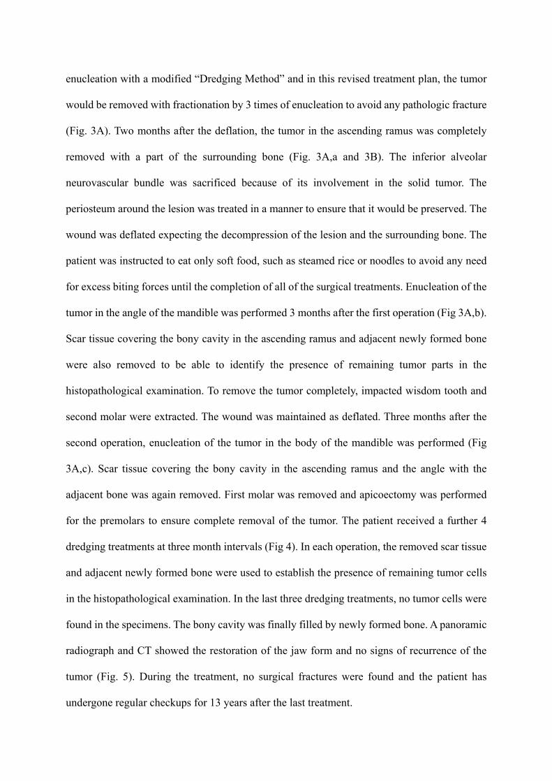
enucleation with a modified “Dredging Method” and in this revised treatment plan, the tumor
would be removed with fractionation by 3 times of enucleation to avoid any pathologic fracture
(Fig. 3A). Two months after the deflation, the tumor in the ascending ramus was completely
removed with a part of the surrounding bone (Fig. 3A,a and 3B). The inferior alveolar
neurovascular bundle was sacrificed because of its involvement in the solid tumor. The
periosteum around the lesion was treated in a manner to ensure that it would be preserved. The
wound was deflated expecting the decompression of the lesion and the surrounding bone. The
patient was instructed to eat only soft food, such as steamed rice or noodles to avoid any need
for excess biting forces until the completion of all of the surgical treatments. Enucleation of the
tumor in the angle of the mandible was performed 3 months after the first operation (Fig 3A,b).
Scar tissue covering the bony cavity in the ascending ramus and adjacent newly formed bone
were also removed to be able to identify the presence of remaining tumor parts in the
histopathological examination. To remove the tumor completely, impacted wisdom tooth and
second molar were extracted. The wound was maintained as deflated. Three months after the
second operation, enucleation of the tumor in the body of the mandible was performed (Fig
3A,c). Scar tissue covering the bony cavity in the ascending ramus and the angle with the
adjacent bone was again removed. First molar was removed and apicoectomy was performed
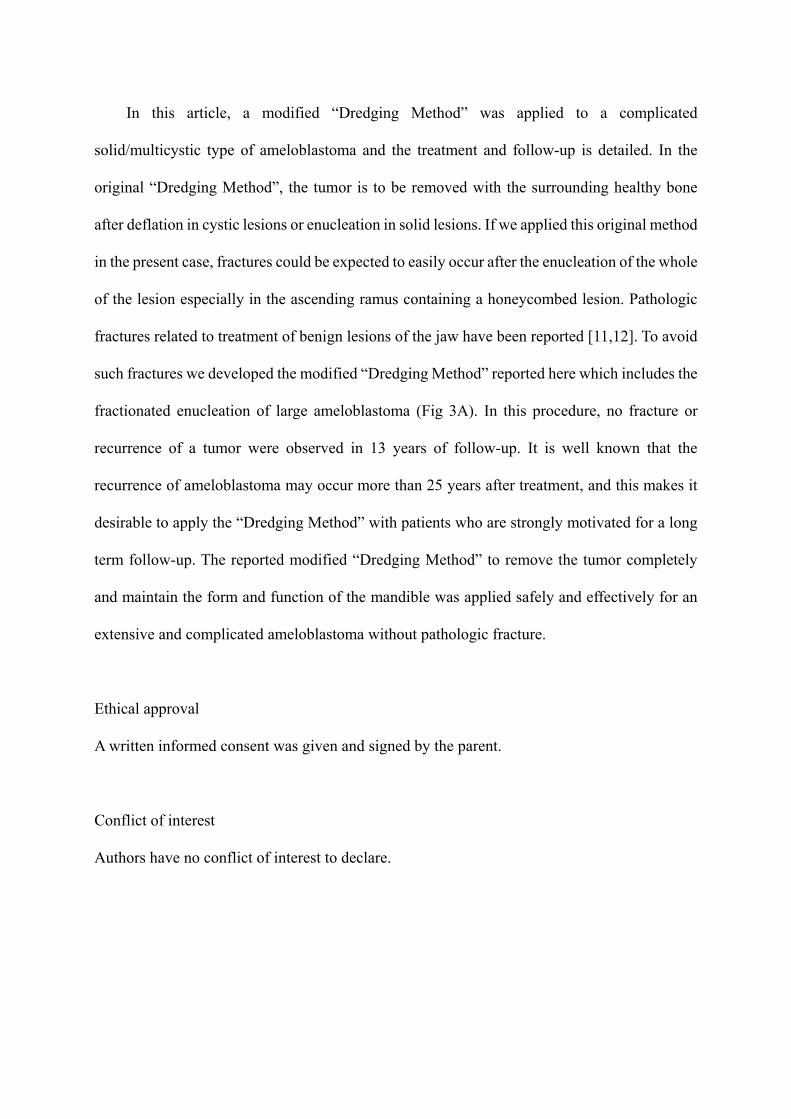
for the premolars to ensure complete removal of the tumor. The patient received a further 4
dredging treatments at three month intervals (Fig 4). In each operation, the removed scar tissue
and adjacent newly formed bone were used to establish the presence of remaining tumor cells
in the histopathological examination. In the last three dredging treatments, no tumor cells were
found in the specimens. The bony cavity was finally filled by newly formed bone. A panoramic
radiograph and CT showed the restoration of the jaw form and no signs of recurrence of the
tumor (Fig. 5). During the treatment, no surgical fractures were found and the patient has
undergone regular checkups for 13 years after the last treatment.

3. Discussion
Ameloblastoma is one of the most common types of odontogenic tumors with a wide variety
of clinical features, and histological patterns. Ameloblastoma is slowly growing histologically
benign tumors, mostly affecting the mandible in a wide age range of patients [1-3]. Although it
is a benign tumor, inadequate treatment results in high recurrence rates because of the
characteristics of the local invasion [1,3]. To ensure the prognosis, radical surgery which
includes the excision with adequate margins rather than conservative enucleation or curettage
is recommended [4-9]. Resection of the mandible, including the condyle causes a number of
complications such as dysfunction, deformities, and psychological distress, even if
reconstruction surgery is performed.
To avoid these complications, our department has established the “Dredging Method” in 1973
to treat odontogenic tumors of the mandible (Fig. 2) [10]. “Dredging Methods” include surgical
procedures with enucleation after deflation or only enucleation followed by repeated dredging
treatments. Deflation is applied to large cystic ameloblastoma to ensure the formation of a bony
outline. Enucleation is performed after the formation of a bony outline in cystic or solid
ameloblastoma. Dredging is performed at 2 to 3 month intervals to accelerate the new bone
formation by removing the scar tissue which has grown to cover the bony cavity. During the
dredging, some part of new bone is also removed with the scar tissue to determine the presence
of remaining tumor cells histopathologically. All of the above procedures are completed in the
deflated wound with irrigation by the patients to maintain the wound as clean. Eventually the
bony cavity is assumed to become filled with newly formed bone. Continuous regular follow-
up is also important to be able to detect recurrences early. In the “Dredging Method”, these
procedures ensure the elimination of the ameloblastoma and maintenance of the form and
function of the mandible.
In this article, a modified “Dredging Method” was applied to a complicated
solid/multicystic type of ameloblastoma and the treatment and follow-up is detailed. In the
original “Dredging Method”, the tumor is to be removed with the surrounding healthy bone
after deflation in cystic lesions or enucleation in solid lesions. If we applied this original method
in the present case, fractures could be expected to easily occur after the enucleation of the whole
of the lesion especially in the ascending ramus containing a honeycombed lesion. Pathologic
fractures related to treatment of benign lesions of the jaw have been reported [11,12]. To avoid
such fractures we developed the modified “Dredging Method” reported here which includes the
fractionated enucleation of large ameloblastoma (Fig 3A). In this procedure, no fracture or
recurrence of a tumor were observed in 13 years of follow-up. It is well known that the
recurrence of ameloblastoma may occur more than 25 years after treatment, and this makes it
desirable to apply the “Dredging Method” with patients who are strongly motivated for a long
term follow-up. The reported modified “Dredging Method” to remove the tumor completely
and maintain the form and function of the mandible was applied safely and effectively for an
extensive and complicated ameloblastoma without pathologic fracture.
Ethical approval
A written informed consent was given and signed by the parent.
Conflict of interest
Authors have no conflict of interest to declare.

References
[1] Gardner DG. Some current concepts on the pathology of ameloblastomas. Oral Surg Oral
Med Oral Pathol Oral Radiol Endod 1996;82:660-9.
[2] Vered M, Muller S, Heikinheimo K, Odell EW, Tilakaratne WM. Ameloblastoma. In: El-
Naggar AK, Chan JKC, Grandis JR, Takata T, Slootweg PJ, editors. WHO Classification
of Head and Neck Tumours (4th edition). Lyon: IARC Press; 2017. p215-8
[3] Reichart PA, Philipsen HP, Sonner S. Ameloblastoma: biological profile of 3677 cases. Eur
J Cancer B Oral Oncol 1995;31B:86-99.
[4] Pogrel MA, Montes DM. Is there a role for enucleation in the management of
ameloblastoma? Int J Oral Maxillofac Surg 2009;38:807-12.
[5] Antonoglou GN, Sándor GK. Recurrence rates of intraosseous ameloblastomas of the jaws:
a systematic review of conservative versus aggressive treatment approaches and meta-
analysis of non-randomized studies. J Craniomaxillofac Surg 2015;43:149-57.
[6] Lau SL, Samman N. Recurrence related to treatment modalities of unicystic
ameloblastoma: a systematic review. Int J Oral Maxillofac Surg 2006;35:681-90.
[7] Laborde A, Nicot R, Wojcik T, Ferri J, Raoul G. Ameloblastoma of the jaws:Management
and recurrence rate. Eur Ann Otorhinolaryngol Head Neck Dis 2017;134:7-11.
[8] Haq J, Siddiqui S, McGurk M. Argument for the conservative management of mandibular
ameloblastomas. Br J Oral Maxillofac Surg 2016;54:1001-1005.
[9] Almeida Rde A, Andrade ES, Barbalho JC, Vajgel A, Vasconcelos BC. Recurrence rate
following treatment for primary multicystic ameloblastoma: systematic review and meta-
analysis. Int J Oral Maxillofac Surg 2016;45:359-67.
[10] Kawamura M, Inoue N, Kobayashi I, Ahmed M. “Dredging Method”-A New Approach
for the Treatment of Ameloblastoma. Asian J Oral Maxillofac Surg 1991;3:81-8
[11] Salmassy DA, Pogrel MA. Liquid nitrogen cryosurgery and immediate bone grafting in
the management of aggressive primary jaw lesions. J Oral Maxillofac Surg 1995;53:784-
90.
[12] Carneiro JT Jr, Carreira AS, Félix VB, da Silva Tabosa AK. Pathologic fracture of jaw in
unicystic ameloblastoma treated with marsupialization. J Craniofac Surg 2012;23:e537-9.
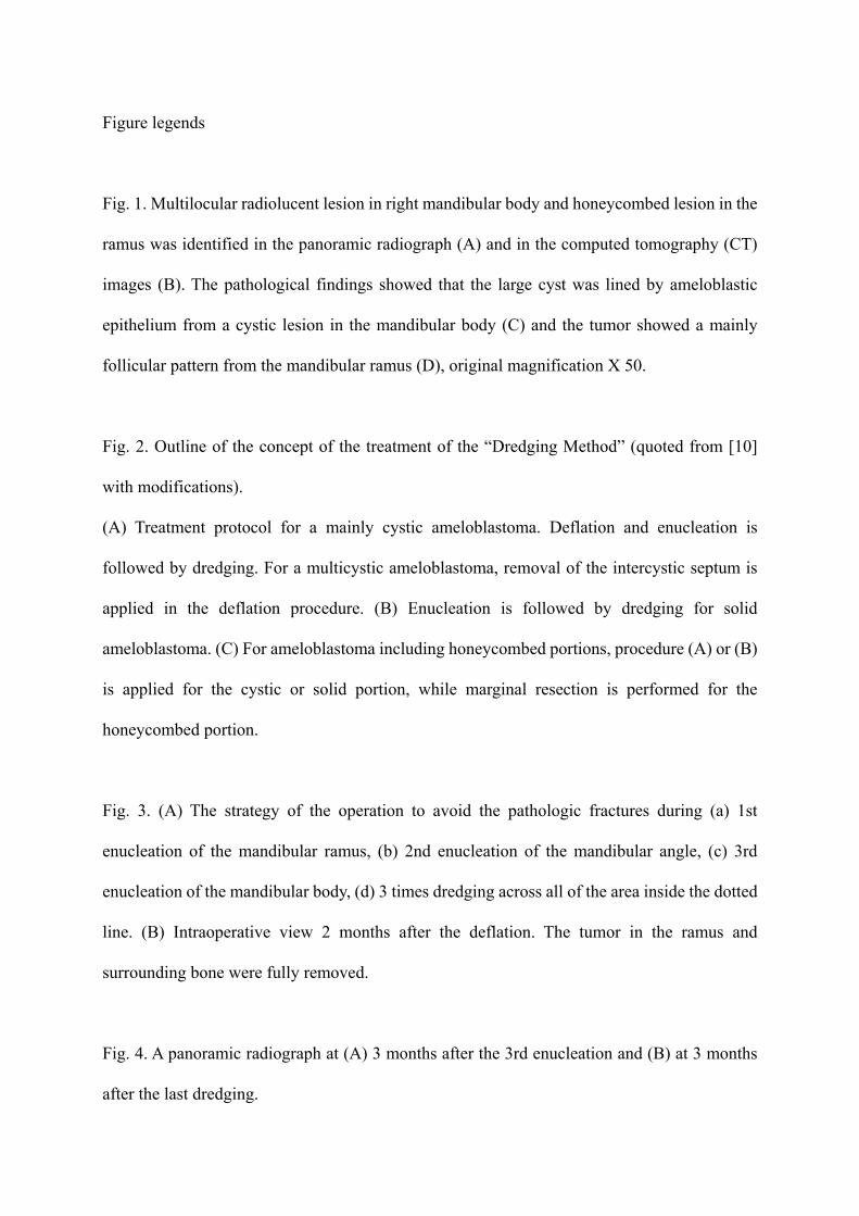
Figure legends
Fig. 1. Multilocular radiolucent lesion in right mandibular body and honeycombed lesion in the
ramus was identified in the panoramic radiograph (A) and in the computed tomography (CT)
images (B). The pathological findings showed that the large cyst was lined by ameloblastic
epithelium from a cystic lesion in the mandibular body (C) and the tumor showed a mainly
follicular pattern from the mandibular ramus (D), original magnification X 50.
Fig. 2. Outline of the concept of the treatment of the “Dredging Method” (quoted from [10]
with modifications).
(A) Treatment protocol for a mainly cystic ameloblastoma. Deflation and enucleation is
followed by dredging. For a multicystic ameloblastoma, removal of the intercystic septum is
applied in the deflation procedure. (B) Enucleation is followed by dredging for solid
ameloblastoma. (C) For ameloblastoma including honeycombed portions, procedure (A) or (B)
is applied for the cystic or solid portion, while marginal resection is performed for the
honeycombed portion.
Fig. 3. (A) The strategy of the operation to avoid the pathologic fractures during (a) 1st
enucleation of the mandibular ramus, (b) 2nd enucleation of the mandibular angle, (c) 3rd
enucleation of the mandibular body, (d) 3 times dredging across all of the area inside the dotted
line. (B) Intraoperative view 2 months after the deflation. The tumor in the ramus and
surrounding bone were fully removed.
Fig. 4. A panoramic radiograph at (A) 3 months after the 3rd enucleation and (B) at 3 months
after the last dredging.
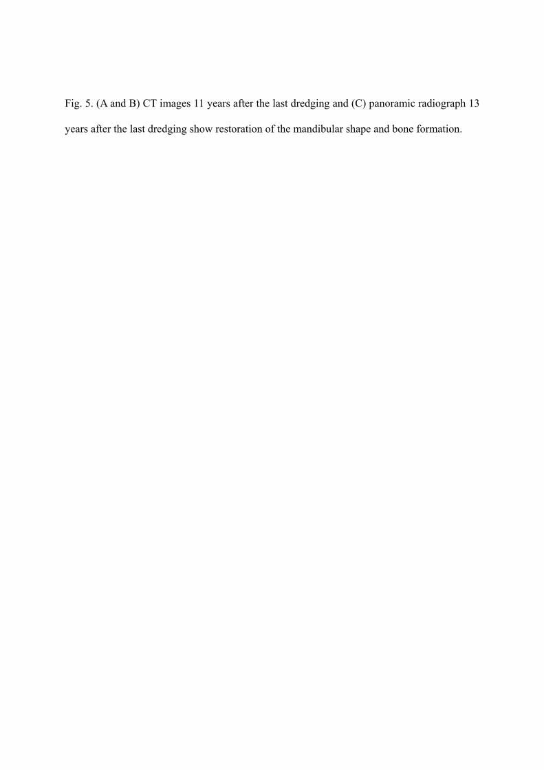
Fig. 5. (A and B) CT images 11 years after the last dredging and (C) panoramic radiograph 13
years after the last dredging show restoration of the mandibular shape and bone formation.
Fig. 1
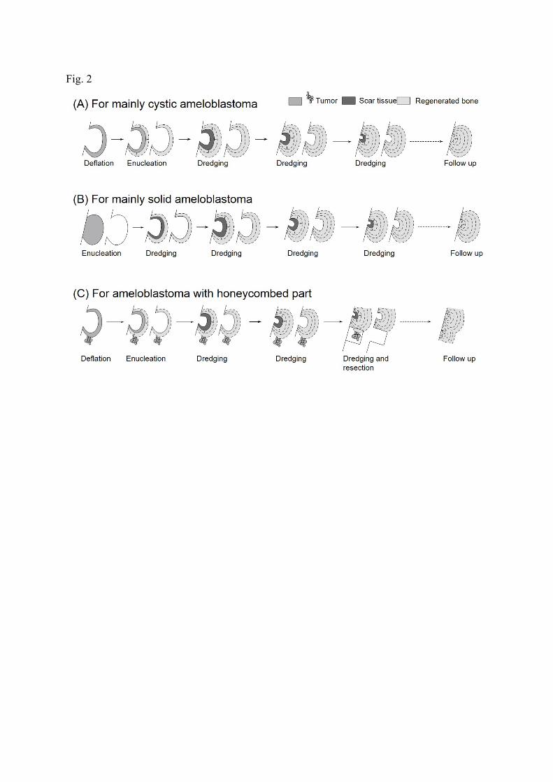
Fig. 2
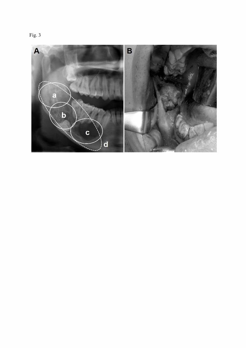
Fig. 3
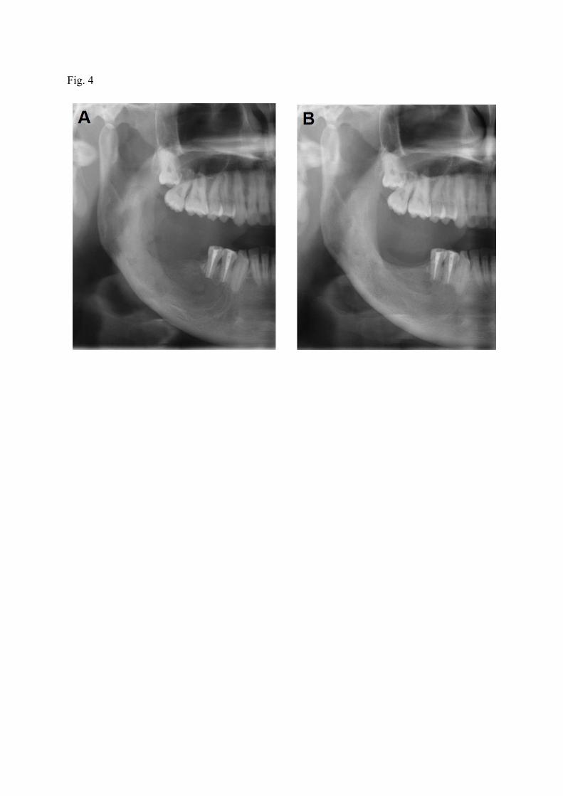
Fig. 4
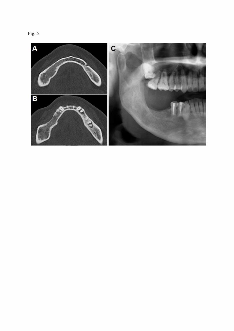
Fig. 5