Embed Size (px)
Citation preview
Foot & Ankle Specialist 1
DOI: 10.1177/1938640012470716. From The Clinic (AS) and “ARS Medica” Clinic (DV), Rome, Italy. Address correspondence to Andrea Scala, The Clinic, 29, Via Cesare Ferrero Di Cambiano, Rome 0091, Italy; e-mail: [email protected].
For reprints and permissions queries, please visit SAGE’s Web site at http://www.sagepub.com/journalsPermissions.nav.
Copyright © 2012 The Author(s)
Modified Minimal Incision Subcapital Osteotomy for Hallux Valgus Correction
Andrea Scala and Domenico Vendettuoli [AQ: 1] [AQ: 2]
vol. x / no. x
The potential advantages of minimal
incision surgery are the reduction in
operating time, minimal surgical exposure,
fewer complications, and early weight
bearing.”
“
Abstract: The potential advantages of minimal incision surgery for hallux valgus (HV) correction are the follow-ing: reduced surgical exposure, dimin-ished soft-tissue stripping, and less blood supply impairment. These advan-tages imply fewer complications. We retrospectively reviewed patients who were consecutively treated with a modi-fied minimally invasive osteotomy from January 2006 until December 2009 for HV deformity. We radiographically assessed the HV angle, 1-2 intermeta-tarsal (IM) angle, and tibial sesamoid position. Clinical outcomes were deter-mined using the American Orthopaedic Foot and Ankle Society Hallux Metatarsophalangeal Interphalangeal (AOFAS HMI) Clinical Rating Scale. A paired Student’s t test was used to deter-mine significance, with P < .01. There were 126 patients (146 feet) with an average age of 52.6 years and an aver-age postoperative follow-up of 29.1 months. Preoperatively, the average HV angle was 32.3°, and postoperatively, it was 4.5° (P < .01). The preoperative average IM angle was 14.4°, whereas postoperatively, it was 4.8° (P < .01). The average tibial sesamoid position was 6.3 preoperatively and 2.5 postop-eratively (P < .01). The average AOFAS HMI score was 54.6 preoperatively and
85.3 postoperatively (P < .01). There were 15 postoperative complications (10.3%) that included hallux varus, painful hardware, and delayed union. The results are comparable with those of traditional open techniques, with the additional advantages of a minimally invasive procedure.
Level of Evidence: Level IV, Therapeutic, Retrospective case series
Keywords: hallux val-gus; minimal incision; percutaneous metatar-sal osteotomy; minimally invasive; foot surgery
IntroductionHallux valgus (HV) is a
common foot deformity of the first meta-tarsophalangeal (MTP) joint (Figure 1).1,2 Although there are more than 130 opera-tive procedures to address this deformity,3 there is a current trend toward mini-mally invasive techniques.4-6 The potential advantages of minimal incision surgery are the reduction in operating time, min-imal surgical exposure, fewer complica-tions, and early weight bearing.4,5,7,8
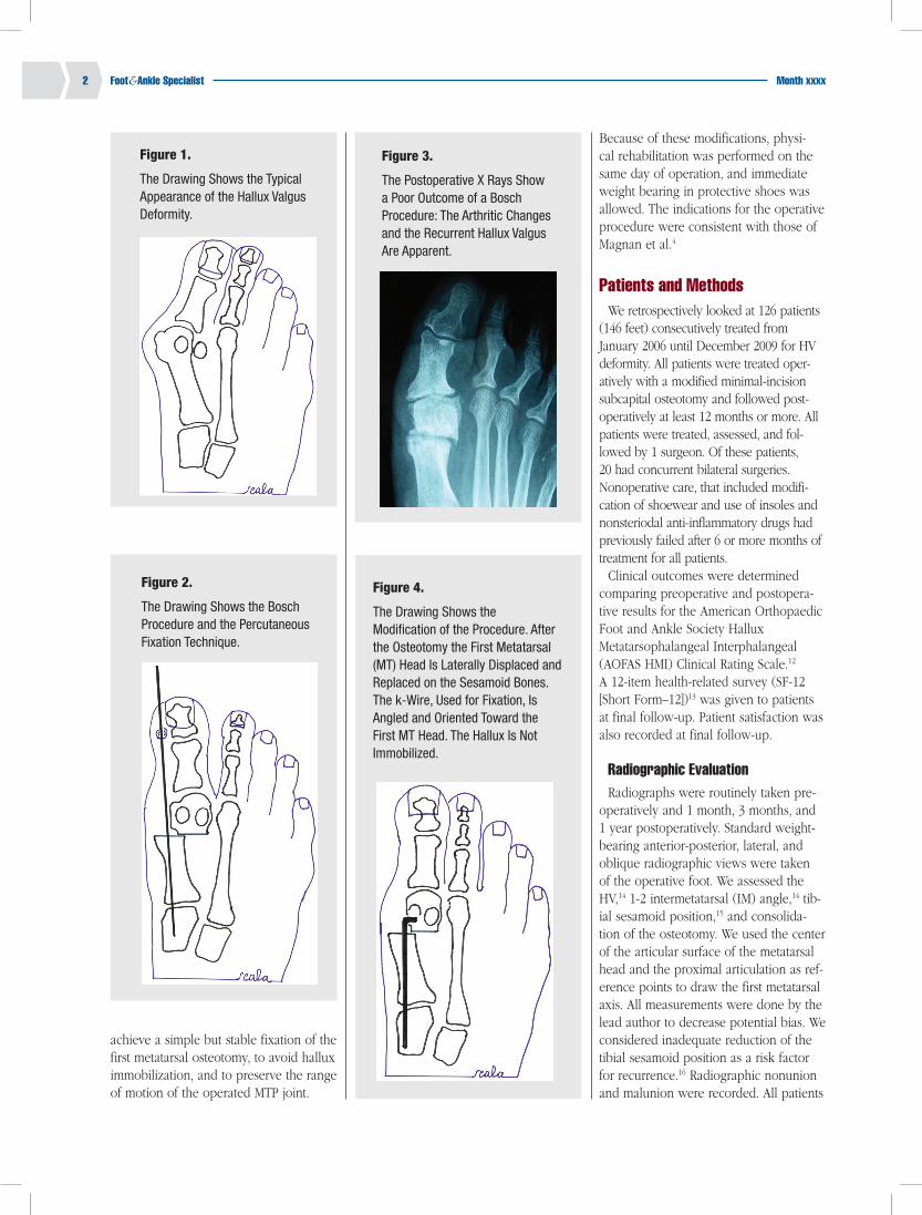
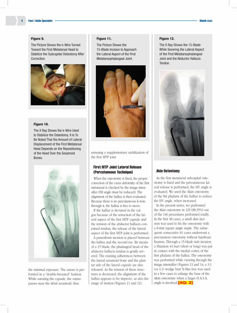
The Bosch osteotomy5 is a single-tract, subcapital osteotomy of the first metatar-sal bone for the correction of HV defor-mity (Figure 2). Some authors have4,5,7,9 published their case series of this sub-capital linear osteotomy and reported effective correction of the HV deformity. However, Kadakia et al10 found a high rate of complications that included osteo-necrosis, nonunion, malunion, and recur-
rence (Figure 3). Huang et al6 found a lower rate of complications but insisted that the operative procedure be limited to deformities with less than 30° of HV angle.
Having reviewed the literature, the lead author modified the traditional minimally invasive procedure for HV repair5,7,11 (Figure 4). The rationale behind the mod-ification of the Bosch osteotomy was to
Month xxxxFoot & Ankle Specialist2
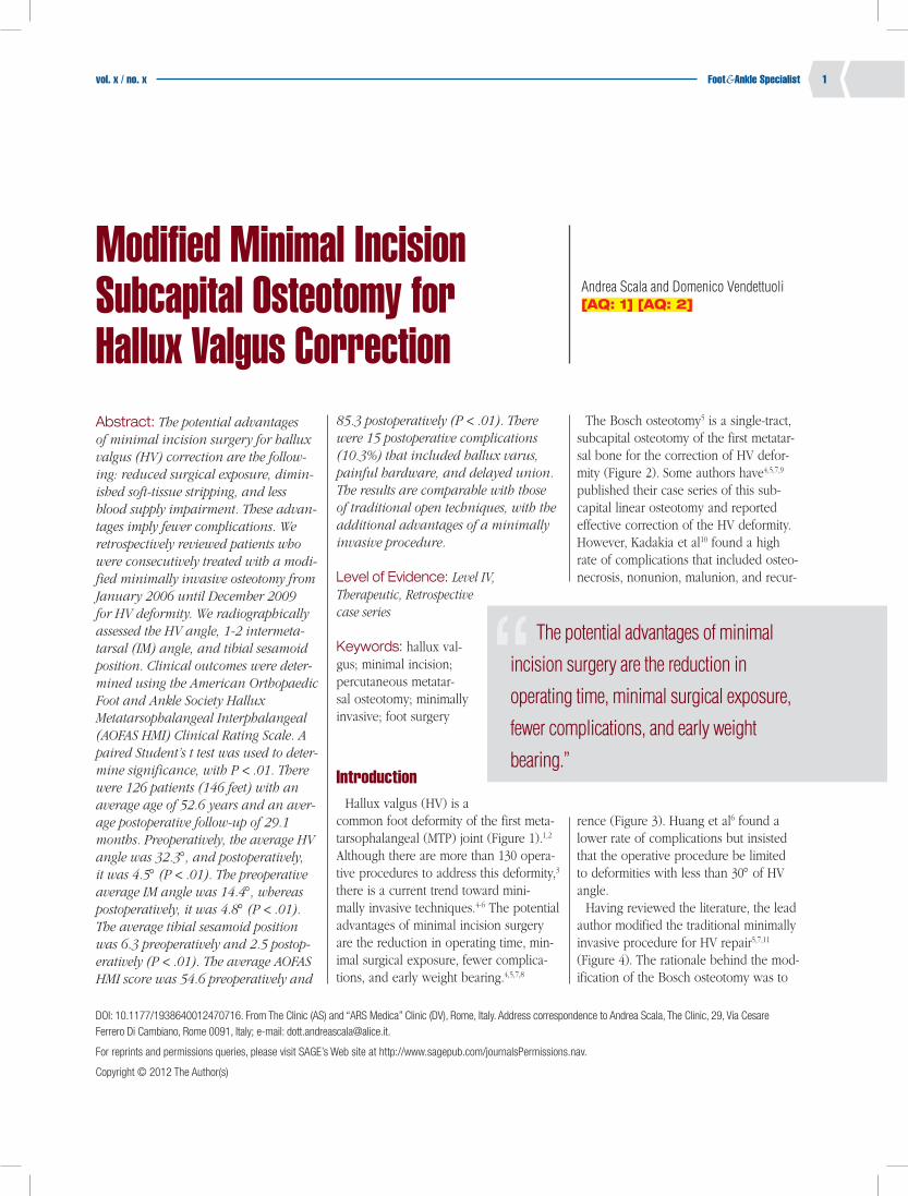
Figure 1.
The Drawing Shows the Typical Appearance of the Hallux Valgus Deformity.
Figure 3.
The Postoperative X Rays Show a Poor Outcome of a Bosch Procedure: The Arthritic Changes and the Recurrent Hallux Valgus Are Apparent.
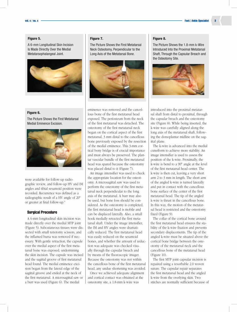
Figure 4.
The Drawing Shows the Modification of the Procedure. After the Osteotomy the First Metatarsal (MT) Head Is Laterally Displaced and Replaced on the Sesamoid Bones. The k-Wire, Used for Fixation, Is Angled and Oriented Toward the First MT Head. The Hallux Is Not Immobilized.
Figure 2.
The Drawing Shows the Bosch Procedure and the Percutaneous Fixation Technique.
achieve a simple but stable fixation of the first metatarsal osteotomy, to avoid hallux immobilization, and to preserve the range of motion of the operated MTP joint.
Because of these modifications, physi-cal rehabilitation was performed on the same day of operation, and immediate weight bearing in protective shoes was allowed. The indications for the operative procedure were consistent with those of Magnan et al.4
Patients and MethodsWe retrospectively looked at 126 patients
(146 feet) consecutively treated from January 2006 until December 2009 for HV deformity. All patients were treated oper-atively with a modified minimal-incision subcapital osteotomy and followed post-operatively at least 12 months or more. All patients were treated, assessed, and fol-lowed by 1 surgeon. Of these patients, 20 had concurrent bilateral surgeries. Nonoperative care, that included modifi-cation of shoewear and use of insoles and nonsteriodal anti-inflammatory drugs had previously failed after 6 or more months of treatment for all patients.
Clinical outcomes were determined comparing preoperative and postopera-tive results for the American Orthopaedic Foot and Ankle Society Hallux Metatarsophalangeal Interphalangeal (AOFAS HMI) Clinical Rating Scale.12 A 12-item health-related survey (SF-12 [Short Form–12])13 was given to patients at final follow-up. Patient satisfaction was also recorded at final follow-up.
Radiographic EvaluationRadiographs were routinely taken pre-
operatively and 1 month, 3 months, and 1 year postoperatively. Standard weight-bearing anterior-posterior, lateral, and oblique radiographic views were taken of the operative foot. We assessed the HV,14 1-2 intermetatarsal (IM) angle,14 tib-ial sesamoid position,15 and consolida-tion of the osteotomy. We used the center of the articular surface of the metatarsal head and the proximal articulation as ref-erence points to draw the first metatarsal axis. All measurements were done by the lead author to decrease potential bias. We considered inadequate reduction of the tibial sesamoid position as a risk factor for recurrence.16 Radiographic nonunion and malunion were recorded. All patients
Foot & Ankle Specialistvol. x / no. x 3
Figure 5.
A 6-mm Longitudinal Skin Incision Is Made Directly Over the Medial Metatarsophalangeal Joint.
Figure 7.
The Picture Shows the First Metatarsal Neck Osteotomy, Perpendicular to the Long Axis of the Metatarsal Bone.
Figure 8.
The Picture Shows the 1.8-mm k-Wire Introduced Into the Proximal Metatarsal Shaft, Through the Capsular Breach and the Osteotomy Site.
Figure 6.
The Picture Shows the First Metatarsal Medial Eminence Excision.
were available for follow-up radio-graphic review, and follow-up HV and IM angles and tibial sesamoid position were recorded. Recurrence was defined as a radiographic result of a HV angle of 20° or greater at final follow-up.6
Surgical ProcedureA 6-mm longitudinal skin incision was
made directly over the medial MTP joint (Figure 5). Subcutaneous tissues were dis-sected with small tenotomy scissors, and the inflamed bursa was removed if nec-essary. With gentle retraction, the capsule over the medial aspect of the first meta-tarsal bone was exposed, undermining the skin incision. The capsule was incised and the sagittal groove of first metatarsal head found. The medial eminence exci-sion began from the lateral edge of the sagittal groove and ended at the neck of the first metatarsal. A microsagittal saw or a burr was used (Figure 6). The medial
eminence was removed and the cancel-lous bone of the first metatarsal head exposed. The periosteum from the neck of the first metatarsal was detached. The osteotomy of the first metatarsal neck began on the cortical aspect of the first metatarsal, 3 mm distal to the cancellous bone previously exposed by the resection of the medial eminence. This 3-mm cor-tical bony bridge is of crucial importance and must always be preserved. The plan-tar vascular bundle of the first metatarsal head was spared because the osteotomy was placed distal to it (Figure 7).
An image intensifier was used to check the appropriate location for the osteot-omy. A microsagittal saw was used to perform the osteotomy of the first meta-tarsal neck perpendicular to the long axis of the metatarsal. A burr may also be used, but bone loss should be con-sidered. As the osteotomy is completed, the first metatarsal head is mobile and can be displaced laterally. Also, a small hook medially retracted the first meta-tarsal shaft. Under the image intensifier, the IM and HV angles were dramati-cally reduced. The first metatarsal head was easily reduced on the sesamoid bones, and whether the amount of reduc-tion was adequate was checked visu-ally through the capsular breach and by means of the fluoroscopic imager. Because the osteotomy was not within the cancellous bone of the first metatarsal head, any undue shortening was avoided.
Once we achieved adequate alignment and cortical contact was obtained at the osteotomy site, a 1.8-mm k-wire was
introduced into the proximal metatar-sal shaft from distal to proximal, through the capsular breach and the osteotomy site (Figure 8). While being inserted, the k-wire was carefully aligned along the long axis of the metatarsal shaft, follow-ing the dorsoplantar midline on the sag-ittal plane.
The k-wire is advanced into the medial cuneiform to achieve more stability. An image intensifier is used to assess the position of the k-wire. Proximally, the k-wire is bend to a 90° angle at the level of the first metatarsal head center. The k-wire is then cut, leaving a very short arm 2 to 3 mm in length. The short arm of the angled k-wire is turned laterally and put in contact with the cancellous bone surface of the center of the first metatarsal head. The tip of the angled k-wire is thrust in the cancellous bone. In this way, the motion of the metatar-sal head is restricted and the osteotomy fixed (Figure 9).
The collar of the cortical bone around the first metatarsal head ensures the sta-bility of the k-wire fixation and prevents secondary displacements. The tip of the angled k-wire must be situated above the cortical bone bridge between the oste-otomy of the metatarsal neck and the cancellous bone of the metatarsal head (Figure 10).
The first MTP joint capsular incision is repaired using a resorbable 2.0 woven suture. The capsular repair separates the first metatarsal head and the angled k-wire from the overlying skin. Two stitches are normally sufficient because of
Month xxxxFoot & Ankle Specialist4
ensuring a supplementary stabilization of the first MTP joint.
First MTP Joint Lateral Release (Percutaneous Technique)When the osteotomy is fixed, the proper
correction of the varus deformity of the first metatarsal is checked by the image inten-sifier (IM angle must be reduced). The alignment of the hallux is then evaluated. Because there is no percutaneous k-wire through it, the hallux is free to move.
If the hallux is deviated in the val-gus because of the retraction of the lat-eral aspect of the first MTP capsule and the tension of the abductor hallucis con-joined tendon, the release of the lateral aspect of the first MTP joint is performed.
A punctiform incision is placed between the hallux and the second toe. By means of a 15 blade, the phalangeal head of the abductor hallucis tendon is gently sev-ered. The existing adherences between the lateral sesamoid bone and the plan-tar side of the lateral capsule are also released. As the tension of these struc-tures is decreased, the alignment of the hallux appears to be improve, as also the range of motion (Figures 11 and 12).
Akin Osteotomy
As the first metatarsal subcapital oste-otomy is fixed and the percutaneous lat-eral release is performed, the HV angle is evaluated. We used the Akin osteotomy of the fist phalanx of the hallux to reduce the HV angle, when increased.
In the present series, we performed the Akin osteotomy in 129 (88.35%) out of the 146 procedures performed totally. In the first 48 cases, a small skin inci-sion was used to fix the osteotomy with a 8-mm square angle staple. The subse-quent consecutive 81 cases underwent a percutaneus osteotomy without hardware fixation. Through a 15-blade stab incision, a Shannon 44 burr (short or long) was put in contact with the medial cortex of the first phalanx of the hallux. The osteotomy was performed while viewing through the image intensifier (Figures 13 and 14; a 3.1 (or 4.1) wedge burr X-Mas tree was used in a few cases to enlarge the base of the Akin osteotomy when a larger D.A.S.A. angle is involved.[AQ: 3]
Figure 9.
The Picture Shows the k-Wire Turned Toward the First Metatarsal Head to Stabilize the Subcapital Osteotomy After Correction.
Figure 11.
The Picture Shows the 15-Blade Incision to Approach the Lateral Aspect of the First Metatarsophalangeal Joint.
Figure 12.
The X Ray Shows the 15-Blade While Severing the Lateral Aspect of the First Metatarsophalangeal Joint and the Abductor Hallucis Tendon.
Figure 10.
The X Ray Shows the k-Wire Used to Stabilize the Osteotomy. It Is To Be Noted That the Amount of Lateral Displacement of the First Metatarsal Head Depends on the Repositioning of the Head Over the Sesamoid Bones.
the minimal exposure. The suture is per-formed in a “double-breasted” fashion. While suturing the capsule, the suture passes near the tibial sesamoid, thus
Foot & Ankle Specialistvol. x / no. x 5
Postoperative ProtocolPostoperatively, the hallux is bandaged
but is not splinted in previous varus-like descriptions.7,11 The hallux remains free to move, and the range of motion is not restricted (Figure 15). The patient is allowed to bear weight immediately after the operation in a surgical shoe and is advised to walk several times during the day (Figure 16).
Limb elevation on the operated foot is advisable. After prolonged walking,
rest with the limb elevated as well as ice application are recommended. The post-operative bandage is removed 1 week after the operation, and a contrast bath (cold water 1 minute, warm water 30 s) is prescribed. The edema and swelling decrease, and patients are able to wear comfortable shoes and sneakers. Driving the car is generally possible 2 weeks after the operation. After a month, patients usually wear regular shoes.
Statistical AnalysisA paired Student t test was used to
determine the significance between pre-operative and postoperative measure-ments, with P < .01. A c2 test was used to compare the complication rates of this study with those of previous studies of minimally invasive osteotomies for hallux valgus deformity.4,6,10
ResultsThe average age of the patients was
52.6 years (range = 16-87 years). There were 115 women and 11 men, and the average follow-up was 29.1 months (range = 12-54 months). Preoperatively, the average HV angle was measured as 32.3° (range = 12°-52°; standard deviation
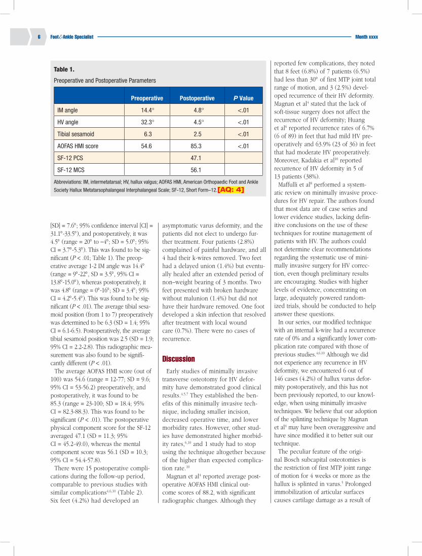
Figure 16.
The Picture Shows a Case of Hallux Valgus Before the Operation.
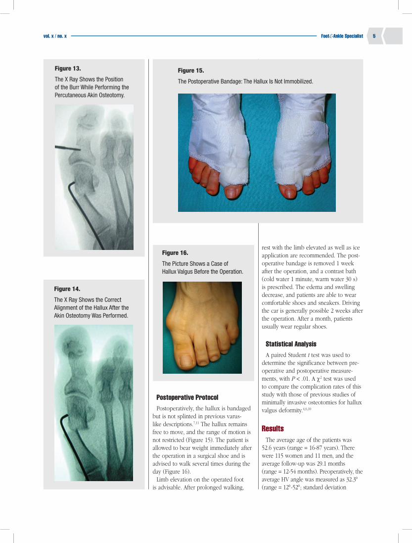
Figure 13.
The X Ray Shows the Position of the Burr While Performing the Percutaneous Akin Osteotomy.
Figure 15.
The Postoperative Bandage: The Hallux Is Not Immobilized.
Figure 14.
The X Ray Shows the Correct Alignment of the Hallux After the Akin Osteotomy Was Performed.
Month xxxxFoot & Ankle Specialist6
[SD] = 7.6°; 95% confidence interval [CI] = 31.1°-33.5°), and postoperatively, it was 4.5° (range = 20° to -4°; SD = 5.0°; 95% CI = 3.7°-5.3°). This was found to be sig-nificant (P < .01; Table 1). The preop-erative average 1-2 IM angle was 14.4° (range = 9°-22°, SD = 3.5°, 95% CI = 13.8°-15.0°), whereas postoperatively, it was 4.8° (range = 0°-16°; SD = 3.4°; 95% CI = 4.2°-5.4°). This was found to be sig-nificant (P < .01). The average tibial sesa-moid position (from 1 to 7) preoperatively was determined to be 6.3 (SD = 1.4; 95% CI = 6.1-6.5). Postoperatively, the average tibial sesamoid position was 2.5 (SD = 1.9; 95% CI = 2.2-2.8). This radiographic mea-surement was also found to be signifi-cantly different (P < .01).
The average AOFAS HMI score (out of 100) was 54.6 (range = 12-77; SD = 9.6; 95% CI = 53-56.2) preoperatively, and postoperatively, it was found to be 85.3 (range = 23-100; SD = 18.4; 95% CI = 82.3-88.3). This was found to be significant (P < .01). The postoperative physical component score for the SF-12 averaged 47.1 (SD = 11.3; 95% CI = 45.2-49.0), whereas the mental component score was 56.1 (SD = 10.3; 95% CI = 54.4-57.8).
There were 15 postoperative compli-cations during the follow-up period, comparable to previous studies with similar complications4,6,10 (Table 2). Six feet (4.2%) had developed an
asymptomatic varus deformity, and the patients did not elect to undergo fur-ther treatment. Four patients (2.8%) complained of painful hardware, and all 4 had their k-wires removed. Two feet had a delayed union (1.4%) but eventu-ally healed after an extended period of non–weight bearing of 3 months. Two feet presented with broken hardware without malunion (1.4%) but did not have their hardware removed. One foot developed a skin infection that resolved after treatment with local wound care (0.7%). There were no cases of recurrence.
DiscussionEarly studies of minimally invasive
transverse osteotomy for HV defor-mity have demonstrated good clinical results.4,5,7 They established the ben-efits of this minimally invasive tech-nique, including smaller incision, decreased operative time, and lower morbidity rates. However, other stud-ies have demonstrated higher morbid-ity rates,6,10 and 1 study had to stop using the technique altogether because of the higher than expected complica-tion rate.10
Magnan et al4 reported average post-operative AOFAS HMI clinical out-come scores of 88.2, with significant radiographic changes. Although they
reported few complications, they noted that 8 feet (6.8%) of 7 patients (6.5%) had less than 30° of first MTP joint total range of motion, and 3 (2.5%) devel-oped recurrence of their HV deformity. Magnan et al4 stated that the lack of soft-tissue surgery does not affect the recurrence of HV deformity; Huang et al6 reported recurrence rates of 6.7% (6 of 89) in feet that had mild HV pre-operatively and 63.9% (23 of 36) in feet that had moderate HV preoperatively. Moreover, Kadakia et al10 reported recurrence of HV deformity in 5 of 13 patients (38%).
Maffulli et al8 performed a system-atic review on minimally invasive proce-dures for HV repair. The authors found that most data are of case series and lower evidence studies, lacking defin-itive conclusions on the use of these techniques for routine management of patients with HV. The authors could not determine clear recommendations regarding the systematic use of mini-mally invasive surgery for HV correc-tion, even though preliminary results are encouraging. Studies with higher levels of evidence, concentrating on large, adequately powered random-ized trials, should be conducted to help answer these questions.
In our series, our modified technique with an internal k-wire had a recurrence rate of 0% and a significantly lower com-plication rate compared with those of previous studies.4,6,10 Although we did not experience any recurrence in HV deformity, we encountered 6 out of 146 cases (4.2%) of hallux varus defor-mity postoperatively, and this has not been previously reported, to our knowl-edge, when using minimally invasive techniques. We believe that our adoption of the splinting technique by Magnan et al4 may have been overaggressive and have since modified it to better suit our technique.
The peculiar feature of the origi-nal Bosch subcapital osteotomies is the restriction of first MTP joint range of motion for 4 weeks or more as the hallux is splinted in varus.5 Prolonged immobilization of articular surfaces causes cartilage damage as a result of
Table 1.
Preoperative and Postoperative Parameters
Preoperative Postoperative P Value
IM angle 14.4° 4.8° <.01
HV angle 32.3° 4.5° <.01
Tibial sesamoid 6.3 2.5 <.01
AOFAS HMI score 54.6 85.3 <.01
SF-12 PCS 47.1
SF-12 MCS 56.1
Abbreviations: IM, intermetatarsal; HV, hallux valgus; AOFAS HMI, American Orthopaedic Foot and Ankle
Society Hallux Metatarsophalangeal Interphalangeal Scale; SF-12, Short Form–12.[AQ: 4]
Foot & Ankle Specialistvol. x / no. x 7
the altered metabolism of cartilaginous tissue.5 The modified osteotomy allows early mobilization and resumption of joint range of motion that is optimal after articular repair or realignment procedures.
This study showed that our modified, minimal-incision subcapital
osteotomy enables the surgeon to achieve good correction of a mild to moderate HV deformity. Each postop-erative outcome measurement showed a significant positive change compared with the preoperative status. Our results reflect positive clinical outcomes as found in other studies,4,5,8,17 except that our
patients did not experience stiffness or recurrence (Figures 16-23).
ConclusionsThe suggested modification has been
shown to be reliable and predictable in treating mild-to-moderate HV deformity. The following features should be outlined:
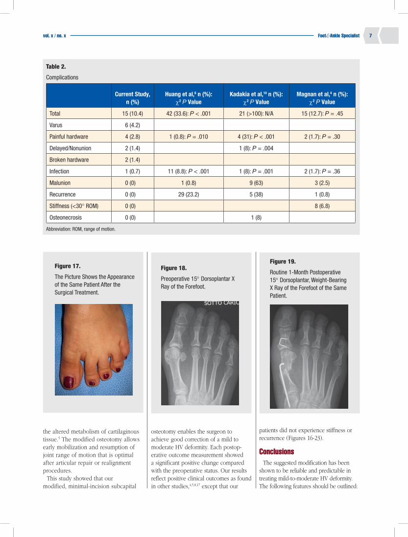
Table 2.
Complications
Current Study, n (%)
Huang et al,6 n (%): c2 P Value
Kadakia et al,10 n (%): c2 P Value
Magnan et al,4 n (%): c2 P Value
Total 15 (10.4) 42 (33.6): P < .001 21 (>100): N/A 15 (12.7): P = .45
Varus 6 (4.2)
Painful hardware 4 (2.8) 1 (0.8): P = .010 4 (31): P < .001 2 (1.7): P = .30
Delayed/Nonunion 2 (1.4) 1 (8): P = .004
Broken hardware 2 (1.4)
Infection 1 (0.7) 11 (8.8): P < .001 1 (8): P = .001 2 (1.7): P = .36
Malunion 0 (0) 1 (0.8) 9 (63) 3 (2.5)
Recurrence 0 (0) 29 (23.2) 5 (38) 1 (0.8)
Stiffness (<30° ROM) 0 (0) 8 (6.8)
Osteonecrosis 0 (0) 1 (8)
Abbreviation: ROM, range of motion.
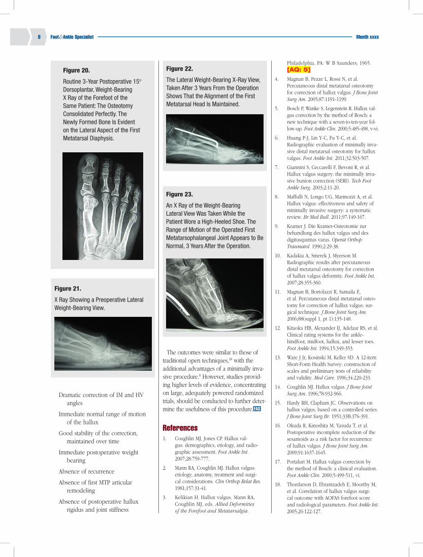
Figure 17.
The Picture Shows the Appearance of the Same Patient After the Surgical Treatment.
Figure 18.
Preoperative 15° Dorsoplantar X Ray of the Forefoot.
Figure 19.
Routine 1-Month Postoperative 15° Dorsoplantar, Weight-Bearing X Ray of the Forefoot of the Same Patient.
Month xxxxFoot & Ankle Specialist8
Dramatic correction of IM and HV angles
Immediate normal range of motion of the hallux
Good stability of the correction, maintained over time
Immediate postoperative weight bearing
Absence of recurrence
Absence of first MTP articular remodeling
Absence of postoperative hallux rigidus and joint stiffness
The outcomes were similar to those of traditional open techniques,18 with the additional advantages of a minimally inva-sive procedure.8 However, studies provid-ing higher levels of evidence, concentrating on large, adequately powered randomized trials, should be conducted to further deter-mine the usefulness of this procedure.
References1. Coughlin MJ, Jones CP. Hallux val-
gus: demographics, etiology, and radio-graphic assessment. Foot Ankle Int. 2007;28:759-777.
2. Mann RA, Coughlin MJ. Hallux valgus: etiology, anatomy, treatment and surgi-cal considerations. Clin Orthop Relat Res. 1981;157:31-41.
3. Kelikian H. Hallux valgus. Mann RA, Coughlin MJ, eds. Allied Deformities of the Forefoot and Metatarsalgia.
Figure 20.
Routine 3-Year Postoperative 15° Dorsoplantar, Weight-Bearing X Ray of the Forefoot of the Same Patient: The Osteotomy Consolidated Perfectly. The Newly Formed Bone Is Evident on the Lateral Aspect of the First Metatarsal Diaphysis.
Figure 21.
X Ray Showing a Preoperative Lateral Weight-Bearing View.
Figure 22.
The Lateral Weight-Bearing X-Ray View, Taken After 3 Years From the Operation Shows That the Alignment of the First Metatarsal Head Is Maintained.
Figure 23.
An X Ray of the Weight-Bearing Lateral View Was Taken While the Patient Wore a High-Heeled Shoe. The Range of Motion of the Operated First Metatarsophalangeal Joint Appears to Be Normal, 3 Years After the Operation.
Philadelphia, PA: W B Saunders; 1965.[AQ: 5]
4. Magnan B, Pezze L, Rossi N, et al. Percutaneous distal metatarsal osteotomy for correction of hallux valgus. J Bone Joint Surg Am. 2005;87:1191-1199.
5. Bosch P, Wanke S, Legenstein R. Hallux val-gus correction by the method of Bosch: a new technique with a seven-to-ten-year fol-low-up. Foot Ankle Clin. 2000;5:485-498, v-vi.
6. Huang P-J, Lin Y-C, Fu Y-C, et al. Radiographic evaluation of minimally inva-sive distal metatarsal osteotomy for hallux valgus. Foot Ankle Int. 2011;32:503-507.
7. Giannini S, Ceccarelli F, Bevoni R, et al. Hallux valgus surgery: the minimally inva-sive bunion correction (SERI). Tech Foot Ankle Surg. 2003;2:11-20.
8. Maffulli N, Longo UG, Marinozzi A, et al. Hallux valgus: effectiveness and safety of minimally invasive surgery: a systematic review. Br Med Bull. 2011;97:149-167.
9. Kramer J. Die Kramer-Osteotomie zur behandlung des hallux valgus und des digitusquintus varus. Operat Orthop Traumatol. 1990;2:29-38.
10. Kadakia A, Smerek J, Myerson M. Radiographic results after percutaneous distal metatarsal osteotomy for correction of hallux valgus deformity. Foot Ankle Int. 2007;28:355-360.
11. Magnan B, Bortolazzi R, Samaila E, et al. Percutaneous distal metatarsal osteo-tomy for correction of hallux valgus: sur-gical technique. J Bone Joint Surg Am. 2006;88(suppl 1, pt 1):135-148.
12. Kitaoka HB, Alexander IJ, Adelaar RS, et al. Clinical rating systems for the ankle- hindfoot, midfoot, hallux, and lesser toes. Foot Ankle Int. 1994;15:349-353.
13. Ware J Jr, Kosinski M, Keller SD. A 12-item Short-Form Health Survey: construction of scales and preliminary tests of reliability and validity. Med Care. 1996;34:220-233.
14. Coughlin MJ. Hallux valgus. J Bone Joint Surg Am. 1996;78:932-966.
15. Hardy RH, Clapham JC. Observations on hallux valgus; based on a controlled series. J Bone Joint Surg Br. 1951;33B:376-391.
16. Okuda R, Kinoshita M, Yasuda T, et al. Postoperative incomplete reduction of the sesamoids as a risk factor for recurrence of hallux valgus. J Bone Joint Surg Am. 2009;91:1637-1645.
17. Portaluri M. Hallux valgus correction by the method of Bosch: a clinical evaluation. Foot Ankle Clin. 2000;5:499-511, vi.
18. Thordarson D, Ebramzadeh E, Moorthy M, et al. Correlation of hallux valgus surgi-cal outcome with AOFAS forefoot score and radiological parameters. Foot Ankle Int. 2005;26:122-127.

![INDEX [media.wiley.com]€¦ · AAEP. See American Association of Equine Practitioners Abaxial fracture, of sesamoidean bone, 598f–599f, 601f Abaxial sesamoid nerve block, in forelimb,](https://img.pdfslide.net/doc/110x75/60011d1dd7b93739b34b8074/index-mediawileycom-aaep-see-american-association-of-equine-practitioners-abaxial.jpg)