Embed Size (px)
Citation preview
Modified PowerPoint from: Aneeq Ahmad -- Henderson State University.
Worth Publishers © 2007
The Brain – Studying &
Structures
Unit 3 – pg. 66-73
Brain Charts and Diagrams●Have out the Brain Structure Chart- you'll fill
in most of it with this powerpoint. What you
don't get, you'll need to find in the book or
from an app called "3D Brain." Download this
on your phone
●You also need "The Brain" 2 pictures of the
brain. Make sure to color code the one on
the front, then label the one on the back.
Techniques to Study the Brain
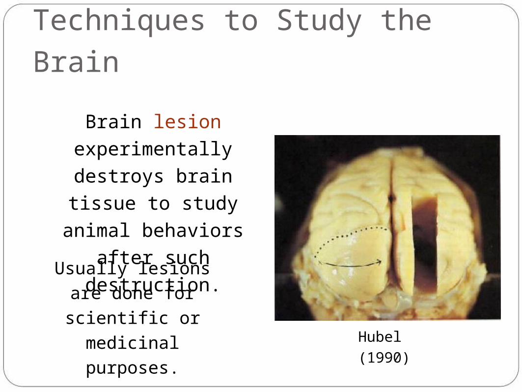
Brain lesion
experimentally destroys
brain tissue to study
animal behaviors after
such destruction.
Hubel
(1990)
Usually lesions are
done for scientific or
medicinal purposes.
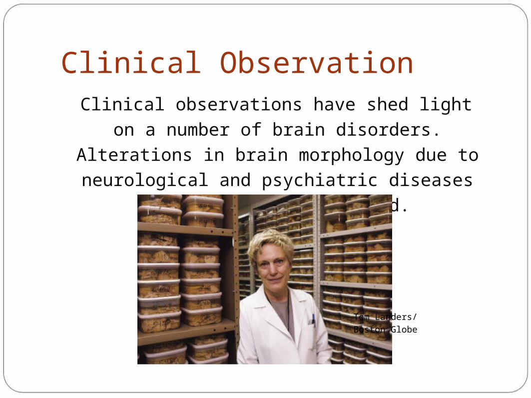
Clinical ObservationClinical observations have shed light on a number of
brain disorders. Alterations in brain morphology due
to neurological and psychiatric diseases are now
being catalogued.
Tom Landers/ Boston
Globe
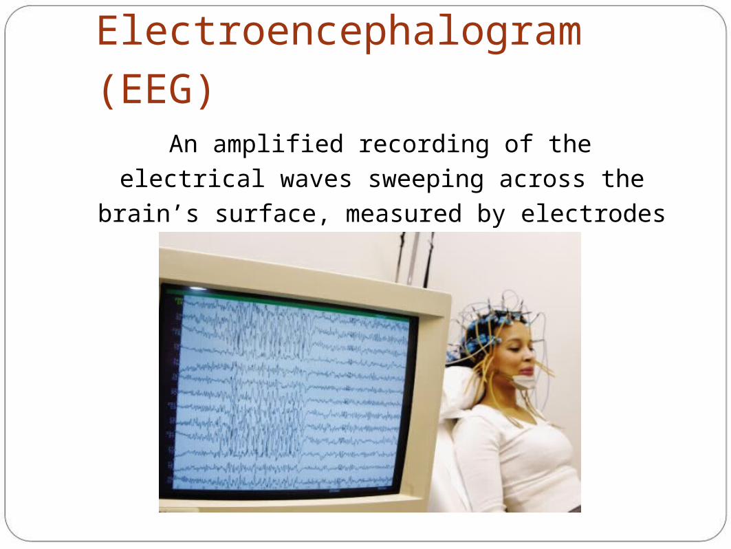
Electroencephalogram (EEG)An amplified recording of the electrical waves
sweeping across the brain’s surface, measured by
electrodes placed on the scalp.
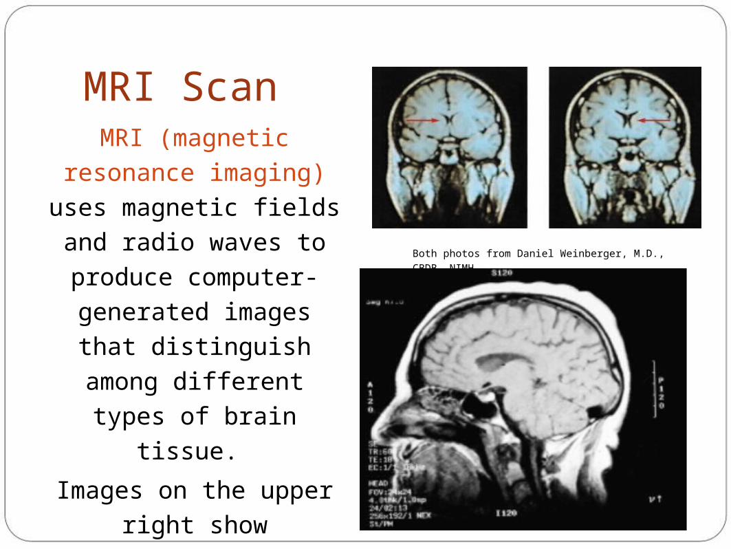
MRI ScanMRI (magnetic resonance
imaging) uses magnetic
fields and radio waves to
produce computer-
generated images that
distinguish among different
types of brain tissue.
Images on the upper right
show ventricular
enlargement in a
schizophrenic patient.
Both photos from Daniel Weinberger, M.D., CBDB,
NIMH
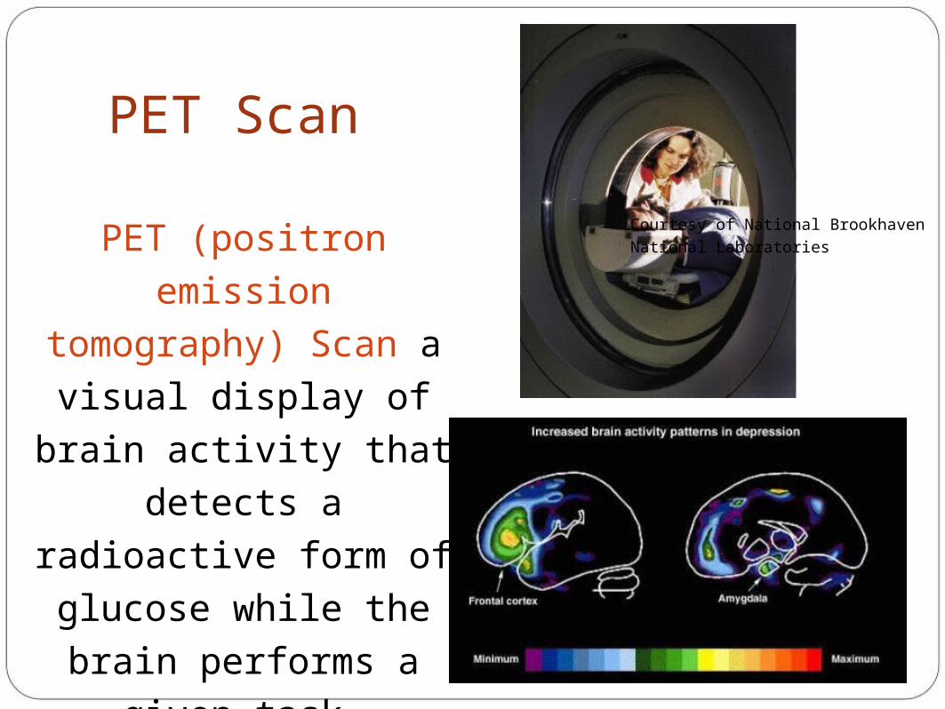
PET Scan
PET (positron emission
tomography) Scan a
visual display of brain
activity that detects a
radioactive form of
glucose while the brain
performs a given task.
Courtesy of National Brookhaven National
Laboratories
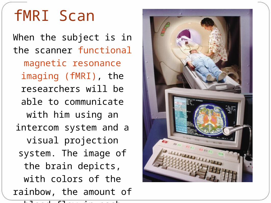
fMRI ScanWhen the subject is in the
scanner functional magnetic
resonance imaging (fMRI), the
researchers will be able to
communicate with him using
an intercom system and a
visual projection system. The
image of the brain depicts,
with colors of the rainbow, the
amount of blood flow in each
part of the brain, which
indicates the amount of neural
activity in each part.
Reticular Formation
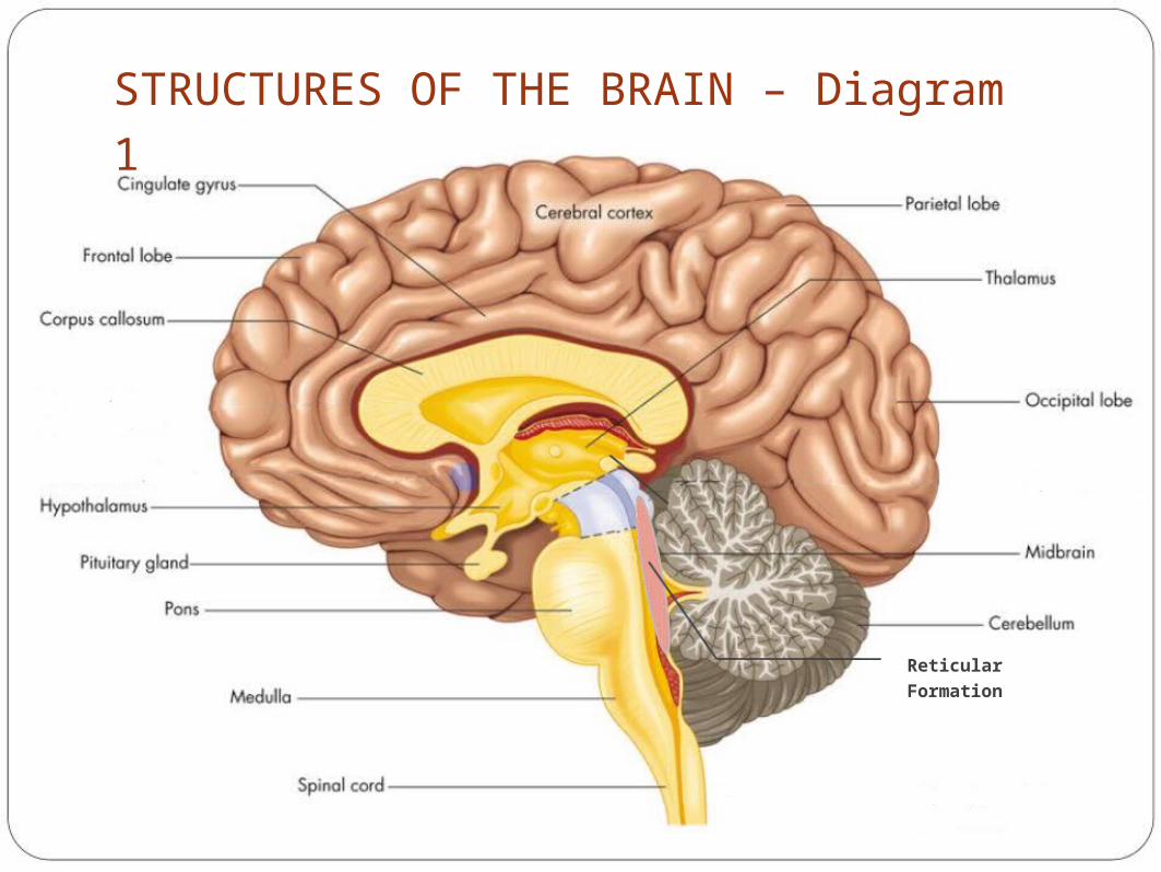
STRUCTURES OF THE BRAIN – Diagram 1
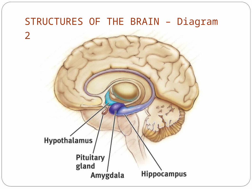
STRUCTURES OF THE BRAIN – Diagram 2
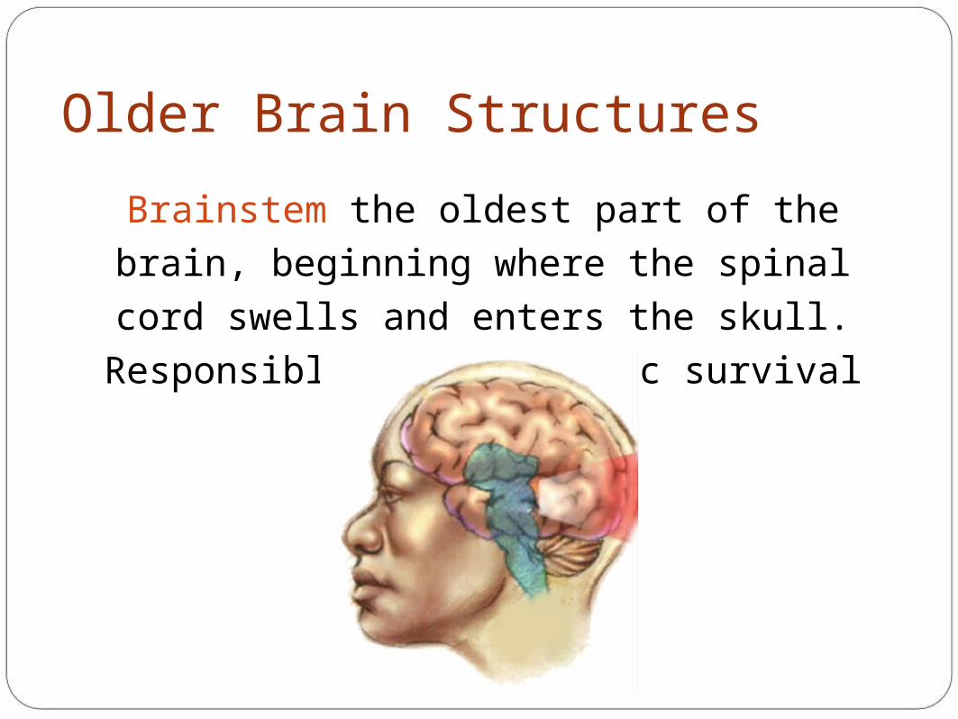
Older Brain Structures
Brainstem the oldest part of the brain, beginning
where the spinal cord swells and enters the skull.
Responsible for automatic survival functions.
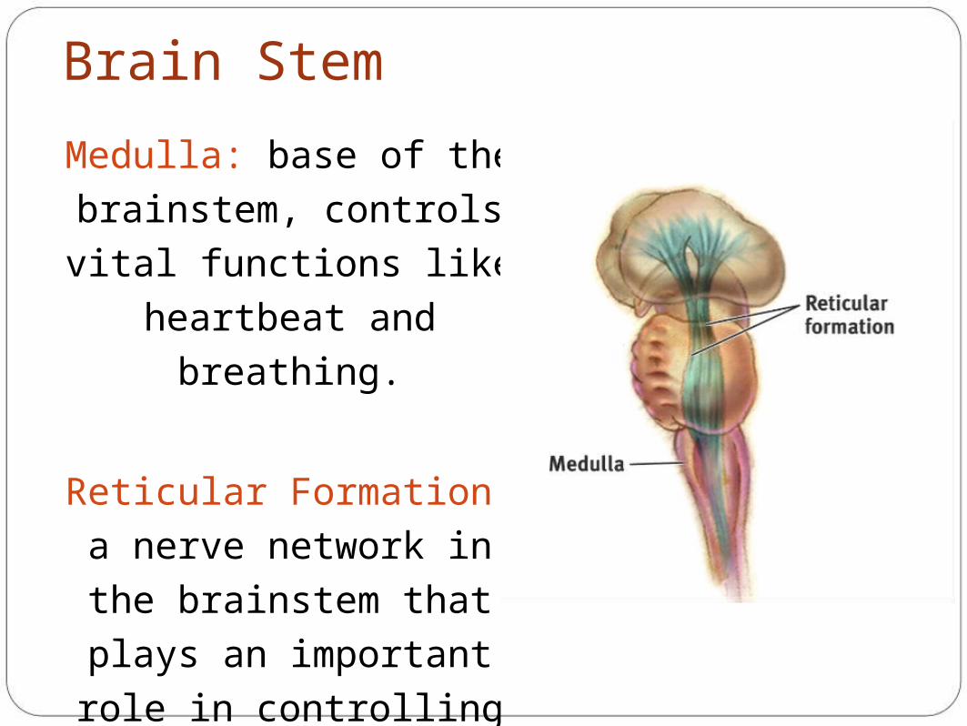
Brain Stem
Medulla: base of the
brainstem, controls vital
functions like heartbeat and
breathing.
Reticular Formation: a nerve
network in the brainstem that
plays an important role in
controlling arousal &
involved in attention and
sleep (filtering out stimuli)
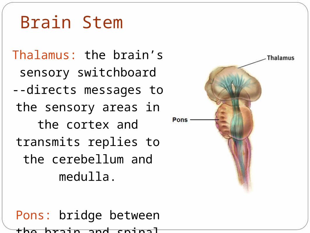
Brain Stem
Thalamus: the brain’s
sensory switchboard --
directs messages to the
sensory areas in the cortex
and transmits replies to the
cerebellum and medulla.
Pons: bridge between the
brain and spinal cord
(especially dealing with
motor messages) and/or
sleeping (sleep cycle)
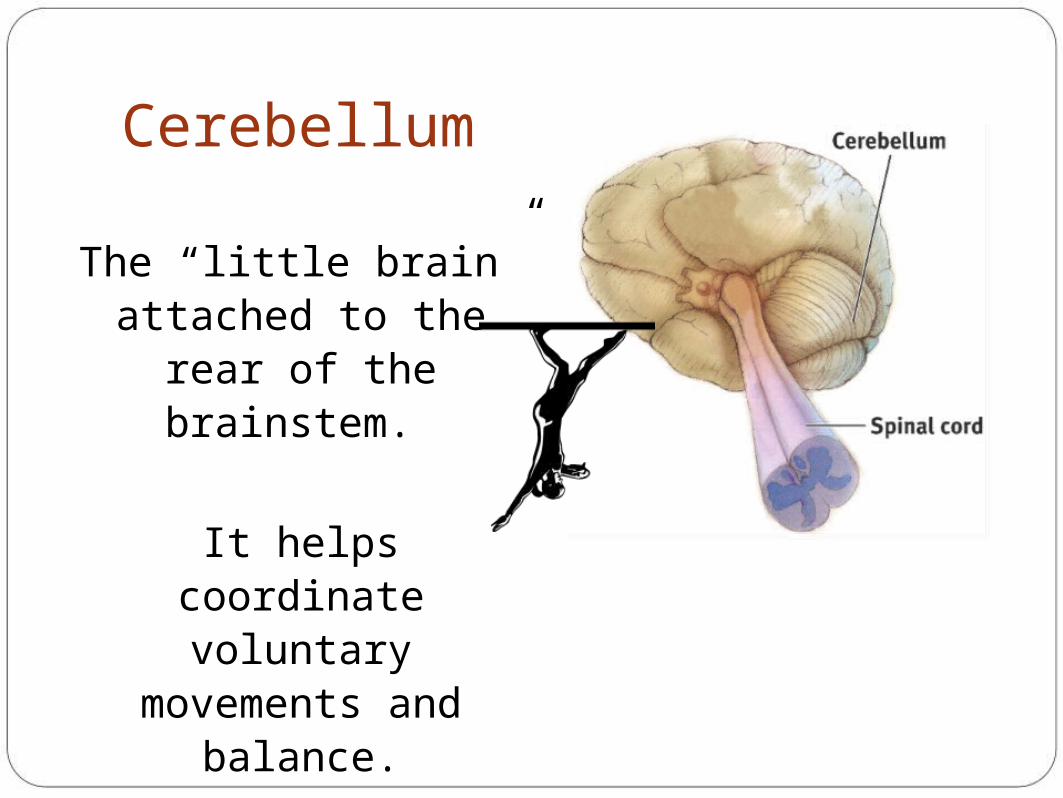
Cerebellum
The “little brain” attached to the rear of
the brainstem.
It helps coordinate voluntary movements
and balance.
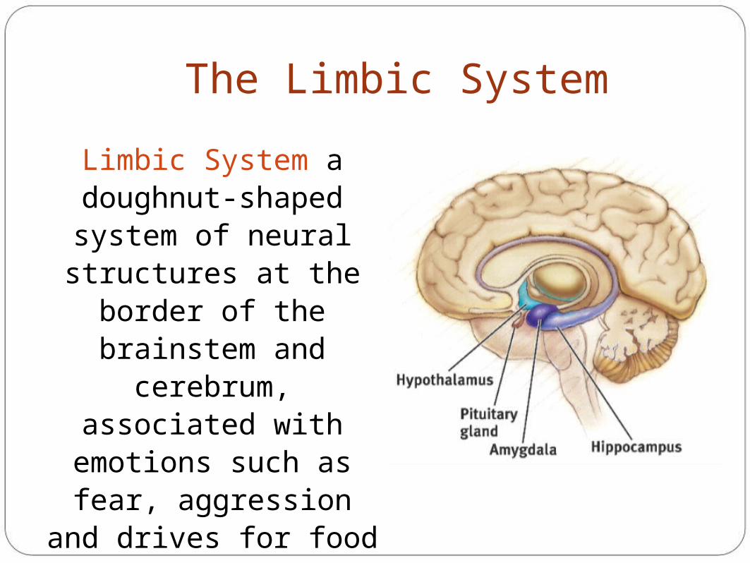
The Limbic System
Limbic System a doughnut-shaped system of neural structures at the border of the brainstem
and cerebrum, associated with emotions such as fear, aggression and
drives for food and sex. It includes the
hippocampus, amygdala, and hypothalamus.
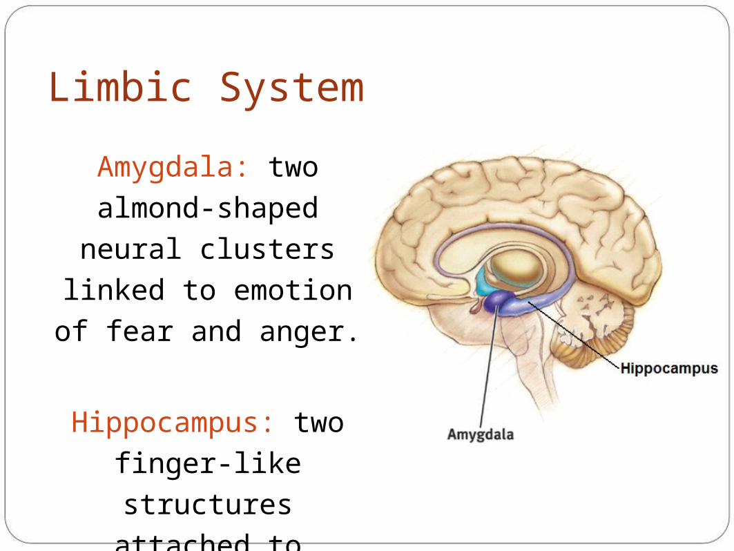
Limbic System
Amygdala: two almond-
shaped neural clusters
linked to emotion of fear
and anger.
Hippocampus: two
finger-like structures
attached to amygdala
involved in processing
(new) memories

Limbic System
Hypothalamus: lies below
(hypo) the thalamus; directs
several maintenance
activities like eating,
drinking, body temperature,
and sex. Helps govern the
endocrine system via the
pituitary gland.
Ventromedial – “vomit” – tells you when to STOP eating
Lateral – “let’s eat” – tell you when you are hungry
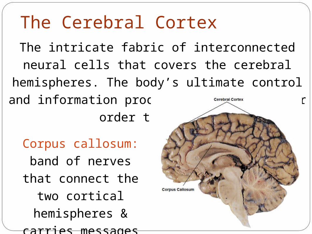
The Cerebral CortexThe intricate fabric of interconnected neural cells that
covers the cerebral hemispheres. The body’s ultimate
control and information processing center (higher order
thoughts).
Corpus callosum: band
of nerves that connect
the two cortical
hemispheres & carries
messages between them