Embed Size (px)
Citation preview
Kurepa et al. BMC Plant Biology (2018) 18:278 https://doi.org/10.1186/s12870-018-1477-0
RESEARCH ARTICLE Open Access
Modulation of auxin and cytokininresponses by early steps of thephenylpropanoid pathway
Jasmina Kurepa, Timothy E. Shull, Sumudu S. Karunadasa and Jan A. Smalle*Abstract
Background: The phenylpropanoid pathway is responsible for the synthesis of numerous compounds importantfor plant growth and responses to the environment. In the first committed step of phenylpropanoid biosynthesis,the enzyme phenylalanine ammonia-lyase (PAL) deaminates L-phenylalanine into trans-cinnamic acid that is thenconverted into p-coumaric acid by cinnamate-4-hydroxylase (C4H). Recent studies showed that the Kelch repeat F-box (KFB) protein family of ubiquitin ligases control phenylpropanoid biosynthesis by promoting the proteolysis ofPAL. However, this ubiquitin ligase family, alternatively named Kiss Me Deadly (KMD), was also implicated incytokinin signaling as it was shown to promote the degradation of type-B ARRs, including the key responseactivator ARR1. Considering that ubiquitin ligases typically have narrow target specificity, this dual targeting ofstructurally and functionally unrelated proteins appeared unusual.
Results: Here we show that the KFBs indeed target PAL but not ARR1. Moreover, we show that changes in earlyphenylpropanoid biosynthesis alter cytokinin sensitivity – as reported earlier - but that the previously documentedcytokinin growth response changes are primarily the result of altered auxin signaling. We found that reduced PALaccumulation decreased, whereas the loss of C4H function increased the strength of the auxin response. Thecombined loss of function of both enzymes led to a decrease in auxin sensitivity, indicating that metabolic eventsupstream of C4H control auxin sensitivity. This auxin/phenylpropanoid interaction impacts both shoot and rootdevelopment and revealed an auxin-dependent stimulatory effect of trans-cinnamic acid feeding on leaf expansionand thus biomass accumulation.
Conclusions: Collectively, our results show that auxin-regulated plant growth is fine-tuned by early steps inphenylpropanoid biosynthesis and suggest that metabolites accumulating upstream of the C4H step impact theauxin response mechanism.
Keywords: Auxin, Cytokinin, F-box proteins, Growth promotion, Phenylpropanoid biosynthesis
BackgroundAuxins are plant hormones that control key aspects ofplant development, including the development of shootand root meristems and cell expansion [1–3]. The auxinresponse pathway includes a repression relief mechanismwherein auxin promotes the degradation of AUX/IAAproteins that repress auxin responses by inhibiting theactivity of the auxin response factors (ARFs), which actas transcriptional regulators of the auxin response [4, 5].
* Correspondence: [email protected] of Plant and Soil Sciences, College of Agriculture, Food andEnvironment, University of Kentucky, Lexington, KY 40546-0236, USA
© The Author(s). 2018 Open Access This articInternational License (http://creativecommonsreproduction in any medium, provided you gthe Creative Commons license, and indicate if(http://creativecommons.org/publicdomain/ze
Auxin acts as a molecular glue and promotes the inter-action between the AUX/IAAs and the SCFTIR1/AFBs E3ligases which commences the degradation of AUX/IAAsby the 26S proteasome [6, 7].The link between the phenylpropanoid (PP) pathway
and auxin responses has already been investigated. Forexample, naringenin, an early intermediate of the flavon-oid branch of the PP pathway, was classified as an auxintransport inhibitor whereas the PP 3,4-(methylenediox-y)cinnamic acid was shown to interfere with auxin efflux[8, 9]. PP biosynthesis starts with L-phenylalanine that isconverted into trans-cinnamic acid (t-CA) by phenyl-alanine ammonia lyase (PAL). t-CA can be converted to
le is distributed under the terms of the Creative Commons Attribution 4.0.org/licenses/by/4.0/), which permits unrestricted use, distribution, andive appropriate credit to the original author(s) and the source, provide a link tochanges were made. The Creative Commons Public Domain Dedication waiverro/1.0/) applies to the data made available in this article, unless otherwise stated.
Kurepa et al. BMC Plant Biology (2018) 18:278 Page 2 of 15
cis-cinnamic acid (c-CA) by light and this photoisomerhas been shown to inhibit auxin transport [10, 11]. Inthe next step of the PP pathway, t-CA is converted top-coumaric acid by the cytochrome P450-dependentmonooxygenase cinnamate-4-hydroxylase (C4H). Thereaction catalyzed by C4H marks the end of the earlysteps of the PP pathway and represents the pathwaybranching point as p-coumaric acid can be diverted to-wards the synthesis of a number of metabolite classes in-cluding lignins and flavonoids. Arabidopsis mutants thatare defective in specific steps of flavonoid biosynthesisalso show auxin-related developmental phenotypes [12].PAL is the first committed enzyme of the PP pathway
and its activity is regulated by environmental and en-dogenous signals at multiple levels [13]. At thepost-translational level, the abundance of PAL isozymesis attuned to metabolic needs by the ubiquitin/prote-asome pathway [14]. In Arabidopsis, PAL degradation isgoverned by the SCF type E3 ligases in which thetarget-specific component, the F-box protein calledKelch Repeat F-Box (KFB), is encoded by four genes[14]. The KFB genes are differentially expressed andcontrol PAL levels in response to developmental and en-vironmental changes. This family of ubiquitin ligases, al-ternatively named Kiss Me Deadly (KMD), was alsoshown to promote the degradation of key transcriptionalactivators of the cytokinin response, the type-B ARRfamily members ARR1 and ARR12. The KMD/KFBgenes are down-regulated by the cytokinin signal andthus are thought to be a feed-forward mechanism thatenhances the cytokinin response [15].Cytokinins are plant growth regulators that control many
agriculturally important processes, including the initiationand development of meristems and the timing of leaf senes-cence [16]. The cytokinin response pathway consists of atwo-component signaling mechanism that involves a se-quence of phosphotransfer reactions. In Arabidopsis, cyto-kinins are perceived by a family of three histidine kinasereceptors that autophosphorylate upon binding with thehormone. The phosphoryl group is then transferred to his-tidine phosphotransfer proteins that in turn phosphorylatemembers of two functionally opposite classes of responseregulators (ARRs), the response-promoting type-B ARRsand the response-inhibiting type-A ARRs. When phosphor-ylated, the type-B ARRs became activated and transcrip-tionally regulate the expression of primary cytokininresponse genes. Both type-A and type-B ARRs are encodedby large gene families. Among the type-B ARRs, the ARR1,ARR10 and ARR12 genes are preeminent because theircombined loss of function leads to a strong cytokinin in-sensitivity and severe growth reduction [17, 18].The finding that KMD/KFBs target two sets of struc-
turally and functionally unrelated proteins was surpris-ing because it implies that KMD/KFBs contain two
different target interaction domains and that they simul-taneously control a hormone signaling pathway inaddition to a secondary metabolite pathway. Here weshow that the KMD/KFBs do not control the stability ofthe type-B ARR member ARR1 but are indeed involvedin the proteasome-dependent degradation of PAL en-zymes. However, we confirm the previous finding thatthe KMD/KFBs modulate the root growth response tocytokinin and demonstrate that this effect on cytokininresponses is a result of changes in auxin signaling. Weshow that loss of function of both PAL and C4H altersthe response to auxin, but in an opposite manner whichindicates that the observed modulation of auxin signal-ing is the result of metabolic changes downstream ofPAL and upstream of the C4H step in the PP pathway.We also show that the product of PAL, t-CA, or its de-rivative(s) enhances auxin signaling and promotesauxin-dependent leaf expansion.
ResultsPAL and the cytokinin responseTo independently test the role of KMD/KFBs in cytokininsignaling, we generated 35S promoter-driven overexpres-sion (OE) lines using the full-length KMD1/KFB20(At1g80440) cDNA. Earlier studies revealed that KMD1/KFB20 OE lines are dwarfed and that the extent of growthretardation is positively correlated with the expressionlevel of the transgene [14, 15]. Indeed, 34 lines out of 52lines we generated were also dwarfed. Both severe cytoki-nin resistance and disruption of the general PP pathwayleads to dwarfism [18, 19]. Thus, this phenotype of the OEplants is not a diagnostic for alteration of the function ofeither cytokinin signaling or PP biosynthesis. Because thePP biosynthesis and cytokinin response pathways are notdirectly linked, we attempted to distinguish between thegrowth inhibition resulting from reduced PP levels andgrowth inhibition induced by reduced cytokinin signalingby feeding severely dwarfed KMD1/KFB20 OE lines withPP pathway intermediates.We grew wild-type and OE plants on media containing
different concentrations of either t-CA, p-coumaric acid,p-coumaraldehyde, caffeic acid or quercetin. Dose-responsecurves are shown in Additional file 1: Figure S1. For thewild type, the feeding experiments with different doses oft-CA show that t-CA is growth promoting at low concen-trations and growth inhibitory and anthocyanin inducing athigh concentrations (Additional file 1: Figure S1a-c).Growth on media supplemented with p-coumaric acid andp-coumaraldehyde did not significantly change the size ofthe wild-type plants (Additional file 1: Figure S1d,e).Caffeic acid and quercetin treatments also did notsignificantly impact wild-type growth at lower doses,but they caused growth inhibition at higher doses(Additional file 1: Figure S1f, g).
Kurepa et al. BMC Plant Biology (2018) 18:278 Page 3 of 15
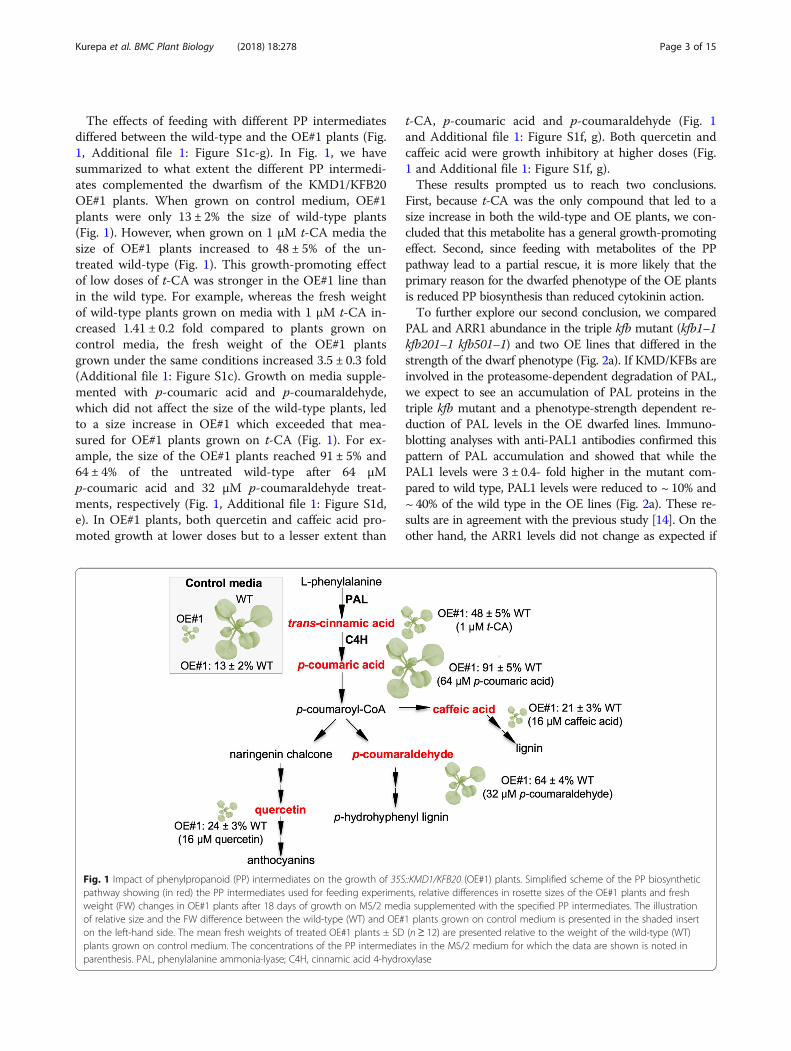
The effects of feeding with different PP intermediatesdiffered between the wild-type and the OE#1 plants (Fig.1, Additional file 1: Figure S1c-g). In Fig. 1, we havesummarized to what extent the different PP intermedi-ates complemented the dwarfism of the KMD1/KFB20OE#1 plants. When grown on control medium, OE#1plants were only 13 ± 2% the size of wild-type plants(Fig. 1). However, when grown on 1 μM t-CA media thesize of OE#1 plants increased to 48 ± 5% of the un-treated wild-type (Fig. 1). This growth-promoting effectof low doses of t-CA was stronger in the OE#1 line thanin the wild type. For example, whereas the fresh weightof wild-type plants grown on media with 1 μM t-CA in-creased 1.41 ± 0.2 fold compared to plants grown oncontrol media, the fresh weight of the OE#1 plantsgrown under the same conditions increased 3.5 ± 0.3 fold(Additional file 1: Figure S1c). Growth on media supple-mented with p-coumaric acid and p-coumaraldehyde,which did not affect the size of the wild-type plants, ledto a size increase in OE#1 which exceeded that mea-sured for OE#1 plants grown on t-CA (Fig. 1). For ex-ample, the size of the OE#1 plants reached 91 ± 5% and64 ± 4% of the untreated wild-type after 64 μMp-coumaric acid and 32 μM p-coumaraldehyde treat-ments, respectively (Fig. 1, Additional file 1: Figure S1d,e). In OE#1 plants, both quercetin and caffeic acid pro-moted growth at lower doses but to a lesser extent than
Fig. 1 Impact of phenylpropanoid (PP) intermediates on the growth of 35Spathway showing (in red) the PP intermediates used for feeding experimenweight (FW) changes in OE#1 plants after 18 days of growth on MS/2 medof relative size and the FW difference between the wild-type (WT) and OE#on the left-hand side. The mean fresh weights of treated OE#1 plants ± SDplants grown on control medium. The concentrations of the PP intermediaparenthesis. PAL, phenylalanine ammonia-lyase; C4H, cinnamic acid 4-hydro
t-CA, p-coumaric acid and p-coumaraldehyde (Fig. 1and Additional file 1: Figure S1f, g). Both quercetin andcaffeic acid were growth inhibitory at higher doses (Fig.1 and Additional file 1: Figure S1f, g).These results prompted us to reach two conclusions.
First, because t-CA was the only compound that led to asize increase in both the wild-type and OE plants, we con-cluded that this metabolite has a general growth-promotingeffect. Second, since feeding with metabolites of the PPpathway lead to a partial rescue, it is more likely that theprimary reason for the dwarfed phenotype of the OE plantsis reduced PP biosynthesis than reduced cytokinin action.To further explore our second conclusion, we compared
PAL and ARR1 abundance in the triple kfb mutant (kfb1–1kfb201–1 kfb501–1) and two OE lines that differed in thestrength of the dwarf phenotype (Fig. 2a). If KMD/KFBs areinvolved in the proteasome-dependent degradation of PAL,we expect to see an accumulation of PAL proteins in thetriple kfb mutant and a phenotype-strength dependent re-duction of PAL levels in the OE dwarfed lines. Immuno-blotting analyses with anti-PAL1 antibodies confirmed thispattern of PAL accumulation and showed that while thePAL1 levels were 3 ± 0.4- fold higher in the mutant com-pared to wild type, PAL1 levels were reduced to ~ 10% and~ 40% of the wild type in the OE lines (Fig. 2a). These re-sults are in agreement with the previous study [14]. On theother hand, the ARR1 levels did not change as expected if
::KMD1/KFB20 (OE#1) plants. Simplified scheme of the PP biosyntheticts, relative differences in rosette sizes of the OE#1 plants and freshia supplemented with the specified PP intermediates. The illustration1 plants grown on control medium is presented in the shaded insert(n ≥ 12) are presented relative to the weight of the wild-type (WT)tes in the MS/2 medium for which the data are shown is noted inxylase
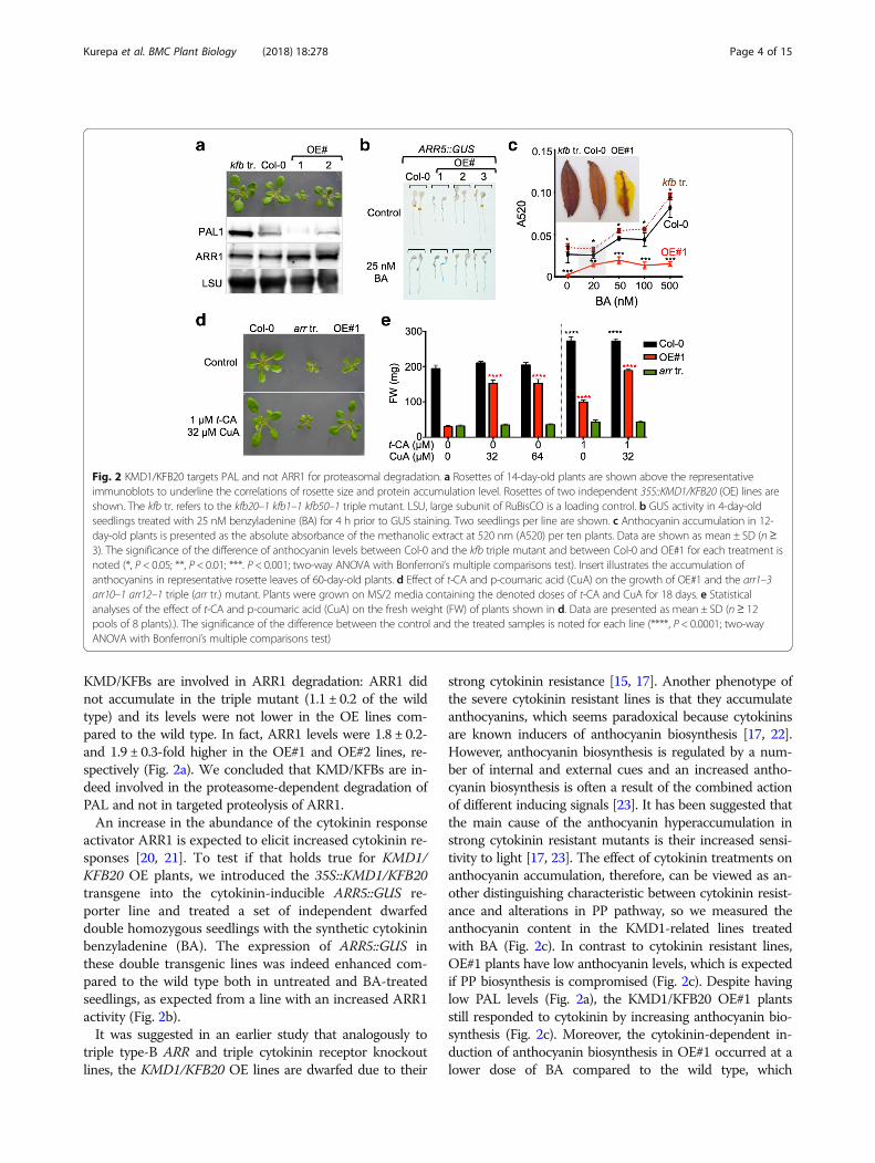
Fig. 2 KMD1/KFB20 targets PAL and not ARR1 for proteasomal degradation. a Rosettes of 14-day-old plants are shown above the representativeimmunoblots to underline the correlations of rosette size and protein accumulation level. Rosettes of two independent 35S::KMD1/KFB20 (OE) lines areshown. The kfb tr. refers to the kfb20–1 kfb1–1 kfb50–1 triple mutant. LSU, large subunit of RuBisCO is a loading control. b GUS activity in 4-day-oldseedlings treated with 25 nM benzyladenine (BA) for 4 h prior to GUS staining. Two seedlings per line are shown. c Anthocyanin accumulation in 12-day-old plants is presented as the absolute absorbance of the methanolic extract at 520 nm (A520) per ten plants. Data are shown as mean ± SD (n≥3). The significance of the difference of anthocyanin levels between Col-0 and the kfb triple mutant and between Col-0 and OE#1 for each treatment isnoted (*, P < 0.05; **, P < 0.01; ***. P < 0.001; two-way ANOVA with Bonferroni’s multiple comparisons test). Insert illustrates the accumulation ofanthocyanins in representative rosette leaves of 60-day-old plants. d Effect of t-CA and p-coumaric acid (CuA) on the growth of OE#1 and the arr1–3arr10–1 arr12–1 triple (arr tr.) mutant. Plants were grown on MS/2 media containing the denoted doses of t-CA and CuA for 18 days. e Statisticalanalyses of the effect of t-CA and p-coumaric acid (CuA) on the fresh weight (FW) of plants shown in d. Data are presented as mean ± SD (n≥ 12pools of 8 plants).). The significance of the difference between the control and the treated samples is noted for each line (****, P < 0.0001; two-wayANOVA with Bonferroni’s multiple comparisons test)
Kurepa et al. BMC Plant Biology (2018) 18:278 Page 4 of 15
KMD/KFBs are involved in ARR1 degradation: ARR1 didnot accumulate in the triple mutant (1.1 ± 0.2 of the wildtype) and its levels were not lower in the OE lines com-pared to the wild type. In fact, ARR1 levels were 1.8 ± 0.2-and 1.9 ± 0.3-fold higher in the OE#1 and OE#2 lines, re-spectively (Fig. 2a). We concluded that KMD/KFBs are in-deed involved in the proteasome-dependent degradation ofPAL and not in targeted proteolysis of ARR1.An increase in the abundance of the cytokinin response
activator ARR1 is expected to elicit increased cytokinin re-sponses [20, 21]. To test if that holds true for KMD1/KFB20 OE plants, we introduced the 35S::KMD1/KFB20transgene into the cytokinin-inducible ARR5::GUS re-porter line and treated a set of independent dwarfeddouble homozygous seedlings with the synthetic cytokininbenzyladenine (BA). The expression of ARR5::GUS inthese double transgenic lines was indeed enhanced com-pared to the wild type both in untreated and BA-treatedseedlings, as expected from a line with an increased ARR1activity (Fig. 2b).It was suggested in an earlier study that analogously to
triple type-B ARR and triple cytokinin receptor knockoutlines, the KMD1/KFB20 OE lines are dwarfed due to their
strong cytokinin resistance [15, 17]. Another phenotype ofthe severe cytokinin resistant lines is that they accumulateanthocyanins, which seems paradoxical because cytokininsare known inducers of anthocyanin biosynthesis [17, 22].However, anthocyanin biosynthesis is regulated by a num-ber of internal and external cues and an increased antho-cyanin biosynthesis is often a result of the combined actionof different inducing signals [23]. It has been suggested thatthe main cause of the anthocyanin hyperaccumulation instrong cytokinin resistant mutants is their increased sensi-tivity to light [17, 23]. The effect of cytokinin treatments onanthocyanin accumulation, therefore, can be viewed as an-other distinguishing characteristic between cytokinin resist-ance and alterations in PP pathway, so we measured theanthocyanin content in the KMD1-related lines treatedwith BA (Fig. 2c). In contrast to cytokinin resistant lines,OE#1 plants have low anthocyanin levels, which is expectedif PP biosynthesis is compromised (Fig. 2c). Despite havinglow PAL levels (Fig. 2a), the KMD1/KFB20 OE#1 plantsstill responded to cytokinin by increasing anthocyanin bio-synthesis (Fig. 2c). Moreover, the cytokinin-dependent in-duction of anthocyanin biosynthesis in OE#1 occurred at alower dose of BA compared to the wild type, which
Kurepa et al. BMC Plant Biology (2018) 18:278 Page 5 of 15
provided another example of cytokinin hypersensitivity ofplants overexpressing KMD1/KFB20. As expected, theanthocyanin levels in OE#1 did not reach wild-type levelsindependent of the BA dose used in the assay. The antho-cyanin levels in the kfb triple mutant were higher thanthose of the wild type, but the dose-response curve had thesame wild-type shape (Fig. 2c). These differences in antho-cyanin accumulation patterns were clearly visible in senes-cing leaves (Fig. 2c).Final confirmation that KMD/KFBs are involved in
proteolysis of PAL but not ARR1 was obtained by com-paring the growth responses of a strong cytokinin resist-ant mutant and the strong KMD1/KFB20 OE line OE#1to PP pathway intermediates. It was previously suggestedthat the severe growth inhibition seen in the strongKMD1/KFB20 OE lines is mechanistically similar to thegrowth inhibition of the strong cytokinin resistant triplemutant arr1–3 arr10–5 arr12–1: both sets of lines werethought to be dwarfed as a result of reduced type-B ARRactivity. If this is correct, then the growth of both arr1–3 arr10–5 arr12–1 and KMD1/KFB20 OE plants shouldbe similarly affected by PP pathway intermediates. How-ever, whereas OE#1 plants reached 97 ± 2% of the un-treated wild-type size on media containing both t-CAand p-coumaric acid, the arr1–3 arr10–5 arr12–1 plantsremained dwarfed and their increase in size was compar-able to that of the increase observed for the wild typegrown on t-CA and p-coumaric acid (40 ± 3% and 34 ±15%, for wild type and triple arr mutant, respectively;Fig. 2d, e). Therefore, it is highly unlikely that the samemechanism that affects growth is operational in both theKMD1/KFB20 OE plants and the triple arr mutant.
C4H and the cytokinin responseIn two hallmark cytokinin response assays, type-A ARRexpression (Fig. 2b) and anthocyanin level analyses (Fig.2c), KMD1/KFB20 OE plants showed cytokinin hyper-sensitivity. A third archetypical cytokinin response assayis the root elongation assay. In this assay, as has beenpreviously reported, KMD1/KFB20 OE plants showeddecreased sensitivity to cytokinin, (Additional file 1: Fig-ure S2a and [15]). To explain this finding, we hypothe-sized that a PP intermediate is required for the wild-typecytokinin root elongation response and tested this hy-pothesis using a genetic approach. We analyzed thecytokinin sensitivity of the ref3 mutants, loss-of-functionmutants of C4H that catalyzes the step immediatelydownstream of PAL [24]. Cytokinin dose-response treat-ments showed that ref3 mutants are cytokinin hypersen-sitive (Fig. 3a and Additional file 1: Figure S2). Thissuggested that a PP intermediate or derivative that accu-mulates in ref3 and is depleted in KMD1/KFB20 OEplants is essential for the wild-type root growth responseto cytokinin. The obvious candidate was t-CA, so we
tested whether reduced cytokinin sensitivity of OE#1plants in root assays is restored to wild-type levels incombined BA/t-CA treatments. Indeed, feeding OE#1seedlings with t-CA reverted the cytokinin sensitivity tothe wild-type level (Fig. 3b).Two of the more prominent visible phenotypes of ref3
plants are increased shoot branching and increased lateralroot formation ([24] and Fig. 3 d, e). Cytokinins promotelateral bud outgrowth and inhibit lateral root formation[25, 26]. Therefore, whereas increased shoot branching inref3 plants could be caused by cytokinin hypersensitivity,the increased lateral root formation is the opposite of whatone would expect in a hypersensitive mutant. To under-stand this apparent contradiction, we separately intro-duced two transgenes into the ref3–1 mutant line:35S::ARR5, which overexpresses the cytokinin response in-hibitor ARR5 and thus causes cytokinin resistance, and35S::KMD1/KFB20, which allows us to test if the develop-mental changes in ref3 plants are caused by a decreasedaccumulation of intermediates downstream of C4H or theaccumulation of intermediates upstream of C4H. Cytoki-nin root growth response assays revealed that both35S::ARR5 and 35S::KMD1/KFB20 transgenes suppressedthe enhanced cytokinin growth response of ref3 seedlings(Fig. 3c). However, only 35S::KMD1/KFB20 suppressedthe increased shoot branching and the increased lateralroot phenotype of the ref3 mutant (Fig. 3d, e). Therefore,these two ref3 phenotypes are not a result of altered cyto-kinin signaling but stem from the altered accumulation ofmetabolites synthesized upstream of the C4H step.
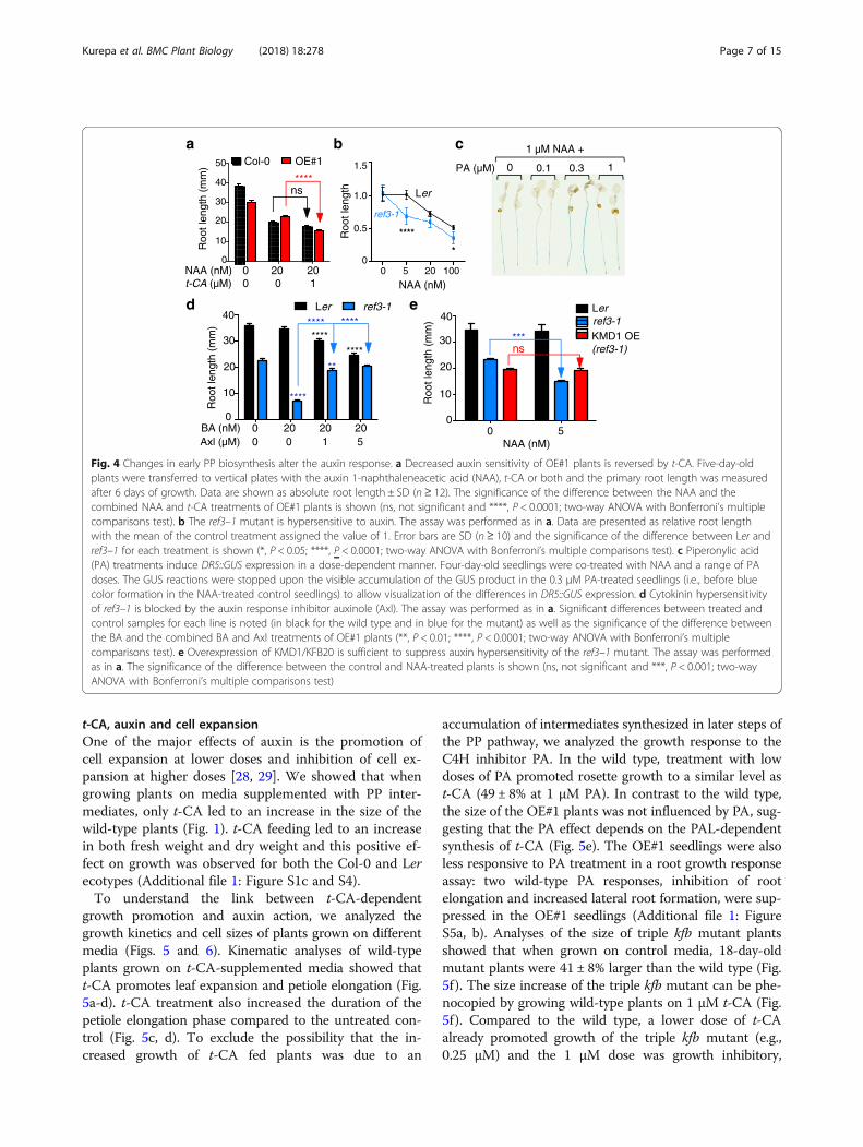
The phenylpropanoid pathway and the auxin responseCytokinin regulates root growth together with auxin[27]. Thus, it was possible that the altered cytokinin rootgrowth responses of 35S::KMD1/KFB20 transgenic linesand ref3 mutants are caused by a change in auxin sensi-tivity. Indeed, the auxin root growth response assayshowed that OE#1 seedlings were less sensitive to auxinand their sensitivity can be restored to wild-type levelsby feeding with t-CA (Fig. 4a). On the other hand, ref3–1 was more sensitive to auxin (Fig. 4b). This suggestedthat similar to cytokinin responses, the altered auxin re-sponses in KMD1/KFB20 OE plants and in ref3 mutantsare caused by changes in the accumulation of metabo-lites synthesized upstream of C4H.Next, we used the C4H inhibitor piperonylic acid
(PA) to test the expression of the cytokinin primary re-sponse reporter ARR5::GUS and of the auxin primaryresponse reporter DR5::GUS. Whereas PA treatmentsdid not alter the cytokinin-dependent induction ofARR5::GUS (Additional file 1: Figure S3), the expressionof the auxin-induced DR5::GUS was affected in adose-responsive manner, with the maximal induction ofDR5::GUS recorded at 0.3 μM PA (Fig. 4c). These
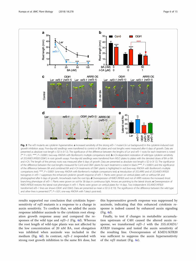
Fig. 3 The ref3mutants are cytokinin hypersensitive. a Increased sensitivity of the strong ref3–1 mutant (in Ler background) in the cytokinin-induced rootgrowth inhibition assay. Five-day-old seedlings were transferred to control or BA plates and root lengths were measured after 6 days of growth. Data arepresented as absolute root length ± SD (n≥ 12). The significance of the difference between the lengths of Ler and ref3–1 roots for each treatment is noted(** P< 0.01, ****, P< 0.0001; two-way ANOVA with Bonferroni’s multiple comparisons test). b t-CA-dependent restoration of wild-type cytokinin sensitivityof 35S::KMD1/KFB20 (OE#1) in root growth assays. Five-day-old seedlings were transferred from MS/2 plates to plates with the denoted doses of BA or BAand t-CA. The length of the primary roots was measured after 6 days of growth. Data are presented as absolute root length ± SD (n≥ 12). The significanceof the difference between the root lengths measured for Col-0 and OE#1 plants for each treatment is noted in black (****, P< 0.0001) and the significanceof the difference between BA and combined BA and t-CA treatments of OE#1 plants is highlighted in red (two-way ANOVA with Bonferroni’s multiplecomparisons test). ****, P< 0.0001 (one-way ANOVA with Bonferroni’s multiple comparisons test). c Introduction of 35S::ARR5 and of 35S::KMD1/KFB20transgenes in ref3–1 suppresses the enhanced cytokinin growth response of ref3–1. Plants were grown on vertical plates with or without BA andphotographed after 6 days of growth. Arrowheads mark the root tips. d Overexpression of KMD1/KFB20 and not of ARR5 restores the increased shootbranching phenotype of ref3–1. Plants were grown on soil for 38 days in continuous light. Arrows are pointing to the lateral shoots. e Overexpression ofKMD1/KFB20 restores the lateral root phenotype in ref3–1. Plants were grown on vertical plates for 14 days. Two independent 35S::KMD1/KFB20transformed ref3–1 lines are shown (OE#1 and OE#2). Data are presented as mean ± SD (n≥ 10). The significance of the difference between the wild typeand other lines is presented (**, P< 0.01; one-way ANOVA with Tukey’s post-test).
Kurepa et al. BMC Plant Biology (2018) 18:278 Page 6 of 15
results supported our conclusion that cytokinin hyper-sensitivity of ref3 mutants is a response to a change inauxin sensitivity. To confirm that, we added the auxinresponse inhibitor auxinole to the cytokinin root elong-ation growth response assay and compared the re-sponses of the wild type and ref3–1 (Fig. 4d). Whereasthe root length of wild-type plants was not affected bythe low concentration of 20 nM BA, root elongationwas inhibited when auxinole was included in themedium (Fig. 4d). In contrast, ref3–1 responded withstrong root growth inhibition to the same BA dose, but
this hypersensitive growth response was suppressed byauxinole, indicating that this enhanced cytokinin re-sponse is indeed caused by enhanced auxin signaling(Fig. 4d).Finally, to test if changes in metabolite accumula-
tion upstream of C4H caused the altered auxin re-sponse, we transformed ref3–1 with the 35S::KMD1/KFB20 transgene and tested the auxin sensitivity ofthe resulting line. Overexpression of KMD1/KFB20was sufficient to suppress the auxin hypersensitivityof the ref3 mutant (Fig. 4e).
PA (µM)
1 µM NAA +ca
d
b
e
BA (nM)
Roo
t len
gth
(mm
)
0
10
30
20
40
Axl (µM)00
200
201
205
Ler ref3-1
****
NAA (nM)
Roo
t len
gth
(mm
)
0
10
30
20
t-CA (µM)00
200
201
****40
50 OE#1Col-0
ns
Roo
t len
gth
(mm
)0
10
30
20
40Lerref3-1KMD1 OE (ref3-1)
0 5NAA (nM)
1 0.3 0.1 0
***ns
Roo
t len
gth
0
0.5
1.0
1.5
0 5 20 100
NAA (nM)
Ler
ref3-1
****
****
*
****
****
**
****
Fig. 4 Changes in early PP biosynthesis alter the auxin response. a Decreased auxin sensitivity of OE#1 plants is reversed by t-CA. Five-day-oldplants were transferred to vertical plates with the auxin 1-naphthaleneacetic acid (NAA), t-CA or both and the primary root length was measuredafter 6 days of growth. Data are shown as absolute root length ± SD (n ≥ 12). The significance of the difference between the NAA and thecombined NAA and t-CA treatments of OE#1 plants is shown (ns, not significant and ****, P < 0.0001; two-way ANOVA with Bonferroni’s multiplecomparisons test). b The ref3–1 mutant is hypersensitive to auxin. The assay was performed as in a. Data are presented as relative root lengthwith the mean of the control treatment assigned the value of 1. Error bars are SD (n ≥ 10) and the significance of the difference between Ler andref3–1 for each treatment is shown (*, P < 0.05; ****, P < 0.0001; two-way ANOVA with Bonferroni’s multiple comparisons test). c Piperonylic acid(PA) treatments induce DR5::GUS expression in a dose-dependent manner. Four-day-old seedlings were co-treated with NAA and a range of PAdoses. The GUS reactions were stopped upon the visible accumulation of the GUS product in the 0.3 μM PA-treated seedlings (i.e., before bluecolor formation in the NAA-treated control seedlings) to allow visualization of the differences in DR5::GUS expression. d Cytokinin hypersensitivityof ref3–1 is blocked by the auxin response inhibitor auxinole (Axl). The assay was performed as in a. Significant differences between treated andcontrol samples for each line is noted (in black for the wild type and in blue for the mutant) as well as the significance of the difference betweenthe BA and the combined BA and Axl treatments of OE#1 plants (**, P < 0.01; ****, P < 0.0001; two-way ANOVA with Bonferroni’s multiplecomparisons test). e Overexpression of KMD1/KFB20 is sufficient to suppress auxin hypersensitivity of the ref3–1 mutant. The assay was performedas in a. The significance of the difference between the control and NAA-treated plants is shown (ns, not significant and ***, P < 0.001; two-wayANOVA with Bonferroni’s multiple comparisons test)
Kurepa et al. BMC Plant Biology (2018) 18:278 Page 7 of 15
t-CA, auxin and cell expansionOne of the major effects of auxin is the promotion ofcell expansion at lower doses and inhibition of cell ex-pansion at higher doses [28, 29]. We showed that whengrowing plants on media supplemented with PP inter-mediates, only t-CA led to an increase in the size of thewild-type plants (Fig. 1). t-CA feeding led to an increasein both fresh weight and dry weight and this positive ef-fect on growth was observed for both the Col-0 and Lerecotypes (Additional file 1: Figure S1c and S4).To understand the link between t-CA-dependent
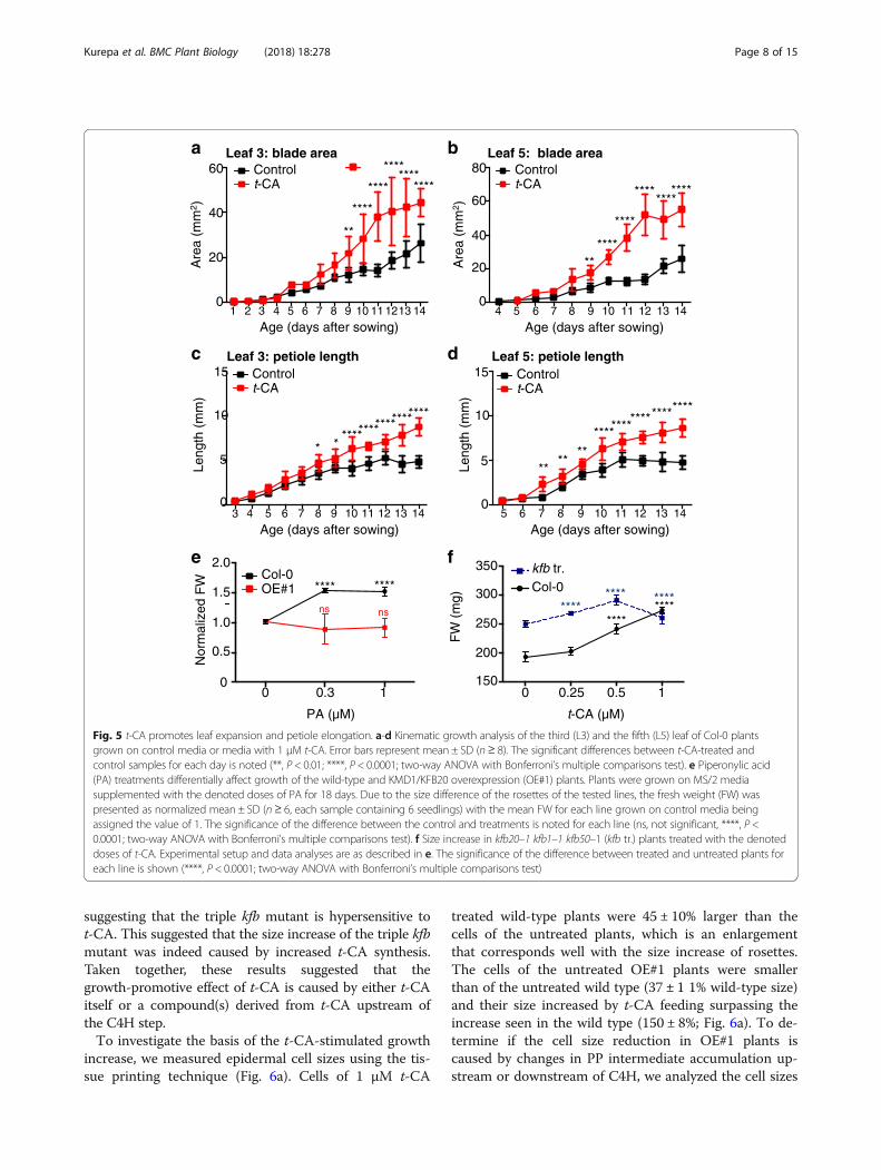
growth promotion and auxin action, we analyzed thegrowth kinetics and cell sizes of plants grown on differentmedia (Figs. 5 and 6). Kinematic analyses of wild-typeplants grown on t-CA-supplemented media showed thatt-CA promotes leaf expansion and petiole elongation (Fig.5a-d). t-CA treatment also increased the duration of thepetiole elongation phase compared to the untreated con-trol (Fig. 5c, d). To exclude the possibility that the in-creased growth of t-CA fed plants was due to an
accumulation of intermediates synthesized in later steps ofthe PP pathway, we analyzed the growth response to theC4H inhibitor PA. In the wild type, treatment with lowdoses of PA promoted rosette growth to a similar level ast-CA (49 ± 8% at 1 μM PA). In contrast to the wild type,the size of the OE#1 plants was not influenced by PA, sug-gesting that the PA effect depends on the PAL-dependentsynthesis of t-CA (Fig. 5e). The OE#1 seedlings were alsoless responsive to PA treatment in a root growth responseassay: two wild-type PA responses, inhibition of rootelongation and increased lateral root formation, were sup-pressed in the OE#1 seedlings (Additional file 1: FigureS5a, b). Analyses of the size of triple kfb mutant plantsshowed that when grown on control media, 18-day-oldmutant plants were 41 ± 8% larger than the wild type (Fig.5f). The size increase of the triple kfb mutant can be phe-nocopied by growing wild-type plants on 1 μM t-CA (Fig.5f). Compared to the wild type, a lower dose of t-CAalready promoted growth of the triple kfb mutant (e.g.,0.25 μM) and the 1 μM dose was growth inhibitory,
ba
dc
fe
aera edalb :5 faeLaera edalb :3 faeL
htgnel eloitep :5 faeLhtgnel eloitep :3 faeL
Controlt-CA
Controlt-CA
Are
a (m
m2 )
0
20
40
60
0
20
40
60
80
Are
a (m
m2 )
1 2 3 4 5 6 7 8 9 10 11 1213 14
Age (days after sowing)4 5 6 7 8 9 10 11 12 13 14
Age (days after sowing)
Controlt-CA
Leng
th (
mm
)
0
5
10
15 Controlt-CA
Leng
th (
mm
)
0
5
10
15
5 6 7 8 9 10 11 12 13 14
Age (days after sowing)4 5 6 7 8 9 10 11 12 13 14
Age (days after sowing)3
Nor
mal
ized
FW
FW
(m
g)
Col-0OE#1 Col-0
kfb tr.
PA (µM) t-CA (µM)
00
15.052.0013.0
0.5
1.0
1.5
2.0
150
200
250
300
350
**** ****
nsns ********
********
********
****
****
****
**
************
****
******
****
**
********************
*
***** **** **** **** ****
****
Fig. 5 t-CA promotes leaf expansion and petiole elongation. a-d Kinematic growth analysis of the third (L3) and the fifth (L5) leaf of Col-0 plantsgrown on control media or media with 1 μM t-CA. Error bars represent mean ± SD (n≥ 8). The significant differences between t-CA-treated andcontrol samples for each day is noted (**, P < 0.01; ****, P < 0.0001; two-way ANOVA with Bonferroni’s multiple comparisons test). e Piperonylic acid(PA) treatments differentially affect growth of the wild-type and KMD1/KFB20 overexpression (OE#1) plants. Plants were grown on MS/2 mediasupplemented with the denoted doses of PA for 18 days. Due to the size difference of the rosettes of the tested lines, the fresh weight (FW) waspresented as normalized mean ± SD (n≥ 6, each sample containing 6 seedlings) with the mean FW for each line grown on control media beingassigned the value of 1. The significance of the difference between the control and treatments is noted for each line (ns, not significant, ****, P <0.0001; two-way ANOVA with Bonferroni’s multiple comparisons test). f Size increase in kfb20–1 kfb1–1 kfb50–1 (kfb tr.) plants treated with the denoteddoses of t-CA. Experimental setup and data analyses are as described in e. The significance of the difference between treated and untreated plants foreach line is shown (****, P < 0.0001; two-way ANOVA with Bonferroni’s multiple comparisons test)
Kurepa et al. BMC Plant Biology (2018) 18:278 Page 8 of 15
suggesting that the triple kfb mutant is hypersensitive tot-CA. This suggested that the size increase of the triple kfbmutant was indeed caused by increased t-CA synthesis.Taken together, these results suggested that thegrowth-promotive effect of t-CA is caused by either t-CAitself or a compound(s) derived from t-CA upstream ofthe C4H step.To investigate the basis of the t-CA-stimulated growth
increase, we measured epidermal cell sizes using the tis-sue printing technique (Fig. 6a). Cells of 1 μM t-CA
treated wild-type plants were 45 ± 10% larger than thecells of the untreated plants, which is an enlargementthat corresponds well with the size increase of rosettes.The cells of the untreated OE#1 plants were smallerthan of the untreated wild type (37 ± 1 1% wild-type size)and their size increased by t-CA feeding surpassing theincrease seen in the wild type (150 ± 8%; Fig. 6a). To de-termine if the cell size reduction in OE#1 plants iscaused by changes in PP intermediate accumulation up-stream or downstream of C4H, we analyzed the cell sizes
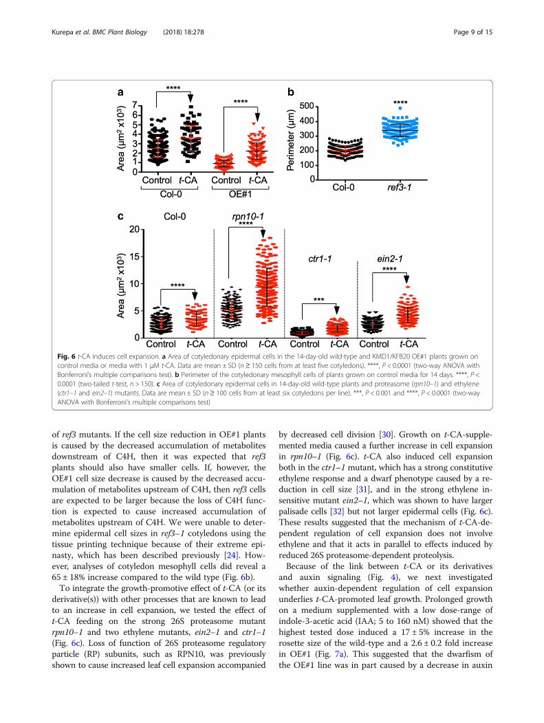
Fig. 6 t-CA induces cell expansion. a Area of cotyledonary epidermal cells in the 14-day-old wild-type and KMD1/KFB20 OE#1 plants grown oncontrol media or media with 1 μM t-CA. Data are mean ± SD (n ≥ 150 cells from at least five cotyledons). ****, P < 0.0001 (two-way ANOVA withBonferroni’s multiple comparisons test). b Perimeter of the cotyledonary mesophyll cells of plants grown on control media for 14 days. ****, P <0.0001 (two-tailed t-test, n > 150). c Area of cotyledonary epidermal cells in 14-day-old wild-type plants and proteasome (rpn10–1) and ethylene(ctr1–1 and ein2–1) mutants. Data are mean ± SD (n≥ 100 cells from at least six cotyledons per line). ***, P < 0.001 and ****, P < 0.0001 (two-wayANOVA with Bonferroni’s multiple comparisons test)
Kurepa et al. BMC Plant Biology (2018) 18:278 Page 9 of 15
of ref3 mutants. If the cell size reduction in OE#1 plantsis caused by the decreased accumulation of metabolitesdownstream of C4H, then it was expected that ref3plants should also have smaller cells. If, however, theOE#1 cell size decrease is caused by the decreased accu-mulation of metabolites upstream of C4H, then ref3 cellsare expected to be larger because the loss of C4H func-tion is expected to cause increased accumulation ofmetabolites upstream of C4H. We were unable to deter-mine epidermal cell sizes in ref3–1 cotyledons using thetissue printing technique because of their extreme epi-nasty, which has been described previously [24]. How-ever, analyses of cotyledon mesophyll cells did reveal a65 ± 18% increase compared to the wild type (Fig. 6b).To integrate the growth-promotive effect of t-CA (or its
derivative(s)) with other processes that are known to leadto an increase in cell expansion, we tested the effect oft-CA feeding on the strong 26S proteasome mutantrpn10–1 and two ethylene mutants, ein2–1 and ctr1–1(Fig. 6c). Loss of function of 26S proteasome regulatoryparticle (RP) subunits, such as RPN10, was previouslyshown to cause increased leaf cell expansion accompanied
by decreased cell division [30]. Growth on t-CA-supple-mented media caused a further increase in cell expansionin rpn10–1 (Fig. 6c). t-CA also induced cell expansionboth in the ctr1–1 mutant, which has a strong constitutiveethylene response and a dwarf phenotype caused by a re-duction in cell size [31], and in the strong ethylene in-sensitive mutant ein2–1, which was shown to have largerpalisade cells [32] but not larger epidermal cells (Fig. 6c).These results suggested that the mechanism of t-CA-de-pendent regulation of cell expansion does not involveethylene and that it acts in parallel to effects induced byreduced 26S proteasome-dependent proteolysis.Because of the link between t-CA or its derivatives
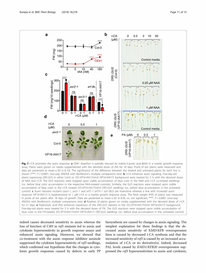
and auxin signaling (Fig. 4), we next investigatedwhether auxin-dependent regulation of cell expansionunderlies t-CA-promoted leaf growth. Prolonged growthon a medium supplemented with a low dose-range ofindole-3-acetic acid (IAA; 5 to 160 nM) showed that thehighest tested dose induced a 17 ± 5% increase in therosette size of the wild-type and a 2.6 ± 0.2 fold increasein OE#1 (Fig. 7a). This suggested that the dwarfism ofthe OE#1 line was in part caused by a decrease in auxin
Kurepa et al. BMC Plant Biology (2018) 18:278 Page 10 of 15
action and that this was complemented by increasingthe auxin concentration. We concluded that t-CA-de-pendent growth promotion is linked to auxin regulationof plant cell expansion implying that t-CA and/or its de-rivatives also promote this auxin response.To reaffirm that t-CA or its derivatives promote
auxin responses, we analyzed the effect of t-CA on theexpression of the auxin-inducible DR5::GUS reporterand on the development of lines with altered auxin sen-sitivity (Fig. 7b, d). For the first experiment, we haveused the synthetic auxin 1-naphthaleneacetic acid(NAA) instead of the natural auxin IAA. Because NAAis known to more readily diffuse into plant cells com-pared to IAA, NAA has been the auxin of choice forshort-term response assays such as the analyses ofDR5::GUS reporter expression [33, 34]. Whereas t-CAalone did not induce the expression of DR5::GUS, it en-hanced the effect of NAA on DR5::GUS expression in acomplex dose-dependent manner (Fig. 7b). The min-imal auxin dose that induces DR5::GUS expression was250 nM NAA (Additional file 1: Figure S6a). Theaddition of t-CA to 250 nM NAA enhanced the expres-sion of DR5::GUS, but only at higher doses of t-CA,such as 10 μM (Fig. 7b). When the t-CA dose-responseexperiments were repeated using 1 μM NAA, the ef-fective dose of t-CA was lower compared to the dosethat enhanced the expression of DR5::GUS at 250 nMNAA (Fig. 7b). These results prove that t-CA (and/ort-CA derivative(s)) indeed promote auxin signaling andsuggest that t-CA leads to plant cell expansion bymodulating auxin responses.Finally, we analyzed the t-CA-induced growth response of
auxin resistant mutants axr2 and axr3 in which the auxinsignaling repressor proteins IAA7 and IAA17 are stabilized,and an arf mutant which carries loss-of-function mutationsin auxin response-regulating ARF genes. Strikingly, all testedmutants (axr2–1, axr3–1 and the arf7–1 arf19–1 doublemutant) were t-CA insensitive in the growth promotionassay (Fig. 7c,d). Treatments with high concentrations ofauxin are growth inhibitory and this can be mimicked byexpression of the 35S::VP16-IAA17mImII transgene thatcauses a strong increase in auxin signaling [35]. The plantsof this transgenic line responded to the t-CA treatment witha further reduction in growth, confirming that t-CA en-hances auxin action also when auxin responses becomegrowth inhibitory (Fig. 7c, d). Next, we introduced theDR5::GUS reporter into 35S::VP16-IAA17mImII back-ground and tested the effect of t-CA on its expression. Asexpected, the DR5::GUS reporter was constitutivelyexpressed in the 35S::VP16-IAA17mImII background (Add-itional file 1: Figure 6b). The expression of the DR5::GUS re-porter was further enhanced both by t-CA and PAtreatments (Fig. 7b, e). We concluded that t-CA or its deriv-ative(s) are de facto modulators of auxin signaling.
DiscussionAlthough the overall conclusion of this study is that earlysteps in the phenylpropanoid pathway are important modu-lators of auxin-regulated plant growth, this work has alsoprompted a number of other discussion points. The firstdiscussion point relates to our analyses of the function ofthe KMD/KFB ubiquitin ligase family. A major step inubiquitin-dependent proteolysis is the interaction of a targetprotein with a ubiquitin E3 ligase that promotes the attach-ment of a polyubiquitin chain to one or more lysine resi-dues within the target [36]. A key feature of a ubiquitinligase is that it binds its target protein in a highly specificmanner and it typically contains a distinct target-interactiondomain. It is not surprising, therefore, that the Arabidopsisgenome encodes for numerous ubiquitin ligases, each hav-ing binding affinity for one target or a highly related familyof target proteins [37]. This complexity and multiplicity ofdifferent E3 ligases reflect the fact that the abundance of nu-merous proteins is controlled by the ubiquitin-proteasomesystem, often in response to specific environmental or en-dogenous signals [38]. It was therefore unusual that theKMD/KFBs were reported to target two structurally andfunctionally unrelated classes of proteins, the PAL enzymesand the type-B ARRs transcription factors [14, 15]. Inaddition, the results of interactomics projects, such asPSICQUIC-View [39], confirmed the binding of KMD/KFBsto PAL, but reported no interactions between KMD/KFBsand type-B ARRs. Here, we show that the endogenousARR1 protein, one of the essential type-B ARRs previouslyshown to be under KMD control, is not targeted forproteasome-dependent degradation by KMDs and that PALis indeed a legitimate target for this F-box protein family.Because we have previously shown that the stability controlof tagged ARR1 versions differs from the stability control ofthe endogenous ARR1 [20], we believe that the use oftagged versions of type-B ARRs is the underlying reason forthis misidentification of KMD targets.A second discussion point involves the interactions of
the PP pathway with auxin and cytokinin. Whereas wecould not confirm any role for the KMD/KFBs in the con-trol of ARR1 stability, our data support the reported find-ings that KFB20/KMD1 overexpression causes a decreasein cytokinin sensitivity in a root elongation response assay[15]. However, we show that the decreased sensitivity isnot caused by a defect in cytokinin signaling and that infact, KMD1/KFB20 overexpression causes an increase inARR1 abundance and consequently, leads to cytokininhypersensitivity. It follows that the decreased cytokininroot growth response is caused by a change acting in par-allel or downstream of the cytokinin signaling pathway. Ithas been shown that the effect of cytokinins on rootgrowth involves auxin regulation and that auxin resistanceimpairs cytokinin-regulated root development [40, 41]. Inthis study, we showed that KMD1/KFB20 overexpression
a b
c
d
FW
(m
g)
Control media
10 40 1600
250
200
e
IAA (nM)
0.25 µM NAA
1 µM NAA
t-CA (µM):
0 0.5 2 10 50
Control media
DR5::G
US
(Col
-0)
DR5::G
US
(VP
16-I
AA
17)
PA (µM): 0 0.3 1
t-CA (µM):
0 0.5 1
axr2-1
Col-0
VP16-IAA17 DR5::G
US
(VP
16-I
AA
17)
Col-0
OE#1
5
FW
(m
g/10
see
dlin
gs)
0
100
300
200
t-CA (µM)
axr2-1
axr3-1
arf db.
ns
ns
ns
****
****V
P16
-IA
A17
150
100
50
0
********
****
0 1 0 1 0 1 0 1 0 1
Fig. 7 t-CA promotes the auxin response. a OE#1 dwarfism is partially rescued by indole-3-acetic acid (IAA) in a rosette growth responseassay. Plants were grown on media supplemented with the denoted doses of IAA for 18 days. Pools of ten plants were measured anddata are presented as mean ± SD (n ≥ 10). The significance of the difference between the treated and untreated plants for each line isshown (****, P < 0.0001; two-way ANOVA with Bonferroni’s multiple comparisons test). b t-CA enhances auxin signaling. Five-day-oldplants expressing DR5::GUS in either Col-0 or 35S::VP16-IAA17mImII (VP16-IAA17) background were treated for 5 h with the denoted dosesof NAA and t-CA. The GUS reactions were stopped upon visible accumulation of blue color in the NAA and t-CA co-treated seedlings(i.e., before blue color accumulation in the respective NAA-treated controls). Similarly, the GUS reactions were stopped upon visibleaccumulation of blue color in the t-CA treated 35S::VP16-IAA17mImII DR5::GUS seedlings (i.e., before blue accumulation in the untreatedcontrol). c Auxin resistant mutants (axr2–1, axr3–1 and arf7–1 arf19–1 (arf db.)) are insensitive whereas a line with increased auxinresponse (VP16-IAA17) is hypersensitive to 1 μM t-CA in a rosette growth response assay. The fresh weight (FW) of plants was measuredin pools of ten plants after 18 days of growth. Data are presented as mean ± SD (n ≥ 8). ns, not significant; ****, P < 0.0001 (one-wayANOVA with Bonferroni’s multiple comparisons test). d Rosettes of plants grown on media supplemented with the denoted doses of t-CAfor 21 days. e Piperonylic acid (PA) enhances expression of the DR5::GUS reporter in the 35S::VP16-IAA17mImII (VP16-IAA17) background.Five-day-old plants were treated for 5 h with the denoted doses of PA. The GUS reactions were stopped upon visible accumulation ofblue color in the PA-treated 35S::VP16-IAA17mImII (VP16-IAA17) DR5::GUS seedlings (i.e., before blue accumulation in the untreated control)
Kurepa et al. BMC Plant Biology (2018) 18:278 Page 11 of 15
indeed causes decreased sensitivity to auxin whereas theloss of function of C4H in ref3 mutants led to auxin andcytokinin hypersensitivity in growth response assays andenhanced auxin signaling. Moreover, we showed thatco-treatment with the auxin response inhibitor auxinolesuppressed the cytokinin hypersensitivity of ref3 seedlings,which confirmed our hypothesis that the changes in cyto-kinin growth responses caused by defects in early PP
biosynthesis are caused by changes in auxin signaling. Thesimplest explanation for these findings is that the de-creased auxin sensitivity of KMD/KFB overexpressionlines is caused by decreased t-CA synthesis and that theincreased sensitivity of ref3 is caused by an increased accu-mulation of t-CA or its derivative(s). Indeed, decreasedPAL levels caused by KMD1/KFB20 overexpression sup-pressed the ref3 hypersensitivities to auxin and cytokinin,
Kurepa et al. BMC Plant Biology (2018) 18:278 Page 12 of 15
which aligns with our hypothesis that these ref3 hypersen-sitive phenotypes are caused by the increased accumula-tion of a metabolite upstream of the C4H step.The third question raised is the identity of the early PP
metabolite that regulates auxin responses. It is currentlyunknown whether ref3 mutants have increased t-CA con-tent [24]. However, it was shown earlier that t-CA doesnot necessarily accumulate when the function of C4H iscompromised [42]. PAL, which catalyzes the first commit-ted step into the PP pathway, is under negative feedbackregulation by t-CA both on the transcriptional and en-zymatic activity levels and blocking the pathway at C4Hleads to both a product feedback-dependent reduction oft-CA synthesis rate and a redirection of carbon flow intobranches that are used less under normal conditions [42].Two examples of metabolic redirection of carbon flowafter C4H inhibition are known: the accumulation of cin-namoylmalate, which is typically not detectable in un-treated wild-type plants [24] and an increased synthesis ofsalicylic acid [43]. Therefore, it is possible that instead oft-CA itself, one or more t-CA derivatives are the actualenhancers of the auxin response.The next question to be addressed is how and at what
level does auxin signaling and responses interact withPP biosynthesis. A previous study showed that C4H in-hibition leads to an increase in auxin biosynthesis that,together with a change in auxin transport, brought aboutthe developmental phenotype of C4H loss-of-functionmutants [9]. Whereas increased auxin biosynthesis doesnot explain the auxin hypersensitivity of ref3 mutants,changes in auxin transport can lead to alterations ofauxin sensitivity [44]. A candidate metabolite that canalter auxin transport is cis-cinnamic acid (c-CA), aphotoisomer of t-CA, that was recently shown to be anauxin efflux inhibitor [10]. However, as already stated,C4H inhibition or loss of function does not necessarilylead to an increase in t-CA level and by extension,should not necessarily lead to an increase in c-CA con-centration. Therefore, it remains possible that anothert-CA derivative that accumulates upstream of C4H dir-ectly impacts the auxin signaling mechanism. The fla-vonoid biosynthetic pathway - one of the downstreambranches of the PP pathway – was also shown to be in-volved in the modulation of auxin transport [12]. If theauxin hypersensitivity of ref3 mutants was caused by thedecreased accumulation of flavonoids then decreasingthe PAL function in the ref3 background would not sup-press but would enhance the auxin hypersensitivity,which is not what we observed. Our results, therefore,reveal that there are multiple interaction points betweenauxin and the PP pathway.The next discussion point and one of the main find-
ings of this study is centered on the strong growth pro-moting effect of t-CA in Arabidopsis. This promotive
effect was, however, detected only when low concentra-tions (e.g., 0.5 and 2 μM) of t-CA were used for treat-ments. The existence of a narrow dose range in whicht-CA acts as a growth promotor after which it becomesgrowth inhibitory may be universal for all plants andmay explain the results of previous studies that describeboth positive and negative effects of t-CA on growth[45–47]. Here we have shown that the positive effect oft-CA on leaf expansion requires an intact auxin responsepathway, thus further strengthening the relation betweenearly PP biosynthesis and auxin regulation. We alsoconcluded that the dwarfism associated with KMD1/KFB20 overexpression is a result of the loss of twogrowth-promoting activities of t-CA: the depletion ofdownstream PP pathway compounds needed for growthand loss of t-CA-dependent promotion of auxin action.Finally, although the primary focus of this study was
the effects of early PP biosynthesis on auxin regulation,the increased ARR1 accumulation and increased cytoki-nin signaling in KMD1/KFB20 overexpression plants areinteresting observations that warrant further discussion.This increased sensitivity at the signaling level was how-ever not accompanied by increased sensitivity at thegrowth response level. Instead, we observed a decreasedsensitivity to cytokinin in a root elongation responseassay and we showed that this is caused by decreasedt-CA synthesis as this insensitivity was reversed in com-bined t-CA/BA treatments. It thus would appear thatthe reduction in PAL accumulation in KMD1/KFB20overexpression plants simultaneously causes an increasein ARR1 abundance and a change that prevents thisARR1 increase to promote the cytokinin growth re-sponse. Currently, we see two ways by which decreasedPAL activity can lead to an increase in ARR1 abundance.The first possibility is that the same early PP metabolitesthat regulate auxin responses directly or indirectly regu-late ARR1 accumulation. The second possibility is thatthe severe growth inhibition of KMD1/KFB20 OE plantscauses an increase in ARR1 levels by simply altering thedevelopmental stage of cells. In this case, the increasedARR1 accumulation would reflect the developmentalregulation of ARR1 gene expression. Future research willhave to address these two hypotheses and reveal if anyother mechanisms are at play.
ConclusionsHere, we have shown that changes in early PP biosyn-thesis alter auxin sensitivity and that these changes,in turn, alter both root and shoot development. Be-cause the early steps in PP biosynthesis are regulatedby environmental and developmental signals, our re-sults suggest that the early steps of the PP pathwayplay a key role in the environmental and developmen-tal control of plant growth.
Kurepa et al. BMC Plant Biology (2018) 18:278 Page 13 of 15
MethodsPlant materialThe wild-type lines used were Columbia (Col-0) and Lans-berg erecta (Ler) dependent on the background of the mu-tations analyzed. The following previously describedmutants and transgenic lines were used: the kfb20–1 kfb1–1 kfb50–1 triple mutant [14], the arr1–3 arr10–5 arr12–1triple mutant [17], ref3–1, ref3–2 and ref3–3 [24], ARR5::-GUS [48], DR5::GUS [33], rpn10–1 [49], ctr1–1 [31], ein2–1[50], axr2–1 [51], axr3–1 [52], arf7–1 arf19–1 [53] and35S::VP16-IAA17mImII [54]. Except for the kfb20–1 kfb1–1kfb50–1 triple mutant, rpn10–1 and 35S:ARR5, all otherlines were obtained from the ABRC Seed Stock Center.The following transgenes were introduced by
Agrobacterium-mediated transformation into the fol-lowing backgrounds: 35S::KMD1/KFB20 into the Col-0wild type (phosphinothricin resistant), 35S::KMD1/KFB20 into ARR5::GUS (phosphinothricin resistant),35S::KMD1/KFB20 into ref3–1 (phosphinothricin resist-ant) and 35S::ARR5 into ref3–1 (phosphinothricin re-sistant). The 35S::ARR5 construct used to generateARR5 overexpression lines was previously described[55]. To generate KMD1/KFB20 overexpression lines,the full-length cDNA clone was amplified usingattB-capped primers. The amplified and verified frag-ment was recombined by BP reaction into pDONR221and transferred to pEarlyGate100 [56] by LR reactionusing the Gateway protocols (Invitrogen). The resultingbinary vector was introduced into Agrobacterium tume-faciens strain C58C1 (Rif-R) by triparental mating andthe plants were transformed by the floral dip method[57]. The 35S::VP16-IAA17mImII DR5::GUS line wasgenerated by introgression of the 35S::VP16-IAA17mI-mII and DR5::GUS transgenic lines, and subsequent se-lection for plants homozygous for both the35S::VP16-IAA17mImII developmental phenotype andGUS activity.
MaterialsThe following chemicals were used for treatments: trans--cinnamic acid (t-CA; Sigma), p-coumaric acid (Sigma),caffeic acid (Sigma), p-coumaraldehyde (Sigma), quercetin(Sigma), benzyladenine (BA; Sigma), 1-naphthaleneaceticacid (NAA; Sigma), piperonylic acid (PA; Sigma) and auxi-nole [58, 59]. All were prepared as stock solutions indimethylsulfoxide (DMSO, Fisher Scientific), which wasused as the mock control in treatments.
Growth conditionsBoth sterile- and soil-grown plants were grown in con-trolled environmental growth chambers at 22 °C undercontinuous light at 80 μmolm− 2 s− 1. For axenic cultures,surface-sterilized and stratified seeds were sown onhalf-strength Murashige and Skoog medium (pH 5.7)
containing 1% sucrose and 0.8% PhytoAgar (MS/2medium). For soil growth, plants were first grown insterile cultures and then transferred to a 1:1 mix of Mir-acle Grow potting soil and vermiculite. For feeding ex-periments, we chose to test a t-CA concentration rangebased on an earlier report that a minimal dose of100 μM is sufficient for increasing the synthesis of ligninin soybean [60]. After initial tests, the test concentra-tions range for Arabidopsis was adjusted to 0 to125 μM t-CA and the doses used for other PP interme-diates were then chosen in a similar range.
Antibody production and immunoblotting analysesThe Arabidopsis ARR1 antibody has been described [20].Monospecific anti-PAL rabbit antibodies were generated(Pacific Immunology, Ramona, CA) against two internalpeptides of PAL1 (At2g37040): Cys-TSHRRTKNGVALQKE(amino acids 126–140) and KVLTTGVNGELHPSRFC(555–571). After affinity purification and specificity testing,the antibodies raised against PAL1 (126–140) were used.Protein extraction and immunoblotting analyses were per-formed as previously described [61]. The secondary anti-bodies used (horseradish peroxidase-conjugated anti-rabbitIgG goat antibodies) were obtained from SantaCruz Bio-technology. Immunoblots were developed using Super-Signal West Femto substrate (Thermo-Pierce) using aChemiDoc™ XRS molecular imager (Bio-Rad). The signalintensities of two independent immunoblots were measuredusing QuantityOne software (Bio-Rad).
GUS stainingFor histochemical GUS analyses, seedlings were trans-ferred to a staining buffer solution (10 mM Na2EDTA,100 mM NaH2PO4, 0.1% Triton X-100) that contained1 mg/ml X-Gluc substrate. To stop the reaction and pre-pare for photography, seedlings were first transferred toethanol, then to a 50% glycerol solution and were finallyarranged on MS/2 plates for photography. Different in-cubation times were used for the GUS activity assaysdependent on the aim of the experiment.
Phenotype analyses and statistical methodsFor all morphometric and kinematic analyses, five-day-oldseedlings germinated and grown on MS/2 plates weretransferred to fresh MS/2 plates containing the test com-pounds. For rosette size analysis, plants were photo-graphed daily and the measurements were done fromphotographs using ImageJ software. Root length analyseswere done as described [55]. For lateral root number, vis-ible lateral roots of any length and developmental stagewere counted. For anthocyanin measurements, 10 plantsper replicate (3 replicates per sample) were collected after12 days of growth on test plates, weighed and used for iso-lation of total flavonoids as described previously [62]. For
Kurepa et al. BMC Plant Biology (2018) 18:278 Page 14 of 15
anthocyanin content measurement, a DTX 880 multi-mode detector (Beckman Coulter) with 520 ± 8 nm filterwas used. Cell size analyses were done either using theagarose print method or by lactophenol clearing [30]. Aminimum of five cotyledons per line was used to deter-mine cell numbers and cell sizes.The descriptive statistics, plotting and hypothesis testing
were done using Prism 6 software (GraphPad SoftwareInc). All data are presented as means ± SD of at least threeindependent experiments. When means of more than twosamples were compared, we used one-way nonparametricANOVA followed by Bonferroni’s posttest to find a signifi-cant difference between pairs of means. The significancelevels, indicated by asterisks in the figures, illustrate theresults of the Bonferroni’s posttest.
Additional file
Additional file 1: Figure S1. Impact of PP intermediates on growth.Figure S2. Sensitivity of ref3 alleles in the cytokinin root elongationgrowth response assay. Figure S3. The C4H inhibitor piperonylic acid(PA) does not alter cytokinin-induced ARR5::GUS expression. Figure S4. t-CA-dependent growth promotion. Figure S5. Piperonylic acid (PA) treat-ments differentially affect growth of the wild-type and KMD1/KFB20 over-expression (OE#1) plants. Figure S6. Expression of the auxin-inducibleDR5::GUS reporter. (DOCX 7659 kb)
AbbreviationsARF: Auxin response factor; ARR: Arabidopsis response regulator;Axl: Auxinole; BA: Benzyladenine; C4H: Cinnamate-4-hydroxylase; c-CA: cis-cinnamic acid; CuA: p-coumaric acid; IAA: Indole-3-acetic acid; KFB: KelchRepeat T F-Box; KMD: Kiss Me Deadly; NAA: 1-naphthaleneacetic acid;OE: Overexpression; PA: Piperonylic acid; PAL: Phenylalanine ammonia-lyase;PP: Phenylpropanoid; RP: Regulatory particle; t-CA: trans-cinnamic acid
AcknowledgmentsWe are grateful to Dr. Ken-ichiro Hayashi for the auxinole. We thank Dr. Chang-JunLiu for providing the kfb triple seeds and the Arabidopsis Biological ResourceCenter at Ohio State University for providing other lines used in this study.
FundingThis work was supported by the USDA National Institute of Food and Agriculture,HATCH project 1009329, by the National Science Foundation (IOS-0919991), andby the Kentucky Tobacco Research and Development Center.
Availability of data and materialsAll materials and data used during the current study are included in thispublished article or are available from the corresponding author upon request.
Authors’ contributionsJ.K., T.E.S, S.S.K. and J.S conducted experiments. S.S.K. did the drawing shownin Fig. 1. J.K. and J.S designed the experiments and wrote the paper. Allauthors read and approved the final manuscript.
Ethics approval and consent to participateNot applicable.
Consent for publicationNot applicable.
Competing interestsThe authors declare that they have no competing interests.
Publisher’s NoteSpringer Nature remains neutral with regard to jurisdictional claims inpublished maps and institutional affiliations.
Received: 26 July 2018 Accepted: 10 October 2018
References1. Sassi M, Vernoux T. Auxin and self-organization at the shoot apical
meristem. J Exp Bot. 2013;64(9):2579–92.2. Di Mambro R, De Ruvo M, Pacifici E, Salvi E, Sozzani R, Benfey PN, Busch W,
Novak O, Ljung K, Di Paola L, et al. Auxin minimum triggers thedevelopmental switch from cell division to cell differentiation in theArabidopsis root. Proc Natl Acad Sci U S A. 2017;114(36):E7641–E49.
3. Majda M, Robert S. The role of auxin in cell wall expansion. Int J Mol Sci.2018;19(4).
4. Salehin M, Bagchi R, Estelle M. SCFTIR1/AFB-based auxin perception:mechanism and role in plant growth and development. Plant Cell. 2015;27(1):9–19.
5. Grigolon S, Bravi B, Martin OC. Responses to auxin signals: an operatingprinciple for dynamical sensitivity yet high resilience. R Soc Open Sci. 2018;5(1):172098.
6. Tan X, Calderon-Villalobos LI, Sharon M, Zheng C, Robinson CV, Estelle M,Zheng N. Mechanism of auxin perception by the TIR1 ubiquitin ligase.Nature. 2007;446(7136):640–5.
7. Calderon-Villalobos LI, Tan X, Zheng N, Estelle M. Auxin perception -structural insights. Cold Spring Harb Perspect Biol. 2010;2(7):a005546.
8. Brown DE, Rashotte AM, Murphy AS, Normanly J, Tague BW, Peer WA, TaizL, Muday GK. Flavonoids act as negative regulators of auxin transport in vivoin Arabidopsis. Plant Physiol. 2001;126(2):524–35.
9. Steenackers W, Cesarino I, Klima P, Quareshy M, Vanholme R, Corneillie S,Kumpf RP, Van de Wouwer D, Ljung K, Goeminne G, et al. Theallelochemical MDCA inhibits lignification and affects auxin homeostasis.Plant Physiol. 2016;172(2):874–88.
10. Steenackers W, Klíma P, Quareshy M, Cesarino I, Kumpf RP, Corneillie S,Araújo P, Viaene T, Goeminne G, Nowack MK, et al. Cis-Cinnamic acid is anovel, natural auxin efflux inhibitor that promotes lateral root formation.Plant Physiol. 2017;173(1):552–65.
11. Yang XX, Choi HW, Yang SF, Li N. A UV-light activated cinnamic acid isomerregulates plant growth and gravitropism via an ethylene receptor-independent pathway. Aust J Plant Physiol. 1999;26(4):325–35.
12. Peer WA, Murphy AS. Flavonoids and auxin transport: modulators orregulators? Trends Plant Sci. 2007;12(12):556–63.
13. Zhang X, Liu CJ. Multifaceted regulations of gateway enzyme phenylalanineammonia-lyase in the biosynthesis of phenylpropanoids. Mol Plant. 2014.
14. Zhang X, Gou M, Liu CJ. Arabidopsis Kelch repeat F-box proteins regulatephenylpropanoid biosynthesis via controlling the turnover of phenylalanineammonia-lyase. Plant Cell. 2013;25(12):4994–5010.
15. Kim HJ, Chiang YH, Kieber JJ, Schaller GE. SCFKMD controls cytokininsignaling by regulating the degradation of type-B response regulators. ProcNatl Acad Sci U S A. 2013;110(24):10028–33.
16. Nadolska-Orczyk A, Rajchel IK, Orczyk W, Gasparis S. Major genesdetermining yield-related traits in wheat and barley. Theor Appl Genet.2017;130(6):1081–98.
17. Argyros RD, Mathews DE, Chiang YH, Palmer CM, Thibault DM, Etheridge N,Argyros DA, Mason MG, Kieber JJ, Schaller GE. Type B response regulators ofArabidopsis play key roles in cytokinin signaling and plant development.Plant Cell. 2008;20(8):2102–16.
18. Ishida K, Yamashino T, Yokoyama A, Mizuno T. Three type-B responseregulators, ARR1, ARR10 and ARR12, play essential but redundant roles incytokinin signal transduction throughout the life cycle of Arabidopsisthaliana. Plant Cell Physiol. 2008;49(1):47–57.
19. Huang J, Gu M, Lai Z, Fan B, Shi K, Zhou YH, Yu JQ, Chen Z. Functionalanalysis of the Arabidopsis PAL gene family in plant growth, development,and response to environmental stress. Plant Physiol. 2010;153(4):1526–38.
20. Kurepa J, Li Y, Smalle JA. Cytokinin signaling stabilizes the responseactivator ARR1. Plant J. 2014;78(1):157–68.
21. Sakai H, Honma T, Aoyama T, Sato S, Kato T, Tabata S, Oka A. ARR1, atranscription factor for genes immediately responsive to cytokinins. Science.2001;294(5546):1519–21.
Kurepa et al. BMC Plant Biology (2018) 18:278 Page 15 of 15
22. Deikman J, Hammer P. Induction of anthocyanin accumulation bycytokinins in Arabidopsis thaliana. Plant Physiol. 1995;108(1):47–57.
23. Winkel-Shirley B. Biosynthesis of flavonoids and effects of stress. Curr OpinPlant Biol. 2002;5(3):218–23.
24. Schilmiller AL, Stout J, Weng JK, Humphreys J, Ruegger MO, Chapple C.Mutations in the cinnamate 4-hydroxylase gene impact metabolism, growthand development in Arabidopsis. Plant J. 2009;60(5):771–82.
25. Waldie T, Leyser O. Cytokinin targets auxin transport to promote shootbranching. Plant Physiol. 2018;177(2):803–18.
26. Laplaze L, Benkova E, Casimiro I, Maes L, Vanneste S, Swarup R, Weijers D,Calvo V, Parizot B, Herrera-Rodriguez MB, et al. Cytokinins act directly onlateral root founder cells to inhibit root initiation. Plant Cell. 2007;19(12):3889–900.
27. Schaller GE, Bishopp A, Kieber JJ. The yin-yang of hormones: cytokinin andauxin interactions in plant development. Plant Cell. 2015;27(1):44–63.
28. Barbier-Brygoo H, Ephritikhine G, Klämbt D, Maurel C, Palme K, Schell J,Guern J. Perception of the auxin signal at the plasma membrane of tobaccomesophyll protoplasts. Plant J. 1991;1(1):83–93.
29. Perrot-Rechenmann C. Cellular responses to auxin: division versusexpansion. Cold Spring Harb Perspect Biol. 2010;2(5):a001446.
30. Kurepa J, Wang S, Li Y, Zaitlin D, Pierce AJ, Smalle JA. Loss of 26Sproteasome function leads to increased cell size and decreased cell numberin Arabidopsis shoot organs. Plant Physiol. 2009;150(1):178–89.
31. Kieber JJ, Rothenberg M, Roman G, Feldmann KA, Ecker JR. CTR1, a negativeregulator of the ethylene response pathway in Arabidopsis, encodes amember of the raf family of protein kinases. Cell. 1993;72(3):427–41.
32. Feng G, Liu G, Xiao J. The Arabidopsis EIN2 restricts organ growth byretarding cell expansion. Plant Signal Behav. 2015;10(5):e1017169.
33. Ulmasov T, Murfett J, Hagen G, Guilfoyle TJ. Aux/IAA proteins repressexpression of reporter genes containing natural and highly active syntheticauxin response elements. Plant Cell. 1997;9(11):1963–71.
34. Delbarre A, Muller P, Imhoff V, Guern J. Comparison of mechanismscontrolling uptake and accumulation of 2,4-dichlorophenoxy acetic acid,naphthalene-1-acetic acid, and indole-3-acetic acid in suspension-culturedtobacco cells. Planta. 1996;198(4):532–41.
35. Li H, Cheng Y, Murphy A, Hagen G, Guilfoyle TJ. Constitutive repression andactivation of auxin signaling in Arabidopsis. Plant Physiol. 2009;149(3):1277–88.
36. Smalle J, Vierstra RD. The ubiquitin 26S proteasome proteolytic pathway.Annu Rev Plant Biol. 2004;55:555–90.
37. Gagne JM, Downes BP, Shiu SH, Durski AM, Vierstra RD. The F-box subunitof the SCF E3 complex is encoded by a diverse superfamily of genes inArabidopsis. Proc Natl Acad Sci U S A. 2002;99(17):11519–24.
38. Vierstra RD. The ubiquitin-26S proteasome system at the nexus of plantbiology. Nat Rev Mol Cell Biol. 2009;10(6):385–97.
39. Aranda B, Blankenburg H, Kerrien S, Brinkman FS, Ceol A, Chautard E, DanaJM, De Las Rivas J, Dumousseau M, Galeota E, et al. PSICQUIC andPSISCORE: accessing and scoring molecular interactions. Nat Methods. 2011;8(7):528–9.
40. Street IH, Mathews DE, Yamburkenko MV, Sorooshzadeh A, John RT, SwarupR, Bennett MJ, Kieber JJ, Schaller GE. Cytokinin acts through the auxin influxcarrier AUX1 to regulate cell elongation in the root. Development. 2016;143(21):3982–93.
41. Rogg LE, Lasswell J, Bartel B. A gain-of-function mutation in IAA28suppresses lateral root development. Plant Cell. 2001;13(3):465–80.
42. Blount JW, Korth KL, Masoud SA, Rasmussen S, Lamb C, Dixon RA. Alteringexpression of cinnamic acid 4-hydroxylase in transgenic plants providesevidence for a feedback loop at the entry point into the phenylpropanoidpathway. Plant Physiol. 2000;122(1):107–16.
43. Schoch GA, Nikov GN, Alworth WL, Werck-Reichhart D. Chemicalinactivation of the cinnamate 4-hydroxylase allows for the accumulation ofsalicylic acid in elicited cells. Plant Physiol. 2002;130(2):1022–31.
44. Geisler M, Murphy AS. The ABC of auxin transport: the role of p-glycoproteins in plant development. FEBS Lett. 2006;580(4):1094–102.
45. Singh NB, Sunaina KY, Amist N: Phytotoxic effects of cinnamic acid oncabbage (Brassica oleracea var capitata), vol 9; 2013.
46. Talaat IM, Balbaa LK. Physiological response of sweet basil plants(Ocimum basilicum L.) to putrescine and trans-cinnamic acid. AEJAES.2010;8(4):438–45.
47. Cheng F, Cheng Z. Research progress on the use of plant allelopathy inagriculture and the physiological and ecological mechanisms of allelopathy.Front Plant Sci. 2015;6(1020).
48. D'Agostino I, Deruère J, Kieber J. Characterization of the response of theArabidopsis ARR gene family to cytokinin. Plant Physiol. 2000;124(4):1706–17.
49. Smalle J, Kurepa J, Yang P, Emborg TJ, Babiychuk E, Kushnir S, Vierstra RD.The pleiotropic role of the 26S proteasome subunit RPN10 in Arabidopsisgrowth and development supports a substrate-specific function in abscisicacid signaling. Plant Cell. 2003;15(4):965–80.
50. Alonso JM, Hirayama T, Roman G, Nourizadeh S, Ecker JR. EIN2, abifunctional transducer of ethylene and stress responses in Arabidopsis.Science. 1999;284(5423):2148–52.
51. Timpte C, Wilson AK, Estelle M. The axr2-1 mutation of Arabidopsis thalianais a gain-of-function mutation that disrupts an early step in auxin response.Genetics. 1994;138(4):1239–49.
52. Leyser HM, Pickett FB, Dharmasiri S, Estelle M. Mutations in the AXR3 geneof Arabidopsis result in altered auxin response including ectopic expressionfrom the SAUR-AC1 promoter. Plant J. 1996;10(3):403–13.
53. Okushima Y, Overvoorde PJ, Arima K, Alonso JM, Chan A, Chang C, Ecker JR,Hughes B, Lui A, Nguyen D, et al. Functional genomic analysis of the AUXINRESPONSE FACTOR gene family members in Arabidopsis thaliana: uniqueand overlapping functions of ARF7 and ARF19. Plant Cell. 2005;17(2):444–63.
54. Tiwari SB, Hagen G, Guilfoyle T. The roles of auxin response factor domainsin auxin-responsive transcription. Plant Cell. 2003;15(2):533–43.
55. Li Y, Kurepa J, Smalle J. AXR1 promotes the Arabidopsis cytokinin responseby facilitating ARR5 proteolysis. Plant J. 2013;74(1):13–24.
56. Earley KW, Haag JR, Pontes O, Opper K, Juehne T, Song K, Pikaard CS.Gateway-compatible vectors for plant functional genomics and proteomics.Plant J. 2006;45(4):616–29.
57. Clough SJ, Bent AF. Floral dip: a simplified method for agrobacterium-mediated transformation of Arabidopsis thaliana. Plant J. 1998;16(6):735–43.
58. Hayashi K, Neve J, Hirose M, Kuboki A, Shimada Y, Kepinski S, Nozaki H.Rational design of an auxin antagonist of the SCFTIR1 auxin receptorcomplex. ACS Chem Biol. 2012;7(3):590–8.
59. Hayashi K, Tan X, Zheng N, Hatate T, Kimura Y, Kepinski S, Nozaki H. Small-molecule agonists and antagonists of F-box protein-substrate interactions inauxin perception and signaling. Proc Natl Acad Sci U S A. 2008;105(14):5632–7.
60. Salvador VH, Lima RB, dos Santos WD, Soares AR, Böhm PA, Marchiosi R, deLoudes Lucio Ferrarese M, Ferrarese-Filho O. Cinnamic acid increases ligninproduction and inhibits soybean root growth. PLoS One. 2013;8(7):e69105.
61. Kurepa J, Smalle JA. Assaying transcription factor stability. Methods Mol Biol.2011;754:219–34.
62. Kubasek WL, Shirley BW, McKillop A, Goodman HM, Briggs W, Ausubel FM.Regulation of flavonoid biosynthetic genes in germinating Arabidopsisseedlings. Plant Cell. 1992;4(10):1229–36.