Embed Size (px)
Citation preview

Modulation of Esterified Drug Metabolism by Tanshinones fromSalvia miltiorrhiza (“Danshen”)M. Jason Hatfield, Lyudmila G. Tsurkan, Janice L. Hyatt, Carol C. Edwards, Andrew Lemoff,Cynthia Jeffries, Bing Yan, and Philip M. Potter*
Department of Chemical Biology and Therapeutics, St. Jude Children’s Research Hospital, Memphis, Tennessee 38105, United States
ABSTRACT: The roots of Salvia miltiorrhiza (“Danshen”) are usedin traditional Chinese medicine for the treatment of numerousailments including cardiovascular disease, hypertension, andischemic stroke. Extracts of S. miltiorrhiza roots in the formulation“Compound Danshen Dripping Pill” are undergoing clinical trials inthe United States. To date, the active components of this materialhave not been conclusively identified. We have determined that S.miltiorrhiza roots contain potent human carboxylesterase (CE)inhibitors, due to the presence of tanshinones. Ki values in the nMrange were determined for inhibition of both the liver and intestinal CEs. As CEs hydrolyze clinically used drugs, the ability oftanshinones and S. miltiorrhiza root extracts to modulate the metabolism of the anticancer prodrug irinotecan (CPT-11) wasassessed. Our results indicate that marked inhibition of human CEs occurs following incubation with both pure compounds andcrude material and that drug hydrolysis is significantly reduced. Consequently, a reduction in the cytotoxicity of irinotecan isobserved following dosing with either purified tanshinones or S. miltiorrhiza root extracts. It is concluded that remediescontaining tanshinones should be avoided when individuals are taking esterified agents and that patients should be warned of thepotential drug−drug interaction that may occur with this material.
In the past, drug discovery approaches traditionally usednatural products as templates for the design of novel
therapeutic agents. However recently, with the advent ofalternative screening and chemical synthesis strategies, aconsiderable number of newly approved drugs have beendeveloped using totally synthetic scaffolds. However, themajority of these molecules tend to be insoluble in aqueousmedia and have relatively poor bioavailability. To overcomethese liabilities, medicinal chemists typically add chemotypessuch as esters, sulfonamides, and amides. Therefore many newclinically used agents (e.g., Lunesta (eszopiclone), Ritalin(methylphenidate), Altace (ramipril)) contain ester moietiesand are, as a consequence, substrates for carboxylesteraseenzymes (CEs).1,2 These proteins hydrolyze these smallmolecules into the corresponding carboxylic acid and thealcohol,3,4 a process that can either activate or inactivate thedrug.In humans, two CE isoforms have been characterized5−8 that
demonstrate markedly different substrate specificities andpatterns of expression. hCE1 (CES1) is primarily expressedin the liver and hydrolyzes small, relatively compact molecules.This includes drugs such as Plavix (clopidogrel),9 methyl-phenidate,1 and Tamiflu (oseltamivir).10 In contrast, hiCE(CES2; hCE2) is localized to the epithelia of the gut, the liver,and the kidney and demonstrates considerably more plasticitywith respect to compounds that can be hydrolyzed.11 Thisincludes the anticancer agent irinotecan (CPT-11, 7-ethyl-10-[4-(1-piperidino)-1-piperidino]carbonyloxycamptothecin)5,8
and the alkaloid cocaine.12 The difference in substratespecificity of these proteins is likely due to the regulated
access of the ester to the catalytic residues that are buried deepwithin the protein, at the bottom of a long hydrophobicgorge.13−15
Recently, we identified several different classes of selectiveCE inhibitors and demonstrated that some of these moleculesare cell permeable.16−20 These compounds resulted in themodulation of drug metabolism and, with irinotecan, led toreduced cytotoxicity in cell culture models.18 These resultsconfirmed that inhibition of CEs can be deleterious withrespect to prodrug hydrolysis and that such activities maycompromise the therapeutic efficacy of clinically used agents.While a few reports have appeared on the occurrence of CE
inhibitors of plant origin, the majority of these compoundsdemonstrate only modest activity, with Ki or IC50 values in thelow micromolar range.21,22 These molecules were identified byscreening different extracts against purified pig liver using acolorimetric substrate. In the present investigation a differentapproach was adopted, using a defined pharmacophore modelto identify structural analogues and then assaying these purifiedcompounds. Using this methodology, the tanshinones havebeen identified as potent CE inhibitors, and it has beendemonstrated that these molecules are present within theChinese herbal medicine “Danshen”. Extracts of the latter, andthe tanshinones, modulate the efficacy of irinotecan-inducedcytotoxicity by inhibiting human CEs.
Received: September 11, 2012Published: January 3, 2013
Article
pubs.acs.org/jnp
© 2013 American Chemical Society andAmerican Society of Pharmacognosy 36 dx.doi.org/10.1021/np300628a | J. Nat. Prod. 2013, 76, 36−44

■ RESULTS AND DISCUSSIONIdentification of Tanshinones As Potential Inhibitors
of Human CEs. On the basis of our previous observationsusing benzils, isatins, 1,2-quinones, alkyl-1,2-diones, and 1-phenylalkyl-1,2-diones,16,19,23−25 our group has identified theessential components of small molecules that account for CEinhibition. These include the necessity for the 1,2-dionechemotype that is not sterically hindered, the presence ofrelatively small hydrophobic domains, and the absence of aheteroatom in the α-position with respect to the carbonylcarbon atom (Figure 1). Using this information, a pharmaco-
phore model was generated based on 10 previously identifiedpotent CE inhibitors using MOE 2011.10 software. Thesemolecules, and their associated Ki values, are presented in Table1. The development of the pharmacophore was drivenprincipally by observations that efficient CE inhibition wasobserved only by molecules containing a 1,2-dione chemotypethat had a relatively high clogP value (typically >3; Figure119,23−25). Therefore, two molecules each of five differentchemical classes were selected that met these criteria (Table 1),and a suitable model was generated that could be used fordatabase searching. Alignment of these molecules using MOEsoftware (Figure 2A) demonstrated that the 1,2-dione moietydemonstrated excellent overlap with the correspondingpharmacophore model (Figure 2B) consisting of essentiallysix different elements (the electrophilic oxygen atoms, thecarbonyl carbon atoms, and two hydrophobic centers derivedfrom the adjacent domains of the compounds).This pharmacophore was then used to search a subset of
natural products present in the ZINC database (the MolecularDiversity Preservation International (MDPI) collection),resulting in 4512 hits. To eliminate molecules that did notcontain the ethane-1,2-dione chemotype, filtering was under-taken using the C(O)C(O) motif (the SMILES notationfor the 1,2-dione). This resulted in 265 candidate compounds,and Table 2 displays the structures and root mean squareddeviation (RMSD) values for the top 20 scoring compounds.
Figure 1. QSAR parameters for CE inhibition by benzil.
Table 1. Ki Values for Human CEs with 1,2-Diones Used for the Development of the Pharmacophore Model
Journal of Natural Products Article
dx.doi.org/10.1021/np300628a | J. Nat. Prod. 2013, 76, 36−4437

Subsequent filtering was then employed to remove amidessince it has been demonstrated previously that the inclusion ofthe nitrogen atom deactivates the ketone, making thesecompounds inactive toward CEs.25 This eliminated compounds5, 6, 8−11, 13, 16, 18, and 19. It should be noted that due tothe symmetry of the pharmacophore, duplicates werefrequently obtained since the model could align with the targetcompounds in at least two different conformations. Examina-tion of the compounds that met all of the search criteria (1−4,7, 12, 14, 15, 17, and 20) indicated that, in general, thesemolecules were polycyclic and contained at least one aromaticring, with the 1,2-dione usually present within a cyclohexylmoiety. Commercial sources for these compounds weredetermined using the ZINC database, and at the time ofthese searches (March 2012), compounds 1−4 could not beobtained. However, compound 7 (tanshinone IIA) was readilyavailable and was used to assess inhibition of the human CEand cholinesterases (ChEs).Inhibition of Human Esterases by Tanshinones.
Inhibition of hiCE, hCE1, hAChE, and hBChE was assessedusing tanshinone IIA (7), and while modest activity was seenwith the CEs when using the generic esterase substrate o-nitrophenyl acetate (o-NPA; 2.4 and 6.9 μM for hiCE andhCE1, respectively; Table 3), 7 was much more potent whenirinotecan was used (69 nM with hiCE). Since irinotecan is avery poor substrate for hCE1, inhibition of hydrolysis of thiscompound was not determined. Interestingly however, 7 didnot inhibit either human AChE or BChE. Since severalanalogues of tanshinone IIA are commercially available, thesecompounds were obtained and assessed for enzyme inhibition.As indicated in Table 3, different levels of activity wereobserved, with the most potent compounds being miltirone(23) and cryptotanshinone (24). Both of these moleculesdemonstrated mid-nanomolar Ki values for hiCE with both o-NPA and irinotecan and modest activity toward hCE1. Indeedthe lowest inhibition constant observed with this series ofcompounds (40 nM for 23 with hiCE and o-NPA) wascomparable with that seen with the benzil analogues.
While most compounds were more potent toward hCE1than hiCE when using o-NPA as a substrate, the most activemolecule, miltirone (23), generated Ki values similar to thoseobserved for compounds used in the development of thepharmacophore. Furthermore, when using irinotecan as thesubstrate, molecules 7, 23, and 24 yielded inhibition constantsthat were similar to that seen for benzil.12 Since our group hasdemonstrated previously that benzil can reduce the toxicity ofirinotecan in cell culture assays (by limiting intracellular drughydrolysis and therefore concentrations of the active metaboliteSN-38), it was considered likely that the tanshinone derivativeswould exhibit similar activity.It was also observed that dihydrotanshinone (22) and
cryptotanshinone (24) demonstrated modest activity towardhAChE, but not BChE. Since both of these small moleculeshave increased saturation of the furan ring, and a chiral carbonin the β-position with respect to the cyclic oxygen (componentsnot seen in the other analogues), this suggest that thesedomains are important for AChE inhibition. Clearly however,this applies only for hAChE, since no inhibition of hBChE wasobserved for any of the tanshinone analogues.Kinetic analyses for all enzyme/substrate/inhibitor combina-
tions indicated that the mode of enzyme inhibition was partiallycompetitive. This indicates that the inhibitor demonstratesstructural similarity to an esterified substrate molecule, but isunable to completely inhibit product formation. This isconsistent with all of the previous results obtained for the1,2-dione-containing inhibitors.18,19,25,26 In addition, the resultspresented here provide further support for the hypothesis thatthe catalytic serine Oγ atom within the enzyme attacks thecarbonyl carbon atom of the inhibitor, but fails to cleave thecarbon−carbon bond within the dione.19,25 This is likely due tothe increased strength and decreased polarity of the latter, ascompared to the weaker, more polar bond present within anester.
Isolation of Tanshinones from Salvia miltiorrhizaRoots. Since tanshinones are abundant in the roots of Salviamiltiorrhiza Bunge (Lamiaceae; red sage) and this material is
Figure 2. Overlays (top two and lower left-hand panel) of the 10 1,2-diones used for the generation of the pharmacophore model. The lower right-hand panel indicates the descriptors for this model that were employed for searching the MDPI natural product database.
Journal of Natural Products Article
dx.doi.org/10.1021/np300628a | J. Nat. Prod. 2013, 76, 36−4438
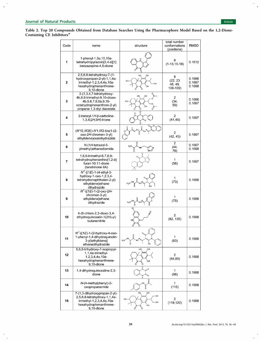
Table 2. Top 20 Compounds Obtained from Database Searches Using the Pharmacophore Model Based on the 1,2-Dione-Containing CE Inhibitorsa
Journal of Natural Products Article
dx.doi.org/10.1021/np300628a | J. Nat. Prod. 2013, 76, 36−4439

used in Chinese herbal medicine under the name Danshen, asample of the latter was obtained and the ability of extracts ofthis material to inhibit CEs was assessed. Three different
extracts were generated using hot water, acetone, or 56%ethanol. The latter was used since in Chinese medicineDanshen is frequently steeped in “Baijui”, a distilled sorghum
Table 2. continued
aScores are ranked based upon RMSD.
Table 3. Inhibition Constants for Tanshinone IIA (7) and Its Analogues with Human Carboxylesterases and Cholinesterases
Table 4. Inhibition of hiCE by Different Salvia miltiorrhiza Root Extracts
IC50 ± SE (μg/mL) IC50 ± SE (μg/mg)
extract/fraction concentration (mg/mL) o-NPA CPT-11 o-NPA CPT-11
water 41.8 4080 ± 990 ND 97.6 ± 23 NDacetone 1.13 0.16 ± 0.05 0.51 ± 0.12 0.14 ± 0.04 0.45 ± 0.10ethanol (56%) 37.1 179 ± 16 41.4 ± 9.3 4.82 ± 0.43 1.12 ± 0.25acetone fraction 16 1a 0.27 ± 0.02 0.52 ± 0.04 0.27 ± 0.02 0.52 ± 0.04
aThe dried chromatography fraction was dissolved in DMSO at 1 mg/mL.
Journal of Natural Products Article
dx.doi.org/10.1021/np300628a | J. Nat. Prod. 2013, 76, 36−4440

liquor that contains high concentrations of ethanol. Asindicated in Table 4, organic solvent extracts of S. miltiorrhizaroots contained large amounts of compounds that inhibitedboth hiCE and hCE1. Indeed, IC50 values as low as 15 ng/mgwere observed for this material. Due to the likely presence ofnumerous tanshinones in these extracts, it is not possible togenerate an accurate Ki value for enzyme inhibition. However, ifwe assume that the active components present within thesample have a molecular weight of ∼300 Da (the molecularweights of tanshinones I and IIA are 276.2 and 294.3,respectively), for the most potent extracts, this would equateto a Ki value of ∼60 nM. Since this is similar to the values seenfor the benzil and benzene sulfonamide-based hiCE inhib-itors,17,19,20 this indicates that very potent compounds arepresent within the S. miltiorrhiza root extracts.
Having established that CE inhibitors were present within S.miltiorrhiza root extracts, chromatographic separation of theacetone sample was undertaken, and the fractions obtainedwere assessed for inhibition of hiCE and hCE1 using o-NPA assubstrate. As depicted in Figure 3A, significant enzymeinhibition was observed in fractions 13−17, with peak activityoccurring in fraction 16. To evaluate the molecule(s)responsible for this inhibition, this fraction was subjected toUPLC/HRMS, and the masses of the major components in thesample were determined. As indicated in Figure 3B, tanshinoneI (21), tanshinone IIA (7), and dihydrotanshinone (22) weredetected in this material, confirming that CE inhibition is likelydue to these compounds. While we do not know the relativecontribution toward enzyme inhibition and the exact amountsof all of the inhibitory compounds in these samples, our
Figure 3. (A) CE inhibition profile of fractions obtained following chromatographic separation of an acetone extract of S. miltiorrhiza root. Fractionswere assayed with either hCE1 (blue) or hiCE (red) at a final concentration of 100 ng/mL. (B) UPLC/HRMS analysis of chromatographic fraction16. High-resolution spectra are included for the major peaks identified in the sample. (a) Dihydrotanshinone (22; HREIMS m/z 279.10210, calcd forC18H14O3, 279.10250). (b) Tanshinone I (21; HREIMS m/z 277.086445, calcd for C18H12O3, 277.08690). (c) Tanshinone IIA (7; HREIMS m/z295.13340, calcd for C19H18O3, 295.13380).
Journal of Natural Products Article
dx.doi.org/10.1021/np300628a | J. Nat. Prod. 2013, 76, 36−4441

preliminary semiquantitative MS analyses indicate that up to 10mg of tanshinone IIA is present in acetone extracts of 5 g of S.miltiorrhiza root. Hence, this herbal material may represent animportant source of tanshinones that might mediate modu-lation of esterified drug metabolism.Intracellular Inhibition of hiCE by Tanshinones and S.
miltiorrhiza Root Extracts. Having demonstrated that thetanshinones are potent CE inhibitors toward purified enzymesin vitro, their ability to effect intracellular inhibition of hiCEwas evaluated using 4-methylumbelifferone acetate (4-MUA) asa substrate. In these studies, a fluorescence-based assay wasused, as previously developed,18 which monitors the in situhydrolysis of 4-MUA in U373MG cells. If reduced substratemetabolism is observed in these assays, this indicates that theinhibitor is cell permeable and can inhibit hiCE intracellularly.As shown in Table 5, all tanshinones except tanshinone
sulfonate (25) were able to inhibit hiCE, indicating that themolecules can cross cell membranes and reduce substratehydrolysis by up to 90%, as compared to the control. Thesevalues are similar to that seen for benzil (Table 518).Furthermore, both acetone and ethanolic (not shown) extractsof S. miltiorrhiza root, as well as fraction 16 of the former, alsoexhibited intracellular inhibition of hiCE in U373MG cells(Table 5). It is not surprising that the sodium salt of tanshinonesulfonate (25) failed to inhibit in these assays since thismolecule is charged and its cell permeability is likely to be verypoor. In addition, since this was one of the least potent
compounds evaluated, it is unlikely that even if the compoundwere able to enter the cell it would effectively inhibit hiCE.On the basis of these data, it was assessed as to whether the
pure tanshinones and S. miltiorrhiza root extracts were capableof modulating irinotecan hydrolysis in vivo. We have previouslydemonstrated that benzil can reduce SN-38 hydrolysis in cellsexpressing hiCE, and this alters the sensitivity of the cells to thedrug.18 Hence, it is likely that the natural product compoundsevaluated here would have similar properties. To assess whetherthe reduction in hiCE activity generated by these compoundswould reduce the sensitivity of cells to CPT-11, growthinhibition assays were undertaken. In these studies, cellsexpressing hiCE were preincubated with either purifiedtanshinones or S. miltiorrhiza root extracts for 1 h and theGI50 values for irinotecan were determined. As indicated inTable 5, changes in cellular sensitivity to the drug wereobserved for all tanshinones and extracts, due to reducedproduction of SN-38. Indeed, with some compounds, such asdihydrotanshinone (22), the GI50 value for CPT-11 increasedas much as 10-fold as compared to cells treated with DMSO(Table 5). Hence, all of the tanshinones (with the exception of25), as well as the acetone root extract of S. miltiorrhiza, wereable to reduce the sensitivity of U373G cells expressing hiCE toirinotecan. These data further confirm that these molecules arecell permeable and can modulate SN-38 production in vivo.The metabolism of esterified drugs by CEs represents a key
process in either the activation or inactivation of numerousclinically used agents.1,5,8−10 Hence compounds that modulatethe activity of these enzymes will likely affect drug efficacy. Wehave identified a class of agents (tanshinones), present withinthe Chinese herbal medicine Danshen (S. miltiorrhiza roots),that are polycyclic compounds containing a 1,2-dione moietythat are potent human CEs inhibitors.Danshen has been recommended for use in Chinese
medicine for numerous conditions including cardiovascularand cerebrovascular disease. However, it has recently beenapproved by the FDA for clinical trials in the United Statesunder the moniker “Compound Danshen Dripping Pills”.According to the clinicaltrials.gov Web site (http://www.clinicaltrials.gov), four clinical trials are being conducted orhave been completed, in the United States using this material.The specific conditions that are being targeted in these studiesare hypertension, coronary heart problems, and polycysticovary disease. To date, the active components in “CompoundDanshen Dripping Pills” have not been identified, although invitro and in vivo experiments have ascribed several differentproperties to S. miltiorrhiza root. These include the cytotoxicityof tanshinones toward tumor cells,27−29 the modulation oflevels of oxidative species in cells treated with these agents,30−32
and specific effects on cellular biochemistry.33−35 Clearlytherefore, S. miltiorrhiza root and the compounds presentwithin the extracts generated have considerable biologicalactivity. Interestingly however, in our cell culture-based assays(either the 4-MUA or the growth inhibition experiments withirinotecan), no adverse effects were observed at 1 μM.However, at these concentrations, significant intracellularinhibition of hiCE and modulation of drug sensitivity toirinotecan were demonstrated (Table 5). This would suggestthat the levels of tanshinones present that are required to affectcell survival are considerably greater than that necessary toinhibit CEs.These results portend that individuals who may take extracts
containing S. miltiorrhiza roots (Danshen) would likely
Table 5. Intracellular CE Inhibition and Reduction ofSensitization of Cells to CPT-11 by Tanshinones and Salviamiltiorrhiza Root (Danshen) Extracts
exptnumbera sample
intracellularCE
inhibitionb
(% ± SD)
CPT-11GI50(μM)
fold change inGI50 (as
compared toDMSO controlfor respectiveexperiment)
1 none (DMSO) 1.1benzil 83.8 ± 4.7 6.0 5.4miltirone (23) 83.0 ± 3.3 8.5 7.7
2 none (DMSO) 0.9tanshinone I (21) 68.7 ± 1.1 3.3 3.9dihydrotanshinone(22)
80.8 ± 2.6 8.9 9.9
3 none (DMSO) 0.44tanshinone IIA (7) 80.8 ± 2.7 2.2 5cryptotanshinone(24)
81.0 ± 7.7 2.0 4.5
4 none (DMSO) 1.3Danshen acetoneextract (2.5 μg/mL)
88.1 ± 0.7 5.1 3.9
5 none (DMSO) 2.0fraction 16 (2.5 μg/mL)
89.4 ± 4.7 7.4 3.7
tanshinone sulfonate(25)
14.9 ± 6.7 ND
aFive independent experiments were conducted for growth inhibitionstudies due to the large number of data points generated by the cell-based assays. The differences in the GI50 values observed aredependent upon the levels of expression of hiCE. Since this varies,each independent experiment has its own DMSO control, and foldchanges are calculated from this value. bData represent percentagereduction in 4-MUA metabolism 30 s after addition of substrate.Results represent the data obtained from 3 individual experiments
Journal of Natural Products Article
dx.doi.org/10.1021/np300628a | J. Nat. Prod. 2013, 76, 36−4442

demonstrate reduced hydrolysis of esterified drugs. Sincenumerous clinically used agents contain this chemotype,potentially significant drug−drug interactions might beobserved when these compounds are combined. Clearly,changes in drug hydrolysis would depend upon thepharmacokinetics of the tanshinones, the formulation andamounts of S. miltiorrhiza roots used (e.g., organic tinctures vsherbal teas), the source of the material (e.g., crude roots vsfinely ground powders), etc. However, due to the potency ofthe inhibition that we have observed and the ubiquitous natureof the ester chemotype in many drugs, we believe that furtherinvestigations of the likelihood for drug−drug interactions arewarranted. Both in vitro and in vivo studies to assess thishypothesis are currently under way.
■ EXPERIMENTAL SECTIONGeneral Experimental Procedures. The glioblastoma line
(U373MG) that has been engineered to overexpress hiCE has beendescribed previously.18 Tanshinone I, tanshinone IIA, tanshinone IIAsulfonate, miltirone, dihydrotanshinone, and cryptotanshinone wereobtained from LKT Laboratories (St. Paul, MN, USA), ChromaDex(Irvine, CA, USA), Carbocore (The Woodlands, TX, USA), or BoscheScientific (New Brunswick, NJ, USA). Benzil, o-NPA, and 4-MUAwere obtained from Sigma Chemicals (St. Louis, MO, USA). CPT-11was a kind gift from Dr. J. P. McGovren (Pfizer, NY, USA).Human CEs (hiCE and hCE1) were purified from baculovirus-
infected Spodoptera f rugiperda cells as previously described.12,36
Human acetyl- and butyrylcholinesterase (hAChE; hBChE) werepurchased from Sigma Chemicals.For identification of components within extracts using HRMS,
samples were separated and analyzed on an Acquity UPLC coupled toa Xevo G2 QToF mass spectrometer (Waters Technology Co.,Milford, MA, USA). Identity was validated by comparison tochromatographic retention times obtained for commercially availabletanshinone standards and by HRMS.Plant Material. Dried roots of S. miltiorrhiza Bunge (lot number
6069902) were purchased from a local Chinese grocery in Memphis,Tennessee, originally provided by South Project Ltd. (Hong Kong). Avoucher specimen (#P0001) was deposited in the Department ofChemical Biology and Therapeutics, St. Jude Children’s ResearchHospital.Extraction and Isolation. Extracts of S. miltiorrhiza root
(Danshen) were generated by incubating 5 g of plant material with100 mL of solvent (water, acetone, or 56% ethanol/44% water).Different approaches (refluxing, microwave extraction), times (up to18 h), and temperatures (ambient to solvent boiling point) were used.Solutions were clarified by filtration, solvents were removed underreduced pressure, and resulting solids were then weighed and dissolvedin methanol. Extracts that demonstrated the highest potency towardthe inhibition of CEs were then subjected to chromatographicseparation using a previously published procedure.37 Briefly, followingremoval of polyphenols, compounds were separated by reversed-phasesemipreparative HPLC on a C18 column (30 × 50 mm, 110 Å, 5 μM)using a water/methanol gradient. This resulted in ∼25 fractions, whichwere dried, weighed, and dissolved in DMSO.Pharmacophore Development and Library Searches. Align-
ment of molecules and development of pharmacophore models wereachieved using MOE 2011.10 software (Chemical Computing Group,Montreal, Canada). Briefly, candidate compounds were minimized,and the Flexible Alignment module was used to overlay compoundswith the default settings, except that the logP option was selected. Thiswas chosen since an inverse correlation between clogP and inhibitorpotency has been demonstrated previously.23,25 The top scoringalignment for these studies was then used for the development of thepharmacophore.Pharmacophore models were calculated using the Pharmacophore
option in MOE with constraints developed for the CO bonds andthe carbon atoms within the 1,2-dione group. Derived models were
then used to search the Molecular Diversity Preservation Internationaldatabase (ca. 22 000 unique molecules). The results were sorted usingthe mean RMSD distances, and compounds lacking the 1,2-dionemoiety were discarded.
Enzyme Inhibition. Inhibition of CE was determined using aspectrophotometric assay with o-NPA as a substrate. Typically at least8 concentrations of inhibitor were used, and data were evaluated usingthe following multifactorial equation:
β α βα α α
=− + −
+ + +i
KK K K
[I]{[s](1 ) ( )}[I]{[s] } { [s] }
s
s i s
where i is the fractional inhibition, [I] is the inhibitor concentration,[s] is the substrate concentration, α is the change in affinity of thesubstrate for enzyme, β is the change in the rate of enzyme substratecomplex decomposition, Ks is the dissociation constant for the enzymesubstrate complex, and Ki is the inhibitor constant.12,19,20,38,39
Examination of the curve fits, where α ranged from 0 to ∞ and βranged from 0 to 1, was performed using GraphPad Prism softwareand Perl data language.19,20 Curves were generated and analyzed usingAkaike’s information criteria.40,41 Finally, using the equation generatedby Prism considered to be the best fit for the data sets, Ki values werethen calculated.19,20 For crude extracts, data are presented as IC50values (amount of sample required to reduce enzymatic activity by50%) since the amounts and molecular weights of the componentspresent within the material are unknown.
Inhibition of hAChE and hBChE were assessed using acetylthiocho-line or butyrylthiocholine as substrates, respectively.42−44
Intracellular Inhibition of hiCE. To assess intracellular inhibitionof CEs, U373MG cells expressing hiCE were exposed to 1 μMinhibitor for 1 h, and, after addition of 4-MUA to a final concentrationof 750 μM, the reduction of the production of 4-methylumbelliferonewas determined by fluorescence spectrometry.18 Data were recordedcontinuously for 2 min, and the percentage of enzyme inhibition 30 sfollowing 4-MUA addition was calculated by comparison to untreated(DMSO only) cells.18
Inhibition of CPT-11 Hydrolysis. In vitro assays to assessinhibition of CPT-11 metabolism were performed as previouslydescribed.20 CPT-11 and SN-38 were detected and quantitated usingHPLC with fluorescence detection.
Growth Inhibition Assays. Growth inhibition assays wereundertaken using previously described methods.18 In combinationstudies, inhibitors were added to cells for 1 h before application ofCPT-11. Two hours later, the medium containing drug and inhibitorwas removed and replaced, and cells were then allowed to grow for afurther 72 h. Typically, data points were determined in triplicate usingat least eight different drug concentrations. After cell counting,concentrations of CPT-11 that resulted in 50% growth inhibition(GI50) were calculated using Prism software (GraphPad, San Diego,CA, USA).
■ AUTHOR INFORMATIONCorresponding Author*Tel: 901-595-6045. Fax: 901-595-4293. E-mail: [email protected] authors declare no competing financial interest.
■ ACKNOWLEDGMENTSThis work was supported in part by NIH grants CA108775, byCancer Center Core grant CA21765, and by the AmericanLebanese Syrian Associated Charities (ALSAC) and St. JudeChildren’s Research Hospital (SJCRH).
■ REFERENCES(1) Sun, Z.; Murry, D. J.; Sanghani, S. P.; Davis, W. I.; Kedishvili, N.Y.; Zou, Q.; Hurley, T. D.; Bosron, W. F. J. Pharmacol. Exp. Ther.2004, 310, 469−476.
Journal of Natural Products Article
dx.doi.org/10.1021/np300628a | J. Nat. Prod. 2013, 76, 36−4443

(2) Grima, M.; Welsch, C.; Michel, B.; Barthelmebs, M.; Imbs, J. L.Hypertension 1991, 17, 492−496.(3) Potter, P. M.; Wadkins, R. M. Curr. Med. Chem. 2006, 13, 1045−1054.(4) Satoh, T.; Hosokawa, M. Annu. Rev. Pharmacol. Toxicol. 1998, 38,257−288.(5) Humerickhouse, R.; Lohrbach, K.; Li, L.; Bosron, W.; Dolan, M.Cancer Res. 2000, 60, 1189−1192.(6) Brzezinski, M. R.; Spink, B. J.; Dean, R. A.; Berkman, C. E.;Cashman, J. R.; Bosron, W. F. Drug Metab. Dispos. 1997, 25, 1089−1096.(7) Schwer, H.; Langmann, T.; Daig, R.; Becker, A.; Aslanidis, C.;Schmitz, G. Biochem. Biophys. Res. Commun. 1997, 233, 117−120.(8) Khanna, R.; Morton, C. L.; Danks, M. K.; Potter, P. M. CancerRes. 2000, 60, 4725−4728.(9) Tang, M.; Mukundan, M.; Yang, J.; Charpentier, N.; LeCluyse, E.L.; Black, C.; Yang, D.; Shi, D.; Yan, B. J. Pharmacol. Exp. Ther. 2006,319, 1467−1476.(10) Shi, D.; Yang, J.; Yang, D.; LeCluyse, E. L.; Black, C.; You, L.;Akhlaghi, F.; Yan, B. J. Pharmacol. Exp. Ther. 2006, 319, 1477−1484.(11) Hatfield, M. J.; Tsurkan, L.; Garrett, M.; Shaver, T.; Edwards, C.C.; Hyatt, J. L.; Hicks, L. D.; Potter, P. M. Biochem. Pharmacol. 2011,81, 24−31.(12) Hatfield, M. J.; Tsurkan, L.; Hyatt, J. L.; Yu, X.; Edwards, C. C.;Hicks, L. D.; Wadkins, R. M.; Potter, P. M. Br. J. Pharmacol. 2010, 160,1916−1928.(13) Bencharit, S.; Morton, C. L.; Howard-Williams, E. L.; Danks, M.K.; Potter, P. M.; Redinbo, M. R. Nat. Struct. Biol. 2002, 9, 337−342.(14) Bencharit, S.; Morton, C. L.; Xue, Y.; Potter, P. M.; Redinbo, M.R. Nat. Struct. Biol. 2003, 10, 349−356.(15) Wierdl, M.; Tsurkan, L.; Hyatt, J. L.; Edwards, C. C.; Hatfield,M. J.; Morton, C. L.; Houghton, P. J.; Danks, M. K.; Redinbo, M. R.;Potter, P. M. Cancer Gene Ther. 2008, 15, 183−92.(16) Hicks, L. D.; Hyatt, J. L.; Moak, T.; Edwards, C. C.; Tsurkan, L.;Wierdl, M.; Ferreira, A. M.; Wadkins, R. M.; Potter, P. M. Bioorg. Med.Chem. 2007, 15, 3801−3817.(17) Hicks, L. D.; Hyatt, J. L.; Stoddard, S.; Tsurkan, L.; Edwards, C.C.; Wadkins, R. M.; Potter, P. M. J. Med. Chem. 2009, 52, 3742−3752.(18) Hyatt, J. L.; Tsurkan, L.; Wierdl, M.; Edwards, C. C.; Danks, M.K.; Potter, P. M. Mol. Cancer Ther. 2006, 5, 2281−2288.(19) Wadkins, R. M.; Hyatt, J. L.; Wei, X.; Yoon, K. J.; Wierdl, M.;Edwards, C. C.; Morton, C. L.; Obenauer, J. C.; Damodaran, K.;Beroza, P.; Danks, M. K.; Potter, P. M. J. Med. Chem. 2005, 48, 2905−2915.(20) Wadkins, R. M.; Hyatt, J. L.; Yoon, K. J.; Morton, C. L.; Lee, R.E.; Damodaran, K.; Beroza, P.; Danks, M. K.; Potter, P. M. Mol.Pharmacol. 2004, 65, 1336−1343.(21) Djeridane, A.; Brunel, J. M.; Vidal, N.; Yousfi, M.; Ajandouz, E.H.; Stocker, P. Chem. Biol. Interact. 2008, 172, 22−26.(22) Djeridane, A.; Yousfi, M.; Nadjemi, B.; Maamri, S.; Djireb, F.;Stocker, P. J. Enzyme Inhib. Med. Chem. 2006, 21, 719−726.(23) Hyatt, J. L.; Moak, T.; Hatfield, J. M.; Tsurkan, L.; Edwards, C.C.; Wierdl, M.; Danks, M. K.; Wadkins, R. M.; Potter, P. M. J. Med.Chem. 2007, 50, 1876−1885.(24) Hyatt, J. L.; Wadkins, R. M.; Tsurkan, L.; Hicks, L. D.; Hatfield,M. J.; Edwards, C. C.; Ii, C. R.; Cantalupo, S. A.; Crundwell, G.;Danks, M. K.; Guy, R. K.; Potter, P. M. J. Med. Chem. 2007, 50, 5727−5734.(25) Parkinson, E. I.; Jason Hatfield, M.; Tsurkan, L.; Hyatt, J. L.;Edwards, C. C.; Hicks, L. D.; Yan, B.; Potter, P. M. Bioorg. Med. Chem.2011, 19, 4635−4643.(26) Hyatt, J. L.; Stacy, V.; Wadkins, R. M.; Yoon, K. J.; Wierdl, M.;Edwards, C. C.; Zeller, M.; Hunter, A. D.; Danks, M. K.; Crundwell,G.; Potter, P. M. J. Med. Chem. 2005, 48, 5543−5550.(27) Yan, M. Y.; Chien, S. Y.; Kuo, S. J.; Chen, D. R.; Su, C. C. Int. J.Mol. Med. 2012, 29, 855−863.(28) Lee, W. Y.; Chiu, L. C.; Yeung, J. H. Food Chem. Toxicol. 2008,46, 328−338.
(29) Wu, W. L.; Chang, W. L.; Chen, C. F. Am. J. Chin. Med. 1991,19, 207−216.(30) Fu, J.; Huang, H.; Liu, J.; Pi, R.; Chen, J.; Liu, P. Eur. J.Pharmacol. 2007, 568, 213−221.(31) Zhang, H. A.; Gao, M.; Zhang, L.; Zhao, Y.; Shi, L. L.; Chen, B.N.; Wang, Y. H.; Wang, S. B.; Du, G. H. Biochem. Biophys. Res.Commun. 2012, 421, 479−483.(32) Qiao, Z.; Ma, J.; Liu, H. Molecules 2011, 16, 10002−10012.(33) Xu, S.; Little, P. J.; Lan, T.; Huang, Y.; Le, K.; Wu, X.; Shen, X.;Huang, H.; Cai, Y.; Tang, F.; Wang, H.; Liu, P. Arch. Biochem. Biophys.2011, 515, 72−79.(34) Koon, C. M.; Woo, K. S.; Leung, P. C.; Fung, K. P. J.Ethnopharmacol. 2011, 138, 175−183.(35) Steinkamp-Fenske, K.; Bollinger, L.; Voller, N.; Xu, H.; Yao, Y.;Bauer, R.; Forstermann, U.; Li, H. Atherosclerosis 2007, 195, e104−111.(36) Morton, C. L.; Potter, P. M. Mol. Biotechnol. 2000, 16, 193−202.(37) Tu, Y.; Jeffries, C.; Ruan, H.; Nelson, C.; Smithson, D.; Shelat,A. A.; Brown, K. M.; Li, X. C.; Hester, J. P.; Smillie, T.; Khan, I. A.;Walker, L.; Guy, K.; Yan, B. J. Nat. Prod. 2010, 73, 751−754.(38) Wadkins, R. M.; Hyatt, J. L.; Edwards, C. C.; Tsurkan, L.;Redinbo, M. R.; Wheelock, C. E.; Jones, P. D.; Hammock, B. D.;Potter, P. M. Mol. Pharmacol. 2007, 71, 713−723.(39) Webb, J. L. Enzyme and Metabolic Inhibitors. Vol. 1. GeneralPrinciples of Inhibition; Academic Press, Inc.: New York, 1963.(40) Akaike, H. In Information Theory and an Extension of theMaximum Likelihood Principle; Second International Symposium onInformation Theory, Budapest, 1973; Petrov, B. N.; Csaki, F., Eds.;Akademiai Kiado: Budapest, 1973; pp 267−281.(41) Akaike, H. IEEE Trans. Automatic Control 1974, AC-19, 716−723.(42) Doctor, B. P.; Toker, L.; Roth, E.; Silman, I. Anal. Biochem.1987, 166, 399−403.(43) Ellman, G. L.; Courtney, K. D.; Anders, V.; Featherstone, R. M.Biochem. Pharmacol. 1961, 7, 88−95.(44) Morton, C. L.; Wadkins, R. M.; Danks, M. K.; Potter, P. M.Cancer Res. 1999, 59, 1458−1463.
Journal of Natural Products Article
dx.doi.org/10.1021/np300628a | J. Nat. Prod. 2013, 76, 36−4444