Embed Size (px)
Citation preview

MOLECULAR AND CELLULAR BIOLOGY, Apr. 1993, p. 2401-24100270-7306/93/042401-10$02.00/0Copyright C 1993, American Society for Microbiology
Modulation of Liver-Specific Transcription by Interactions betweenHepatocyte Nuclear Factor 3 and Nuclear Factor 1
Binding DNA in Close AppositionDAVID A. JACKSON, KATHLEEN E. ROWADER, KIMBERLY STEVENS, CHENGYU JIANG,
PATRICE MILOS, AND KENNETH S. ZARET*
Section ofBiochemistry, Box G, Brown University, Providenice, Rhode Island 02912
Received 25 November 1992/Returned for modification 4 January 1993/Accepted 18 January 1993
The liver-specific enhancer of the serum albumin gene contains an essential segment, designated eH, whichbinds the hepatocyte nuclear factor 3a (HNF3a) and ubiquitous nuclear factor 1/CCAAT transcription factor(NF1/CTF) proteins in tight apposition. We previously showed that activation of transcription by the eH sitewas correlated with an increase in intracellular HNF3ae levels during the in vitro differentiation of the hepaticcell line H2.35. We now show that transfection of an HNF3oa cDNA expression vector into dedifferentiatedH2.35 cells is sufficient to induce transcription from the eH site. Mutational analysis of the enhancerdemonstrates that NF1/CTF cooperates with HNF3a to induce enhancer activity. However, when the eH siteis removed from the context of the enhancer, NF1/CTF can inhibit transcriptional activation by HNF3a. Weconclude that the ternary complex of HNF3a, NF1/CTF, and the eH site forms a novel, composite regulatoryelement that is sensitive to the local DNA sequence environment and suggest that the transcriptionalstimulatory activity ofNF1/CTF depends on its higher-order interactions with other proteins during hepatocytedifferentiation.
Cellular differentiation is governed by the binding oftranscription factors to DNA in specific combinations, oftenleading to the formation of large arrays of protein-DNAcomplexes. Interactions between different transcription fac-tors bound to adjacent sites can lead to striking changes intheir respective activities. For example, the yeast proteinMCM1 (PRTF) activates a-cell-specific promoters whenbound adjacent to the al transcription factor, but MCM1represses a-cell-specific promoters when bound next to thea2 protein (1, 29). In this report, we study the interactionsbetween two liver transcription factors when they are tightlyjuxtaposed at a site on DNA and explore how the geneticcontext of the proteins' DNA binding sites governs theirinteractions during hepatic differentiation.Serum albumin gene transcription is an excellent marker
for liver differentiation in mammals because it is activatedearly during liver development (8, 49, 51, 53) and increasesto a rate 1,000-fold greater in the liver than in other tissues(33, 44). Tissue specificity is controlled in part by an en-hancer element that lies 10 kb upstream of the transcriptionstart site and functions selectively in the liver of transgenicmice (43). The albumin enhancer contains three sites, des-ignated eE, eG, and eH, that are essential for enhanceractivity in various hepatocyte-derived cell lines, such asH2.35 cells (24, 25, 34). The eE site binds liver-enrichedC/EBP-related proteins (6, 11, 56), and the eG site binds afamily of transcription factors, hepatocyte nuclear factor 3a(HNF3a), HNF3,, and HNF3y, expressed in liver and lung(31). The HNF3 family contains a conserved DNA bindingdomain found in the developmentally important Drosophilafork head protein (54). The eH site was defined by a largeDNase I footprint with nuclear extracts from mouse liverand H2.35 cells (34, 58); only a portion of the eH sequence
was protected in kidney and spleen extracts, suggesting theinvolvement of a tissue-restricted factor.The eH site, like the albumin enhancer itself, responds to
extracellular signals that control hepatocyte differentiation(13). When freshly isolated hepatocytes are cultured in thepresence of serum and on a plastic substratum, they rapidlydedifferentiate, and the transcription of liver-specific genesdeclines dramatically (9). In H2.35 cells cultured under theseconditions, the eH site footprinting activity is at low abun-dance, and neither the albumin enhancer nor multimers ofthe eH site stimulate the albumin promoter from 800 bpupstream. However, when hepatocytes are cultured in se-rum-free medium and on extracellular matrix gels, theymaintain higher levels of liver-specific gene transcription (3,7, 15, 17), and these conditions increase eH site footprintingactivity and activate both the intact enhancer and multimersof the eH site in H2.35 cells (13, 34). By contrast, multimersof the enhancer eG HNF3 binding site function as enhancersin H2.35 cells even when the cells are cultured in serum andon a plastic substratum (13). Thus, understanding the controlof the eH site would provide insight into the regulation ofliver gene expression and differentiation.
In this study, we identify two transcription factors thatbind simultaneously to the eH site, creating a compositeregulatory element with properties that differ from those ofeither factor bound alone. These findings explain key fea-tures of the regulation of transcription by a liver-specificenhancer and further reveal how the genetic context of thefactors' binding sites can govern whether one of the factorsinhibits or augments liver-specific transcription.
MATERIALS AND METHODS
Preparation of nuclear extracts and in vitro translations.Mouse liver nuclear extracts were prepared by the method ofGorski et al. (21, 32), with minor modifications describedelsewhere (38). H2.35 cell nuclear extracts were prepared for* Corresponding author.
2401
Vol. 13, No. 4
2402 JACKSON ET AL.
TABLE 1. Sequences of eH site oligonucleotides
Oligonucleotide Sequencea
eH-WVT.................... CCGAACGTGTTTGCCTTGGCCAGTTTTCCATGTACATGCAeH-M4 (TGT3-).....................CCGAACGACCA TCACTTGGCCAGTTTTCCATGTACATGCAeH-M5 (NF1+).................... CCGAACGTGTTTGCCTTGGCCAGTTAGGT_GACACATGCAeH-M7 (NF1-).................... CCGAACGTGTTTGCCTATATCAGTTTTA9TTGTACATGCAeH-M4/M7 (TGT3-, NF1-) ....................CCGAACGACCATCACTATATCAGTTTTAGCTGTACATGCA
a Conserved TGT3 and NF1/CTF binding sequences are shown in bold; mutated sequences are underlined.
electromobility shift assays (EMSAs) as described previ-ously (13); for immunoblots, nuclei were lysed in sodiumdodecyl sulfate (SDS) sample buffer (47) and sonicated.HNF3ac and nuclear factor 1 (NF1) proteins were translatedin vitro from their respective cDNAs (30, 36) in micrococcalnuclease-treated reticulocyte lysates as recommended by themanufacturer (Promega Biotec).EMSAs. EMSAs were performed as previously described
(13), with the following modifications. Nuclear extracts wereadded to binding reactions at 2 ,ug per reaction in thepresence of 5 ,ug of bovine serum albumin and 80 to 100 ngof poly(dI-dC), depending on the particular extract prepara-tion. After 10 min, oligonucleotide probe, with or withoutunlabeled competitor oligonucleotide, was added to thebinding reaction, and incubation was continued for 20 min.One microliter of reticulocyte lysate translation product wasadded to binding reactions unless indicated otherwise. Thereticulocyte lysate contained a nonspecific binding activitywhich was competed for by the inclusion of 35 to 150 ng ofsonicated salmon sperm DNA per reaction. Binding reac-tions using purified NF1 were performed by using theamounts of protein and nonspecific competitor indicated inthe figures and in the presence of 5 ,ug of bovine serumalbumin.
Oligonucleotides and probes. Except for the TTR (mousetransthyretin gene) oligonucleotide (10) and eH-M4, thedouble-stranded oligonucleotides used were synthesized ascomplementary single strands spanning the sequencesshown in Table 1 (top strand is shown). In addition to thesequences shown, double-stranded oligonucleotides con-tained 5' extensions that created the sequence GCT at eitherend after fill-in with Klenow DNA polymerase. eH-M4 wascreated by annealing a 26-bp oligonucleotide containing theTGT3 sequence mutation to a 29-bp wild-type bottom strandand filling in the single-stranded regions with Klenow en-zyme. Oligonucleotide probes were labeled by filling in 5'extensions with [0t-32P]dATP or [ct-32P]dCTP and Klenowpolzrmerase or by incubating filled-in oligonucleotides with[y- 2P]ATP and polynucleotide kinase.
Antibodies and immunoblotting. Rabbit antibody that rec-ognized the amino termini of HNF3aL and HNF3I wasgenerously provided by E. Lai (30). Rabbit antibody againstbacterially expressed human NFl/cellular transcription fac-tor 1 (CTF1) was a gift from N. Tanese and R. Tjian. ForEMSAs, 1 VI of specific antiserum or normal rabbit serumwas added to binding reactions as described above. Forimmunoblots, proteins were boiled in SDS sample buffer,electrophoresed in a reducing SDS-polyacrylamide gel,transferred to nitrocellulose filters, exposed to a 1:750 dilu-tion of the NF1/CTF antiserum, and subsequently incubatedwith 125I-protein A, using standard protocols (47).
Purification of NFl. A pig liver was minced and exten-sively washed in phosphate-buffered saline on ice, macer-ated with a tissue grinder, and homogenized in a blender inthe tissue homogenization buffer of Fritton et al. (16) con-
taining 0.45 M sucrose, 1 ,ug each of soybean trypsininhibitor, leupeptin, and antipain per ml, 100 ,uM benzami-dine, and 1 mM phenylmethylsulfonyl fluoride. Followinghomogenization, nuclei were pelleted through a 0.6 M su-crose cushion, resuspended, and washed once with tissuehomogenization buffer. The nuclei were again collected bycentrifugation, and the pellet was resuspended by Douncehomogenization in buffer C of Dignam et al. (12) containingprotease inhibitors as specified above. After a 30-min incu-bation on ice, the nuclei were pelleted, and the protein in thesupernatant was precipitated with (NH4)2SO4. The precipi-tate was collected by centrifugation, dissolved in the bufferD of Dignam et al. (12), and dialyzed against a large volumeof the same buffer. Approximately 8 g of protein wasrecovered.Two grams of the crude extract was fractionated on a
20-ml heparin-agarose column (IBF) and eluted with KCl insteps. The majority of the eH binding activity eluted at 400mM KCl. After dialysis against buffer D, 24 mg of thisfraction was passed over a 5-ml DEAE-Sepharose column;the 14-mg flowthrough fraction contained the bulk of the eHbinding activity separated from a nuclease activity. TheDEAE flowthrough (11.5 mg) was fractionated on a double-stranded DNA-cellulose column by step elution with KCl.The eH binding activity eluted as a sharp peak (880 jig) at the400 mM KCl step. The enrichment of the NF1 DNA bindingactivity was approximately 400-fold.
Plasmid constructs. Single copies of double-stranded oligo-nucleotides (see Table 1) were inserted at a site 54 bpupstream of the albumin transcription start site in plasmidpAN6 (38), which contains albumin sequences up to the +8position, fused to the neomycin resistance gene. The struc-tures of all inserts were confirmed by DNA sequencing. ThepRSV-tk internal transfection control plasmid has beendescribed elsewhere (57). The HNF3a expression plasmidwas created by inserting the 1.7-kb EcoRI cDNA fragment(30) into plasmid pRG, which contains the Rous sarcomavirus (RSV) long terminal repeat as a promoter and thebovine growth hormone polyadenylation site downstream(provided by Mitchell Reff, Smith Kline Beecham Labora-tories). The NF1- version of the albumin enhancer inpAT2-NA (34) was created by using one of the eH-M7oligonucleotides with the polymerase chain reaction-basedmethod described in reference 58; DNA sequencing showedthat the plasmid contained the expected M7 base pairchanges except the A-to-C transversion in the second NF1half-site (Table 1).
Transfection of H2.35 cells. H2.35 cells (6.5 x 10⁵ to 10 x105) were seeded onto 60-mm-diameter plastic dishes at 33°Cin Dulbecco's modified Eagle's medium (GIBCO) containing4% fetal bovine serum (HyClone), 2 x 10⁻⁷ M dexametha-sone, and 100 U each of penicillin and streptomycin per ml.One day later, 3 jig of pAN6-based vector, 0.1 jig of pRT1,and 3 jig of pRG-HNF3a or pRG control vector, for a totalof 6 to 8 jig of DNA per dish, were transfected into the cells
MOL. CELL. BIOL.
INTERACTIONS BETWEEN LIVER TRANSCRIPTION FACTORS 2403
eH eH-M4 eH-M5 eH-M7 probe
-R- xse+, i +R-A iNI R< compelitor-A\ \-Z- -"\~
H/N[ E
h- , 11 ]H
1 2 3 4 5 6 7 8 9 10 11 12FIG. 1. Evidence that HNF3 and NF1/CTF form a ternary
complex with the albumin enhancer eH site. EMSAs were per-formed with mouse liver nuclear extracts and wild-type and mutatedeH site probes, as listed on the top row (see Table 1). The followingunlabeled competitor oligonucleotides were added at a 75-fold molarexcess (second row): TTR, HNF3 site (10); and NFlb, NF1/CTFsite (2). Protein-DNA complexes H/N, N, and H are indicated at thesides of the autoradiograph; free probe migrated to the bottom of thegel. The specific activity of the eH probe was about half of that forthe other probes. The band migrating slightly faster than H in lanes4 to 6 is nonspecific.
by the calcium phosphate procedure, using a kit fromBRL/GIBCO. Alternatively, the pAT2-NA plasmids weretransfected with pRT1, and 18 h later the cells were subcul-tured onto plastic and collagen gel substrata as describedpreviously (13). Two days later, total cellular RNA wasisolated and experimental and control transcripts were sub-jected to quantitative primer extension analysis, using radio-actively labeled neo- and tk-specific primers simultaneouslyin the same reaction (34). Primer extension products weredisplayed on DNA sequencing gels with size standardsconsisting of dideoxy sequencing reactions initiated with theneo primer. Dried gels were exposed to Kodak XAR or FujiRX film in the presence of intensifying screens. Differentexposures of autoradiographs (12 to 72 h) were scanned withan LKB laser densitometer, test signals were normalized tothe RSV internal control in the same lane, and data fromdifferent experiments were subjected to statistical analysis.
RESULTS
Distinct TGT3 and NF1 sequence motifs and binding activ-ities at the eH site. In EMSA, an eH site oligonucleotideprobe produces a complex pattern of bands with mouse livernuclear extracts (13, 34) (Fig. 1, lane 1), suggesting that thesite is bound by more than one protein. Inspection of the eHsequence revealed the TGT3 motif (50) (Table 1), TGTTTGC, which occurs in regulatory elements of diverse liver-specific genes (50, 57) and which, in some cases, bindsHNF3 proteins (14, 40). Two base pairs downstream ofTGT3 is the recognition sequence for the NF1/CTF family oftranscription factors, TGGCN7CCA (5, 18, 36, 41, 46, 48).Mutational substitution of the TGT3 motif in eH (eH-M4)caused a loss of both the lowest-mobility species (designatedH/N) and the highest-mobility species (designated H) of the
c,,-HNF3 o(- NF1
- N - N
H/N['i.; --A
N [ !!w1
*. .
j ]Ab-NF1
1 2 3 4 5 6FIG. 2. Antibody (Ab) recognition of HNF3 and NF1/CTF pro-
teins in the H/N complex. Binding reactions were performed as forFig. 1 except that either immune serum (I) against HNF3a andHNF30 (lane 3) or against NF1 (lane 6) or nonimmune serum (N;lanes 2 and 5) was added to the binding reactions. Anti-HNF3antibody (a-HNF3) was added to the binding reaction 9.5 min priorto the addition of probe; anti-NFl antibody (a-NFl) was added 5min after the addition of probe. Anti-HNF3 antibody disrupted theformation of the H/N complex, and anti-NF1 supershifted both H/Nand N complexes. Free probe is not shown.
EMSA pattern (Fig. 1, lane 4). A known HNF3 binding sitefrom the mouse 'l'R gene (10) produced the same effectwhen used as a competitor in a binding reaction with thewild-type eH site oligonucleotide (lane 2). Mutational sub-stitution of the 3' half of the NF1/CTF consensus (eH-M5)led to a reduction of both the H/N complex and the centralcluster of bands and to an increase in the H band (N; lane 7);an NF1/CTF binding site from the hepatitis B virus enhancer(NFlb [2]) produced the same effect when used as a com-petitor with the wild-type eH site (lane 3). Mutation of bothNF1/CTF half-sites (eH-M7) completely eliminated the H/Nand N complexes and led to a sharp increase in the Hcomplex, which was competed for by an HNF3 site oligo-nucleotide. The increase in HNF3 binding to the eH-M7oligonucleotide appears to be due to sequence changes in theNF1 half-site closest to the TGT3 motif, because the strongincrease in binding was not observed with the eH-WT probein the presence of the NF1/CTF competitor (lane 3). TheeH-M7 sequence changes may affect the selectivity or affin-ity of the site for particular HNF3 family members (31). Inconclusion, HNF3 and NF1/CTF proteins in liver nuclearextracts appear to bind to the TGT3 and NF1 motifs of theeH site, respectively.HNF3 and NF1/CTF proteins form a ternary complex with
eH site DNA in liver nuclear extracts. To establish definitivelythat HNF3 and NF1 proteins form a ternary complex withthe eH site in liver nuclear extracts, we added antiseraspecific for HNF3aL and HNF3I (31), and for NF1/CTF, toeH site binding reactions. Anti-HNF3 antibody caused theselective loss of the low-mobility H/N complex withoutmarkedly affecting the central NFl-specific cluster of bands(Fig. 2, lane 3). Addition of anti-NF1/CTF antiserum causedthe loss of the NF1 bands and the H/N band, concomitantwith the formation of a supershifted band (designated Ab-NF1; lane 6). In contrast, the presence of normal rabbitserum in the binding reactions did not significantly alter thebinding patterns (lanes 2 and 3). We conclude that HNF3 andNF1/CTF proteins in liver extracts can bind simultaneouslyto the eH site to form the H/N ternary complex.
VOL. 13, 1993
2404 JACKSON ET AL.
AeH probe
- e\ comp
Banti-NF1
NF1b eH probe,,'- N A - N A serum
U ]Ab-NF1
CeH probe
- 01 0.7 1&3 ug fraction C-.'- 1.
M H M H - M H - M H - r lysate
a* HNF3/NF1
_> ]~~~~~
HNF3
a ]NF1 ]NF1
1 2 3 4
mm
1 2 3 4 5 6 7 8 9 10 i11 2 3 4 5 6
D e H
C
025 050 075
M H M H M H M
eH-M4 NFlb probeB C fraictio
100 Lil r lys
H H M H M H M H ri ysat
D n (07 ug)
ate
te
I HNF3!/NF1
j HNF3
NF1
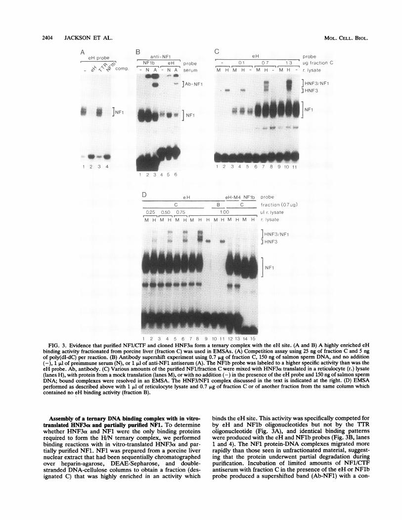
1 2 3 4 5 6 7 8 9 10 11 12 13 14 15FIG. 3. Evidence that purified NF1/CTF and cloned HNF3a form a ternary complex with the eH site. (A and B) A highly enriched eH
binding activity fractionated from porcine liver (fraction C) was used in EMSAs. (A) Competition assay using 25 ng of fraction C and 5 ngof poly(dI-dC) per reaction. (B) Antibody supershift experiment using 0.7 SLg of fraction C, 150 ng of salmon sperm DNA, and no addition(-), 1 ,ul of preimmune serum (N), or 1 pl of anti-NF1 antiserum (A). The NFlb probe was labeled to a higher specific activity than was theeH probe. Ab, antibody. (C) Various amounts of the purified NFl/fraction C were mixed with HNF3a translated in a reticulocyte (r.) lysate(lanes H), with protein from a mock translation (lanes M), or with no addition (-) in the presence of the eH probe and 150 ng of salmon spermDNA; bound complexes were resolved in an EMSA. The HNF3/NF1 complex discussed in the text is indicated at the right. (D) EMSAperformed as described above with 1 ,ul of reticulocyte lysate and 0.7 ,ug of fraction C or of another fraction from the same column whichcontained no eH binding activity (fraction B).
Assembly of a ternary DNA binding complex with in vitro-translated HNF3ae and partially purified NFl. To determinewhether HNF3a and NF1 were the only binding proteinsrequired to form the H/N ternary complex, we performedbinding reactions with in vitro-translated HNF3a and par-tially purified NFl. NF1 was prepared from a porcine livernuclear extract that had been sequentially chromatographedover heparin-agarose, DEAE-Sepharose, and double-stranded DNA-cellulose columns to obtain a fraction (des-ignated C) that was highly enriched in an activity which
binds the eH site. This activity was specifically competed forby eH and NFlb oligonucleotides but not by the TTRoligonucleotide (Fig. 3A), and identical binding patternswere produced with the eH and NFlb probes (Fig. 3B, lanes1 and 4). The NF1 protein-DNA complexes migrated more
rapidly than those seen in unfractionated material, suggest-ing that the protein underwent partial degradation duringpurification. Incubation of limited amounts of NF1/CTFantiserum with fraction C in the presence of the eH or NFlbprobe produced a supershifted band (Ab-NF1) with a con-
I NF1
MOL. CELL. BIOL.
INTERACTIONS BETWEEN LIVER TRANSCRIPTION FACTORS 2405
current reduction in the NFl-like bands (Fig. 3B, lanes 3 and6), indicating that at least the bulk of the binding activity isNF1/CTF.
Next, in vitro-translated HNF3a was mixed with theNFl-containing fraction C in the presence of excess eHprobe. The appearance of a specific binding complex formedwith the HNF3at translation product, and not with a mock-translated reticulocyte lysate, demonstrates definitively thatHNF3ot binds the eH site (Fig. 3C, lanes 1 and 2). Uponaddition of increasing amounts of NF1, there was a progres-sive increase in the formation of a new, slowly migratingcomplex (HNF3/NF1; Fig. 3C, lanes 4, 7 and 10). Indeed,the amount of HNF3/NF1 complex eventually exceeded theamount of HNF3ao-DNA complex that formed in the absenceof NF1, indicating that the presence of NF1 increased thetotal amount of HNF3a bound. The formation of the HNF3/NF1 complex also increased progressively when increasingamounts of HNF3a were added to a constant amount of NF1(Fig. 3D, lanes 2, 4, 6, and 8). The HNF3/NF1 complex wasnot detected when mock-translated reticulocyte lysate wasadded to the binding reaction (lanes M) or when HNF3ao wasmixed with protein eluted from the DNA-cellulose column ata different salt step from NF1 (fraction B; Fig. 3D, lane 11),indicating that production of this species depends on bothNF1 and HNF3a. Formation of the complex also requiredthe presence of binding sites for both factors, as no low-mobility species was detected when either the eH-M4 orNFlb oligonucleotide was used as a probe (Fig. 3D, lanes 13and 15). We conclude that HNF3a and NF1/CTF togetherare sufficient to form a ternary complex with the albumin eHsite.NF1/CTF binding inhibits activation by HNF3a at the eH
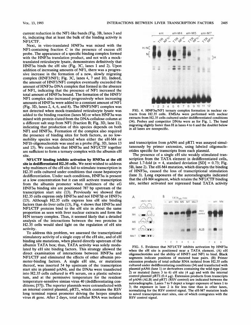
site in dedifferentiated H2.35 cells. We next wished to addresswhy multimers of the eH site fail to stimulate transcription inH2.35 cells cultured under conditions that cause hepatocytededifferentiation. Under such conditions, HNF3a is presentat a low concentration but it can still activate transcriptionfrom the albumin promoter when multimers of the eGHNF3cx binding site are positioned 787 bp upstream of thetranscription start site (13). Previously we showed thatH2.35 cells express only HNF3a and not HNF30 or HNF3-y(13). Although H2.35 cells express less eH site bindingfactors than do liver cells (13), Fig. 4 shows that HNF3ct andNF1/CTF proteins bind to the eH site in about the sameproportion as seen with liver nuclear extracts and form theH/N ternary complex. Thus, it seemed likely that a detailedanalysis of the interactions between the two proteins inH2.35 cells would shed light on the regulation of eH siteactivity.To address this problem, we assessed the transcriptional
stimulatory activity of a single copy of the eH site, and of eHbinding site mutations, when placed directly upstream of thealbumin TATA box; thus, TATA activity was solely modu-lated by eH site binding factors. This strategy allowed thedirect examination of interactions between HNF3aL andNF1/CTF and eliminated the effects of other albumin pro-moter-binding factors. A single eH site, or mutationsthereof, was inserted 54 bp upstream of the transcriptionstart site in plasmid pAN6, and the DNAs were transfectedinto H2.35 cells cultured in 4% serum, on a plastic substra-tum, and at the permissive temperature for the residenttemperature-sensitive simian virus 40 (dedifferentiating con-ditions; [57]). The reporter plasmids were cotransfected withan internal control plasmid, pRT1, which contains the RSVlong terminal repeat promoter driving the herpes simplexvirus tk gene. After 2 days, total cellular RNA was isolated
eH eH-M4 eH-M5 eH-M7
H/NE
, IH
1 2 3 4 5 6 7 8 9 10 11 12FIG. 4. HNF3a/NF1 ternary complex formation in nuclear ex-
tracts from H2.35 cells. EMSAs were performed with nuclearextracts from H2.35 cells cultured under dedifferentiated conditions(34). Probes and competitor DNAs were as for Fig. 1. The bandmigrating slightly faster than H in lanes 4 to 6 and the doublet belowin all lanes are nonspecific.
and transcription from pAN6 and pRT1 was assayed simul-taneously by primer extension, using labeled oligonucle-otides specific for transcripts from each plasmid.The presence of a single eH site weakly stimulated tran-
scription from the TATA element in dedifferentiated cells,about 1.7-fold (n = 4, standard deviation [SD] = 0.73; Fig.SB, lane 2). The eH-M4 mutation, which disrupts the bindingof HNF3a, caused the loss of transcriptional stimulation(lane 3). Long exposures of the autoradiographs indicatedthat the eH-M4 segment, which retains the NF1/CTF bindingsite, neither activated nor repressed basal TATA activity
AHNr3 NF1
eH
MA - -i
M57
M7 117'.°11:X
B TATAaibflEo-- eH M4 M5 M7 M4.7 -- eH M4
* * sm to tcofrol
o ALB aM4:7 ' -Al:-
1 2 3 4 5 6 8 9
FIG. 5. Evidence that NF1/CTF inhibits activation by HNF3awhen the eH site is positioned near a TATA element. (A) eHoligonucleotides and the ability of HNF3 and NF1 to bind. Blacksegments indicate positions of mutated base pairs. (B) Primerextension products of total cellular RNA isolated from H2.35 cellscultured under dedifferentiating conditions (34) and transfected withplasmid pAN6 (lane 1) or derivatives containing the wild-type (lane2) or mutated (lanes 3 to 6) eH site (4 ,ug) and with the internalcontrol plasmid pRT1 (0.4 ,ug). Extension products from transcriptsof pAN6 (ALB) and pRT1 (RSV control) are indicated between theautoradiographs. Lanes 7 to 9 depict a longer exposure of lanes 1 to3; the exposure in lane 2 is for less time than in other lanes,normalizing for the RSV control signal. The eH-M7 mutation leadsto novel transcription start sites, one of which comigrates with theRSV control signal.
VOL. 13, 1993

2406 JACKSON ET AL.
I'A A!a' -fnlo
eH M4 wop-JC M5 N.;'-+ - + C.o-trarsfac!rcrHINF30(RaS3-.- * il P wco ii,
Alb Promoter
plastic collagenWI NF1 WI NF1- < Alb enhancer
RSVam41%ww- -~ control
;4 Al B
2 3 4
FIG. 6. Activation of the eH site in dedifferentiated H2.35 cellsby cotransfected HNF3a expression vector. pRG-HNF3a expres-sion vector (lanes 2, 4, and 6) or the pRG control vector (lanes 1, 3,and 5) (4 pLg of each) was cotransfected with pAN6 derivatives asshown (4 ,ug) and the RSV control plasmid (0.4 ,ug). Primerextension products of the resulting RNAs are shown. ALB, albu-min.
(lane 9). Strikingly, the eH-M5 mutation, which reducesbinding of NF1/CTF but not of HNF3ax, increased transcrip-tion 2.5-fold more than did eH-WT (n = 4, SD = 0.28; lane4). The eH-M7 mutation, which eliminates NF1 binding,further increased transcription from the normal transcrip-tional start site, and from various other sites up to 15nucleotides upstream, a total of 3.5-fold more than dideH-WT (n = 3, SD = 1.8; lane 5). Most of these start sitesare evident on long exposures of the wild-type template (lane7). An abundant primer extension product arises from a startsite 15 bp upstream of the normal site and comigrates withthe pRT1 product (data not shown); the use of these up-stream initiation sites may be controlled by an A/T-richregion 15 bp upstream of the albumin TATA box in plasmidpAN6-eH-M7.
It is possible that the different transcription start sites withthe eH-M7 plasmid could be due to the creation of a bindingsite for an unexpected protein, although no novel bindingactivities were discovered in an EMSA (Fig. 1 and 4, lanes9). To address this possibility further, we tested the eH-M4/7sequence, which disrupts both the TGT3 and NF1 motifs;transcriptional stimulation was reduced to the eH level (n =2, SD = 0.4; Fig. 5, lane 6). Considering the results withtemplates derived from both the eH-M5 and eH-M7 muta-tions, we conclude that in dedifferentiated H2.35 cells,HNF3a can activate transcription from the eH site TGT3motif but that it is inhibited from doing so by the binding ofNFl.
Increased expression of HNF3a can activate the eH site indedifferentiated H2.35 cells. The inverse relationship be-tween NF1/CTF binding and transcriptional activation byHNF3a suggested that increasing the fraction of eH sitesoccupied by HNF3ao alone, by increasing the cellular con-centration of the protein, should increase transcriptionalactivity. This model could account for the increased activityof multimers of the eG and eH sites, and the albuminenhancer itself, during hepatic differentiation, when theconcentration of HNF3at increases (13, 34) but that ofNF1/CTF does not (see below). To test this model directly,we transfected H2.35 cells cultured under dedifferentiatingconditions with the HNF3a expression vector pRG-HNF3a,or the pRG plasmid lacking the HNF3ao insert, and with asubset of the pAN6 plasmids and pRT1. Cotransfection ofpRG-HNF3a reproducibly caused a 4.1-fold stimulation (n =3, SD = 1.4) of pAN6-eH transcription, whereas the pRGcontrol vector had no effect (Fig. 6, lane 2). The stimulatory
ALB
1 2 3 4
FIG. 7. Evidence that NF1/CTF binding to the eH site is re-quired for the activity of the albumin (Alb) enhancer. H2.35 cellswere transfected on plastic substrata with pAT2-NA (34) containingthe wild-type albumin enhancer (WT) or with pAT2-NA/M7, con-taining a mutation of the NF1 site (NF1-), and the control plasmidpRT1. After 18 h, the cells were released from the plates andsubcultured either on plastic or on collagen gels. Two days later,RNA was isolated from the cells and subjected to the primerextension assay.
effect of HNF3a was dependent upon an intact TGT3 motif(eH-M4; lane 4) and persisted when NF1 binding wasweakened in eH-M5 (3.5-fold; n = 2, SD = 1.8; lane 6).These findings demonstrate that the eH TGT3 motif repre-sents a functional binding site for HNF3a and that increasedexpression of HNF3o is sufficient to activate the albumin eHsite in dedifferentiated H2.35 cells.NF1/CTF binding is required for albumin enhancer activity
in differentiated H2.35 cells. The aforementioned resultssuggest a simple model for the role of NF1 during hepatocytedifferentiation. In dedifferentiated cells, bound NF1 inhibitsactivation by HNF3, blocking enhancer activity that couldbe mediated by low amounts of liver-enriched transcriptionfactors. During differentiation, increased expression ofHNF3aL increases the chance that the eH site is occupied bythis factor alone, contributing to an active enhancer. There-fore, disruption of the NF1 site in the intact enhancer shouldlead to greater transcriptional stimulatory activity. To testthis hypothesis, we introduced the eH-M7 sequence into an830-bp enhancer segment that contains the essential eE, eG,and eH sites described above (34). This NF1- version of theenhancer and the wild-type segment were placed 787 bpupstream of the albumin transcription start site in the testplasmid pAT2 (34). These plasmids and the pRT1 controlwere transfected into H2.35 cells cultured on a plasticsubstratum; the cells were subsequently subcultured ontoplastic and collagen gel substrata; the latter condition acti-vates the albumin enhancer (13).These experiments led to an unexpected finding. As seen
in Fig. 7 (lane 2), mutation of the NF1 binding site did notlead to activation of the albumin enhancer in H2.35 cellscultured on plastic. This finding suggests that in dedifferen-tiated cells, there may be insufficient amounts of HNF3a orother enhancer binding proteins for enhancer activity. Asexpected, the wild-type enhancer stimulated the albuminpromoter in the cells cultured on collagen (lane 3). However,contrary to the prediction of the model described above,mutation of the NF1 binding site did not increase enhanceractivity; rather, enhancer activity was abolished (lane 4).Thus, in the context of the albumin enhancer, binding of
MOL. CELL. BIOL.

INTERACTIONS BETWEEN LIVER TRANSCRIPTION FACTORS 2407
A. NF1 Inhibits Activation by HNF3at a TATA-Proxirnal Site
7HNF3 NFI
eHNF1
B. NFI Cooperates with HNF3 in theContext of the Albumin Enhancer
? LAP/CRP2 HNF3 HNF3 NFIeD eE eG eH
1 2 3 4
FIG. 8. Immunoblot analysis of NF1/CTf proteins. Proteinsfrom an in vitro transcription/translation reaction with an NF1/CTFcDNA (36) (lane 1) or 75-,ug amounts of protein from liver nuclearextracts (lane 2) and from nuclear extracts of H2.35 cells cultured oneither plastic (lane 3) or collagen gel (lane 4) substrata wereseparated by SDS-polyacrylamide gel electrophoresis. After trans-fer to nitrocellulose, NF1/CTF proteins were visualized with an
NFl/COF-specific antibody and 1"I-protein A.
NF1/CTF is required for transcriptional induction duringhepatic differentiation.
Considering that different NF1/CTF species might possessdistinct regulatory properties during differentiation, we nextwished to determine whether different NF1/CGT familymembers are expressed in H2.35 cells on plastic versuscollagen gel substrata. When nuclear proteins were analyzedby immunoblotting with the NF1/TEF-specific antibody, thedetected antigens in H2.35 cells and mouse liver migratedtogether as a smear (Fig. 8, lanes 2 to 4), which could beresolved to three bands in some experiments. NF1/CTFproteins from all cellular samples migrated more slowly thanNF1 translated in a reticulocyte lysate (lane 1), suggestingthat the protein is modified posttranslationally in vivo. Nochange in the distribution or amount of the NFl/CTF specieswas evident when H2.35 cells were shifted from plastic to acollagen gel substratum. While these experiments could notdetect subtle changes in NF1/CTF species, they support thehypothesis that it is the induction of HNF3 during hepaticdifferentiation, not of NF1/CTF, that is a critical switch forenhancer activation.
DISCUSSION
Regulation of transcription by the albumin enhancer eHsite provides a useful model for dissecting the mechanisms ofhepatocyte differentiation. The eH site is essential for theactivity of the enhancer, by itself it functions only inhepatocyte-derived cells, and it is sensitive to cues thatactivate the transcription of a variety of liver-specific genes(13, 34). We found that the eH site binds and is activated bythe liver transcription factor HNF3a, which accounts for thesite's liver-specific activity. We also found that the eH sitebinds members of the NF1/CTF family of ubiquitous tran-scription factors. The interaction between HNF3a and NF1at the eH site creates a composite regulatory element thatpermits the modulation of liver-specific transcription.At an isolated eH sequence next to the TATA element,
NF1/CT1F inhibits transcriptional activation by HNF3a (Fig.9A). To our knowledge, this is the first instance in whichNFl-related proteins have been shown to negatively regulatetranscription. It is unlikely that they do so, in this case, bydirectly occluding the binding of TATA-binding protein
FIG. 9. Model for transcription factor interactions at an isolatedeH site (A) or in the context of the albumin enhancer (B). The largearrow in panel B indicates that the factors work cooperatively toenhance transcription from the promoter.
because the NF1 binding site is upstream by four turns of theDNA helix and because the NF1 binding site alone (pAN6-eH/M4) did not reduce basal TATA activity. The lack ofeffect of eH multimers distal to TATA in HeLa cells (13),which lack HNF3, further demonstrates that NFl does notgenerally repress transcription of the albumin promoter.Multimers of the albumin enhancer eG site, which bind onlyHNF3, function as an enhancer at a distal position indedifferentiated cells, whereas multimers of the eH site donot (13); hence, NF1 may also be inhibitory to HNF3 atisolated eH elements distal to TATA. The increased activityof pAN6-eH in dedifferentiated cells when an HNF3a ex-pression vector is cotransfected likely reflects increasedoccupancy of the eH site by HNF3a. Given the tightjuxtaposition of HNF3 and NF1/CTF at the eH site, wesuggest that at an isolated eH site (Fig. 9A), NF1/CTF candirectly inhibit a transcriptional activation domain of HNF3(39).By contrast, at the albumin enhancer, NF1/CTF and
HNF3 are among a host of different proteins that form anactive complex in differentiated cells (Fig. 9B). The on-or-offnature of the albumin enhancer (34) suggests that complexformation, transcriptional stimulation, or both is a coopera-tive process dependent upon the binding of NF1/1C'E.During hepatic differentiation, increased binding by HNF3and other proteins at the enhancer may introduce protein-protein interactions that preclude inhibition of HNF3 byNF1/CTF and that utilize an inherent transcriptional stimu-latory property of NF1/CTF (37).The modulation of cell-type-specific transcription by NFl/
COF proteins, as seen at the albumin eH site, may prove tobe a relatively widespread phenomenon, as other tissue-specific enhancers containing NFl binding sites have beendescribed (19, 23, 27, 52) and in one case an increase in theexpression of an NFl mRNA has been correlated with theactivation of an enhancer in differentiating cells (23). West-ern immunoblot analysis failed to detect changes in theabundance or migration of NF1/CTF proteins when H2.35cells were induced to differentiate on a collagen gel substra-tum. We doubt that covalent alteration of an NF1/CTFfamily member during differentiation is sufficient to explainthe difference in the protein's function at the enhancerversus an isolated eH site, because preliminary studies showthat NF1 binding to an isolated eH site inhibits HNF3 in bothdifferentiated and dedifferentiated cells (26).However, NF1/CTF proteins are products of a multigene
family (18, 37, 41, 46, 48), and it is possible that the bindingof a particular positive-acting isoform is stabilized by pro-tein-protein interactions at the albumin enhancer, while adifferent, negative-acting isoform binds the isolated eH site.
H2.35
:. ... a~2., .II,.J,1 ?
97 -
68
45
VOL. 13, 1993
2408 JACKSON ET AL.
For example, the Oct-2 protein does not normally activate aU2 small nuclear RNA promoter, but the Oct-2B variantprotein, which contains a different carboxy-terminal domain,recognizes the same sequence as does Oct-2 and can activatethe promoter (51a). Similarly, specific domains of retinoicacid receptors confer interactions with adjacent bindingproteins in one genetic context but not another (38a). Thesystem that we have described should allow mapping ofNF1/CTF protein domains that confer sensitivity to thegenetic context of eH site.The TGT3 class of HNF3 binding sites, like that found at
the eH site, is often found in close association with bindingsites for other transcription factors (4, 14, 20, 22, 35, 42, 50,55, 58). The TGT3 motif does not resemble the 1TR consen-sus from the TIR gene promoter (10); however, TGT3 andTTR sites can compete with one another in binding assays(this report; 34, 40), suggesting that HNF3 proteins have asingle DNA recognition domain. Preliminary experimentsindicate that HNF3 dissociates significantly faster fromTGT3 elements than from the TTR site (26). Hence, occu-pancy of TGT3 sites by HNF3 could be more sensitive tovariations in the abundance of HNF3 than is occupancy ofTTR sites. In vitro reconstitution experiments of the H/Ncomplex suggested that the binding of HNF3aL and NF1/CTFmay be cooperative at the eH site. The combination of arelatively weak HNF3 binding site and cooperative interac-tion with NF1 could make the eH site, in the context of thealbumin enhancer, a highly sensitive switch for respondingto the increase in HNF3aL concentration that occurs duringhepatocyte differentiation.HNF3 binding at other TGT3 sites may also be more
readily subject to modulation by other proteins boundnearby. For example, a TGT3 sequence occurs within aglucocorticoid response element (GRE) 2.5 kb upstream ofthe rat tyrosine aminotransferase gene (TAT [28]). Bindingof the steroid receptor to the GRE in vivo appears to betransient, and it causes the subsequent binding of a liver-enriched factor to an overlapping TGT3 sequence within theGRE (45). On the basis of the similarity of DNase I hyper-sensitivities over the TGT3 sequence in footprinting assaysof the eG site, the eH site (34, 58), and the TAT -2.5 site(45), we suggest that the factor binding to the TAT sequenceis HNF3. The interactions between HNF3 and the glucocor-ticoid receptor provide an explanation for the liver-specificactivity of the TAT hormone response element.These studies demonstrate that the assembly of transcrip-
tion factors at a complex regulatory sequence creates a localenvironment that influences the functional properties of thebound proteins. To understand the role of the proteins ingene regulation, it therefore becomes imperative to assessthe function of transcription factors bound to their naturalsequence context and not solely to isolated elements. Ourcomparison of the interaction between NF1/CTF and aliver-specific factor in isolated and natural contexts contrib-utes to an understanding of how tissue-specific regulatoryelements, such as the albumin enhancer, are constructedfrom segments that bind ubiquitous and tissue-restrictedproteins.
ACKNOWLEDGMENTS
We thank Eseng Lai, Naoko Tanese, and Robert Tjian forgenerously supplying antibodies; E. L. Winnacker and Emily Slaterfor the NF1 cDNA; John Burch, Kelly LaMarco, and AlbrechtSippel for useful comments; and Jen-kuei Liu for early contributionsto the work.
The research was supported by grant GM36477 from the NationalInstitutes of Health to K.S.Z.
REFERENCES
1. Bender, A., and G. F. Sprague, Jr. 1987. MATot1 protein, ayeast transcription activator, binds synergistically to a set ofcell-type-specific genes. Cell 50:681-691.
2. Ben-Levy, R., 0. Faktor, I. Berger, and Y. Shaul. 1989. Cellularfactors that interact with the hepatitis B virus enhancer. Mol.Cell. Biol. 9:180-1809.
3. Bissell, D. M., J. M. Caron, L. E. Babiss, and J. M. Friedman.1990. Transcriptional regulation of the albumin gene in culturedrat hepatocytes: role of basement membrane matrix. Mol. Biol.Med. 7:187-197.
4. Boissier, F., C. Auge-Gouillou, E. Schaeffer, and M. Zakin. 1991.The enhancer of the human transferrin gene is organized in twostructural and functional domains. J. Biol. Chem. 266:9822-9828.
5. Borgmeyer, U., J. Nowock, and A. E. Sippel. 1984. The TGGCA-binding protein: a eukaryotic nuclear protein recognizing asymmetrical sequence on double-stranded DNA. Nucleic AcidsRes. 12:4295-4311.
6. Cao, Z., R. M. Umek, and S. L. McKnight. 1991. Regulatedexpression of three C/EBP isoforms during adipose conversionof 3T3-L1 cells. Genes Dev. 5:1538-1552.
7. Caron, J. M. 1990. Induction of albumin gene transcription inhepatocytes by extracellular matrix proteins. Mol. Cell. Biol.10:1239-1243.
8. Cascio, S., and K. S. Zaret. 1991. Hepatocyte differentiationinitiates during endodermal-mesenchymal interactions prior toliver formation. Development 113:217-225.
9. Clayton, D. F., and J. E. Darnell, Jr. 1983. Changes in liver-specific compared to common gene transcription during pri-mary culture of mouse hepatocytes. Mol. Cell. Biol. 3:1552-1561.
10. Costa, R. H., D. R. Grayson, and J. E. Darnell, Jr. 1989.Multiple hepatocyte-enriched nuclear factors function in theregulation of transthyretin and al-antitrypsin genes. Mol. Cell.Biol. 9:1415-1425.
11. Descombes, P., M. Chojkier, M. Lichtsteiner, E. Falvey, and U.Schibler. 1990. LAP, a novel member of the C/EBP gene family,encodes a liver-enriched transcriptional activator protein.Genes Dev. 4:1541-1551.
12. Dignam, J. D., R. M. Lebowitz, and R. G. Roeder. 1983.Accurate transcription initiation by RNA polymerase II in asoluble extract from isolated mammalian nuclei. Nucleic AcidsRes. 11:1475-1489.
13. Dipersio, C. M., D. A. Jackson, and K. S. Zaret. 1991. Theextracellular matrix coordinately modulates liver transcriptionfactors and hepatocyte morphology. Mol. Cell. Biol. 11:4405-4414.
14. Drewes, T., L. Klein-Hitpass, and G. U. Ryffel. 1991. Liverspecific transcription factors of the HNF3, C/EBP, and LFB1families interact with the A-activator binding site. Nucleic AcidsRes. 23:6383-6389.
15. Enat, R., D. M. Jefferson, N. Ruiz-Opazo, Z. Gatmaitan, L. A.Leinwand, and L. M. Reid. 1984. Hepatocyte proliferation invitro: its dependence on the use of serum-free, hormonallydefined medium and substrata of extracellular matrix. Proc.Natl. Acad. Sci. USA 81:1411-1415.
16. Fritton, H. P., A. E. Sippel, and T. Igo-Kemenes. 1983. Nucle-ase-hypersensitive sites in the chromatin domain of the chicklysozyme gene. Nucleic Acids Res. 11:3467-3485.
17. Georgoff, I., T. Scott, and H. C. Isom. 1984. Effect of simianvirus 40 infection on albumin production by hepatocytes cul-tured in chemically defined medium and plated on collagen andnon-collagen attachment surfaces. J. Biol. Chem. 259:9595-9602.
18. Gil, G., J. R. Smith, J. L. Goldstein, C. A. Slaughter, K. Orth,M. S. Brown, and T. F. Osborne. 1988. Multiple genes encodenuclear factor 1-like proteins that bind to the promoter for3-hydroxy-3-methyl-glutaryl-coenzyme A reductase. Proc.
MOL. CELL. BIOL.
INTERACTIONS BETWEEN LIVER TRANSCRIPTION FACTORS 2409
Natl. Acad. Sci. USA 85:8963-8967.19. Gloss, B., G. M. Yeo, M. Meisterernst, L. Rogge, E.-L. Win-
nacker, and H. U. Bernard. 1989. Clusters of nuclear factor Ibinding sites identify enhancers of several papilloma viruses butalone are not sufficient for enhancer function. Nucleic AcidsRes. 17:3519-3533.
20. Godbout. R., R. S. Ingram, and S. M. Tilghman. 1988. Finestructure mapping of the three mouse a-fetoprotein gene en-hancers. Mol. Cell. Biol. 8:1169-1178.
21. Gorski, K., M. Carneiro, and U. Schibler. 1986. Tissue specificin vitro transcription from the mouse albumin promoter. Cell47:767-776.
22. Grange, T., J. Roux, G. Rigaud, and R. Pictet. 1991. Cell-typespecific activity of two glucocorticoid responsive units of rattyrosine aminotransferase gene is associated with multiple bind-ing sites for C/EBP and a novel liver-specific nuclear factor.Nucleic Acids Res. 19:131-139.
23. Graves, R. A., P. Tontonoz, S. R. Ross, and B. M. Spiegelman.1991. Identification of a potent adipocyte-specific enhancer:involvement of an NF-1-like factor. Genes Dev. 5:428-437.
24. Herbst, R. S., N. Friedman, J. E. Darnell, Jr., and L. E. Babiss.1989. Positive and negative regulatory elements in the mousealbumin enhancer. Proc. Natl. Acad. Sci. USA 86:1553-1557.
25. Hu, J.-M., S. A. Camper, S. M. Tilghman, T. Miller, I. Georgoff,R. Serra, and H. C. Isom. 1992. Functional analyses of albuminexpression in a series of hepatocyte cell lines and in primaryhepatocytes. Cell Growth Differ. 3:577-588.
26. Jackson, D. A., K. E. Rowader, and K. S. Zaret. Unpublisheddata.
27. Jackson, P. D., T. Evans, J. M. Nickol, and G. Felsenfeld. 1989.Developmental modulation of protein binding to beta-globinregulatory sites within chicken regulatory loci. Genes Dev.3:1860-1873.
28. Jantzen, H. M., U. Strahle, B. Gloss, F. Stewart, W. Schmid, M.Boshart, R. Miksicek, and G. Schutz. 1987. Cooperativity ofglucocorticoid response elements located far upstream of thetyrosine amino transferase gene. Cell 49:29-38.
29. Kelleher, C. A., C. Goutte, and A. D. Johnson. 1988. The yeastcell-type-specific repressor a2 acts cooperatively with a non-cell-type-specific protein. Cell 53:927-936.
30. Lai, E., V. R. Prezioso, E. Smith, 0. Litvin, R. H. Costa, andJ. E. Darnell, Jr. 1990. HNF3A, a hepatocyte-enriched tran-scription factor of novel structure is regulated transcriptionally.Genes Dev. 4:1427-1436.
31. Lai, E., V. R. Prezioso, W. Tao, W. S. Chen, and J. E. Darnell,Jr. 1991. Hepatocyte nuclear factor 3a belongs to a gene familyin mammals that is homologous to the Drosophila homeoticgene fork head. Genes Dev. 5:416-427.
32. Lichtsteiner, S., J. Wuarin, and U. Schibler. 1987. The interplayof DNA-binding proteins on the promoter of the mouse albumingene. Cell 51:963-973.
33. Liu, J.-K., Y. Bergman, and K. S. Zaret. 1988. The mousealbumin promoter and a distal upstream site are simultaneouslyDNase I hypersensitive in liver chromatin and bind similar liverabundant factors in vitro. Genes Dev. 2:528-541.
34. Liu, J.-K., C. M. DiPersio, and K. S. Zaret. 1991. Extracellularsignals that regulate liver transcription factors during hepaticdifferentiation in vitro. Mol. Cell. Biol. 11:773-784.
35. Magnuson, M. A., T. L. Andreone, R. L. Printz, S. Koch, andD. K. Granner. 1989. Rat glucokinase gene: structure andregulation by insulin. Proc. Natl. Acad. Sci. USA 86:4838-4842.
36. Meisterernst, M., L. Rogge, R. Foeckler, M. Karaghiosoff, andE.-L. Winnacker. 1989. Structural and functional organizationof a porcine gene coding for nuclear factor I. Biochemistry28:8191-8200.
37. Mermod, N., E. A. O'Neill, T. J. Kelly, and T. Tjian. 1989. Theproline-rich transcriptional activator of CTF/NF-1 is distinctfrom the replication and DNA binding domain. Cell 48:741-753.
38. Milos, P. M., and K. S. Zaret. 1992. A ubiquitous factor isrequired for C/EBP-related proteins to form stable transcrip-tion complexes on an albumin promoter segment in vitro.
Genes Dev. 6:991-1004.38aNagpal, S., M. Saunders, P. Kastern, B. Durand, H. Nakshatri,
and P. Chambon. 1992. Promoter context- and response ele-ment-dependent specificity of the transcriptional activation andmodulating functions of retinoic acid receptors. Cell 70:1007-1019.
39. Pani, L., D. G. Overdier, A. Porcella, X. Qian, E. Lai, and R.Costa. 1992. Hepatocyte nuclear factor 3p contains two tran-scriptional activation domains, one of which is novel andconserved with the Drosophila fork head protein. Mol. Cell.Biol. 12:3723-3732.
40. Pani, L., X. Qian, D. Clevidence, and R. Costa. 1992. Therestricted promoter activity of the liver transcription factorhepatocyte nuclear factor 3p involves a cell-specific factor andpositive autoactivation. Mol. Cell. Biol. 12:552-562.
41. Paonessa, G., F. Gounari, R. Frank, and R. Cortese. 1988.Purification of a NFl-like DNA-binding protein from rat liverand cloning of the corresponding cDNA. EMBO J. 7:3115-3123.
42. Petropoulos, I., C. Auge-Gouillou, and M. M. Zakin. 1991.Characterization of the active part of the human transferrin geneenhancer and purification of two liver nuclear factors interactingwith the TG'TTGC motif present in this region. J. Biol. Chem.266:24220-24231.
43. Pinkert, C. A., D. M. Ornitz, R. L. Brinster, and R. D. Palmiter.1987. An albumin enhancer located 10 kb upstream functionsalong with its promoter to direct efficient liver-specific expres-sion in transgenic mice. Genes Dev. 1:268-276.
44. Powell, D. J., J. M. Friedman, A. J. Oulette, K. S. Krauter, andJ. E. Darnell, Jr. 1984. Transcriptional and post-transcriptionalcontrol of specific messenger RNAs in adult and embryonicliver. Mol. Biol. 170:21-35.
45. Rigaud, G., J. Roux, R. Pictet, and T. Grange. 1991. In vivofootprinting of rat TAT gene: dynamic interplay between theglucocorticoid receptor and a liver-specific factor. Cell 67:977-986.
46. Rupp, R. A. W., U. Kruse, G. Multhaup, U. Gobel, K. Bey-reuther, and A. E. Sippel. 1990. Chicken FN1/TGGCA proteinsare encoded by at least three independent genes: NF1/A, NFl-Band NFl-C with homologies in mammalian genomes. NucleicAcids Res. 18:2607-2616.
47. Sambrook, J., E. F. Fritsch, and T. Maniatis. 1989. Molecularcloning: a laboratory manual. Cold Spring Harbor Laboratory,Cold Spring Harbor, N.Y.
48. Santoro, C., N. Mermod, P. C. Andrews, and R. Tjian. 1988. Afamily of human CAAT-box-binding proteins active in transcrip-tion and DNA replication: cloning and expression of multiplecDNAs. Nature (London) 334:218-224.
49. Schmid, P., and W. A. Schulz. 1990. Coexpression of theC-MYC protooncogene with a-fetoprotein and albumin in fetalmouse liver. Differentiation 45:96-102.
50. Shaul, Y., and R. Ben-Levy. 1987. Multiple nuclear proteins inliver cells are bound to hepatitis B virus enhancer element andits upstream sequences. EMBO J. 6:1913-1920.
51. Shiojiri, N., J. M. Lemire, and N. Fausto. 1990. Cell lineages andoval cell progenitors in rat liver development. Cancer Res.51:2611-2620.
51a.Tanaka, M., J.-S. Lai, and W. Herr. 1992. Promoter-selectiveactivation domains in Oct-1 and Oct-2 direct differential activa-tion of an snRNA and mRNA promoter. Cell 68:755-767.
52. Theisen, M., A. Steif, and A. E. Sippel. 1986. The lysozymeenhancer: cell-specific activation of the chicken lysozyme geneby a far-upstream DNA element. EMBO J. 5:719-724.
53. Tilghman, S. M., and A. Belayew. 1982. Transcriptional controlof the murine albumin/a-fetoprotein locus during development.Proc. Natl. Acad. Sci. USA 79:5254-5257.
54. Weigel, D., and H. Jackle. 1990. Thefork head domain: a novelDNA binding motif of eukaryotic transcription factors. Cell63:455-456.
55. Widom, R. L., J. A. A. Ladias, S. Kouidou, and S. Karathana-sias. 1991. Synergistic interactions between transcription factorscontrol expression of the apolipoprotein Al gene in liver cells.Mol. Cell. Biol. 11:677-687.
VOL. 13, 1993

2410 JACKSON ET AL. MOL. CELL. BIOL.
56. Williams, S. C., C. A. Cantwell, and P. F. Johnson. 1991. Afamily of C/EBP-related proteins capable of forming covalent-ly linked leucine zipper dimers in vitro. Genes Dev. 5:1553-1567.
57. Zaret, K. S., C. M. DiPersio, D. A. Jackson, W. J. Montigny,and D.L. Weinstat. 1988. Conditional enhancement of liver-
specific gene transcription. Proc. Natl. Acad. Sci. USA 85:9076-9080.
58. Zaret, K. S., J.-k. Liu, and C. M. DiPersio. 1990. Site-directedmutagenesis of the albumin transcriptional enhancer with thepolymerase chain reaction. Proc. Natl. Acad. Sci. USA 87:5469-5473.