Embed Size (px)
Citation preview
ChemicalScience
EDGE ARTICLE
Ope
n A
cces
s A
rtic
le. P
ublis
hed
on 1
4 Ju
ly 2
020.
Dow
nloa
ded
on 1
0/9/
2021
11:
13:5
3 A
M.
Thi
s ar
ticle
is li
cens
ed u
nder
a C
reat
ive
Com
mon
s A
ttrib
utio
n 3.
0 U
npor
ted
Lic
ence
.
View Article OnlineView Journal | View Issue
Modulation of m
aInstitute of Physical and Theoretical Che
Stremayrgasse 9, Graz 8010, Austria. E-maibSchool of Physical Sciences, Faculty of
Australia 5005, Australia. E-mail: christian.cInstitute of Analytical Chemistry and Food C
8010 Graz, AustriadSchool of Science, Nanobiotechnology Resea
3001 Melbourne, AustraliaeThe University of Utah, College of PharmacfSymic. Bio, Inc., 1400 Pine St., #640505, SagAdvanced Porous Materials Unit (APMU), IM
E-28935 Mostoles, Madrid, SpainhInstitute of Inorganic Chemistry, Graz Univ
† Electronic supplementary information (Eadditional gures. See DOI: 10.1039/d0sc0
Cite this: Chem. Sci., 2020, 11, 10835
All publication charges for this articlehave been paid for by the Royal Societyof Chemistry
Received 28th February 2020Accepted 11th July 2020
DOI: 10.1039/d0sc01204a
rsc.li/chemical-science
This journal is © The Royal Society o
etal-azolate frameworks for thetunable release of encapsulatedglycosaminoglycans†
Miriam de J. Velasquez-Hernandez, a Efwita Astria, a Sarah Winkler,a
Weibin Liang, b Helmar Wiltsche,c Arpita Poddar, d Ravi Shukla, d
Glenn Prestwich,e John Paderi,f Pablo Salcedo-Abraira, g Heinz Amenitsch, h
Patricia Horcajada, g Christian J. Doonan *b and Paolo Falcaro *a
Glycosaminoglycans (GAGs) are biomacromolecules necessary for the regulation of different biological
functions. In medicine, GAGs are important commercial therapeutics widely used for the treatment of
thrombosis, inflammation, osteoarthritis and wound healing. However, protocols for the encapsulation of
GAGs in MOFs carriers are not yet available. Here, we successfully encapsulated GAG-based clinical
drugs (heparin, hyaluronic acid, chondroitin sulfate, dermatan sulfate) and two new biotherapeutics in
preclinical stage (GM-1111 and HepSYL proteoglycan) in three different pH-responsive metal-azolate
frameworks (ZIF-8, ZIF-90, and MAF-7). The resultant GAG@MOF biocomposites present significant
differences in terms of crystallinity, particle size, and spatial distribution of the cargo, which influences
the drug-release kinetics upon applying an acidic stimulus. For a selected system, heparin@MOF, the
released therapeutic retained its antithrombotic activity while the MOF shell effectively protects the drug
from heparin lyase. By using different MOF shells, the present approach enables the preparation of GAG-
based biocomposites with tunable properties such as encapsulation efficiency, protection and release.
Introduction
Metal–organic frameworks (MOFs) are a class of extendedmaterials composed of metal nodes connected viamultidentateorganic linkers.1,2 The chemical mutability of these buildingblocks permits tailoring the properties of MOFs for applicationsranging from gas storage to chemical sensing and catalysis.3,4
More recently, MOFs have been studied for drug deliverybecause of their high encapsulation efficiency, tunable releaseprole, high selectivity toward specic cells and tissues, and lowcytotoxicity.5 MOFs can be loaded with different therapeutics,
mistry, Graz University of Technology,
Sciences, University of Adelaide, South
hemistry, Graz University of Technology,
rch Laboratory (NBRL), RMIT University,
y, Salt Lake City, Utah 84112-5820, USA
n Francisco, CA 94164, USA
DEA Energy, Avda. Ramon de la Sagra 3,
ersity of Technology, 8010 Graz, Austria
SI) available: Experimental details, and1204a
f Chemistry 2020
including biomacromolecules and assembly of thereof(proteins, DNA, viruses and cells).6–8 In these cases, the MOFcoating acts as a protective carrier that can be dissolved undercontrolled conditions to release the bioentities.7,9–12 Theimmobilisation of large biomolecules within Zn-based zeoliticimidazolate frameworks (ZIFs),13–16 including ZIF-8 and relatedtopologies, ZIF-90 and MAF-7,17–20 is commonly achieved viaencapsulation.6–8 The widespread interest in ZIFs for theencapsulation of biomacromolecules is due to their compati-bility with aqueous synthetic conditions,6 their hydrolyticstability,21 and their controlled release properties (e.g. via acidicpH or addition of chelating agents).22 These properties havebeen exploited for the design of stimulus-responsive drugdelivery systems.23–26
Despite the versatility of the encapsulation protocol, werecently demonstrated that not all the biomacromolecules areprone to induce the spontaneous crystallisation of ZIFs.27 Forexample, negatively charged molecules trigger the growth ofbiocomposites, while their positively charged counterpartsprevent their spontaneous formation.27 These results under-score the importance of electrostatic interactions betweentarget biomolecules and Zn2+ ions, as an increased localconcentrations of Zn2+ on the surface of the biomolecule trig-gers the self-assembly of the framework. We hypothesize thatthis approach can be applied to the design of ZIF-based drugcarriers for highly-negative charged clinical biotherapeutics
Chem. Sci., 2020, 11, 10835–10843 | 10835

Scheme 1 Schematic representation of one-pot synthesis of GAG@-MOFs biocomposites based on three different metal-azolateframeworks.
Chemical Science Edge Article
Ope
n A
cces
s A
rtic
le. P
ublis
hed
on 1
4 Ju
ly 2
020.
Dow
nloa
ded
on 1
0/9/
2021
11:
13:5
3 A
M.
Thi
s ar
ticle
is li
cens
ed u
nder
a C
reat
ive
Com
mon
s A
ttrib
utio
n 3.
0 U
npor
ted
Lic
ence
.View Article Online
such as glycosaminoglycans,28 it is worth noting that, so far,carbohydrate@ZIF composites have been prepared only withCM-dextran (a model drug) and ZIF-8.29 Thus, the encapsulationof real carbohydrate-based therapeutics in azolate frameworkswould progress MOF-based carriers to drug delivery applica-tions. Glycosaminoglycans (GAGs) are unbranched high-molecular weight polysaccharides formed from disaccharideunits that consist of an amino sugar (D-glucosamine or D-galactosamine), and uronic acid (D-glucuronic acid or L-iduronicacid).28 The multiple carboxylate and sulfate moieties attachedto the carbohydrate backbone impart the negative charge toGAGs.28 The most common GAGs are heparin (HP), hyaluronicacid (HA), chondroitin sulfate (CS), and dermatan sulfate(DS).28,30 They naturally occur either covalently linked toproteins, forming proteoglycans, or free within the extracellularmatrix.28,30 In living organisms, GAGs are involved in a variety ofbiological roles, including anti-coagulation, wound healing,lubrication of synovial joints, cell signalling, angiogenesis, andaxonal growth.28,30–32 GAGs can be used as therapeutics toprevent the proliferation of bacteria (e.g. Mycobacterium tuber-culosis), and viruses (e.g. Herpes simplex).28,30–33 Recently, therelevance of GAGs to vaccines, protein, and antibody modi-cations, and polyvalent glycan therapeutics has been high-lighted by Paderi and co-workers.34 Furthermore, due to theimportant role of proteoglycans in tumour progression andmetastasis, GAGs have been applied to the design of novelanticancer therapeutics.28,30,32
GAGs-based therapeutics are typically administered via theparenteral route as their bioavailability is compromised in thegastrointestinal tract.28,35,36 Dosing of GAGs, via the parenteralroute requires careful monitoring, as an excess of the drug canlead to bleeding as result of their anticoagulant properties.35,36
This method of administration is not compatible with alldisease treatments such as wound healing and anti-inammatory applications that require efficient local adminis-tration.37 As a consequence, novel carriers with customisabledelivery properties for the administration of GAGs are desirable.
This study presents a straightforward approach to circum-vent those problems through themodulation of the drug releasekinetics of the resultant biocomposites by tuning the physico-chemical properties of the MOF shell. Three different Zn-basedmetal-azolate frameworks (ZIF-8, ZIF-90, and MAF-7), of mark-edly different hydro-phobicity/-philicity,38,39 were employed toencapsulate a selected set of GAGs-based therapeutics (HA, HP,CS, DS, GM-1111, and HepSYL, where the last two are syntheticdrugs in preclinical development).34,40,41 The encapsulationefficiencies (EE%) and therapeutic release proles of each bio-composite were assessed as these are crucial information forthe development of drug delivery systems.25
As a case study, we focused on HP, a GAG with anticoagulantactivity mediated by its affinity for binding to antithrombin III(AT) leading to the inhibition of serine proteases involved in thecoagulation process.42 However, in this process, the therapeuticactivity is strongly dependent on the preservation of specicpentasaccharide sequence of HP. Thus, subtle structuralmodications on the pentasaccharide sequence might alter theanticoagulant activity of HP.42 In this context, the current
10836 | Chem. Sci., 2020, 11, 10835–10843
delivery of HP is predominantly based on covalent surfaceimmobilisation on carriers, an immobilisation method thatcompromises the activity of this GAG.43 Thus, new protocols forencapsulation and delivery of GAGs are highly desired.42 Herewe examine the activity of HP released by HP@ZIF-8, HP@ZIF-90, and HP@MAF-7 demonstrating that MAF-7 fully preserve HPbioactivity.
For the rst time we demonstrated that the encapsulation,protection and release of pharmacologically activecarbohydrate-based therapeutics can be performed usingazolate-based MOF particles.
Results and discussion
ZIF-8-based biocomposites have been intensively studied fortheir drug release properties, however limited or no attentionhas been paid to ZIF-90 and MAF-7 as drug carriers.25,44,45
Although, ZIF-8, ZIF-90 and MAF-7 are isoreticular, they arecomposed of different organic linkers and possess distinctchemical properties.17,38,39 For example, ZIF-8 (2-methyl-imidazole; HmIM) is more hydrophobic than ZIF-90 (2-imid-azole carboxaldehyde; HICA) and MAF-7 (3-methyl-1,2,4-triazole; Hmtz).38,39 We posit that the different properties ofthese ZIFs could inuence their performance as drug deliverycarriers. To verify this hypothesis, we encapsulated six differentGAGs-based therapeutics (HA, HP, CS, DS, GM-1111, and Hep-SYL) within ZIF-8, ZIF-90, and MAF-7, respectively (Scheme 1)and examined the performance characteristics of the GAG@ZIFbiocomposites as carriers for pH-responsive delivery. For eachsystem, we determined the encapsulation efficiency (EE%) andthe drug release proles.46 However, we rst focused ourattention on nding the synthetic conditions to the loadingcapacity and release properties of the biocomposites derivedfrom the three different MOF systems (ZIF-8, ZIF-90, and MAF-7).
This journal is © The Royal Society of Chemistry 2020

Edge Article Chemical Science
Ope
n A
cces
s A
rtic
le. P
ublis
hed
on 1
4 Ju
ly 2
020.
Dow
nloa
ded
on 1
0/9/
2021
11:
13:5
3 A
M.
Thi
s ar
ticle
is li
cens
ed u
nder
a C
reat
ive
Com
mon
s A
ttrib
utio
n 3.
0 U
npor
ted
Lic
ence
.View Article Online
Given that there are no previous studies describing theencapsulation of carbohydrates in ZIF-90 and MAF-7, we usedcarboxymethyl-dextran tagged with uorescein isothiocyanate(FITC–CMD), as a model therapeutic to determine ifcarbohydrate-based biocomposites of these ZIFs could be ob-tained. FITC–CMD was selected as it is an inexpensive carbo-hydrate that closely mimics GAGs, and the uorescein tagpermits quantication of the amount of CM-dextran encapsu-lated (Fig. S1–S3, ESI†).22
The synthesis of FITC–CMD@ZIF-8, FITC–CMD@ZIF-90 andFITC–CMD@MAF-7 was performed by varying the concentra-tion of the biomolecule ([FITC–CMD] ¼ 0 (1), 0.18 (2), 0.36 (3),0.72 (4), 1.44 (5) mgmL�1) and themetal to ligand ratio (Zn2+ : L¼ 1 : 4 (A), 1 : 3.47 (B) and 1 : 2.52 (C)) (Tables S1 and S2†). Thedata shows that for FITC–CMD@ZIF-8 biocomposites, theoptimal encapsulation efficiencies (>90%) were reached using0.36 and 0.72 mg mL�1 of FITC–CMD and metal to ligand ratiosof 1 : 4 (A) and 1 : 3.47 (B) (Fig. S1, ESI†). In the case of FITC–CMD@ZIF-90 biocomposites the higher EE% values (>90%)were observed for samples obtained from the lowest concen-tration of FITC–CMD (0.18 mg mL�1), for both 1 : 3.47 (B) and1 : 2.52 (C) Zn2+ : HICA ratios (Fig. S2, ESI†). Conversely, FITC–CMD@MAF-7 biocomposites present exceptional poly-saccharide payloads regardless of the initial concentration ofFITC–CMD, when using 1 : 3.47 (B) Zn2+ : Hmtz ratio (Fig. S3,ESI†). However, It should be pointed out that, unlike previousreports describing the synthesis of protein@MAF-7 bio-composites,19,38 in this work FITC–CMD@MAF-7 is synthesisedin absence of ammonia, a deprotonating agent with lowbiocompatibility.47 Furthermore, we note that the encapsulationefficiency of FITC–CMD increases concomitantly with a reduc-tion in the amount of NH3$H2O.
The drug release kinetics of the FITC–CMD@ZIF-8, FITC–CMD@ZIF-90 and FITC–CMD@MAF-7 biocomposites were ob-tained upon applying an external acidic stimulus (Fig. S4–S6, ESI†).The release proles obtained from FITC–CMD@MAF-7 bio-composites show that the higher the concentration of the FITC–CMD the faster the delivery of the cargo (Fig. S7†). Similar behav-iour was observed for the samples obtained from FITC–CMD@ZIF-90 when using 1 : 3.47metal-to-ligand ratio (90DXBn; where n¼ 2–5). However, for FITC–CMD@ZIF-90 biocomposites obtained fromZn2+ : HICA¼ 1 : 4 and Zn2+ : HICA¼ 1 : 2.52 ratios (90DXAn and90DXCn, respectively; where n ¼ 2–5), as well as for the FITC–CMD@ZIF-8 biocomposites, a clear trend is not evident (Fig. S7†).For FITC–CMD@MAF-7 and FITC–CMD@ZIF-90 biocomposites,the release rate increases as the Zn2+ : L ratio decreases (Fig. S4–S7†). For FITC–CMD@ZIF-8 biocomposites, this trend wasobserved only for the samples obtained with FITC–CMD > 0.36 mgmL�1; however, the FITC–CMD@ZIF-8 samples obtained withFITC–CMD ¼ 0.18 mg mL�1 show the slowest release rate withZn2+ : HmIM ¼ 1 : 3.47 (Fig. S4–S7†).
Additionally, our stability tests in water (pH ¼ 7.0) demon-strate that for FITC–CMD@MOF biocomposites obtained from0.36 mg mL�1 of FITC–CMD keeping the Zn2+ : L ¼ 1 : 3.47(8DXB3, 90DXB3 and 7DXB3) do not release FITC–CMD aerbeing stored in water (pH ¼ 7.0) for 24 h (Fig. S24, ESI†).However, for its analogues obtained from 0.72 mg mL�1 and
This journal is © The Royal Society of Chemistry 2020
1.44mgmL�1 of FITC–CMDwemeasured leaching of the modeldrug during the incubation of the samples in DI water for 24 hat room temperature.
Thus, the maximum concentration of FITC–CMD thatafforded controlled stimulus-response drug release was 0.36 mgmL�1. The stability of the samples 8DXB3, 90DXB3 and 7DXB3was further conrmed by inductively coupled plasma-opticalemission spectrometry (ICP-OES) through the determinationof Zn2+ released upon the incubation of the samples in DI waterfor 24 h (Fig. S24d, ESI†).
In summary, employing FITC–CMD, as a model drug, wefound that a metal to ligand ratio Zn2+ : L ¼ 1 : 3.47 anda carbohydrate concentration of 0.36 mg mL�1 yielded accept-able EE% and facilitates the release of the model therapeutic ondemand. Accordingly, these synthetic conditions (Zn2+ : L ¼1 : 3.47, [GAG] ¼ 0.36 mg mL�1) were employed for the encap-sulation of selected GAG-based therapeutics (i.e. HA, HP, CS,DS, see Scheme 1).
The biocomposites derived from ZIF-8 and ZIF-90 were isolatedas crystalline precipitates (HA@ZIF-n, HP@ZIF-n, CS@ZIF-n, andDS@ZIF-n, where n ¼ 8 and 90; respectively). However, the MAF-7-based biocomposites formed either viscous solutions with a gel-likeconsistency (HP@MAF-7, CS@MAF-7, and DS@MAF-7) or non-owing gels (HA@MAF-7). The formation of metal–organic gelshas been previously explained as a result of the rapid formation ofMOF nanoparticles, which aggregate through weak van der Waalsinteractions, H-bonding or p–p stacking.48–50
Aer the 24 h of reaction, GAG@MOF samples were washedwith water and ethanol (see ESI† for details) and the air-driedsolids were analysed by powder X-ray diffraction (PXRD)(Fig. 1). The diffraction patterns show that the control sample(ZIF-8 without biotherapeutic) possess predominantly a dia-mondoid topology (dia), while the GAG@ZIF-8 biocompositesexhibit a sodalite (sod) topology (Fig. 1a). These data indicatethat the presence of GAGs enhance the formation rate of the ZIFyielding the less thermodynamically stable sod topology.51 ThePXRD pattern of ZIF-90, prepared in the absence of GAGs, isconsistent with the dia polymorph. However, in presence of thebiomolecule the resultant GAG@ZIF-90 biocomposites showa mixture of kinetic sod-ZIF-90 and the thermodynamic poly-morph dia-ZIF-90 (Fig. 1b). Finally, the diffraction pattern ofpure MAF-7 synthesised in presence of NH3$H2O (10%) showsthe formation of a crystalline phase with sod topology, which isconsistent with previous reports.19,38 Conversely, the PXRDpattern of MAF-7 prepared in absence of ammonia showsdiminished crystallinity (Fig. 1c). The diffraction patterns ob-tained from the GAG@MAF-7 biocomposites indicate thatHA@MAF-7 and HP@MAF-7 are predominantly amorphous,whereas DS@MAF-7 shows a mixture of crystalline and amor-phous phase. The sample CS@MAF-7 gives rise to a PXRDpattern with broad diffraction peaks that can be attributed tothe presence of nanoparticles with domain sizes between (3.3 �2 to 92.7 � 5 nm) as determined by the Scherrer equation(Fig. 1c). The crystal size and the morphology of the controlsamples of MOFs and their corresponding GAG@MOF bio-composites were assessed by scanning electron microscopy(SEM) (Fig. 1). The micrographs obtained from the control
Chem. Sci., 2020, 11, 10835–10843 | 10837

Fig. 1 (a) Comparison of the diffraction patterns of ZIF-8 and GAG@ZIF-8 biocomposites (GAGs ¼ heparin (HP), hyaluronic acid (HA), chon-droitin sulfate (CS), and dermatan sulfate (DS)). (b) Diffraction patterns obtained from ZIF-90 and GAG@ZIF-90 biocomposites. The dashed linesrepresent the diffraction peaks associated to the formation of dia phase. (c) Diffraction patterns of sod-MAF-7 obtained in presence of NH3$H2O(10%), as well as the air-dried xerogel obtained from the synthesis of MAF-7 and GAG@MAF-7 biocomposites without ammonia. (d) SEM imagesof pure ZIF-8 and its corresponding GAG@ZIF-8 biocomposites. (e) SEM images of ZIF-90 and GAG@ZIF-90 biocomposites. (f) TEM images ofMAF-7 and GAG@MAF-7 biocomposites obtained without the addition of deprotonating agents (e.g. NH3$H2O).
Chemical Science Edge Article
Ope
n A
cces
s A
rtic
le. P
ublis
hed
on 1
4 Ju
ly 2
020.
Dow
nloa
ded
on 1
0/9/
2021
11:
13:5
3 A
M.
Thi
s ar
ticle
is li
cens
ed u
nder
a C
reat
ive
Com
mon
s A
ttrib
utio
n 3.
0 U
npor
ted
Lic
ence
.View Article Online
sample of ZIF-8 show the formation of plate-like crystals, whichis the typical morphology observed in dia-ZIF-8 topology(Fig. S8†).18,20,51 For GAG@ZIF-8 (Fig. 1d), the characteristicrhombic dodecahedron morphology of sod-ZIF-8 topology ispresent.18,20,51 HA@ZIF-8, CS@ZIF-8, and DS@ZIF-8 haveparticle sizes below 500 nm, while HP@ZIF-8 shows wider sizedistribution up to 1 mm (Fig. 1d, S8 and S9†).
SEM images obtained from pure ZIF-90 shows sphericalclusters of prismatic crystals (Fig. 1e). For all the other samplesprepared in presence of GAGs (GAG@ZIF-90) a rhombicdodecahedron morphology is observed (Fig. 1e).18 The crystal-line powder obtained from HP@ZIF-90, CS@ZIF-90, andDS@ZIF-90 possesses particle sizes ranging from ca. 5 mm to ca.7 mm; whereas HA@ZIF-90 presents a wider particle sizedistribution ranging from ca. 500 nm to ca. 4 mm (Fig. S10 andS11, ESI†). Furthermore, mesopores are observed on the surfaceof some of the GAG@ZIF-90 crystals. Although this is moreevident for HP@ZIF-90, this can be seen for CS@ZIF-90, andDS@ZIF-90 (Fig. 1e and S11†). Similar textural features havebeen previously found in other MOFs, prepared in the presenceof long-chain carboxylic acids.52,53 Due to the small particle sizesof the MAF-7 materials obtained without NH3$H2O (10%), theywere studied using transmission electron microscopy (TEM)(Fig. 1f). The images show that the solid materials obtained forpure MAF-7 and GAGs@MAF-7 consist of aggregated nano-particles with an average size below 100 nm (Fig. S12 and S13†).
10838 | Chem. Sci., 2020, 11, 10835–10843
To ascertain the encapsulation of GAGs within the ZIFmatrices, the samples were washed with water (2 mL, 3�) andethanol (2 mL, 3�) to ensure the complete removal of GAGsloosely attached to the particle surface.22 Then the collectedsolids were analysed by Fourier transform infrared spectroscopy(FTIR) (Fig. S14–S18†). IR spectra obtained from GAGs@ZIFsbiocomposites show the vibration bands typically attributed tothe ZIF framework including the Zn–N stretching mode(421 cm�1) and characteristic vibrational modes of the azolateligands (1584 cm�1 (nC]N), 1500–1350 cm�1 (nring) and 800–650 cm�1 (dring)) (Fig. S14†). For each GAG used, additionalbands originating from the specic pendant groups wereobserved.54 For example, the vibrational bands attributed to thecarboxylic groups are found around 1610–1620 cm�1 nas(COO
�)and 1410–1420 cm�1 ns(COO
�). Furthermore, all of the spectraalso display a broad band around 2850–3600 cm�1, that resultsfrom the stretching modes of the OH group, as well as the bandattributed to C–O stretching vibration in 1020–1040 cm�1.54
Finally, those biocomposites obtained from sulfated bio-macromolecules (HP, CS, and DS) present additional weakvibrational bands at 1220–1240 cm�1 nas(S]O), and 1000–822 cm�1 (OSO3
�) (Fig. S16–S18†).54
The EE% of each GAG@MOF biocomposite was assessedusing UV-vis spectroscopy using the carbazole assay; which isa direct method to quantify glycosaminoglycans by colorimetry(lmax ¼ 520 nm) (see ESI† for details).55,56 The GAG@MOF
This journal is © The Royal Society of Chemistry 2020

Edge Article Chemical Science
Ope
n A
cces
s A
rtic
le. P
ublis
hed
on 1
4 Ju
ly 2
020.
Dow
nloa
ded
on 1
0/9/
2021
11:
13:5
3 A
M.
Thi
s ar
ticle
is li
cens
ed u
nder
a C
reat
ive
Com
mon
s A
ttrib
utio
n 3.
0 U
npor
ted
Lic
ence
.View Article Online
samples, were soaked, separately, in citrate buffer (2 mL,80 mM, pH¼ 6) to dissolve the MOFs. Once a clear solution wasobtained, a Sephadex column was used to separate the GAGsfrom the MOF precursors. For these clinical biotherapeutics,the MAF-7-based biocomposites present the highest EE%reaching values above 80% (Fig. 2a) and the GAG@ZIF-90 bio-composites display the lowest EE% (ca. 50%) (Fig. 2a). In thecase of ZIF-8, the biocomposites obtained from HP and CSpresent exceptional EE% (ca. 100%); however, those derivedfrom HA and DS show an EE% of ca. 60% (Fig. 2a).
The amount of the commercial GAGs (HP, CS, DS, HA)encapsulated in MOFs was conrmed by thermogravimetricanalysis (TGA) (Fig. S28, ESI†) and high loading capacity of HPwere calculated (e.g. 19 wt% HP for HP@MAF-7).
The drug-release prole studies were determined by quan-tifying the amount of GAG delivered in citrate buffer (80 mM,pH ¼ 6) as a function of time (see ESI†). The citrate buffer wasemployed with the aim of emulating the interstitial tissue pHfound in inammatory diseases and in cancer cells.57,58 All therelease proles present an initial rapid release of the bio-therapeutic, followed by a slower sustained delivery. Neverthe-less, each MOF-system shows unique release behaviour(Fig. 2b–e). For instance, the release proles of GAG@MAF-7biocomposites exhibit a large initial burst release, where ca.50% of the cargo was liberated within the rst minute, reachingthe complete delivery within 30 min (Fig. S19†). The observedburst effect for MAF-7-based biocomposites, irrespective of theGAG used, can be explained by the rapid degradation of thesmall nanoparticles. In the case of GAG@ZIF-8 biocomposites,the initial release rate in the burst stage varies: CS@ZIF-8 andDS@ZIF-8 present the fastest initial drug release (ca. 50%within the rst minute), reaching the complete delivery of thecargo aer 1 h (Fig. 2d and e).
Fig. 2 (a) Encapsulation efficiency (%) of the GAGs-based biocompositesrelease profiles of the biocomposites: (b) HA@MOFs, (c) HP@MOFs, (d) D
This journal is © The Royal Society of Chemistry 2020
HP@ZIF-8 and HA@ZIF-8 show a 50% release of the cargowithin 5 min, and 100% release aer 40 min (Fig. 2b and c).GAG@ZIF-90 composites display a sustained longer-termrelease prole. The initial burst stage is observed in 10 to15 min, and ca. 50% of the loaded drug was released, followedby a gradual delivery of the cargo where the complete releaseranges from 50min (HP@ZIF-90) to 1.5 h (HA@ZIF-90, CS@ZIF-90 and DS@ZIF-90) (Fig. 2 and S19†).
In summary, by using different azolate-based MOFs weproved that we can design systems for the customised release ofcarbohydrate-based therapeutics from fast delivery, useful incase of infections,59 to longer delivery desired in case of anti-coagulant administration.60 For example, for heparin, the poordosage control via intravenous administration could lead toeither fast clearance from the body (under-dosage) or sponta-neous haemorrhages (over-dosage).61,62 An initial rapid releaseof HP followed by a more sustained delivery is most suitable forthe treatment of urgent clinical situations, such as vascularsurgery, frostbite, dialysis, etc.37,43,63 Thus, the development ofHP delivery systems with fast responsive rate have attractedsignicant attention.64–66 The here prepared HP@MOFcomposites show release proles that are relevant for urgentmedical treatments.37,43,63
To test possible alteration in the biotherapeutic properties ofHP due the encapsulation and recovery processes, we useda chromogenic anti-IIa assay to evaluate the anticoagulantactivity of heparin before and aer being encapsulated withinthe three different MOFs (see ESI† for details). The collecteddata reveals that the HP released from ZIF-8 retainsz98% of itsinitial activity, whereas the HP released from ZIF-90 and MAF-7retainsz95% andz97% activity, respectively (Fig. 3a and S26,ESI†). To verify the successful encapsulation of HP, we exposedthe biocomposites to heparinase I, which is a heparin lyase over
based on three different MOFs (ZIF-8, ZIF-90 andMAF-7). ComparativeS@MOFs, and (e) CS@MOFs, upon applying an acidic stimulus (pH ¼ 6).
Chem. Sci., 2020, 11, 10835–10843 | 10839

Fig. 3 (a) Determination of the remaining anticoagulant activity of theHP released from the HP@MOFs biocomposites. (b) Comparison of theanticoagulant activity of unprotected HP and HP encapsulated withinthe MOF particles after being exposed to heparinase I.
Chemical Science Edge Article
Ope
n A
cces
s A
rtic
le. P
ublis
hed
on 1
4 Ju
ly 2
020.
Dow
nloa
ded
on 1
0/9/
2021
11:
13:5
3 A
M.
Thi
s ar
ticle
is li
cens
ed u
nder
a C
reat
ive
Com
mon
s A
ttrib
utio
n 3.
0 U
npor
ted
Lic
ence
.View Article Online
expressed in infected human organs and tissues.67 Thus, herein,HP@MOFs biocomposites and the free HP were exposed toheparinase I for 1 h at 30 �C. Subsequently, the encapsulated HPwas recovered from the HP@MOF biocomposites and theanticoagulant activity was determined using anti-IIa chromo-genic assay and compared with the activity of unprotectedheparin exposed to the enzyme and pure HP as a control(Fig. S26, ESI†). The results showed that the unprotectedheparin loses completely its anticoagulant activity. In contrast,the HP released from the HP@ZIF-8 and HP@ZIF-90 partiallyretains the antithrombotic activity (z67%, z84%; respec-tively), whereas the activity HP released from MAF-7 is fullypreserved (z99%) (Fig. 3b). These results demonstrate that HPis predominantly located within the MOF shells that protect HPfrom lyases.
To this point we have established that carbohydrate-baseddrugs can be encapsulated with high efficiency and theirrelease can be controlled by the judicious selection of the MOFmatrix (ZIF-8, ZIF-90 and MAF-7), we expanded our study to theassessment of ZIFs for the delivery of carbohydrate-based drugsin late-stage clinical trials. Thus, we employed two preclinicalstage biotherapeutics: GM-1111 and HepSYL.34,40,41 GM-1111 isan anti-inammatory agent engineered to treat chronic
10840 | Chem. Sci., 2020, 11, 10835–10843
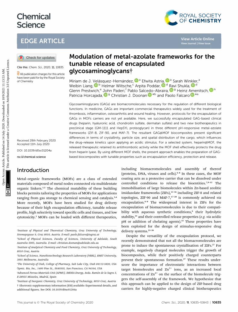
rhinosinusitis: it inhibits multiple inammatory mediators andrequires topical intranasal administration route.68 HepSYL isa new synthetic proteoglycan designed for oncotherapy appli-cations. As such, a parenteral administration route is needed.GM-1111 and HepSYL were encapsulated within ZIF-8, ZIF-90and MAF-7 following the synthetic protocol used for the GAGsbased therapeutics (see ESI†). Aer washing and drying, thepowders were examined with PXRD. The diffraction patternsindicate that GM-1111@ZIF-8 and HepSYL@ZIF-8 are a mixtureof different crystalline phases, sod, dia, and ZIF-C,20,69 with sodas the predominant phase (Fig. 4a). By contrast, the diffractionpattern of GM-1111@ZIF-90 and HepSYL@ZIF-90 show that thesamples are pure sod phase (Fig. 4a). GM-1111@MAF-7 andHepSYL@MAF-7 yield amorphous materials (Fig. 4a).
SEM analysis reveals that the crystalline particles of GM-1111@ZIF-8 and HepSYL@ZIF-8 are of rhombic dodecahedronmorphology (Fig. 4b). For GM-1111@ZIF-8 we observed inho-mogeneous particles with average size of ca. 500 nm; while forHepSYL@ZIF-8 the particles are homogeneous with size isbelow 200 nm. Likewise, the particle morphology observed inGM-1111@ZIF-90 and HepSYL@ZIF-90 samples corresponds torhombic dodecahedron, with particle sizes of ca. 8 mm and ca. 2mm, respectively (Fig. 4b). Due to the small particle size, GM-1111@MAF-7 and HepSYL@MAF-7 samples were analysed byTEM (Fig. 4b). The images reveal the formation of aggregatescomprised of nanoparticles with an average size below 50 nm.Finally, confocal laser microscopy (CLSM) was employed toascertain the location of HepSYL within the ZIF particles(Fig. 4c, S20, and S21†). The CLSM images show that theproteoglycan is homogeneously distributed within ZIF-8 andMAF-7 (Fig. 4c, and S20†). However, in the case of HepSYL@ZIF-90, the proteoglycan is predominantly localised towards thesurface region of crystalline particles (Fig. 4c and S20†).
The EE% and the drug-release kinetics of GM-1111@MOFand HepSYL@MOF were assessed using UV-vis spectroscopy(Fig. 5a). Following the protocol previously described for GAG-based biocomposites, the amount of GM-1111 encapsulatedwithin the MOF shell was determined using the carbazole assay(lmax ¼ 520 nm).55,56
In the case of HepSYL@ZIFs biocomposites, the EE% wasdetermined by monitoring the absorbance of the colorant usedto label the protein (CF633, l ¼ 633 nm) (Fig. S22†).
The data collected reveals that the best performance, interms of EE%, was found when using MAF-7, followed by ZIF-90and then ZIF-8 (Fig. 5a). The EE% is also inuenced by thebiomacromolecule. GM-1111, that is more structurally similarto GAGs, shows a higher EE% than HepSYL, which containspositively charged peptides.
The drug release proles of GM-1111@ZIFs reveal that GM-1111@MAF-7 and GM-1111@ZIF-8 present a rapid burstrelease upon applying an external acidic stimulus (pH ¼ 6), andcomplete release was achieved within the rst 20 min (Fig. 5b).
The release prole of GM-1111@ZIF-90 presents a long-termcontrolled drug delivery, with complete release observed aer2.5 h (Fig. 5b). Finally, the HepSYL@ZIF release proles revealthat the fastest drug delivery is determined for HepSYL@MAF-7,while HepSYL@ZIF-8 and HepSYL@ZIF-90 exhibit a longer-
This journal is © The Royal Society of Chemistry 2020
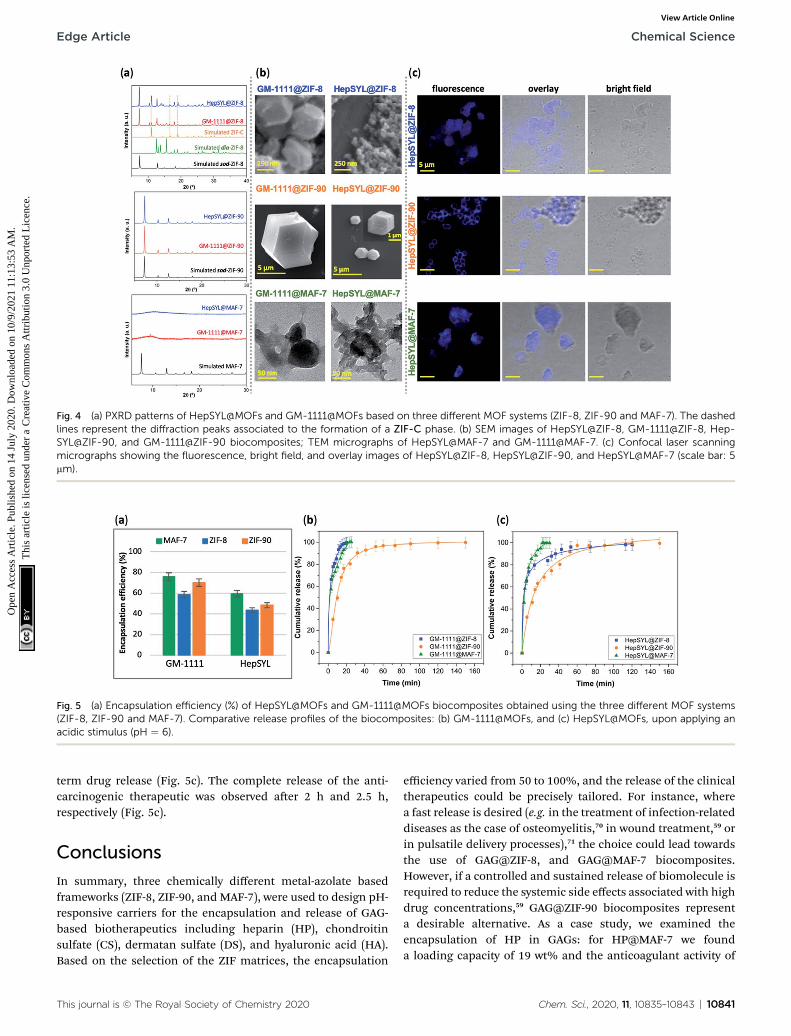
Fig. 4 (a) PXRD patterns of HepSYL@MOFs and GM-1111@MOFs based on three different MOF systems (ZIF-8, ZIF-90 and MAF-7). The dashedlines represent the diffraction peaks associated to the formation of a ZIF-C phase. (b) SEM images of HepSYL@ZIF-8, GM-1111@ZIF-8, Hep-SYL@ZIF-90, and GM-1111@ZIF-90 biocomposites; TEM micrographs of HepSYL@MAF-7 and GM-1111@MAF-7. (c) Confocal laser scanningmicrographs showing the fluorescence, bright field, and overlay images of HepSYL@ZIF-8, HepSYL@ZIF-90, and HepSYL@MAF-7 (scale bar: 5mm).
Fig. 5 (a) Encapsulation efficiency (%) of HepSYL@MOFs and GM-1111@MOFs biocomposites obtained using the three different MOF systems(ZIF-8, ZIF-90 and MAF-7). Comparative release profiles of the biocomposites: (b) GM-1111@MOFs, and (c) HepSYL@MOFs, upon applying anacidic stimulus (pH ¼ 6).
Edge Article Chemical Science
Ope
n A
cces
s A
rtic
le. P
ublis
hed
on 1
4 Ju
ly 2
020.
Dow
nloa
ded
on 1
0/9/
2021
11:
13:5
3 A
M.
Thi
s ar
ticle
is li
cens
ed u
nder
a C
reat
ive
Com
mon
s A
ttrib
utio
n 3.
0 U
npor
ted
Lic
ence
.View Article Online
term drug release (Fig. 5c). The complete release of the anti-carcinogenic therapeutic was observed aer 2 h and 2.5 h,respectively (Fig. 5c).
Conclusions
In summary, three chemically different metal-azolate basedframeworks (ZIF-8, ZIF-90, and MAF-7), were used to design pH-responsive carriers for the encapsulation and release of GAG-based biotherapeutics including heparin (HP), chondroitinsulfate (CS), dermatan sulfate (DS), and hyaluronic acid (HA).Based on the selection of the ZIF matrices, the encapsulation
This journal is © The Royal Society of Chemistry 2020
efficiency varied from 50 to 100%, and the release of the clinicaltherapeutics could be precisely tailored. For instance, wherea fast release is desired (e.g. in the treatment of infection-relateddiseases as the case of osteomyelitis,70 in wound treatment,59 orin pulsatile delivery processes),71 the choice could lead towardsthe use of GAG@ZIF-8, and GAG@MAF-7 biocomposites.However, if a controlled and sustained release of biomolecule isrequired to reduce the systemic side effects associated with highdrug concentrations,59 GAG@ZIF-90 biocomposites representa desirable alternative. As a case study, we examined theencapsulation of HP in GAGs: for HP@MAF-7 we founda loading capacity of 19 wt% and the anticoagulant activity of
Chem. Sci., 2020, 11, 10835–10843 | 10841
Chemical Science Edge Article
Ope
n A
cces
s A
rtic
le. P
ublis
hed
on 1
4 Ju
ly 2
020.
Dow
nloa
ded
on 1
0/9/
2021
11:
13:5
3 A
M.
Thi
s ar
ticle
is li
cens
ed u
nder
a C
reat
ive
Com
mon
s A
ttrib
utio
n 3.
0 U
npor
ted
Lic
ence
.View Article Online
the released heparin was fully preserved, even aer exposure tolyase agents.
Finally, the azolate-based MOF carriers were employed forthe encapsulation and release of pre-clinical therapeutics usedas anti-inammatory and anticarcinogenic agents. Similar tothe GAG-based biocomposites the EE% and release prolescould be tailored by the judicious selection of the MOF matrix.We anticipate that our ndings will facilitate progress in theburgeoning area of MOF-based drug delivery.
Conflicts of interest
There are no conicts to declare.
Acknowledgements
The authors acknowledge support from the European Union'sHorizon 2020 FETOPEN-1-2016-2017 research and innovationprogram under grant agreement 801464 and LP-03. P. H.acknowledges the Spanish Ramon y Cajal Programme (2014-16823). M. J. V. H. acknowledges The National Council ofScience and Technology (CONACyT, Mexico) for the post-doctoral scholarship (CVU 419210). E. A. acknowledges AustrianAgency for International Cooperation in Education andResearch (OeAD-GmbH) for the PhD scholarship. The authorsacknowledge the CERIC-ERIC Consortium for the access toexperimental facilities and nancial support. C. J. D. and P. F.acknowledge ARC DP170103531 for nancial support. A. P.acknowledge the travel scholarship support from AustralianNanotechnology Network (ANN) to visit EU and carry outexperiments.
Notes and references
1 O. M. Yaghi, M. O'Keeffe, N. W. Ockwig, H. K. Chae,M. Eddaoudi and J. Kim, Nature, 2003, 423, 705–714.
2 C. S. Diercks, M. J. Kalmutzki, N. J. Diercks and O. M. Yaghi,ACS Cent. Sci., 2018, 4, 1457–1464.
3 H. Furukawa, K. E. Cordova, M. O'Keeffe and O. M. Yaghi,Science, 2013, 341, 1230444.
4 C. A. Trickett, A. Helal, B. A. Al-Maythalony, Z. H. Yamani,K. E. Cordova and O. M. Yaghi, Nat. Rev. Mater., 2017, 2,17045.
5 T. Simon-Yarza, A. Mielcarek, P. Couvreur and C. Serre, Adv.Mater., 2018, 30, 1707365.
6 K. Liang, R. Ricco, C. M. Doherty, M. J. Styles, S. Bell,N. Kirby, S. Mudie, D. Haylock, A. J. Hill, C. J. Doonan andP. Falcaro, Nat. Commun., 2015, 6, 7240.
7 X. Lian, Y. Fang, E. Joseph, Q. Wang, J. Li, S. Banerjee,C. Lollar, X. Wang and H.-C. Zhou, Chem. Soc. Rev., 2017,46, 3386–3401.
8 C. Doonan, R. Ricco, K. Liang, D. Bradshaw and P. Falcaro,Acc. Chem. Res., 2017, 50, 1423–1432.
9 S. Li, M. Dharmarwardana, R. P. Welch, C. E. Benjamin,A. M. Shamir, S. O. Nielsen and J. J. Gassensmith, ACSAppl. Mater. Interfaces, 2018, 10, 18161–18169.
10842 | Chem. Sci., 2020, 11, 10835–10843
10 R. Ricco, W. Liang, S. Li, J. J. Gassensmith, F. Caruso,C. Doonan and P. Falcaro, ACS Nano, 2018, 12, 13–23.
11 C. Wang, H. Sun, J. Luan, Q. Jiang, S. Tadepalli,J. J. Morrissey, E. D. Kharasch and S. Singamaneni, Chem.Mater., 2018, 30, 1291–1300.
12 Y. Feng, H. Wang, S. Zhang, Y. Zhao, J. Gao, Y. Zheng,P. Zhao, Z. Zhang, M. J. Zaworotko, P. Cheng, S. Ma andY. Chen, Adv. Mater., 2019, 31, 1805148.
13 B. Chen, Z. Yang, Y. Zhu and Y. Xia, J. Mater. Chem. A, 2014,2, 16811–16831.
14 F. Lyu, Y. Zhang, R. N. Zare, J. Ge and Z. Liu, Nano Lett., 2014,14, 5761–5765.
15 F.-S. Liao, W.-S. Lo, Y.-S. Hsu, C.-C. Wu, S.-C. Wang,F.-K. Shieh, J. V. Morabito, L.-Y. Chou, K. C.-W. Wu andC.-K. Tsung, J. Am. Chem. Soc., 2017, 139, 6530–6533.
16 C. Wang, G. Sudlow, Z. Wang, S. Cao, Q. Jiang, A. Neiner,J. J. Morrissey, E. D. Kharasch, S. Achilefu andS. Singamaneni, Adv. Healthcare Mater., 2018, 7, 1800950.
17 J.-P. Zhang, Y.-B. Zhang, J.-B. Lin and X.-M. Chen, Chem.Rev., 2012, 112, 1001–1033.
18 W. Liang, R. Ricco, N. K. Maddigan, R. P. Dickinson, H. Xu,Q. Li, C. J. Sumby, S. G. Bell, P. Falcaro and C. J. Doonan,Chem. Mater., 2018, 30, 1069–1077.
19 W. Liang, H. Xu, F. Carraro, N. K. Maddigan, Q. Li, S. G. Bell,D. M. Huang, A. Tarzia, M. B. Solomon, H. Amenitsch,L. Vaccari, C. J. Sumby, P. Falcaro and C. J. Doonan, J. Am.Chem. Soc., 2019, 141, 2348–2355.
20 F. Carraro, M. de J. Velasquez-Hernandez, E. Astria,W. Liang, L. Twight, C. Parise, M. Ge, Z. Huang, R. Ricco,X. Zou, L. Villanova, C. O. Kappe, C. Doonan andP. Falcaro, Chem. Sci., 2020, 11, 3397–3404.
21 K. S. Park, Z. Ni, A. P. Cote, J. Y. Choi, R. Huang, F. J. Uribe-Romo, H. K. Chae, M. O'Keeffe and O. M. Yaghi, Proc. Natl.Acad. Sci. U. S. A., 2006, 103, 10186–10191.
22 E. Astria, M. Thonhofer, R. Ricco, W. Liang, A. Chemelli,A. Tarzia, K. Alt, C. E. Hagemeyer, J. Rattenberger,H. Schroettner, T. Wrodnigg, H. Amenitsch, D. M. Huang,C. J. Doonan and P. Falcaro, Mater. Horiz., 2019, 6, 969–977.
23 T.-T. Chen, J.-T. Yi, Y.-Y. Zhao and X. Chu, J. Am. Chem. Soc.,2018, 140, 9912–9920.
24 W.-H. Chen, G.-F. Luo, M. Vazquez-Gonzalez, R. Cazelles,Y. S. Sohn, R. Nechushtai, Y. Mandel and I. Willner, ACSNano, 2018, 12, 7538–7545.
25 W. Cai, J. Wang, C. Chu, W. Chen, C. Wu and G. Liu, Adv.Sci., 2019, 6, 1801526.
26 M. A. Luzuriaga, R. P. Welch, M. Dharmarwardana,C. E. Benjamin, S. Li, A. Shahrivarkevishahi, S. Popal,L. H. Tuong, C. T. Creswell and J. J. Gassensmith, ACSAppl. Mater. Interfaces, 2019, 11, 9740–9746.
27 N. K. Maddigan, A. Tarzia, D. M. Huang, C. J. Sumby,S. G. Bell, P. Falcaro and C. J. Doonan, Chem. Sci., 2018, 9,4217–4223.
28 N. S. Gandhi and R. L. Mancera, Chem. Biol. Drug Des., 2008,72, 455–482.
29 E. Astria, M. Thonhofer, R. Ricco, W. Liang, A. Chemelli,A. Tarzia, K. Alt, C. E. Hagemeyer, J. Rattenberger,
This journal is © The Royal Society of Chemistry 2020

Edge Article Chemical Science
Ope
n A
cces
s A
rtic
le. P
ublis
hed
on 1
4 Ju
ly 2
020.
Dow
nloa
ded
on 1
0/9/
2021
11:
13:5
3 A
M.
Thi
s ar
ticle
is li
cens
ed u
nder
a C
reat
ive
Com
mon
s A
ttrib
utio
n 3.
0 U
npor
ted
Lic
ence
.View Article Online
H. Schroettner, T. Wrodnigg, H. Amenitsch, D. M. Huang,C. J. Doonan and P. Falcaro, Mater. Horiz., 2019, 6, 969–977.
30 V. Pomin and B. Mulloy, Pharmaceuticals, 2018, 11, 27.31 A. Varki, Glycobiology, 2017, 27, 3–49.32 G. Ruiz-Gomez, S. Vogel, S. Moller, M. T. Pisabarro and
U. Hempel, Sci. Rep., 2019, 9, 4905.33 R. D. Astronomo and D. R. Burton, Nat. Rev. Drug Discovery,
2010, 9, 308–324.34 J. Paderi, G. D. Prestwich, A. Panitch, T. Boone and K. Stuart,
Adv. Ther., 2018, 1, 1800082.35 N. S. Gandhi and R. L. Mancera, Drug Discovery Today, 2010,
15, 1058–1069.36 A. Neves, M. Correia-da-Silva, E. Sousa and M. Pinto,
Pharmaceuticals, 2016, 9, 37.37 K. Vaghasiya, A. Sharma, K. Kumar, E. Ray, S. Adlakha,
O. P. Katare, S. K. Hota and R. K. Verma, ACS Biomater. Sci.Eng., 2019, 5, 6617–6631.
38 J.-P. Zhang, A.-X. Zhu, R.-B. Lin, X.-L. Qi and X.-M. Chen, Adv.Mater., 2011, 23, 1268–1271.
39 A. U. Ortiz, A. P. Freitas, A. Boutin, A. H. Fuchs andF.-X. Coudert, Phys. Chem. Chem. Phys., 2014, 16, 9940–9949.
40 J. A. Alt, W. Y. Lee, B. M. Davis, J. R. Savage, T. P. Kennedy,G. D. Prestwich and A. Pulsipher, PLoS One, 2018, 13,e0204709.
41 J. Zhang, X. Xu, N. V. Rao, B. Argyle, L. McCoard, W. J. Rusho,T. P. Kennedy, G. D. Prestwich and G. Krueger, PLoS One,2011, 6, e16658.
42 T. Suzuki, A. Ishii-Watabe, N. Hashii, Y. Nakagawa,T. Takahashi, A. Ebisawa, S. Nishi, N. Fujita, A. Bando,Y. Sekimoto, K. Miyata, T. Endo, T. Otsu, S. Sugimoto,T. Kondou, Y. Fujita, N. Miyanaga, M. Mashimo,N. Shimada, H. Yoden, H. Shimamura, Y. Kurata,S. Koyama and N. Kawasaki, Biologicals, 2013, 41, 415–423.
43 J. Rnjak-Kovacina, F. Tang, J. M. Whitelock and M. S. Lord,Adv. Healthcare Mater., 2018, 7, 1701042.
44 F.-M. Zhang, H. Dong, X. Zhang, X.-J. Sun, M. Liu,D.-D. Yang, X. Liu and J.-Z. Wei, ACS Appl. Mater.Interfaces, 2017, 9, 27332–27337.
45 Z. Xie, X. Cai, C. Sun, S. Liang, S. Shao, S. Huang, Z. Cheng,M. Pang, B. Xing, A. A. A. Kheraif and J. Lin, Chem. Mater.,2019, 31, 483–490.
46 J.-W. Yoo, D. J. Irvine, D. E. Discher and S. Mitragotri, Nat.Rev. Drug Discovery, 2011, 10, 521–535.
47 Y. K. Ip, S. F. Chew and D. J. Randall, Fish Physiology,Elsevier, 2001, vol. 20, pp. 109–148.
48 A. Mahmood, W. Xia, N. Mahmood, Q. Wang and R. Zou, Sci.Rep., 2015, 5, 10556.
49 B. Bueken, N. Van Velthoven, T. Willhammar, T. Stassin,I. Stassen, D. A. Keen, G. V. Baron, J. F. M. Denayer,R. Ameloot, S. Bals, D. De Vos and T. D. Bennett, Chem.Sci., 2017, 8, 3939–3948.
This journal is © The Royal Society of Chemistry 2020
50 A. Chakraborty, P. Sutar, P. Yadav, M. Eswaramoorthy andT. K. Maji, Inorg. Chem., 2018, 57, 14480–14483.
51 Z. Akimbekov, A. D. Katsenis, G. P. Nagabhushana, G. Ayoub,M. Arhangelskis, A. J. Morris, T. Friscic and A. Navrotsky, J.Am. Chem. Soc., 2017, 139, 7952–7957.
52 Z. Fang, B. Bueken, D. E. De Vos and R. A. Fischer, Angew.Chem., Int. Ed., 2015, 54, 7234–7254.
53 K. M. Choi, H. J. Jeon, J. K. Kang and O. M. Yaghi, J. Am.Chem. Soc., 2011, 133, 11920–11923.
54 N. Mainreck, S. Brezillon, G. D. Sockalingum, F.-X. Maquart,M. Manfait and Y. Wegrowski, J. Pharm. Sci., 2011, 100, 441–450.
55 S. B. Frazier, K. A. Roodhouse, D. E. Hourcade and L. Zhang,Open Glycosci., 2008, 1, 31–39.
56 T. M. C. C. Filisetti-Cozzi and N. C. Carpita, Anal. Biochem.,1991, 197, 157–162.
57 A. Riemann, A. Ihling, J. Thomas, B. Schneider, O. Thewsand M. Gekle, Biochim. Biophys. Acta, Mol. Cell Res., 2015,1853, 299–307.
58 G. Hao, Z. P. Xu and L. Li, RSC Adv., 2018, 8, 22182–22192.59 X. Huang and C. S. Brazel, J. Controlled Release, 2001, 73,
121–136.60 H. Ouyang, Z. Zheng, Y. Chen, Y. Liu, C. Hong, Y. Zhu,
J. Deng, X. Ding, W. Zhou and X. Wang, J. Mater. Chem. B,2019, 7, 6099–6108.
61 P. E. Nolan and T. C. Trujillo, in Complications ofPercutaneous Coronary Interventions, ed. S. M. Butman,Springer, New York, NY, 2005, pp. 6–16.
62 P. Prandoni, A. W. A. Lensing, A. Piccioli, E. Bernardi,P. Simioni, B. Girolami, A. Marchiori, P. Sabbion,M. H. Prins, F. Noventa and A. Girolami, Blood, 2002, 100,3484–3488.
63 Y. Zhang, J. Yu, J. Wang, N. J. Hanne, Z. Cui, C. Qian,C. Wang, H. Xin, J. H. Cole, C. M. Gallippi, Y. Zhu andZ. Gu, Adv. Mater., 2017, 29, 1604043.
64 R. Bhat, A. Ribes, N. Mas, E. Aznar, F. Sancenon,M. D. Marcos, J. R. Murguıa, A. Venkataraman andR. Martınez-Manez, Langmuir, 2016, 32, 1195–1200.
65 C. Argyo, V. Cauda, H. Engelke, J. Radler, G. Bein andT. Bein, Chem. – Eur. J., 2012, 18, 428–432.
66 N. S. Jones, M. G. Glenn, L. A. Orloff andM. R. Mayberg, Arch.Otolaryngol., 1990, 116, 779–785.
67 P. Montalto, J. Vlachogiannakos, D. J. Cox, S. Pastacaldi,D. Patch and A. K. Burroughs, J. Hepatol., 2002, 37, 463–470.
68 S. Albu, Drug Des., Dev. Ther., 2012, 125–132.69 S. A. Basnayake, J. Su, X. Zou and K. J. Balkus, Inorg. Chem.,
2015, 54, 1816–1821.70 D. Puppi, D. Dinucci, C. Bartoli, C. Mota, C. Migone, F. Dini,
G. Barsotti, F. Carlucci and F. Chiellini, J. Bioact. Compat.Polym., 2011, 26, 478–492.
71 D. Jain, R. Raturi, V. Jain, P. Bansal and R. Singh, Biomatter,2011, 1, 57–65.
Chem. Sci., 2020, 11, 10835–10843 | 10843