Embed Size (px)
Citation preview
NPTEL – Biotechnology – Cell Biology
Joint initiative of IITs and IISc – Funded by MHRD Page 29 of 169
Module 1 Lecture 3 Principles of membrane organization, membrane proteins Introduction
All living cells possess a cell membrane. These membranes serve to contain and
protect cell components from the surroundings as well as regulate the transport of
material into and out of the cell. Cell membranes are the selectively permeable lipid
bilayers inclusive of membrane proteins which delimits all prokaryotic and eukaryotic
cells. In prokaryotes and plants, the plasma membrane is an inner layer of protection
bounded to the inner side of a rigid cell wall. Eukaryotes lack this external layer of
protection or the cell wall. In eukaryotes the membrane also forms boundary of cell
organelles. The cell membrane has been given different specific names based on their
lipid and protein composition such as “sarcolemma” in myocytes and “oolemma” in
oocytes. The plasma membrane is just 5-10nm wide thus cannot be detected under the
light microscope. It can only be observed under the Transmission electron microscope as
a trilaminar structure which is a layer of hydrophobic tails of phospholipids sandwiched
between two layers of hydrophillic heads.
Functions
Functionally membranes take part in several cellular activities covering motility, energy
transduction in lower unicellular organisms to immunorecognition in higher eukaryotes.
The most valuable function is segregation of the cell into compartments. This functional
diversity is due to the variability in lipid and protein composition of the membranes. The
various functions can be summarized as given below.
1. Diffusion: Diffusion of small molecules such as carbon dioxide, oxygen (O2), and
water happens by passive transport.
2. Osmosis: Cell membrane is semipermeable thus it sets up an osmotic flow for solvent
such as water, which can be transported across the membrane by osmosis.
3. Mediated Transport: Nutrients are moved across the membrane by special proteins
called transport proteins or permeases which are quite specific, recognizing and
transporting only a limited group of chemical substances, often even only a single
substance.
NPTEL – Biotechnology – Cell Biology
Joint initiative of IITs and IISc – Funded by MHRD Page 30 of 169
4. Endocytosis: Endocytosis is the process in which cells absorb molecules by engulfing
them small molecules and ions and macromolecules through active transport which
requires ATP.
5. Exocytosis: The plasma membrane can extrude its contents to the surrounding medium
to remove undigested residues of substances brought in by endocytosis, to secrete
substances such as hormones and enzymes, and to transport a substance completely
across a cellular barrier.
6. Cell adhesion.
7. Cell signaling.
Theories:
Quincke first perceived the lipid nature of the cell membranes and proposed it to be less
than 100 nm thick. With time many researchers have proposed models for cell
membrane.
In 1935, Danielli and Davson, proposed a model, called sandwich model, for membrane
structure in which a lipid bilayer was coated on its either side with hydrated proteins
(globular proteins). Mutual attraction between the hydrocarbonchains of the lipids and
electrostatic forces between the protein and the “head” of the lipid molecules, were
thought to maintain the stability of the membrane. From the speed at which various
molecules penetrate the membrane, they predicted the lipid bilayer to be about 6.0 nm in
thickness, and each of the protein layer of about 1.0 nm thickness, giving a total thickness
of about 8.0 nm. The Danielli-Davson model got support from electron microscopy.
Electron micrographs of the plasma membrane showed that it consists of two dark layers
(electron dense granular protein layers), both separated by a lighter area in between (the
central clear area of lipid bilayer). The total thickness of the membranes too turned out to
be about 7.5 nm.
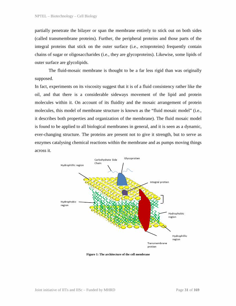
Currently, the most accepted model for cell membrane is fluid mosaic model proposed
by S.J.Singer and G.L.Nicolson (1972). According to this model, the plasma membrane
contains a bimolecular lipid layer, both surfaces of which are interrupted by protein
molecules. Proteins occur in the form of globular molecules and they are dotted about
here and there in a mosaic pattern (see Figure 1). Some proteins are attached at the polar
surface of the lipid (i.e., the extrinsic proteins); while others (i.e., integral proteins) either
NPTEL – Biotechnology – Cell Biology
Joint initiative of IITs and IISc – Funded by MHRD Page 31 of 169
partially penetrate the bilayer or span the membrane entirely to stick out on both sides
(called transmembrane proteins). Further, the peripheral proteins and those parts of the
integral proteins that stick on the outer surface (i.e., ectoproteins) frequently contain
chains of sugar or oligosaccharides (i.e., they are glycoproteins). Likewise, some lipids of
outer surface are glycolipids.
The fluid-mosaic membrane is thought to be a far less rigid than was originally
supposed.
In fact, experiments on its viscosity suggest that it is of a fluid consistency rather like the
oil, and that there is a considerable sideways movement of the lipid and protein
molecules within it. On account of its fluidity and the mosaic arrangement of protein
molecules, this model of membrane structure is known as the “fluid mosaic model” (i.e.,
it describes both properties and organization of the membrane). The fluid mosaic model
is found to be applied to all biological membranes in general, and it is seen as a dynamic,
ever-changing structure. The proteins are present not to give it strength, but to serve as
enzymes catalysing chemical reactions within the membrane and as pumps moving things
across it.
Figure 1: The architecture of the cell membrane
NPTEL – Biotechnology – Cell Biology
Joint initiative of IITs and IISc – Funded by MHRD Page 32 of 169
Biochemistry of the cell membrane
Membrane lipids
The cell membrane lipids are highly complex comprising of
• Phospholipids,
• Glycolipids,
• Cholesterols.
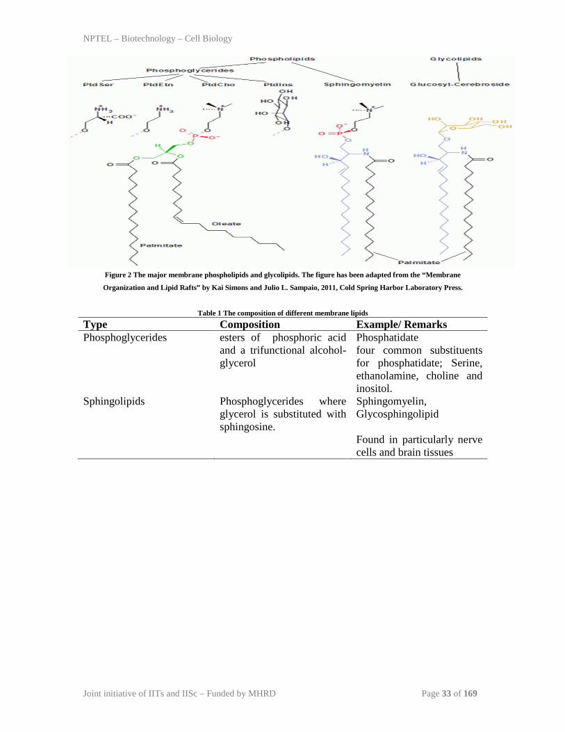
The major membrane phospholipids and glycolipids are phosphatidylcholine
(PtdCho), phosphatidylethanolamine (PtdEtn), phosphatidylinositol (PtdIns) and
phosphatidylserine (PtdSer) (Figure 2, Table 1). Eukaryotic membrane lipids are
glycerophospholipids, sphingolipids, and sterols. Sphingolipids (SPs) and sterols enable
eukaryotic cellular membranes with the property of vesicular trafficking important for the
establishment and maintenance of distinct organelles. Mammalian cell membranes
contain cholesterol which imparts stiffening and strengthening effect on the membrane,
along with glycerophospholipids and sphingolipids. The head group of
glycerophospholipids can vary, the fatty acids can differ in length (16- and 18-carbon
fatty acids are the most common) Fatty acids can be saturated or unsaturated with the
double bonds always in cis configuration in the later. The unsaturated fatty acids prevent
tight packing of the fatty acid chains leading to lowering of melting temperature and
increase in membrane fluidity. Also, the sphingolipids have the combinatorial propensity
to create diversity by different ceramide backbones. Lipid molecules are free exhibit
lateral diffusion along the layer in which they are present. However, the exchange of
phospholipid molecules between intracellular and extracellular leaflets of the bilayer is a
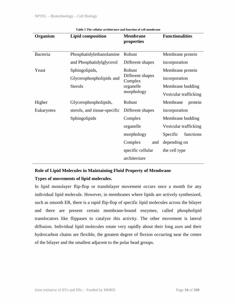
very slow process. The lipid composition, cellular architecture and
function of cell membrane from unicellular bacteria to yeast and higher eukaryotes is
presented in Table 2.
NPTEL – Biotechnology – Cell Biology
Joint initiative of IITs and IISc – Funded by MHRD Page 33 of 169
Figure 2 The major membrane phospholipids and glycolipids. The figure has been adapted from the “Membrane
Organization and Lipid Rafts” by Kai Simons and Julio L. Sampaio, 2011, Cold Spring Harbor Laboratory Press.
Table 1 The composition of different membrane lipids
Type Composition Example/ Remarks Phosphoglycerides esters of phosphoric acid
and a trifunctional alcohol- glycerol
Phosphatidate four common substituents for phosphatidate; Serine, ethanolamine, choline and inositol.
Sphingolipids Phosphoglycerides where glycerol is substituted with sphingosine.
Sphingomyelin, Glycosphingolipid Found in particularly nerve cells and brain tissues
NPTEL – Biotechnology – Cell Biology
Joint initiative of IITs and IISc – Funded by MHRD Page 34 of 169
Table 2 The cellular architecture and function of cell membrane
Organism Lipid composition Membrane properties
Functionalities
Bacteria Phosphatidylethanolamine
and Phosphatidylglycerol
Robust
Different shapes
Membrane protein
incorporation
Yeast Sphingolipids,
Glycerophospholipids and
Sterols
Robust Different shapes Complex organelle morphology
Membrane protein
incorporation
Membrane budding
Vesicular trafficking
Higher
Eukaryotes
Glycerophospholipids,
sterols, and tissue-specific
Sphingolipids
Robust
Different shapes
Complex
organelle
morphology
Complex and
specific cellular
architecture
Membrane protein
incorporation
Membrane budding
Vesicular trafficking
Specific functions
depending on
the cell type
Role of Lipid Molecules in Maintaining Fluid Property of Membrane
Types of movements of lipid molecules.
In lipid monolayer flip-flop or transbilayer movement occurs once a month for any
individual lipid molecule. However, in membranes where lipids are actively synthesized,
such as smooth ER, there is a rapid flip-flop of specific lipid molecules across the bilayer
and there are present certain membrane-bound enzymes, called phospholipid
translocators like flippases to catalyze this activity. The other movement is lateral
diffusion. Individual lipid molecules rotate very rapidly about their long axes and their
hydrocarbon chains are flexible, the greatest degree of flexion occurring near the centre
of the bilayer and the smallest adjacent to the polar head groups.
NPTEL – Biotechnology – Cell Biology
Joint initiative of IITs and IISc – Funded by MHRD Page 35 of 169
Role of unsaturated fats in increasing membrane fluidity.
A synthetic bilayer made from a single type of phospholipid changes from a liquid state
to a rigid crystalline state at a characteristic freezing point. This change of state is called a
phase transition
and the temperature at which it occurs becomes lower if the hydrocarbon chains are short
or have double bonds. Double bonds in unsaturated hydrocarbon chains tend to increase
the fluidity of a phospholipid bilayer by making it more difficult to pack the chains
together. Thus, to maintain fluidity of the membrane, cells of organisms living at low
temperatures have high proportions of unsaturated fatty acids in their membranes, than do
cells at higher temperatures.
Role of cholesterol in maintaining fluidity of membrane
Eukaryotic plasma membranes are found to contain a large amount of cholesterol; up to
one molecule for every phospholipid molecule. Cholesterol inhibits phase transition by
preventing hydrocarbon chains from coming together and crystallizing. Cholesterol also
tends to decrease the permeability of lipid bilayers to small water-soluble molecules and
is thought to enhance both the flexibility and the mechanical stability of the bilayer.
Membrane proteins
In addition to the lipid bilayer, the cell membrane also contains a number of proteins.
35% of the genes in any genome encode membrane proteins, and many other proteins
spend part of their lifetime bound to membranes. The amount of protein differs between
species and according to function, however the typical amount in a cell membrane is
50%. Membrane proteins are free to move within the lipid bilayer as a result of its
fluidity. Although this is true for most proteins, they can also be confined to certain areas
of the bilayer with enzymes.
They can be classified into
• Integral (intrinsic)
• Peripheral (extrinsic)
which is based on the nature of the membrane-protein interactions (Figure 3). Integral
proteins have one or more segments that are embedded in the phospholipid bilayer from
four to several hundred residues long, extending into the aqueous medium on each side of
the bilayer. The transmembrane embedded in the hydrophobic core of the bilayer are α
NPTEL – Biotechnology – Cell Biology
Joint initiative of IITs and IISc – Funded by MHRD Page 36 of 169
helices or multiple β strands interacting with the lipid bilayer with hydrophobic and ionic
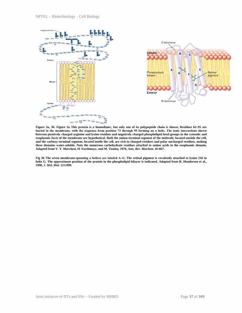
interactions. An example is Glycophorin which is a major erythrocyte membrane protein
and bacteriorhodopsin, a protein found in a photosynthetic bacterium (Figure 3a, 3b).
Glycophorin is a homodimer containing α helix in coiled-coiled conformation, composed
of uncharged amino acids. Few positively charged amino acids (lysine and arginine)
prevent it from slipping across the membrane by interacting with negatively charged
phospholipid head groups. Most of these charged residues are adjacent to the cytosolic
face of the lipid bilayer. Bacteriorhodopsins have serpentine membrane spanning domain.
Other examples of seven-spanning membrane proteins include the opsins (eye proteins
that absorb light), cell-surface receptors for many hormones, and receptors for odorous
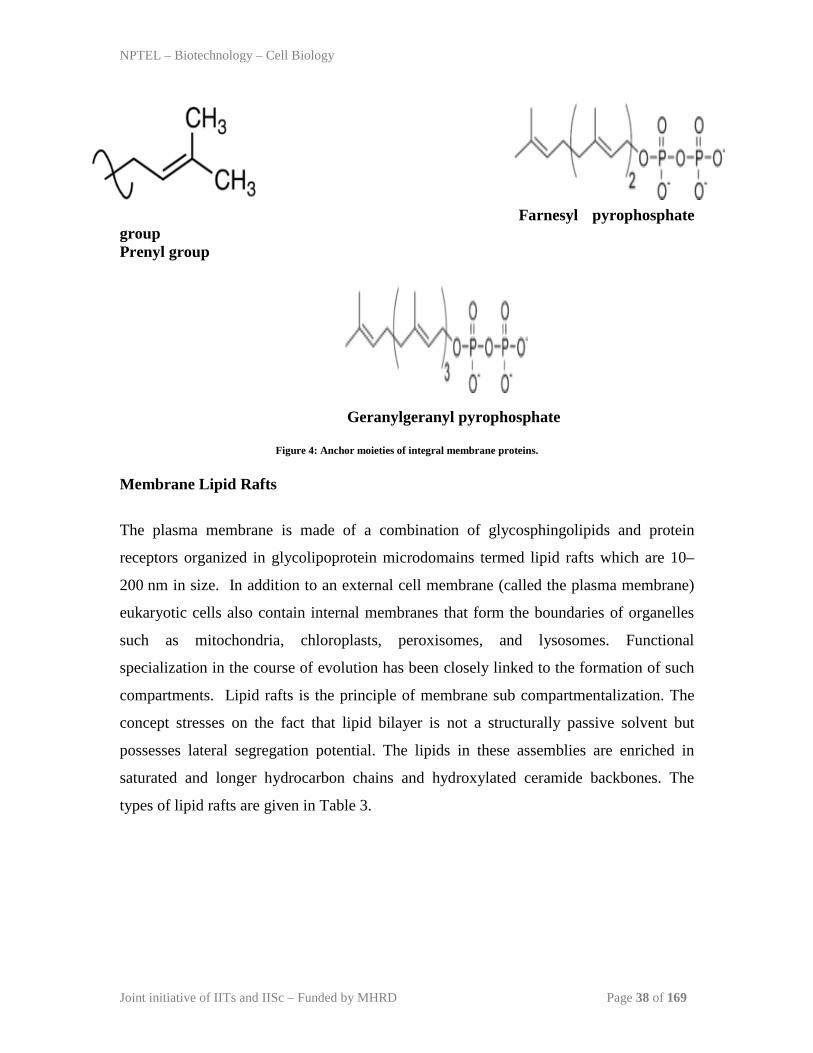
molecules. Some integral proteins are anchored to the exoplasmic face of the plasma
membrane by a complex glycosylated phospholipid that is linked to the C-terminus. A
common example of this type of anchor is glycosylphosphatidylinositol, which contains
two fatty acyl groups, N-acetylglucosamine, mannose, and inositol for example alkaline
phosphatase. Whereas some are attached by a hydrocarbon moiety covalently attached to
a cysteine near the C-terminus. The most common anchors are prenyl, farnesyl, and
geranylgeranyl groups.
Peripheral membrane proteins do not interact with the hydrophobic core and are bound to
the membrane indirectly by interactions with integral membrane proteins or directly by
interactions with lipid polar head groups. Peripheral proteins localized to the cytosolic
face of the plasma membrane include the cytoskeletal proteins spectrin and actin in
erythrocytes and the enzyme protein kinase C involved in cell signaling. An important
group of peripheral membrane proteins are water-soluble enzymes that associate with the
polar head groups of membrane phospholipids.
NPTEL – Biotechnology – Cell Biology
Joint initiative of IITs and IISc – Funded by MHRD Page 37 of 169
Figure 3a, 3b. Figure 3a This protein is a homodimer, but only one of its polypeptide chain is shown. Residues 62–95 are buried in the membrane, with the sequence from position 73 through 95 forming an α helix. The ionic interactions shown between positively charged arginine and lysine residues and negatively charged phospholipid head groups in the cytosolic and exoplasmic faces of the membrane are hypothetical. Both the amino-terminal segment of the molecule, located outside the cell, and the carboxy-terminal segment, located inside the cell, are rich in charged residues and polar uncharged residues, making these domains water-soluble. Note the numerous carbohydrate residues attached to amino acids in the exoplasmic domain. Adapted from V. T. Marchesi, H. Furthmayr, and M. Tomita, 1976, Ann. Rev. Biochem. 45:667. Fig 3b The seven membrane-spanning α helices are labeled A–G. The retinal pigment is covalently attached to lysine 216 in helix G. The approximate position of the protein in the phospholipid bilayer is indicated. Adapted from R. Henderson et al., 1990, J. Mol. Biol. 213:899.
NPTEL – Biotechnology – Cell Biology
Joint initiative of IITs and IISc – Funded by MHRD Page 38 of 169
Farnesyl pyrophosphate group Prenyl group Geranylgeranyl pyrophosphate
Figure 4: Anchor moieties of integral membrane proteins. Membrane Lipid Rafts
The plasma membrane is made of a combination of glycosphingolipids and protein
receptors organized in glycolipoprotein microdomains termed lipid rafts which are 10–
200 nm in size. In addition to an external cell membrane (called the plasma membrane)
eukaryotic cells also contain internal membranes that form the boundaries of organelles
such as mitochondria, chloroplasts, peroxisomes, and lysosomes. Functional
specialization in the course of evolution has been closely linked to the formation of such
compartments. Lipid rafts is the principle of membrane sub compartmentalization. The
concept stresses on the fact that lipid bilayer is not a structurally passive solvent but
possesses lateral segregation potential. The lipids in these assemblies are enriched in
saturated and longer hydrocarbon chains and hydroxylated ceramide backbones. The
types of lipid rafts are given in Table 3.
NPTEL – Biotechnology – Cell Biology
Joint initiative of IITs and IISc – Funded by MHRD Page 39 of 169
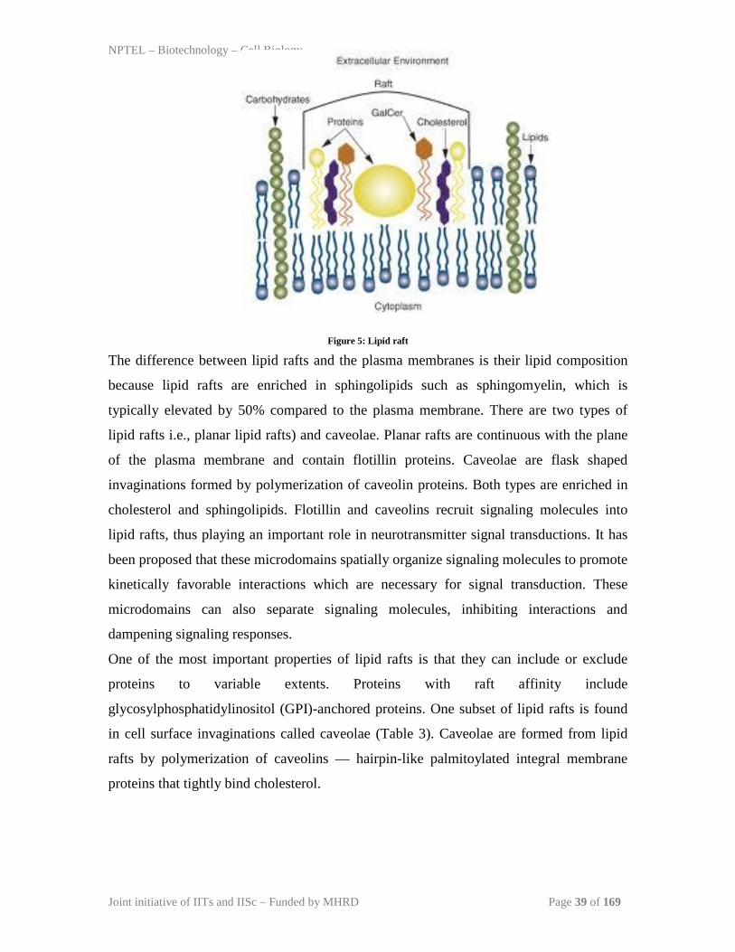
Figure 5: Lipid raft
The difference between lipid rafts and the plasma membranes is their lipid composition
because lipid rafts are enriched in sphingolipids such as sphingomyelin, which is
typically elevated by 50% compared to the plasma membrane. There are two types of
lipid rafts i.e., planar lipid rafts) and caveolae. Planar rafts are continuous with the plane
of the plasma membrane and contain flotillin proteins. Caveolae are flask shaped
invaginations formed by polymerization of caveolin proteins. Both types are enriched in
cholesterol and sphingolipids. Flotillin and caveolins recruit signaling molecules into
lipid rafts, thus playing an important role in neurotransmitter signal transductions. It has
been proposed that these microdomains spatially organize signaling molecules to promote
kinetically favorable interactions which are necessary for signal transduction. These
microdomains can also separate signaling molecules, inhibiting interactions and
dampening signaling responses.
One of the most important properties of lipid rafts is that they can include or exclude
proteins to variable extents. Proteins with raft affinity include
glycosylphosphatidylinositol (GPI)-anchored proteins. One subset of lipid rafts is found
in cell surface invaginations called caveolae (Table 3). Caveolae are formed from lipid
rafts by polymerization of caveolins — hairpin-like palmitoylated integral membrane
proteins that tightly bind cholesterol.
NPTEL – Biotechnology – Cell Biology
Joint initiative of IITs and IISc – Funded by MHRD Page 40 of 169
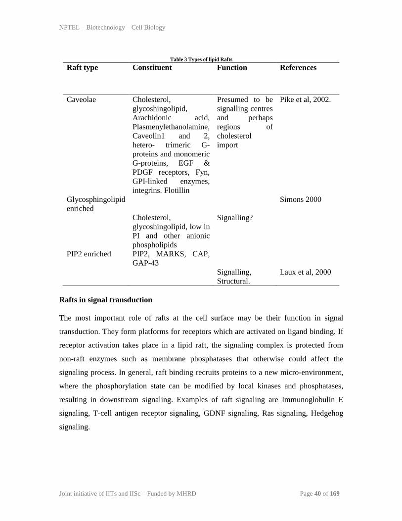
Table 3 Types of lipid Rafts
Raft type Constituent Function
References
Caveolae Cholesterol,
glycoshingolipid, Arachidonic acid, Plasmenylethanolamine, Caveolin1 and 2, hetero- trimeric G-proteins and monomeric G-proteins, EGF & PDGF receptors, Fyn, GPI-linked enzymes, integrins. Flotillin
Presumed to be signalling centres and perhaps regions of cholesterol import
Pike et al, 2002.
Glycosphingolipid enriched
Cholesterol, glycoshingolipid, low in PI and other anionic phospholipids
Signalling?
Simons 2000
PIP2 enriched PIP2, MARKS, CAP, GAP-43
Signalling, Structural.
Laux et al, 2000
Rafts in signal transduction The most important role of rafts at the cell surface may be their function in signal
transduction. They form platforms for receptors which are activated on ligand binding. If
receptor activation takes place in a lipid raft, the signaling complex is protected from
non-raft enzymes such as membrane phosphatases that otherwise could affect the
signaling process. In general, raft binding recruits proteins to a new micro-environment,
where the phosphorylation state can be modified by local kinases and phosphatases,
resulting in downstream signaling. Examples of raft signaling are Immunoglobulin E
signaling, T-cell antigen receptor signaling, GDNF signaling, Ras signaling, Hedgehog
signaling.
NPTEL – Biotechnology – Cell Biology
Joint initiative of IITs and IISc – Funded by MHRD Page 41 of 169
Models for signal initiation in rafts
A common theme of signal transduction is that individual rafts cluster together to connect
raft proteins and interacting proteins into a signalling complex. Receptors have at least
three different options in rafts for signal transduction (Figure 6). First, receptors could be
activated through ligand binding (Figure 6). Second, individual receptors possessing
weak raft affinity can oligomerize on ligand binding (Figure 6). Last, crosslinking
proteins can be recruited to bind to proteins in other rafts (Figure 6). The formation of
clustered rafts would lead to amplification of signal. The interactions that drive raft
assembly are dynamic and reversible. Raft clusters can be also be disassembled by
removal of raft components from the cell surface by endocytosis. The coalescence of
individual rafts to form raft clusters has been observed when crosslinking raft
components with antibodies. The movement and behavior of the raft clusters can also be
influenced by interaction with cytoskeletal elements and second messengers, which help
organize actin assemblies on the cytoplasmic surface of the rafts.

NPTEL – Biotechnology – Cell Biology
Joint initiative of IITs and IISc – Funded by MHRD Page 42 of 169
Figure 6: Models of how signalling could be initiated through rafts. A. In these models, signalling occurs in either single rafts (Model 1) or clustered rafts (Model 2). Following dimerization the protein becomes phosphorylated in rafts. B. In the second model we assume that there are several rafts in the membrane, which differ in protein composition (shown in orange, purple or blue). Clustering would coalesce rafts (red), so that they would now contain a new mixture of molecules, such as crosslinkers and enzymes. Clustering could occur either extracellularly, within the membrane, or in the cytosol (a–c, respectively). Raft clustering could also occur through GPIanchored proteins (yellow), either as a primary or co-stimulatory response. Notably, models 1 and 2 are not mutually exclusive. For instance, extracellular signals could increase a protein’s raft affinity (for example, similar to the effect of single versus dual acylation) therefore drawing more of the protein into the raft where it can be activated and recruit other proteins, such as LAT, which would crosslink several rafts. Printed with permission from Simons K Sampaio J L. Membrane Organization and Lipid Rafts. Cold Spring Harbor Perspective Biology. 2011.


![[Marcel Mulder] Basic Principles of Membrane Techn(BookZZ.org)](https://img.pdfslide.net/doc/110x75/563db968550346aa9a9d0aab/marcel-mulder-basic-principles-of-membrane-technbookzzorg.jpg)