Embed Size (px)
Citation preview
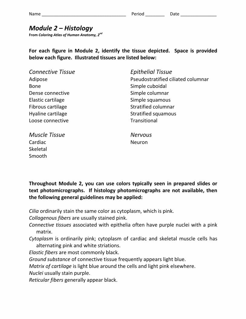
Name ___________________________________ Period ________ Date _______________
Module 2 – Histology From Coloring Atlas of Human Anatomy, 2
nd
For each figure in Module 2, identify the tissue depicted. Space is provided below each figure. Illustrated tissues are listed below:
Connective Tissue Adipose Bone Dense connective Elastic cartilage Fibrous cartilage Hyaline cartilage Loose connective
Muscle Tissue Cardiac Skeletal Smooth
Epithelial Tissue Pseudostratified ciliated columnar Simple cuboidal Simple columnar Simple squamous Stratified columnar Stratified squamous Transitional
Nervous Neuron
Throughout Module 2, you can use colors typically seen in prepared slides or text photomicrographs. If histology photomicrographs are not available, then the following general guidelines may be applied: Cilia ordinarily stain the same color as cytoplasm, which is pink. Collagenous fibers are usually stained pink. Connective tissues associated with epithelia often have purple nuclei with a pink
matrix. Cytoplasm is ordinarily pink; cytoplasm of cardiac and skeletal muscle cells has
alternating pink and white striations. Elastic fibers are most commonly black. Ground substance of connective tissue frequently appears light blue. Matrix of cartilage is light blue around the cells and light pink elsewhere. Nuclei usually stain purple. Reticular fibers generally appear black.

Name ___________________________________ Period ________ Date _______________

Name ___________________________________ Period ________ Date _______________

Name ___________________________________ Period ________ Date _______________

Name ___________________________________ Period ________ Date _______________

Name ___________________________________ Period ________ Date _______________

Name ___________________________________ Period ________ Date _______________