Embed Size (px)
Citation preview

NPTEL – Biotechnology – Gene Therapy
Joint initiative of IITs and IISc – Funded by MHRD Page 1 of 34
Module 2 In vivo gene therapy
Lecture 7
In-situ, in-vivo and ex-vivo gene therapy (part I)
Somatic cell gene therapy involves the transfer of gene to a diseased somatic cell
either within the body or outside the body with the help of a viral or non viral gene
therapy vector.
Ex vivo is any procedure accomplished outside. In gene therapy clinical trials
cells are modified in a variety of ways to correct the gene. In ex vivo cells are
modified outside the patient’s body and the corrected version is transplanted back in
to the patient. The cells are treated with either a viral or non viral gene therapy vector
carrying the corrected copy of the gene.
Opposite of ex vivo is what we call in vivo where cells are treated inside the
patient’s body. The corrected copy of the genes is transferred into the body of the
patient. The cells may be treated either with a viral or non viral vector carrying the
corrected copy of the gene. If the patient is weak or the cell cannot be extracted out
from the body, the gene is introduced directly into the body.
Gene therapy done in a restricted area or to a particular site is called in-situ. In
situ gene therapy requires the vector to be placed directly into the affected tissues. In
vivo gene therapy involves injecting the vector into the blood stream. The vector then
must find the target tissue and deliver the therapeutic genes.
7.1 In situ gene therapy
In situ gene therapy comprises transfer of corrected copy of the gene into the
targeted organ or tissue. The major concern of current time gene therapy protocol is
the lack of efficient transduction of the targeted organ. The method is effectively used
against cystic fibrosis, a disease of airway epithelium (discussed in later chapters).
The method is also explored for cancer gene therapy where the viral vector is
engineered to contain the herpes simplex virus thymidine kinase gene. After injection
of the viral vector the patient is treated with a prodrug such as Ganciclovir, which
causes 75% reduction in the tumor cell population.

NPTEL – Biotechnology – Gene Therapy
Joint initiative of IITs and IISc – Funded by MHRD Page 2 of 34
7.2 In vivo gene therapy
Delivery of corrected copy of the gene systemically through injection is a
highly efficient way to transfer a transgene to the patient’s body. The major problem
of in vivo method is its inefficient targeting. The transgene delivered into the body by
means of viral or non viral vector also evokes the immune response. The immune
response against the vector leads to its clearance and only transient expression of
transgene. The neutralizing antibody does not allow the second injection of the vector.
Reducing the neutralizing antibody is the current area of research in order to improve
the delivery of gene therapy vector. All gene therapy delivery protocols require the
transgene to cross the plasma membrane and enter inside the nucleus. The major
obstacle is still to deliver the transgene effectively to the intracellular compartment.
Many modifications have been suggested into the viral vectors and also non viral
vectors to target the gene to the tissue. VP22, a protein of herpes simplex virus has a
property to spread from one cell to the other, and this property has been successfully
implemented in designing the vectors.
Figure 7.1 Ex vivo, in vivo, and in situ gene therapy:
NPTEL – Biotechnology – Gene Therapy
Joint initiative of IITs and IISc – Funded by MHRD Page 3 of 34
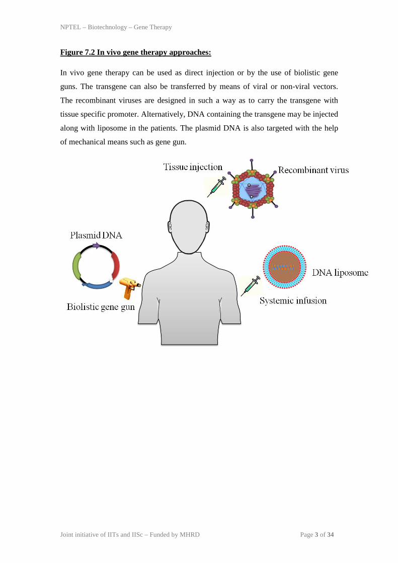
Figure 7.2 In vivo gene therapy approaches:
In vivo gene therapy can be used as direct injection or by the use of biolistic gene
guns. The transgene can also be transferred by means of viral or non-viral vectors.
The recombinant viruses are designed in such a way as to carry the transgene with
tissue specific promoter. Alternatively, DNA containing the transgene may be injected
along with liposome in the patients. The plasmid DNA is also targeted with the help
of mechanical means such as gene gun.

NPTEL – Biotechnology – Gene Therapy
Joint initiative of IITs and IISc – Funded by MHRD Page 4 of 34
Figure 7.3 Clinical ex vivo and in vivo approach:
In vivo, the transgene can be directly injected by various routes based on the clinical
condition of the patient and design of the vector. For instance, transducing the lung
airway epithelium is targeted. The target specific gene therapy is possible with the
help of unique promoters. The liver directed gene therapy is done usually by thyroid
binding globulin promoter (TBG) while cytomegalovirus (CMV) promoter can be
used to direct the muscle cells. Ex vivo on the other hand requires the manipulation of
the cells outside the body and the transducer cells are injected back after quality
assurance.

NPTEL – Biotechnology – Gene Therapy
Joint initiative of IITs and IISc – Funded by MHRD Page 5 of 34
Lecture 8
In-situ, in-vivo and ex-vivo gene therapy (part II)
8.1 Modification by in vivo and ex vivo gene therapy
Somatic cells are modified in a number of different ways
8.1.1 Gene supplementation
This method is also called as gene augmentation. It aims to supply a functional
copy of the defective gene. The method is generally employed for a gene product that
has lost its function or is showing inadequate expression of protein. The process can
be used when there is irreversible damage of the gene. The gene supplementation can
be used for cancer therapy to increase the immune response against the tumor cells.
Alternatively it can be used to replace the defective tumor suppressor gene.
8.1.2 Gene replacement
In this the mutated or nonfunctional copy of the gene is replaced by the correct
functional copy of the same gene. The gene replacement is performed for a mutated
gene which is harmful for the host. In general gene replacement aims for gain of
function.
8.1.3 Targeted inhibition of gene function
The targeted inhibition of gene function is relevant for the infectious diseases
where specific gene of pathogens is targeted. The pathogen associated antigenic gene
is knocked down in order to avoid the harmful effect of the protein. It also aims for
targeted inhibition of tumor antigen to reduce the autoimmune response. The gene is
silenced by various means including siRNA, RNAi, etc.

NPTEL – Biotechnology – Gene Therapy
Joint initiative of IITs and IISc – Funded by MHRD Page 6 of 34
8.1.4 Targeted killing of the cells
The targeted killing aims specifically for cancer cells where the metastatic
form of the tumors are targeted and killed in situ. Many novel viruses called oncolytic
viruses are targeted to kill the cancerous cells. Paramyxoviruses belong to such group
of promising oncolytic viruses. Many studies using paramyxoviruses have shown
encouraging results in reducing the cancerous condition by specifically targeting and
killing the cancer cells by apoptosis.
Facts about gene therapy
The beginning of gene therapy trial for humans started in 1990 for Severe Combined
Immune Disorder (SCID).
Majority of the gene therapy trials are conducted in United States and more than 60%
of those are approved for cancer based gene therapy trials.
8.2 Mechanism of suicidal gene therapy
Retroviruses or lentiviral vectors are designed to carry a therapeutic gene.
Since the retroviruses only grow in dividing cells so they will specifically multiply in
tumor cells. Retroviral vectors are designed to express the thymidine kinase gene (tk)
from herpes virus.
The tk gene sensitizes the tumor cells for a prodrug ganciclovir. Herepes virus
tk is a normal substrate for the ganciclovir while the host tk is not affected by the
drug. Therefore host cells surrounding the tumors will not have any effect of
ganciclovir.
Ganciclovir is phosphorylated by the herpes virus tk into monophosphate
form; the ganciclovir is then converted into triphosphate form which inhibits the DNA
synthesis in tumor cells. The tumors cells are specifically killed by this process as the
host tk has little affinity with the ganciclovir.

NPTEL – Biotechnology – Gene Therapy
Joint initiative of IITs and IISc – Funded by MHRD Page 7 of 34
Figure 8.1 Schematic representation of suicidal gene therapy:
Herpes simplex virus contains an enzyme called THYMIDINE KINASE which is
used in targeting cancer cells for suicidal gene therapy. The Ganciclovir, an antiviral
drug is used for suicidal gene therapy. The underlying mechanism behind the killing
of a cancer cells is by phosphorylation of Ganciclovir.

NPTEL – Biotechnology – Gene Therapy
Joint initiative of IITs and IISc – Funded by MHRD Page 8 of 34
Figure 8.2 Application of viral and non viral vectors for in vivo and in vitro
approach:
Viral vectors are categorized into integrating or non-integrating vectors based on their
recombination capacity with the host cell chromosome. Adeno-associated viruses are
known to target the genetic material to human chromosome number 19 (19q13.4). The
incorporation of genes into the chromosome can lead to STABLE expression of the
protein of interest. The other type is called as TEMPORARY where the proteins are
expressed only for a short period of time and gene is not integrated with the
chromosome.

NPTEL – Biotechnology – Gene Therapy
Joint initiative of IITs and IISc – Funded by MHRD Page 9 of 34
Lecture 9
Transgenic animal models (part I)
Genetically modified animal models also called as transgenic animals
represent a promising tool in biology to understand the host pathogen interactions and
gene function in the purview of disease susceptibility and its progression. Apart from
many animal models mice represent one of the best tools to understand many of the
above important roles in the discovery and development of new disease treatments.
9.1 Types of Transgenic Animals
Transgenic animals are genetically altered with specific characteristics which
otherwise would not be present in that specific animal. In general, transgenic animals
have either DNA added (to express an additional gene) or have their genome altered
(to abolish or modify the expression of an existing gene). Rodents particularly mice
comprise of over 95% of transgenic animals used in biomedical research. The mouse
is the model organism of choice because of the following reasons:
Complete mice genome sequence is available.
Easy genetic manipulation of mice cells and embryos.
Short gestation period and large litter size.
Availability of major antibodies and other molecular biology tools.
Possibility to perform physiologic and behavioral tests that can be directly
linked to human disease.
Other transgenic species include cattle, pig, sheep and rats. Their use in
pharmaceutical research has so far been limited due to technical constraints. Recent
advances in molecular biology techniques may allow us to use transgenic rat for the
development of many human therapeutics where the rat is a better model than the
mouse.

NPTEL – Biotechnology – Gene Therapy
Joint initiative of IITs and IISc – Funded by MHRD Page 10 of 34
9.2 Uses of transgenic animals:
Transgenic animals are useful in the discovery of new therapy for important
human diseases.
Transgenic animals are fundamentally similar to their counter wild type
variety except for some genomic heterogeneity.
Transgenic animals are used in the gene therapy experiment to understand the
importance of malfunctioned or mutated gene.
Transgenic animals are also used to check the efficacy and safety of the drugs
or vaccines used in the clinical trials.
Transgenic mice can be generated to express human targeted gene that can be
further used to design new therapy. Transgenic mice can also obviate the use of
animals such as monkeys for testing drugs for many human diseases, eg.
hypercholesterolemia and HIV.
Some terminology:
The number of offsprings produced at one birth by an animal is called Litter size.
Normal pregnancy period is called as gestation period.
Presence of high levels of cholesterol in the blood is called as hypercholesterolemia.
LDL receptor (LDLR) present in liver helps to eliminate the excess cholesterol from
the body. The deficiency of LDLR in human body leads to increase in blood
cholesterol level, condition called as HYPERCHOLESTEREMIA.
In hypercholesteremia chances of getting heart attack is higher because of the
deposition of cholesterol in the lining of blood vessels. Blood plasma is purified for
any unwanted substance in order to prolong the life of a patient (eg low density
lipoprotein [LDL] from blood) by a method called PLASMAPHERESIS.

NPTEL – Biotechnology – Gene Therapy
Joint initiative of IITs and IISc – Funded by MHRD Page 11 of 34
The historical example of a transgenic animal goes to successful generation of
a mice where the mouse gene for metallothionein-I was fused to the human growth
hormone (GH) and introduced into mice. The resultant transgenic mice showed
altered growth characteristics and served as a valuable resource for human disease
condition where production of excess growth hormone modulates many physiological
processes.
9.3 Generation of transgenic animal
Transgenic animals can be generated by following methods:
Retroviral infection of pre- or pro-implantation embryos
DNA injection of embryos at pronuclear stage.
Microinjection of genetically modified embryonic stem cells into blastocysts.
The transgenic animals are created mostly by a well- known technique where
fertilized embryos were microinjected by plasmid DNA containing a fused protein in
vitro. A micro needle is used to inject the targeted plasmid DNA into the embryo that
leads to integration of foreign DNA into the host genome. The pronuclear embryo
containing the foreign DNA is then implanted into the recipient animal (Figure 9.1).
Figure 9.1 Standard transgenic approach:

NPTEL – Biotechnology – Gene Therapy
Joint initiative of IITs and IISc – Funded by MHRD Page 12 of 34
In another approach, the pluripotent stem cells derived from embryonic
blastocysts are elctroporated by foreign DNA containing the gene of interest. The
microinjected stem cells are reintroduced into the blastocysts which are then
transferred into the uterus of a pseudopregnant recipient animal (Figure 9.2).
Figure 9.2 Alternate approach:
Both the methods are successful only after multiple generations of breeding
and selection of the most stable transgenic line. Plasmid DNA containing gene of
interest can be coupled with a tissue specific promoter to make it as an inducible
system. Cytomegalovirus promoter is used widely as a promoter for the inducible
system. Alternatively the recombination of gene of interest can also be achieved by
Cre-lox pathway.

NPTEL – Biotechnology – Gene Therapy
Joint initiative of IITs and IISc – Funded by MHRD Page 13 of 34
Lecture 10
Transgenic animal models (part II)
10.1 Cre-lox system: This system allows the genetic manipulation of target cells to control its gene
expression, delete specific DNA sequences, or modify the genomic content. The Cre
recombinase is a site-specific integrase isolated from bacteriophage P1. It catalyzes
the recombination of DNA between specific loxP sites in DNA. Generally, this
system is created after generating two strains, one expressing Cre recombinase and
the other having loxP site flanked with the gene of interest. Both the strains are
crossed in order to allow independent recombination and their outcome is determined
by the location and orientation of loxP site containing gene of interest. If the loxP
sites are oriented in opposite directions, Cre recombinase mediates the inversion of
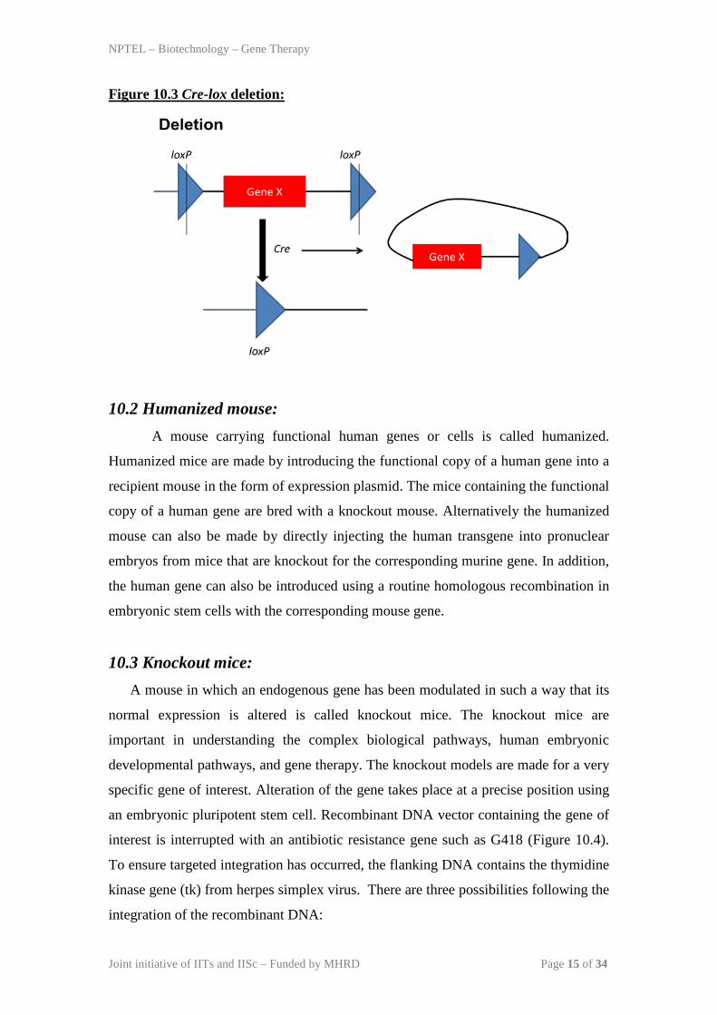
the gene of interest (Figure 10.1). However, Cre recombinase mediates a translocation
event if the loxP sites are located on different chromosomes (Figure 10.2) and
deletion event if the loxP sites are oriented in the same direction on a chromosome
segment (Figure 10.3).
Some terminology:
Pluripotent embryonic stem cells are undifferentiated early embryonic cells derived
from the inner cell mass of mouse blastocysts.
Nuclear localization sequences (NLS) are important for directing a protein to nucleus.
Generally NLS contains PK3RKV amino acid residues in the protein. The proteins are
directed to the nucleus with the help of nuclear pore complex and with the combined
effect of RAN-GTPase and IMPORTIN molecules.

NPTEL – Biotechnology – Gene Therapy
Joint initiative of IITs and IISc – Funded by MHRD Page 14 of 34
Figure 10.1 Cre-lox inversion:
Figure 10.2 Cre-lox translocation:
NPTEL – Biotechnology – Gene Therapy
Joint initiative of IITs and IISc – Funded by MHRD Page 15 of 34
Figure 10.3 Cre-lox deletion:
10.2 Humanized mouse: A mouse carrying functional human genes or cells is called humanized.
Humanized mice are made by introducing the functional copy of a human gene into a
recipient mouse in the form of expression plasmid. The mice containing the functional
copy of a human gene are bred with a knockout mouse. Alternatively the humanized
mouse can also be made by directly injecting the human transgene into pronuclear
embryos from mice that are knockout for the corresponding murine gene. In addition,
the human gene can also be introduced using a routine homologous recombination in
embryonic stem cells with the corresponding mouse gene.
10.3 Knockout mice: A mouse in which an endogenous gene has been modulated in such a way that its
normal expression is altered is called knockout mice. The knockout mice are
important in understanding the complex biological pathways, human embryonic
developmental pathways, and gene therapy. The knockout models are made for a very
specific gene of interest. Alteration of the gene takes place at a precise position using
an embryonic pluripotent stem cell. Recombinant DNA vector containing the gene of
interest is interrupted with an antibiotic resistance gene such as G418 (Figure 10.4).
To ensure targeted integration has occurred, the flanking DNA contains the thymidine
kinase gene (tk) from herpes simplex virus. There are three possibilities following the
integration of the recombinant DNA:

NPTEL – Biotechnology – Gene Therapy
Joint initiative of IITs and IISc – Funded by MHRD Page 16 of 34
1. Cells or embryo will die if they fail to integrate when grown in presence of
neomycin and in absence of resistance gene.
2. The cells or embryo will die under Ganciclovir if the integration occurs at
random site, since the expression of tk will kill it.
3. Cells and embryo will survive if the integration is site specific since the
knocked out cells will survive both in G418 and Ganciclovir.
Figure 10.4 knockout mice generation:
NPTEL – Biotechnology – Gene Therapy
Joint initiative of IITs and IISc – Funded by MHRD Page 17 of 34
Lecture 11
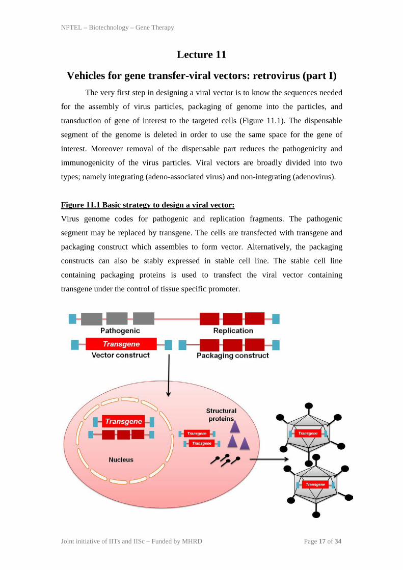
Vehicles for gene transfer-viral vectors: retrovirus (part I) The very first step in designing a viral vector is to know the sequences needed
for the assembly of virus particles, packaging of genome into the particles, and
transduction of gene of interest to the targeted cells (Figure 11.1). The dispensable
segment of the genome is deleted in order to use the same space for the gene of
interest. Moreover removal of the dispensable part reduces the pathogenicity and
immunogenicity of the virus particles. Viral vectors are broadly divided into two
types; namely integrating (adeno-associated virus) and non-integrating (adenovirus).
Figure 11.1 Basic strategy to design a viral vector:
Virus genome codes for pathogenic and replication fragments. The pathogenic
segment may be replaced by transgene. The cells are transfected with transgene and
packaging construct which assembles to form vector. Alternatively, the packaging
constructs can also be stably expressed in stable cell line. The stable cell line
containing packaging proteins is used to transfect the viral vector containing
transgene under the control of tissue specific promoter.
NPTEL – Biotechnology – Gene Therapy
Joint initiative of IITs and IISc – Funded by MHRD Page 18 of 34
11.1 Retrovirus Retroviruses are a large family of enveloped RNA viruses found in all
vertebrates. The enveloped virus particle contains two copies of the viral RNA
genome, which are surrounded by a cone-shaped core. The viral RNA contains three
essential genes, gag, pol, and env, and is flanked by long terminal repeats (LTRs).
The env gene encodes for the envelope glycoproteins (gP120 and gP41), which
mediates virus entry.
Functions of gag, pol, and env
– Gag protein is proteolytically processed into
• MA (matrix)
• CA (capsid)
• NC (nucleocapsid)
– Pol protein encodes enzymes
• PR (protease)
• RT (Reverse Transcriptase which has both DNA polymerase
and RNase H activities)
• IN (Integrase)
– Env protein encodes
• SU surface glycoprotein (gP120)
• TM transmembrane protein (gP41)

NPTEL – Biotechnology – Gene Therapy
Joint initiative of IITs and IISc – Funded by MHRD Page 19 of 34
Figure 11.2 Schematic representation of a retrovirus:
The use of each coreceptor corresponds to viruses with different biological
properties and pathogenicity. Viruses isolated at the beginning of infection use the
CCR5 co-receptor, which is the major coreceptor for macrophage-tropic strains (M-
tropic). In full-blown AIDS cases, new viral species appear with high level of
replication, cytopathic effects and they use the coreceptor CXCR4, which is the major
receptor for T-cell strains (T-cell tropic). There are also dual tropic viruses that can
use both CXCR4 and CCR5 coreceptors and alternative chemokine coreceptors. The
fusion between the viral membrane and the cellular membrane involves binding of
CD4 receptor with gp120 and a change in conformation of gp41, which enables it to
insert into the cellular phospholipids bilayer. The retrovirus that binds to the receptor
of mice cells are called ECOTROPIC virus while those binding to both human and
mice is called as AMPHOTROPIC virus.
Points to remember:
Reverse transcriptase is an enzyme present in retrovirus (eg HIV) that converts RNA
to DNA. The phenomenon is also called as TEMINISM (Discovered by Temin and
Baltimore)
NPTEL – Biotechnology – Gene Therapy
Joint initiative of IITs and IISc – Funded by MHRD Page 20 of 34
11.2 Replication cycle of retrovirus Virion attachment to a specific cell surface receptor
Virion penetration into the cell
Reverse transcription of ther genome.
Transfer of viral DNA to the infected cell nucleus
Integration of viral DNA randomly to the cellular DNA to form the provirus
Viral RNA synthesis by cellular RNA polymerase II using the proviral
template
Transcripts processing to viral genome and mRNAs
Virus protein synthesis
Assembly and budding from cell surface
Processing of capsid proteins
After binding to its receptor (CD4 receptor on T cells), the viral capsid containing
the RNA genome enters the cell through membrane fusion. The viral RNA genome is
subsequently converted into a double-stranded viral DNA by the viral enzyme reverse
transcriptase. The viral DNA is heavily associated with viral proteins like
nucleocapsid, reverse transcriptase, and integrase, and translocates to the nucleus
where the viral enzyme integrase mediates integration of the viral DNA into the host
cell genome to form PROVIRUS.

NPTEL – Biotechnology – Gene Therapy
Joint initiative of IITs and IISc – Funded by MHRD Page 21 of 34
Lecture 12
Vehicles for gene transfer-viral vectors: retrovirus (part II) 12.1 Retroviral vectors
Among the viral vectors used for gene therapy trials, retrovirus is the most
commonly used RNA virus. Retroviruses are the first to develop for the application of
gene therapy related studies. Retroviruses are the RNA containing enveloped viruses
and are classified into oncoretroviruses, lentiviruses, and spumaviruses.
Retroviruses are enveloped viruses having two copies of ssRNA genome. The
viral genome codes for three essential proteins; namely gag, pol, and env (Figure 2).
The genome is flanked with long terminal repeats (LTR) in its terminal end. The gag
gene in retroviruses codes for capsid, matrix, and nucleocapsid proteins. The pol gene
codes for viral enzymes protease, integrase, and reverse transcriptase. The env gene
codes for surface glycoproteins, which mediates the virus entry into the cells.
Oncoretroviruses only codes for these three proteins while lentiviruses and
spumaviruses are more complex in nature (discussed in later chapter).
Figure 12.1 Schematic diagram of retrovirus:
Entry of virus particle is accomplished by the fusion of viral surface
glycoprotein with the host cell receptor. The viral RNA genome is converted into
DNA by the enzyme reverse transcriptase. The DNA after combining with the viral
proteins then migrates to nucleus and integrates with the host cell chromosome with
the help of enzyme integrase. The transcription of the viral mRNA starts from LTR
using host cell transcription factors. The new virus particles are formed after
assembling of two copies of ssRNA along with the essential enzyme inside core. The
mature virus particles are released by budding from the host cell membrane.

NPTEL – Biotechnology – Gene Therapy
Joint initiative of IITs and IISc – Funded by MHRD Page 22 of 34
Retroviral vectors are based on replication deficient retroviruses. The vectors
are mainly derived from Rous sarcoma virus, avian leukosis virus, and murine
leukemia virus. The retroviral vectors are made by replacing the viral proteins with
the gene of interest driven by a tissue specific promoter. The vector also contains the
long terminal repeat (LTR) which is an essential packaging signal. In addition, the
vector also contains essential enzyme such as reverse transcriptase and integrase.
Vector RNA production is achieved by either the LTR or promoters present upstream
to the transgene. The packaging of the vector is completed by the incorporation of
viral structural proteins in trans from packaging cell lines. Alternatively, the vector
can also be produced by the transfection of plasmid expressing the structural proteins.
The latter method is less time consuming as it avoids the use of packaging cell line.
Viruses are recovered from the supernatants of actively growing producer cells.
Major issue of using retroviral vectors is the possibility of the defective
genome to be recombined with the host cell chromosome. The concern leads to the
development of self inactivating retroviral vectors by using tissue specific promoter
for transgene transcription. In self inactivating vectors, the transcription of the
transgene is carried out by using an internal promoter instead of LTR. Apart for gene
delivery, retroviral vectors are extensively used for many other applications.
Retroviruses have ability to integrate with the host cell chromosome; the property is
explored to express the protein of interest in a cell. Long term expression of a protein
in a suitable cell is a very useful way to make a stable cell line. The replication
depends on actively dividing cells making these an important tool to manipulate stem
cells and tumor cells. On the other hand lentiviral vectors are used for gene delivery in
many organs including brain, eye, liver, muscles, and hematopoietic cells.
The transduction of retroviral vector is limited because of its ability to have
limited cellular tropism. The cellular tropism of the retrovirus is broadened by the
incorporation of envelope from related or unrelated viruses, making them pseudotype
virus. Incorporation of vesicular stomatitis virus glycoproteins into the retrovirus
virion or the envelope of murine leukemia virus allows the broad host range to the
transducing vectors. Pseudotyping of retrovirus by the use of lyssavirus glycoprotein
makes it transducible to the brain while incorporation of surface protein of ebola virus
helps in transducing airway epithelium.

NPTEL – Biotechnology – Gene Therapy
Joint initiative of IITs and IISc – Funded by MHRD Page 23 of 34
Lecture 13
Adenovirus
Adenoviruses are isolated from wide varieties of animals and human beings.
The list of adenovirus serotypes that affects human are more than 50. Adenoviruses
are associated with the respiratory diseases, conjunctivitis and gastroenteritis in
humans. Adenoviruses are also associated with the tumor formation in animals.
Adenoviruses are non-enveloped and icosahedral particles usually around 90 nm in
diameter (Figure 13.1). Adenovirus has a double stranded DNA (ds-DNA) genome of
approximately 35-36 kb in length. The ds-DNA genome contains transcription
segments in an overlapping fashion. The genome contains more than 50 proteins that
are formed by splicing, 11 of which are structural proteins.
Figure 13.1 Schematic representation of adenovirus:

NPTEL – Biotechnology – Gene Therapy
Joint initiative of IITs and IISc – Funded by MHRD Page 24 of 34
Mature virion contains penton base, which is surrounded by 5 different
proteins and hexon proteins, which are surrounded by 6 different proteins. The penton
base contains a fiber that interacts with the host cell receptors. The fiber protein
projects from the virion, and the carboxy-terminal (knob) forms a high-affinity
complex with the receptor present on the host cell. The virion consists of a shell called
capsid which surrounds DNA containing core (Figure 13.1). The DNA is packaged in
such a way that it touches all the penton proteins. The Coxsackie virus and
adenovirus receptor (CAR) present in respiratory epithelium, nervous system, liver,
lung, and intestinal lining acts as a receptor for the adenovirus. While entering the cell
through CAR, the virions follow caveolae mediated endocytosis (clathrin
independent). It has been shown that efficient virus entry requires the interaction of
penton proteins and the cellular integrins receptor.
Some facts:
There are certain enzymes like fillipin and methyl β-cyclodextrin which destroys
cholesterol on the cell surface.
13.1 Virus replication
The virus replication and life cycle is divided into late and early stages. After
entering the cell, the virion is endocytosed in lysosome which has acidic environment.
The capsid of the virion gets disrupted by the acidic environment releasing the
subvirion into the cytoplasm. Subvirion of the adenovirus interacts with the nuclear
pore complex leading to its migration into the infected cell nucleus. As soon as it
reaches the nucleus, early proteins are synthesized which are required for viral
genome replication, transcription, and translation. These early proteins make their
way out in the cytoplasm in order to facilitate the process. The late proteins are
formed later during the infectious cycle and comprise mainly of capsid or structural
proteins (L1–L5). The adenovirus uses most of the host cell machinery in order to
make a suitable niche inside host cells. There are five early proteins which are E1A,
E1B, E2, E3 and E4 (Figure 13.2). Among these E1 and E4 are responsible for DNA

NPTEL – Biotechnology – Gene Therapy
Joint initiative of IITs and IISc – Funded by MHRD Page 25 of 34
replication, E2 for RNA polymerase while E3 is responsible for virus specific
immune response. Inverted terminal repeats (ITRs) are present on the 5’ and 3’ ends
of the viral genome and are the packaging signals responsible for packaging of viral
genome into the icosahedral capsid. Adenovirus also forms an RNA intermediate
called virus-associated (VA) RNA. The VA RNA is not fully functional because it
doesn’t form any protein. The viral transcription segments are transcribed by the help
of cellular RNA polymerase II, whereas the VA is transcribed by RNA polymerase
III. Infection of adenovirus induces a high immune response in the host body. The
initial immune response comprises of cytokines such as tumor necrosis factor and
interleukins 1 and 6 followed by specific cytotoxic T lymphocyte. The high level of
immune response against the adenovirus is a major hurdle in making adenoviral
vector as a gene therapy tool.
Figure 13.2 Adenoviral genome organization:
13.2 Adenovirus as a vector
Adenoviruses have been explored as a gene therapy vector for quite some time
with varying degree of success. Generally adenovirus serotype 5 is widely used but
serotypes 2, 4, 7, and non human adenovirus isolates were also explored for gene
therapy vector. Adenovirus is made replication deficient after removing the coding
regions of the viral genome. The first generation of adenoviral vectors is made by
removing E1 region of the genome with a transgene (gene of interest). The E1 protein
in adenovirus is essential to activate the expression of genome specific transcripts;
absence of E1 makes the virus replication deficient in most of the cell lines.
Alternatively, the E1 deficient virus can be grown in cells that contain the E1 proteins
in trans, such as 293 cells. Removal of E1 from the genome reduces its size which is
further used to put the transgene of around 4.5 kb at the same position. The cloning

NPTEL – Biotechnology – Gene Therapy
Joint initiative of IITs and IISc – Funded by MHRD Page 26 of 34
capacity of E1 deleted adenoviral vectors can be further increased by deleting the
nonessential region of E3 protein (Figure 13.3). Minimum quantity of viral gene in
the vector reduces the chances of recombination with the host cell DNA.
Figure 13.3 E1/E3 depleted helper virus:
The second generations of adenovirus vectors are generated by deleting E1 as
well as part of E2 and/or E4 gene segments. The deletion of E2 along with E1 further
increases the capacity of adenovirus vector to accommodate a larger transgene. E2
proteins are essential for viral genome replication and for successful rescue of
adenoviral vector it has to be supplied in trans in the packaging cells. Many deletions
in other genes on the adenovirus genome have been tried by scientists in order to
increase the capacity to clone a transgene. The deleted regions of the adenovirus
genome must be provided in trans in order to rescue recombinant virus particles
carrying the gene of interest. In theory it is possible to make an adenovirus that lacks
almost all of its proteins except the ITRs. The vectors made in this way don’t have
any virus specific sequences and are termed as GUTLESS vectors (Figure 13.4).
Figure 13.4 Gutless vector:
A third generation adenovirus vector is made by generating a cell line that can
stably express the essential adenovirus proteins. This is achieved by Cre-lox
recombination method where adenovirus genome is integrated at the loxp site of 293
cells. The recombinant vector is made by transfecting the transgene driven by a
suitable promoter in 293 cells expressing the adenoviral proteins (Figure 13.5). The
process is very laborious but can still induce an immune response that can kick out the

NPTEL – Biotechnology – Gene Therapy
Joint initiative of IITs and IISc – Funded by MHRD Page 27 of 34
vector from the host cell. The major breakthrough in the field of gene therapy can be
achieved by making an adenovirus vector that is devoid of any host immune response.
Figure 13.5 Adenovirus vector production:
Gutless adenovirus vector contains cis-acting ITRs and the packaging signal ψ
along with the transgene insert and promoter sequence. Helper adenoviral vector
without E3 and E1 with loxP sites flanks ends of the packaging sequence. The helper
vector is transfected in the 293 producer cells because they express cre recombinase.
Here ψ sequence is excised to prevent packaging of helper construct.

NPTEL – Biotechnology – Gene Therapy
Joint initiative of IITs and IISc – Funded by MHRD Page 28 of 34

NPTEL – Biotechnology – Gene Therapy
Joint initiative of IITs and IISc – Funded by MHRD Page 29 of 34
Lecture 14
Adeno-associated virus
Adeno-associated virus (AAV) is a non pathogenic virus. It was isolated
during the screening of sputum from of an infected patient. The AAV is mostly
associated as a contaminant of adeno and herpesvirus infection. It is also called as
dependovirus because it depends on other helper virus for replication and infection
and belongs to the family parvoviridae. Infection of AAV into the host cell requires
helper function of adenovirus or herpesvirus. In addition to helper virus infection the
AAV infection in the cells can also be achieved under stress conditions such as
radiation or any drug that can cause toxicity to the host genome. In the absence of
conducive condition the AAV genome can integrate to the host cell chromosome to
establish the latent infection. Different serotypes of AAVs are isolated from different
species and their number keeps on increasing.
All AAV serotypes are having similar size and genome characteristics. Among
all AAV-2 is the most characterized and is mostly used as a backbone for the
preparation of recombinant AAV. AAV virions are on an average 25 nanometer in
diameter with an icosahedral capsid. The genome of AAV is single stranded DNA
(ssDNA) of about 4.7kb in size. The genome encodes for two open reading frame
(ORF); rep and cap. The rep and cap genes are flanked by inverted terminal repeats
(ITR) similar to that of an adenovirus (Figure 14.1). The ITR sequences in the AAV
genomes are very important and are responsible for replication, packaging and
integration of the virus genome. The recombinant AAVs are made by removing all the
internal genes except the ITRs. All the essential genes are supplied in trans or a
packaging cell line expressing rep and cap proteins. There are four rep proteins
required for replication and packaging of the viral genome. The AAV cap codes for 3
structural proteins; VP1, VP2, and VP3. The VP1, VP2, and VP3 form the viral
capsid and usually presents in the ratio of 1:1:10, respectively.

NPTEL – Biotechnology – Gene Therapy
Joint initiative of IITs and IISc – Funded by MHRD Page 30 of 34
Figure 14.1 Schematic representation of adeno-associated virus genome:
Different AAV serotypes are reported to use different cell surface receptors
for their entry. The specificity of the host cell receptor makes the serotype specific for
a particular host. The heparan sulfate proteoglycans present in a variety of cell types
is a receptor used by AAV-2. In addition, fibroblast growth factor receptor-1 and
integrin αvβ5 are also reported as a co-receptor for the AAV infection in the host
cells. Sialic acid is used as receptor by AAV-4 and AAV-5. Usually after binding to
cell receptors, virus particles are internalized by receptor mediated endocytosis and
the ssDNA is transported to the host cell nucleus.
The ITRs of AAV contains the self complementary sequences which are hair
pin like structure and provides a binding site for the viral rep protein. In addition to
the rep protein mediated virus genome replication, host cell machinery also helps in
its replication. The genome replication takes place in the nucleus and both +ve and –
ve sense genome is packaged with equal frequency in the virion capsid. The ssDNA is
converted to dsDNA form in the nucleus to facilitate the expression of viral proteins
and genome replication. The dsDNA is either formed by the complementary DNA
strand from another virus or mediated by the host cell machinery. The AAV genome
is known to integrate specifically to the chromosome 19 at the location q13.4 by
nonhomologous recombination. The integration is mediated by the rep proteins, and
remains in the cell throughout its life span. Further AAV genome replication depends
on the infection of helper virus or their gene products. Upon infection of adeno or
herpesvirus, the viral protein regulates the replication and transcription of AAV
genome. The new virus particle gets assembled and packaged in the nucleus and
released from the cells along with the helper viruses (adeno or herpesvirus).
NPTEL – Biotechnology – Gene Therapy
Joint initiative of IITs and IISc – Funded by MHRD Page 31 of 34
14.1 Adeno-associated virus as a gene therapy vector
The first AAV gene therapy vector was generated by replacing internal rep
and cap gene with the transgene. The recombinant virus was produced by the co-
infection of helper virus and transfection of plasmid expressing rep and cap proteins
in trans. The method still generates the vector having ITR sequences (around 400bps)
of the viral genome. The presence of ITR sequences may lead to production of wild
type virus in the subsequent generation.
The next generation of AAV contains only part of ITR sequences essential for
the production and integration. Essentially the AAV vector system contains a
promoter and a transgene flanked by the ITRs, the method prevents the production of
replication competent AAV production. The adenovirus E1A, E1B, E2A, VA, and E4
proteins are essential for the production of a successful virus particle. To avoid the
use of replication adenovirus particles, scientists have developed a system that
requires only E2A, VA, and E4 proteins to produce viable AAV vector. The system
requires transfection of E2A, VA, and E4 gene expressing plasmid in 293 cells.
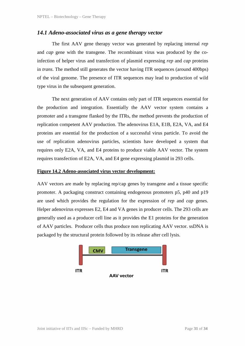
Figure 14.2 Adeno-associated virus vector development:
AAV vectors are made by replacing rep/cap genes by transgene and a tissue specific
promoter. A packaging construct containing endogenous promoters p5, p40 and p19
are used which provides the regulation for the expression of rep and cap genes.
Helper adenovirus expresses E2, E4 and VA genes in producer cells. The 293 cells are
generally used as a producer cell line as it provides the E1 proteins for the generation
of AAV particles. Producer cells thus produce non replicating AAV vector. ssDNA is
packaged by the structural protein followed by its release after cell lysis.

NPTEL – Biotechnology – Gene Therapy
Joint initiative of IITs and IISc – Funded by MHRD Page 32 of 34
Interesting points:
293 cells are known to express adenovirus E1A and E1B gene products.

NPTEL – Biotechnology – Gene Therapy
Joint initiative of IITs and IISc – Funded by MHRD Page 33 of 34
Table 14.1 Difference between adeno and adeno-associated virus:
Character Adenovirus Adeno-associated
virus
Integration in host No Yes
Expression of vector Transient Stable
Transfection
efficiency
High Low
Immune response High No
Genome 36kb
ds DNA
4.7kb
ssDNA
Envelope No No
Receptor CAR Heparan Sulphate
Cell line 293 HEK 293 HEK
Entry Caveolae mediated Clathrin mediated
Proteins Five early and five late Four for replicase and three for capsid
Capsid size 40 nm 25 nm
14.2 Tropism of adeno-associated virus
AAV-2 is the most common serotype used to develop a gene therapy vector.
The cap protein of AAV-1, -2 and -3 share a great homology with each other and
hence are known to use the heparan sulfate receptor. The capsid proteins of AAV-4
and -5 are different from other serotypes and probably involved in using different
cellular receptors to infect the susceptible host. The AAV-1 is more tropic towards
muscle and liver while AAV-5 is more tropic to retina (eye). AAV-3 is more tropic
towards the hematopoietic stem cells. The tropism of AAV serotypes can by
modulated by shuffling the capsid proteins. To be more efficient AAV genome should
be of dsDNA instead of parental ssDNA, the expression of transgene increases to a
large extent after the production of dsDNA from ssDNA by host cell machinery. The

NPTEL – Biotechnology – Gene Therapy
Joint initiative of IITs and IISc – Funded by MHRD Page 34 of 34
dsDNA form of the AAV vector can persist for a long time in a transduced cell by
head to tails recombination with ITR sequences.
The major hurdle in AAV mediated gene therapy is regarding the limitation of
transgene capacity. Another hurdle is regarding the presence of neutralizing
antibodies against the AAV serotypes in the human population because of natural
AAV infection. In addition a single injection of AAV can mount a huge humoral
immune response that inhibits the second injection of the same serotype. Using AAV
serotypes of different capsid protein may overcome this hurdle. The AAV offers a
very promising tool for the gene therapy vector and its efficacy and potential is
increasing day by day. Currently many gene therapy trials are going on using AAV as
a vector expressing different transgene.