Embed Size (px)
Citation preview
MODULE 2.4. NONCLINICAL OVERVIEW
CONFIDENTIALm2.4. Nonclinical Overview 2013N179645_00
2
TABLE OF CONTENTS
PAGEList of Abbreviations .................................................................................................7
1. OVERVIEW OF THE NONCLINICAL TESTING STRATEGY...................................81.1. Introduction...................................................................................................8
1.1.1. Rationale for the use of DTG/ABC/3TC FDC in the treatment of HIV.............................................................................9
1.2. Nonclinical Development Programme .........................................................101.2.1. DTG/ABC/3TC in combination .....................................................101.2.2. DTG, ABC and 3TC as monotherapies ........................................10
1.2.2.1. DTG ...........................................................................111.2.2.2. ABC............................................................................111.2.2.3. 3TC ............................................................................12
2. PHARMACOLOGY.................................................................................................132.1. Presentation Order of Pharmacology Summaries .......................................132.2. DTG............................................................................................................13
2.2.1. Virology (primary pharmacodynamics).........................................132.2.1.1. Mechanism of action...................................................132.2.1.2. In vitro antiviral activity and potential resistance .........13
2.2.2. Secondary pharmacology ............................................................142.2.3. Safety pharmacology ...................................................................142.2.4. Pharmacodynamic drug interactions ............................................15
2.3. ABC............................................................................................................152.3.1. Virology (primary pharmacodynamics).........................................15
2.3.1.1. Mechanism of action...................................................162.3.1.2. In vitro antiviral activity and potential resistance .........16
2.3.2. Secondary pharmacology ............................................................162.3.3. Safety pharmacology ...................................................................162.3.4. Pharmacodynamic drug interactions ............................................18
2.4. 3TC.............................................................................................................182.4.1. Virology (primary pharmacodynamics).........................................18
2.4.1.1. Mechanism of action...................................................182.4.1.2. In vitro antiviral activity and potential resistance .........18
2.4.2. Secondary pharmacology ............................................................192.4.3. Safety pharmacology ...................................................................192.4.4. Pharmacodynamic drug interactions ............................................19
3. PHARMACOKINETICS ..........................................................................................203.1. Presentation Order of Pharmacokinetics Summaries..................................203.2. DTG............................................................................................................20
3.2.1. Analytical methods and validation ................................................203.2.2. Absorption and pharmacokinetics ................................................21
3.2.2.1. Single dose.................................................................213.2.2.2. Repeat dose toxicokinetics .........................................21
3.2.3. Distribution...................................................................................223.2.3.1. Protein binding and blood cell association ..................223.2.3.2. Efflux-mediated transport and cell membrane
permeability ................................................................22

CONFIDENTIALm2.4. Nonclinical Overview 2013N179645_00
3
3.2.3.3. Tissue distribution.......................................................223.2.4. Metabolism ..................................................................................23
3.2.4.1. In vitro biotransformation ............................................233.2.4.2. In vivo studies.............................................................23
3.2.5. Excretion......................................................................................263.2.6. Pharmacokinetic drug interactions ...............................................26
3.2.6.1. Potential effect of co-administered agents on DTG ...........................................................................27
3.2.6.2. Effect of DTG on co-administered agents ...................273.3. ABC............................................................................................................28
3.3.1. Analytical methods and validation ................................................283.3.2. Absorption and pharmacokinetics ................................................29
3.3.2.1. Single dose.................................................................293.3.2.2. Repeat dose toxicokinetics .........................................29
3.3.3. Distribution...................................................................................293.3.3.1. Protein binding and blood cell association ..................293.3.3.2. Efflux-mediated transport and cell membrane
permeability ................................................................293.3.3.3. Tissue distribution.......................................................30
3.3.4. Metabolism ..................................................................................303.3.4.1. In vitro biotransformation ............................................303.3.4.2. In vivo studies.............................................................31
3.3.5. Excretion......................................................................................333.3.6. Pharmacokinetic drug-drug interactions.......................................33
3.3.6.1. In vitro drug interaction studies with ABC ...................333.3.6.2. Potential effect of co-administered agents on
ABC............................................................................333.3.6.3. Potential effect of ABC on co-administered
agents ........................................................................343.4. 3TC.............................................................................................................34
3.4.1. Analytical methods and validation ................................................343.4.2. Absorption and pharmacokinetics ................................................34
3.4.2.1. Single dose.................................................................343.4.2.2. Repeat dose toxicokinetics .........................................35
3.4.3. Distribution...................................................................................353.4.3.1. Protein binding and blood cell association ..................353.4.3.2. Efflux-mediated transport and cell membrane
permeability ................................................................363.4.3.3. Tissue distribution.......................................................36
3.4.4. Metabolism ..................................................................................363.4.4.1. In vivo biotransformation.............................................36
3.4.5. Excretion......................................................................................383.4.6. Pharmacokinetic drug-drug interactions.......................................39
3.4.6.1. In vitro drug interaction studies with 3TC....................393.4.6.2. In vivo drug interaction studies with 3TC ....................393.4.6.3. Potential effect of co-administered agents on
3TC ............................................................................393.4.6.4. Effect of 3TC on co-administered agents....................39
4. TOXICOLOGY .......................................................................................................414.1. Presentation Order of Toxicology Summaries.............................................414.2. DTG............................................................................................................41

CONFIDENTIALm2.4. Nonclinical Overview 2013N179645_00
4
4.2.1. Choice of species ........................................................................414.2.2. Single dose toxicity ......................................................................414.2.3. Repeat dose toxicity ....................................................................42
4.2.3.1. Treatment-related mortality/morbidity .........................424.2.3.2. Gastrointestinal effects ...............................................424.2.3.3. Hepatic effects............................................................444.2.3.4. Renal effects ..............................................................454.2.3.5. Bone marrow and lymph node effects.........................46
4.2.4. Genetic toxicology .......................................................................474.2.5. Carcinogenicity ............................................................................474.2.6. Reproductive toxicology...............................................................474.2.7. Juvenile toxicology.......................................................................494.2.8. Irritancy........................................................................................494.2.9. Immunotoxicity.............................................................................49
4.2.9.1. Immunotoxicity assessment in adults..........................494.2.9.2. Immunotoxicity assessment in juveniles .....................49
4.2.10. Phototoxicity ................................................................................504.3. ABC............................................................................................................57
4.3.1. Choice of species ........................................................................574.3.2. Single dose toxicity ......................................................................574.3.3. Repeat dose toxicity ....................................................................57
4.3.3.1. General condition and mortality ..................................584.3.3.2. Hematology effects.....................................................584.3.3.3. Liver effects ................................................................594.3.3.4. Testes effects .............................................................604.3.3.5. Other findings .............................................................61
4.3.4. Genetic toxicology .......................................................................624.3.5. Carcinogenicity ............................................................................644.3.6. Reproductive toxicology...............................................................654.3.7. Mitochondrial toxicity....................................................................674.3.8. Juvenile toxicology.......................................................................674.3.9. Hypersensitivity............................................................................684.3.10. Phototoxicity ................................................................................68
4.4. 3TC.............................................................................................................704.4.1. Choice of species ........................................................................704.4.2. Single dose toxicity ......................................................................704.4.3. Repeat dose toxicity ....................................................................714.4.4. Genetic toxicology .......................................................................724.4.5. Carcinogenicity ............................................................................734.4.6. Reproductive and developmental toxicity .....................................744.4.7. Mitochondrial toxicity....................................................................764.4.8. Phototoxicity ................................................................................78
4.5. Impurities and Excipients ............................................................................804.5.1. Combination drug product............................................................80
5. INTEGRATED ASSESSMENT OF THE FIXED DOSE COMBINATION PRODUCT .............................................................................................................82
5.1.1. Potential for pharmacodynamic interactions.................................835.1.2. Potential for pharmacokinetic interactions....................................835.1.3. Potential for toxicologic interactions .............................................83
5.1.3.1. Repeat dose toxicity studies .......................................835.1.3.2. Genotoxicity and carcinogenicity ................................84

CONFIDENTIALm2.4. Nonclinical Overview 2013N179645_00
5
5.1.3.3. Fertility, pregnancy and lactation ................................85
6. OVERALL CONCLUSIONS....................................................................................86
7. LIST OF LITERATURE CITATIONS.......................................................................87
CONFIDENTIALm2.4. Nonclinical Overview 2013N179645_00
6
LIST OF TABLES
PAGE
Table 1 Principal Toxicological Findings in Rats and Monkeys Following Oral Administration of DTG ....................................................................51
Table 2 Comparative Assessment of Mean Systemic Exposure Following Oral Administration of DTG ....................................................................52
Table 3 Comparative Assessment of Mean Animal to Human Exposure Ratios (AUC, mg/kg and mg/M2) Following Oral Administration of DTG in the 4 and 26 Week Rat Toxicology Studies................................55
Table 4 Comparative Assessment of Mean Animal to Human Exposure Ratios (AUC, mg/kg and mg/M2) Following Oral Administration of DTG in the 14 Day and 4 and 38 Week Monkey Toxicology Studies...................................................................................................56
Table 5 Comparative Assessment of Mean Systemic Exposure Following Oral Administration of ABC.....................................................................69
Table 6 Comparative Assessment of Mean Systemic Exposure Following Oral Administration of 3TC .....................................................................79
LIST OF FIGURES
PAGE
Figure 1 Structure of DTG, ABC and 3TC ..............................................................8
Figure 2 Comparative Metabolic Profile of DTG Between Nonclinical Species and Humans .............................................................................25
Figure 3 Comparative Metabolic Profile of Primary ABC Metabolites Between Nonclinical Species and Humans ............................................32
Figure 4 Comparative Metabolic Profile of 3TC Metabolites Between Nonclinical Species and Humans ...........................................................38

CONFIDENTIALm2.4. Nonclinical Overview 2013N179645_00
7
List of Abbreviations
ABC AbacavirAIDS Acquired immunodeficiency syndromeAPI Active pharmaceutical ingredientBID Twice dailyCFR Code of Federal Regulations (US)DRV DarunavirDTG DolutegravirEVG ElvitegravirFC Fold changeFDC Fixed dose combinationGI GastrointestinalHCV Hepatitis C virusHIV Human immunodeficiency virusIN IntegraseINI Integrase inhibitorNNRTI Non-nucleoside reverse transcriptase inhibitorNOAEL No observed adverse effect levelNRTI Nucleoside reverse transcriptase inhibitorQD Once dailyRAL RaltegravirRT Reverse transcriptaseRTV RitonavirSTR Single tablet regimenTK Toxicokinetics3TC Lamivudine

CONFIDENTIALm2.4. Nonclinical Overview 2013N179645_00
8
1. OVERVIEW OF THE NONCLINICAL TESTINGSTRATEGY
1.1. Introduction
Dolutegravir (DTG) is an integrase inhibitor (INI) and abacavir (ABC) and lamivudine (3TC) are nucleoside reverse transcriptase inhibitors (NRTIs). A once daily fixed dose combination (FDC) single tablet regimen (STR) that combines DTG with ABC and 3TC is being developed for use in the treatment of human immunodeficiency virus (HIV) infection (the STR is referred to in this document as DTG/ABC/3TC FDC).
This current application is for DTG/ABC/3TC FDC as a complete regimen for the treatment of HIV infection in adults and adolescents from 12 years of age who are antiretroviral treatment-naïve or are infected with HIV without documented or clinically suspected resistance to any of the 3 antiretroviral agents in DTG/ABC/3TC FDC.
The dose will be orally administered as an FDC tablet. Each film coated tablet contains 50 mg of dolutegravir as dolutegravir sodium, 600 mg of abacavir as abacavir sulfate and 300 mg of lamivudine.
Figure 1 Structure of DTG, ABC and 3TC
Dolutegravir (DTG)
NH2
N
S
HO
O
O
N
Chiral
Lamivudine (3TC) Abacavir (ABC)

CONFIDENTIALm2.4. Nonclinical Overview 2013N179645_00
9
1.1.1. Rationale for the use of DTG/ABC/3TC FDC in the treatment of HIV
HIV is a retrovirus which is recognised as the aetiological agent of the acquired immunodeficiency syndrome (AIDS). The initiation and progression of AIDS is characterised by the infection of CD4 expressing cells, for example, lymphocytes, monocytes and macrophages via the binding of the viral envelope protein gp120 to the cellular receptor CD4. When these CD4+ cells have been infected with HIV, a proviral DNA copy of the ribonucleic acid must be synthesised before the viral genome can be integrated into the host genome and viral replication commence. This essential step is dependent upon the presence of the virus coded enzyme reverse transcriptase. HIV-reverse transcriptase is therefore an excellent target for antiviral therapy and indeed several dideoxynucleosides are metabolised intracellularly to their triphosphate derivatives which interact with HIV-reverse transcriptase and block the replication of the virus and its induced cytopathic effects in vitro [Mitsuya, 1985; Mitsuya, 1986].
The antiviral activity of integrase inhibitors has been demonstrated in short-term monotherapy studies for raltegravir (MK-0518, Merck; RAL) and elvitegravir (GS-9137, Gilead; EVG) with a ~2 log drop in HIV RNA-1 [Markowitz, 2006; DeJesus, 2006]. Longer-term data with RAL demonstrated a significant antiviral effect in treatment experienced patients when added to an optimized background regimen of antiretroviral therapy [Grinsztejn, 2007] and in treatment-naïve patients when co-administered with a nucleoside backbone [Merck Research Laboratories, 2007]. Clinical resistance to both RAL and EVG has been reported in treatment experienced patients [Hazuda, 2007; McColl, 2007]. Therefore, the development of new integrase inhibitors (INI) with different resistance profiles is desirable, and in the case of many treatment-experienced patients with clinical resistance to RAL and EVG, is essential for providing HIV-infected individuals an option for constructing an effective antiretroviral regimen. DTG is a potent, low nanomolar inhibitor of HIV integrase which provides the excellent antiviral activity and tolerability demonstrated for the INI class.
DTG is currently available in the United States (US) and has marketing applications under review in the European Union (EU) and other countries worldwide; it received US approval in August 2013 and is marketed as TIVICAY™ (NDA 204790).
There is extensive clinical experience with abacavir and lamivudine marketed as individual agents (ZIAGEN™, EPIVIR™ NDA 020977 and 020564, respectively), in dual combination (KIVEXA™/EPZICOM™) or as a triple combination with zidovudine (TRIZIVIR™ NDA 021205) in Europe, the US and many other countries around theworld.
Co-formulated ABC 600 mg/3TC 300 mg is available as a once daily FDC in the US, EU and many countries worldwide; it was first approved in 2004 and is marketed as KIVEXA (EPZICOM in the US and Japan). The ABC/3TC FDC is approved for once daily dosing in some markets down to 12 years, while the individual components (ABC and 3TC) are approved down to 3 months of age [EPZICOM US Prescribing Information, 2012; KIVEXA EU Summary of Product Characteristics, 2013].

CONFIDENTIALm2.4. Nonclinical Overview 2013N179645_00
10
DTG/ABC/3TC FDC will be a valuable new therapeutic option for patients and prescribers as it allows the benefits of a maximally simplified daily treatment (i.e., a once daily single tablet regimen). The DTG/ABC/3TC FDC tablet may also represent the best choice for patients with HIV infection who would be optimally treated with a DTG based regimen, either due to tolerance concerns or resistance concerns (e.g., those with virusresistant to NNRTIs or protease inhibitors [PIs]), and who would benefit from the adherence advantages associated with STRs.
Taken together, this Nonclinical Overview provides support to the therapeutic benefit of DTG/ABC/3TC FDC in the treatment of HIV.
1.2. Nonclinical Development Programme
1.2.1. DTG/ABC/3TC in combination
In agreement with ICH Guidance M3 (R2), nonclinical combination studies with DTG/ABC/3TC were not considered warranted to support the current application. However, a combination genetic toxicology study (micronucleus assay) was performed with ABC + 3TC as part of the development programme for KIVEXA.
Refer to Section 5 for a complete discussion of the nonclinical considerations regarding the combined use of DTG/ABC/3TC FDC as an STR.
The key nonclinical information relating to the virology of DTG, ABC and 3TC, effects in repeat dose toxicity, genotoxicity, carcinogenicity and reproductive toxicity studies, and use during pregnancy and lactation is addressed in the proposed product label[m1.14.1 (US) or m1.3 (EU)].
1.2.2. DTG, ABC and 3TC as monotherapies
Full packages of nonclinical safety studies have been completed with DTG, ABC and 3TC individually to support their development as monotherapies. Nonclinical reports have been previously submitted and reviewed as part of the original NDA applicationsfor DTG, ABC and 3TC (NDAs 204790, 020977 and 020564, respectively), and cross-reference is made to these NDAs. Therefore, reports are not resubmitted with this application, but the reports are available upon request. Two recent transporter studies were reported for DTG and these are included in this current submission [Section 3.2.6]. Cross-reference is made to tabulated summaries [m2.6.5] for these two studies in Section 3.2.6.2. Throughout this module, m2.4, report numbers are indicated within the text to serve as reference for the reviewer.
Preliminary nonclinical toxicity studies, experiments undertaken to determine thevirology of DTG, ABC and 3TC, and to establish suitable dose levels for use in repeat dose toxicity and pharmacokinetics studies, were conducted in line with Company Divisional Standard Operating Procedures and Policies, and in general accordance with the principles of Good Laboratory Practice (GLP). All definitive toxicity studies were carried out in full compliance with GLP regulations in an OECD member country in accordance with the OECD Test Guidelines.
CONFIDENTIALm2.4. Nonclinical Overview 2013N179645_00
11
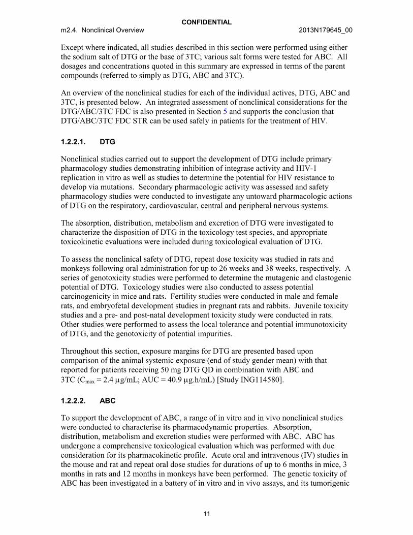
Except where indicated, all studies described in this section were performed using either the sodium salt of DTG or the base of 3TC; various salt forms were tested for ABC. All dosages and concentrations quoted in this summary are expressed in terms of the parent compounds (referred to simply as DTG, ABC and 3TC).
An overview of the nonclinical studies for each of the individual actives, DTG, ABC and 3TC, is presented below. An integrated assessment of nonclinical considerations for the DTG/ABC/3TC FDC is also presented in Section 5 and supports the conclusion that DTG/ABC/3TC FDC STR can be used safely in patients for the treatment of HIV.
1.2.2.1. DTG
Nonclinical studies carried out to support the development of DTG include primary pharmacology studies demonstrating inhibition of integrase activity and HIV-1 replication in vitro as well as studies to determine the potential for HIV resistance to develop via mutations. Secondary pharmacologic activity was assessed and safety pharmacology studies were conducted to investigate any untoward pharmacologic actions of DTG on the respiratory, cardiovascular, central and peripheral nervous systems.
The absorption, distribution, metabolism and excretion of DTG were investigated to characterize the disposition of DTG in the toxicology test species, and appropriate toxicokinetic evaluations were included during toxicological evaluation of DTG.
To assess the nonclinical safety of DTG, repeat dose toxicity was studied in rats and monkeys following oral administration for up to 26 weeks and 38 weeks, respectively. A series of genotoxicity studies were performed to determine the mutagenic and clastogenic potential of DTG. Toxicology studies were also conducted to assess potential carcinogenicity in mice and rats. Fertility studies were conducted in male and female rats, and embryofetal development studies in pregnant rats and rabbits. Juvenile toxicity studies and a pre- and post-natal development toxicity study were conducted in rats. Other studies were performed to assess the local tolerance and potential immunotoxicity of DTG, and the genotoxicity of potential impurities.
Throughout this section, exposure margins for DTG are presented based upon comparison of the animal systemic exposure (end of study gender mean) with that reported for patients receiving 50 mg DTG QD in combination with ABC and 3TC (Cmax = 2.4 g/mL; AUC = 40.9 g.h/mL) [Study ING114580].
1.2.2.2. ABC
To support the development of ABC, a range of in vitro and in vivo nonclinical studies were conducted to characterise its pharmacodynamic properties. Absorption, distribution, metabolism and excretion studies were performed with ABC. ABC has undergone a comprehensive toxicological evaluation which was performed with due consideration for its pharmacokinetic profile. Acute oral and intravenous (IV) studies in the mouse and rat and repeat oral dose studies for durations of up to 6 months in mice, 3 months in rats and 12 months in monkeys have been performed. The genetic toxicity of ABC has been investigated in a battery of in vitro and in vivo assays, and its tumorigenic
CONFIDENTIALm2.4. Nonclinical Overview 2013N179645_00
12
potential has been studied in oral carcinogenicity studies in the mouse and the rat. A range of reproductive toxicity studies have been performed in the rat and rabbit following oral administration. Additionally, studies investigating the mechanism of toxicity, impurities and local tolerance have been performed.
Throughout this section, exposure margins for ABC are presented based upon comparison of the animal systemic exposure with that reported for patients receiving 600 mg ABC QD in combination with DTG and 3TC (Cmax = 4.0 g/mL; AUC = 13.9 g.h/mL) [Study ING114580].
1.2.2.3. 3TC
A range of in vitro and in vivo nonclinical studies were conducted to characterise the pharmacodynamic properties of 3TC. Absorption, distribution, metabolism and excretion studies were performed with 3TC. Although the programme was initiated using the racemate, gsk001* , in most cases the pharmacokinetic studies were subsequently repeated with 3TC itself, except for the distribution profile in the rat which was obtained using the racemate. 3TC has undergone a comprehensive toxicological evaluation. Acute oral studies in the mouse and rat and repeat oral dose studies for durations of up to 52 weeks in the dog and 26 weeks in the rat have been performed. The genetic toxicity of 3TC has been investigated in a battery of in vitro and in vivo assays, and its tumorigenic potential has been studied in oral carcinogenicity studies in the mouse and the rat. A range of reproductive toxicity studies have been performed in the rat and rabbit following oral administration. Additionally, studies investigating haematotoxicity have been conducted, and impurities studies and local tolerance studies have also been performed.
Throughout this section, exposure margins for 3TC are presented based upon comparison of the animal systemic exposure with that reported for patients receiving 300 mg 3TC QD in combination with DTG and ABC (Cmax = 2.1 g/mL; AUC = 12.3 g.h/mL) [Study ING114580].
*新薬承認情報提供時に置き換え

CONFIDENTIALm2.4. Nonclinical Overview 2013N179645_00
13
2. PHARMACOLOGY
2.1. Presentation Order of Pharmacology Summaries
Pharmacology Summary Section
Pharmacology for DTG, ABC and 3TC in combination Not applicable.
Pharmacology of DTG alone Section 2.2
Pharmacology of ABC alone Section 2.3
Pharmacology of 3TC alone Section 2.4
Overall summary of all pharmacological studies performed with DTG, ABC or 3TC
Section 5, Integrated Assessment of the Fixed Dose Combination Product
2.2. DTG
2.2.1. Virology (primary pharmacodynamics)
A range of in vitro virology studies have been conducted to determine the mechanism of action, antiviral activity and the potential for development of drug resistance via mutations. An overview of these studies is provided within the Clinical Overview [m2.5, Section 4.1]. However, for the reviewer’s convenience, a brief overview of the key findings from these studies is also provided below.
2.2.1.1. Mechanism of action
Integration of viral DNA into the host chromosome of infected cells is an important step in the HIV replication cycle and is facilitated by viral integrase protein [Pommier, 2005]. Integration requires two metal-dependent consecutive steps in the viral replication cycle: 3'-processing and strand transfer. Viral cDNA is primed for integration in the cytoplasm by integrase-mediated trimming of the 3'-ends of the viral cDNA. Integrase remains bound to the viral cDNA ends in the pre-integration complexes (PICs). Following nuclear translocation of the PICs, integrase catalyzes the insertion of the viral cDNA ends into the host chromosomes. DTG inhibits HIV integrase by binding to the integrase active site and blocking the strand transfer step of retroviral DNA integration which is essential for the HIV replication cycle.
2.2.1.2. In vitro antiviral activity and potential resistance
DTG has low nM activity against wildtype HIV-1 and HIV-2 in a variety of cell lines, regardless of subtype. DTG has little activity against non-HIV viruses, displaying the

CONFIDENTIALm2.4. Nonclinical Overview 2013N179645_00
14
highest antiviral activity against HCV. Human serum causes an approximately 75-fold increase in the DTG IC50. DTG is additive or synergistic when assayed in combination with other antiretroviral agents.
When HIV-1 Strain IIIB was passaged in the presence of DTG for 112 days, viruses with a 4.1-fold maximum increase in IC50 and S153Y or S153F substitutions in integrase polymorphic sites were observed. Passage of the wildtype HIV-1 NL432 in the presence of 6.4 nM DTG selected for E92Q (FC=3.1) and G193E (FC=3.2) substitutions in the IN region on Day 56. Passage of HIV-1 NL432 with Q148H, Q148K or Q148R RAL-resistant mutations resulted in selection of additional mutations and an increase in DTG FC. Passage of HIV-1 subtypes B and A/G in TZM-bl cells selected for integrase mutation R263K.
Comparative susceptibilities to DTG and RAL were obtained from 60 RAL-resistant site directed HIV-1 mutants and 6 site directed HIV-2 mutants. DTG retained activity against a vast majority of these mutants. Additionally, susceptibilities to DTG and RAL were determined for over 700 RAL-resistant clinical isolates, with DTG retaining activity (<10 FC) against >90% of them.
The dissociation of DTG, RAL and EVG from wildtype and mutant IN proteins complexed with DNA was investigated to obtain a better understanding of INI dissociation kinetics. DTG demonstrated slower dissociation from all IN-DNA complexes tested, including those with single and double residue IN substitutions.
2.2.2. Secondary pharmacology
DTG (up to 10 M) was tested in vitro against a variety of proteins which included16 enzyme assays and 65 physiological receptors and ion channels binding sites [Report RH2007/00072]. DTG at 10 M did not significantly affect (defined as 50%)80 of the 81 in vitro assays. The only effect greater than 50% was a 64% inhibition in the melanocortin (MC4) receptor binding assay. Inhibition at 10 M is approximately 100-fold above the free clinical Cmax for DTG when administered 50 mg BID (Cmax = 4.2 g/mL = ~10 M, DTG is ~99% protein bound, therefore, 0.042 g/mL = free unbound concentration). No findings associated with MC4R agonism or antagonism have been observed in toxicity or clinical studies with DTG. No significant effects on body weight in healthy or HIV-infected subjects administered DTG have been observed to date. Taken together, these data indicate a lack of apparent biological activity at the MC4 receptor.
2.2.3. Safety pharmacology
No treatment-related behavioral or overt pharmacological effects were noted in conscious male rats at 500 mg/kg (the highest dose tested) [Report RD2007/01038]. Systemic exposure at 500 mg/kg is estimated to be ~36X above the expected human Cmax of DTGadministered 50 mg in combination with ABC and 3TC, based on extrapolation from Day 1 exposure in the rat 14 day toxicity study (87.1 g/mL).

CONFIDENTIALm2.4. Nonclinical Overview 2013N179645_00
15
Single oral doses of DTG at 500 mg/kg did not produce any effect on respiratory functional parameters in male rats when monitored for up to 6 hours following dosing [Report RD2007/01037]. Systemic exposure at 500 mg/kg is estimated to be ~36X above the expected human Cmax of DTG administered 50 mg in combination with ABC and 3TC, based on extrapolation from Day 1 exposure in the rat 14 day toxicity study (87.1 g/mL).
In male monkeys, single oral doses of DTG at doses up to 1000 mg/kg (Cmax = 20.1 g/mL; AUC0-24 = 259 g.h/mL) had no effect on arterial blood pressures, heart rate or electrocardiographic (ECG) parameters when monitored for 24 hours after dosing at a Cmax ~8X above the expected human Cmax of DTG when administered 50 mg in combination with ABC and 3TC [Report RD2007/01141]. Additionally, there were no treatment-related effects in ECG parameters measured during the repeat dose monkey toxicity studies up to 38 weeks at doses 1000 mg/kg/day.
The effect of a series of DTG concentrations (8.38 g/mL) on hERG tail current was studied [Report RD2007/01039]. An IC50 could not be determined as only 16.1% inhibition of hERG channel tail current occurred at the highest concentration, 20 M. The high dose (20 M or 8.4 g/mL) is ~227X above the free Cmax obtained with a 50 mg QD dose of DTG in combination with ABC and 3TC (0.037 g/mL for 50 mg QD based on 99% protein binding).
There were no findings from safety pharmacology studies that indicate an unacceptable risk for oral administration of DTG to patients in accordance with the proposed indication. Additionally, a supratherapeutic dose of DTG (250 mg as a suspension, which achieved exposures ~3X higher than a 50 mg QD dose) was well tolerated and had no effect on cardiac repolarization [see m2.5, Section 5.6.6.5].
2.2.4. Pharmacodynamic drug interactions
A number of in vitro studies have been conducted with DTG in combination with approved agents from all anti-HIV therapy classes (e.g., nucleoside/nucleotide reverse transcriptase [RT] inhibitors, non-nucleoside RT inhibitors and protease inhibitors) andwas shown to be additive or synergistic in all cases. These studies are discussed as part of the virology discussion [see m2.5, Section 4.1].
2.3. ABC
2.3.1. Virology (primary pharmacodynamics)
A range of in vitro virology studies have been conducted to determine the mechanism of action, antiviral activity and the potential for development of drug resistance via mutations. An overview of these studies is provided within the Clinical Overview [m2.5, Section 4.1]. However, for the reviewer’s convenience, a brief overview of the key findings from these studies is also provided below.
CONFIDENTIALm2.4. Nonclinical Overview 2013N179645_00
16
2.3.1.1. Mechanism of action
ABC is a carbocyclic synthetic nucleoside analogue. ABC is converted by cellular enzymes to the active metabolite, carbovir triphosphate (CBV-TP), an analogue of deoxyguanosine-t’-triphosphate (dGTP). CBV-TP inhibits the activity of HIV-1 reverse transcriptase (RT) by competing with the natural substrate dGTP and by its incorporation into viral DNA. The lack of a 3’-OH group in the incorporated nucleotide analogue prevents the formation of the 5’ to 3’ phosphodiester linkage essential for DNA chain elongation, and therefore, the viral DNA growth is terminated. CBV-TP is a weak inhibitor of cellular DNA polymerase , and .
2.3.1.2. In vitro antiviral activity and potential resistance
ABC has low M activity against wildtype HIV-1 in a variety of cell lines, regardless of subtype. ABC has little activity against non-HIV viruses, displaying the highest antiviral activity against Hepatitis B virus. ABC is additive or synergistic when assayed in combination with other antiretroviral agents, including 3TC.
HIV-1 isolates with reduced susceptibility to ABC have been selected in cell culture and were also obtained from subjects treated with ABC. Genotypic analysis of isolates selected in cell culture and recovered from ABC-treated subjects demonstrated that amino acid substitutions K65R, L74V, Y115F and M184V/I in RT contributed to ABC resistance.
The substitution at M184I/V causes almost complete resistance to 3TC and only mild resistance to ABC without the additional substitutions K65R, L74M and Y115F. For 3TC, the M184 substitution confers high fold change in IC50 and clinical resistance, while for ABC, the single M184V change confers low fold change increases which alone do not confer clinical resistance. Rather, sequential addition of additional mutations increases FC and decreases clinical response for ABC in a stepwise fashion.
2.3.2. Secondary pharmacology
There was no pharmacologically significant binding of ABC to any of a battery of 19 receptors and ion channels [Report TPZZ/93/0004].
The effect of ABC at the following receptors was assessed in isolated tissue preparations: cholinergic (guinea pig ileum), adrenergic (rabbit aorta, guinea pig atria and trachea), histaminergic (guinea pig atria) and serotonergic (rat fundus). In addition, the ability of ABC to affect tissue responsiveness to arachidonic acid (rat fundus), bradykinin (guinea pig ileum) and angiotensin II (rabbit aorta) was determined. There were no direct effects of ABC on any of the isolated tissue preparations, and no significant effects on contractile responses to any of the substances studied.
2.3.3. Safety pharmacology
The general pharmacological and gross behavioural effects of ABC were investigated in mice and rats by the oral and intraperitoneal routes; effects on the conditioned avoidance

CONFIDENTIALm2.4. Nonclinical Overview 2013N179645_00
17
reflex were determined in rats. In initial studies, the compound was formulated in 50% PEG 400 and was well tolerated at oral doses of up to 1000 mg/kg and 500 mg/kg in mice and rats, respectively; the only effects observed were hypothermia, hypoactivity and blepharospasm, in mice only [Report TPZZ/93/0005]. Following administration by the intraperitoneal route, the compound showed minimal effects in both species; the maximum tolerated doses were 500 mg/kg and 250 mg/kg in mice and rats, respectively. At these doses, which themselves produced no mortality, behavioral effects were limited to slight depression of traction and co-ordination reflexes and a slight decrease in respiration rate. Mortality occurred at high doses in the 50% PEG 400 vehicle and was not seen at the same doses when ABC was formulated in water only [ReportTPZZ/93/0120]. In rats trained to respond to an audio visual cue to avoid foot shock in a shuttle box, ABC had no effect at doses of up to 300 mg/kg when administered orally[Report TPZZ/93/0021]. The autonomic effects of ABC (50 mg/kg intravenously, infused over 50 minutes) were determined in anaesthetised dogs. There was a slight inhibition of responses to vagal stimulation but not to injected acetylcholine [ReportTPZZ/92/0072]. Similarly, there was a small reduction in the response to bilateral carotid artery occlusion while the response to injected noradrenaline was unchanged.
The effects of ABC (50 mg/kg intravenously, infused over 50 minutes) on respiration were determined in anaesthetised dogs. There were no compound-related effects on respiratory rate or minute volume [Report TPZZ/92/0072].
The cardiovascular effects of ABC were determined in isolated cardiac muscle in conscious normotensive rats and in anaesthetised dogs. ABC had no chronotropic effectsin the rat isolated perfused heart or guinea pig isolated spontaneously beating right atria, no inotropic effect in cat isolated papillary muscle or guinea pig isolated spontaneously beating left atria, and it did not induce dysrhythmias in rat isolated perfused heart [ReportTPZZ/92/0071].
In other studies, ABC has been investigated in vitro (in human cells) and in vivo (in anaesthetized rats) for its ability to recruit leukocytes, as leukocyte accumulation is a hallmark of certain vascular diseases [De Pablo, 2010]. Leukocyte endothelial cell interactions were induced in rat mesenteric microvenules following intraperitoneal administration of 10 M ABC, a dose chosen to compare with plasma levels of ABC seen in human patients. However, these results obtained from local administration to the vessels under investigation are difficult to relate to patients.
In vivo, treatment with ABC for 5 weeks showed no cardiotoxicity in adult inbred wildtype C57/BL6 or transgenic mice models of mitochondrial oxidative stress and cardiac function [Kohler, 2010].
In the conscious normotensive rat, ABC had no effect on arterial blood pressure or on heart rate following oral administration at a dose of 100 mg/kg, either as the free base or as the succinate salt [Report TPZZ/92/0058].
Studies were carried out in rats to investigate the potential mechanisms of an increase in ABC associated cardiovascular risk in humans [Li, 2010a; Li, 2010b]. Sprague Dawley rats were administered ABC up to 160 mg/kg/day for 28 days. At this dose, exposures

CONFIDENTIALm2.4. Nonclinical Overview 2013N179645_00
18
achieved were 15 to 20 times higher than those seen in humans on standard clinical treatment. ABC had no affect on vascular contractility nor did it induce endothelial inflammation. However, ABC upregulated platelet activity (measured by elevated plasma levels of CD40L, a marker of platelet activation), which could theoretically increase the risk of thrombosis.
In the anaesthetised dog, ABC infused at a dose of 50 mg/kg over 50 minutes produced small decreases in mean arterial blood pressure. These returned to pre-dose levels within 1 hour of the end of the infusion. There was a gradual increase in heart rate during infusion of ABC; these began to reverse during the infusion and had returned to pre-dose levels with 1 hour of the end of the infusion. There were no dysrhythmias in the electrocardiogram [Report TPZZ/92/0072].
2.3.4. Pharmacodynamic drug interactions
No specific nonclinical pharmacodynamic drug interaction studies have been performed with ABC.
2.4. 3TC
2.4.1. Virology (primary pharmacodynamics)
A range of in vitro virology studies have been conducted to determine the mechanism of action, antiviral activity and the potential for development of drug resistance via mutations. An overview of these studies is provided within the Clinical Overview [m2.5, Section 4.1]. However, for the reviewer’s convenience, a brief overview of the key findings from these studies is also provided below.
2.4.1.1. Mechanism of action
Intracellularly, 3TC is phosphorylated to its active 5’-triphosphate metabolite, 3TC triphosphate (3TC-TP). The principal mode of action of 3TC-TP is the inhibition of HIV-1 reverse transcriptase (RT) via DNA chain termination after incorporation of the nucleotide analogue into viral DNA 3TC-TP is a weak inhibitor of mammalian DNA polymerases , and .
2.4.1.2. In vitro antiviral activity and potential resistance
3TC has low M activity against wildtype HIV-1 in a variety of cell lines, regardless of subtype. 3TC has little activity against non-HIV viruses, displaying the highest antiviral activity against Hepatitis B virus. 3TC is additive or synergistic when assayed in combination with other antiretroviral agents, including ABC.
3TC resistant variants have been isolated in cell culture. Genotypic analysis showed that the resistance was due to a specific amino acid substitution in the HIV-1 reverse transcriptase at codon 184 changing the methionine to either isoleucine or valine (M184V/I). The substitution at M184I/V causes almost complete resistance to 3TC and

CONFIDENTIALm2.4. Nonclinical Overview 2013N179645_00
19
only mild resistance to ABC without the additional substitutions K65R, L74M and Y115F.
Studies demonstrate that although 3TC 5'-triphosphate is a substrate for the DNA-dependent DNA polymerase activity of DNA polymerase , the product of this reaction is also a substrate for the 3'-5' exonuclease activity of DNA polymerase . This may explain the low levels of mitochondrial toxicity observed with 3TC.
2.4.2. Secondary pharmacology
No specific nonclinical secondary pharmacology studies have been performed with 3TC.
2.4.3. Safety pharmacology
There were no significant overt pharmacodynamic actions of 3TC following acute oral administration in the rat and dog at doses up to 600 mg/kg [Report WBA/91/005]. Similarly, there were no effects on intestinal transport (up to 300 mg/kg oral in mice), or on respiration, blood pressure, heart rate or the electrocardiogram (up to 100 mg/kg IV in anaesthetised cats and 600 mg/kg oral in dogs) [Reports NPY/96/006, WBA/91/003 and WPT/94/218].
In saline loaded rats, a single oral dose of 3TC at 300 mg/kg slightly increased the urinary excretion of potassium ions with an accompanying small increase in urine osmolarity [Report NPY/96/006]. This slight effect on urinary potassium excretion has also been observed, though not consistently, at the high dose levels in repeat dose toxicity studies in rats and dogs. The possibility that this reflects a transient change in potassium excretion in rats cannot be discounted.
2.4.4. Pharmacodynamic drug interactions
No specific nonclinical pharmacodynamic drug interaction studies have been performed with 3TC.

CONFIDENTIALm2.4. Nonclinical Overview 2013N179645_00
20
3. PHARMACOKINETICS
Extensive programs of studies investigating the absorption, distribution, metabolism and excretion have been carried out individually with DTG, ABC and 3TC in animals used in toxicity studies. In general, the systemic exposure and metabolism defined in the animal species used for toxicological assessment indicates that the species used were appropriate for predicting the safety of DTG, ABC and 3TC, and their metabolites in humans.
3.1. Presentation Order of Pharmacokinetics Summaries
Pharmacokinetics Summary Section
Pharmacokinetics for DTG, ABC and 3TC in combination
Not applicable.
Pharmacokinetics of DTG alone Section 3.2
Pharmacokinetics of ABC alone Section 3.3
Pharmacokinetics of 3TC alone Section 3.4
Overall summary of all pharmacokinetics studies performed with DTG, ABC or 3TC
Section 5, Integrated Assessment of the Fixed Dose Combination Product
3.2. DTG
3.2.1. Analytical methods and validation
In pharmacokinetic and toxicity studies, plasma DTG concentrations were measuredfollowing protein precipitation with chiral or achiral liquid chromatographic tandem mass spectrometric (LC/MS/MS) methods. For toxicity and human studies, the chiral and achiral methods used for analysis were fully validated across each calibration range. Allmethods and limits of quantification were adequate with regard to specificity and sensitivity to support the pharmacokinetic analyses of DTG.
In investigations where [14C]-DTG was used, determination of the radioactivity in in vitro or in vivo biological samples was carried out by either direct liquid scintillation counting (LSC) or by LSC following combustion of the sample. For analysis of radioactivity concentrations in tissues, quantitative whole body autoradiography was used. The profiling and identification of metabolites of DTG was performed using LC-MSn. Nuclear magnetic resonance (NMR) methods were used to confirm structures not confirmed by mass spectrometric methods.
CONFIDENTIALm2.4. Nonclinical Overview 2013N179645_00
21
3.2.2. Absorption and pharmacokinetics
The nonclinical pharmacokinetics of DTG are characterized by low plasma clearance and low volume of distribution. Absorption was rapid with high oral bioavailability from a solution formulation. In repeat oral administration studies, systemic exposure to DTG was dissolution or solubility limited leading to an increase that was less than proportional with dose.
3.2.2.1. Single dose
Intravenous: Following a single intravenous administration, DTG exhibited low plasma clearance (<15% liver plasma flow) in the rat, dog and monkey [Reports RH2007/00101, RH2007/00102 and RH2007/00103]. The low steady state volume of distribution reflects the restrictive high protein binding of the compound (Section 3.2.3.1). The terminal half-life in rats and monkeys was 5.2 to 6.2 hours [m2.5, Section 3.2].
Oral: DTG absorption from an oral solution was rapid, reaching peak plasma concentrations within 2 hours with high oral bioavailability (76 to 87%) in fasted rats and monkeys [Reports RH2007/00101 and RH2007/00103]. When DTG was administered as a suspension, the increase in systemic exposure (Cmax and AUC0-t) was less than proportional to the increase in dose. The oral bioavailability of DTG from a suspension formulation was lower (bioavailability range of 25% to 52%) and suggested that the absorption is limited by dissolution rate or solubility. Administration of DTG with food to rats reduced exposure whereas in humans exposure was increased [see m2.5, Section 3.2 for human effects].
3.2.2.2. Repeat dose toxicokinetics
Oral: The repeat dose toxicokinetics of DTG were assessed as part of general toxicity,reproductive toxicity and juvenile toxicity studies. A comparison of systemic exposure values (Cmax and AUC0-24) for DTG is presented in Table 2.
The increase in systemic exposure (Cmax and AUC0-24) to DTG was less than proportionalwith the increase in dose during repeated oral administration toxicity studies in mice, non-pregnant rats, rabbits and monkeys. Differences (>2-fold) in systemic exposure between single and repeated administration, regardless of pregnancy status, or between the sexes were generally not observed.
Higher systemic exposure to DTG was observed in pre-weaning rat pups (Day 13 post partum) compared to juvenile rats on Day 32 post partum. Because DTG is primarily metabolized by uridine glucuronosyl transferase (UGT) in the rat (Section 4.2.7), this difference reflects the early differential expression of UGT in the rat [Kishi, 2008; Saghir, 2012; de Zwart, 2008]. No apparent sex-related differences (>2-fold) in systemic exposure were observed in juvenile rats.

CONFIDENTIALm2.4. Nonclinical Overview 2013N179645_00
22
3.2.3. Distribution
DTG has high passive membrane permeability, is highly protein bound and is widely distributed. DTG crosses the placental barrier and is secreted into the milk of lactating rats.
3.2.3.1. Protein binding and blood cell association
The in vitro protein binding of DTG was high (99%) across species (rat, monkey and human) and similar to an ex-vivo assessment (>99%) in plasma from healthy human subjects [Reports RH2007/00106 and 2011N119355; m2.5, Section 3.1]. The association of DTG-related material with blood cellular components was minimal [Reports RD2009/00562, RD2008/00108, CD2008/00195 and RD2008/01300; m2.5, Section 3.1].
3.2.3.2. Efflux-mediated transport and cell membrane permeability
In vitro, DTG was a substrate for the human efflux transporters P-glycoprotein (P-gp) and human breast cancer resistance protein (BCRP). DTG was determined to have high passive membrane permeability (333 nm/s at pH 7.4). The absorptive membrane permeabilities also were high in the presence of FaSSIF at pH 7.4 and pH 5.5 (P7.4[abs]
value of 253 nm/s and a P5.5[abs] value of 265 nm/s, respectively) [Report RD2008/00360]. Based on solubility and permeability determinations, DTG sodium is classified as a Biopharmaceutics Classification System (BCS) Class 2 drug.
3.2.3.3. Tissue distribution
After a single oral dose of [14C]-DTG, radioactivity was widely distributed in a similar pattern between male Lister Hooded partially pigmented rats and pregnant Sprague Dawley rats. Radioactivity in tissues generally peaked 4 to 6 hours post dose with concentrations typically less than those in blood [Reports CD2008/00195 and 2012N137348]. The concentration of radioactivity in the brain was low (~2% of the blood radiocarbon concentration) due in part to restrictive protein binding. By 28 days post dose, only bone and pigmented skin contained quantifiable concentrations of radioactivity. Radioactivity was not associated with melanin in the uveal tract, lowering the concern for a phototoxicity liability (Section 4.2.10). DTG crossed the placental barrier but appeared to exert no adverse effects on fetal development [Section 4.2.6,m1.14.1 (US) or m1.3 (EU)]. Radioactivity rapidly equilibrated to fetal tissues with fetal tissue to fetal blood ratios generally higher than corresponding maternal tissue to blood ratios. DTG concentrations in fetal bone marrow exceeded those in fetal blood.
In lactating rats at 10 days post partum, radioactivity was detected in milk at concentrations typically higher than blood, with unchanged DTG constituting most (97% to 83%) of the drug-related material [Report 2012N132387]. These results suggested that pre-weaned pups were exposed to DTG (Section 4.2.7) by nursing in the pre- and post-natal development study.
CONFIDENTIALm2.4. Nonclinical Overview 2013N179645_00
23
3.2.4. Metabolism
The comparative biotransformation pathways between animals and humans are presented in Figure 2. The predominant circulating component in the nonclinical species and humans is unchanged DTG with no disproportionate human metabolites observed. The main metabolic route in each species and humans was by conjugation to form the ether glucuronide (M3). These studies confirm the suitability of the mouse, rat and monkey for the toxicological assessment of DTG.
3.2.4.1. In vitro biotransformation
The in vitro metabolic turnover of DTG was low (<10%), indicating low intrinsic clearance consistent with the low plasma clearance. The primary biotransformation common to all species was glucuronidation to form the ether glucuronide (M3) that also was observed in vivo. Other common metabolic products included a glucose conjugate (M2) and an N-dealkylated product (M1) [Reports RH2007/00076, RD2007/01557, RD2007/01496 and RH2007/00058]. The generation of a glutathione or cysteine conjugate through oxidative defluorination and microsomal binding with rat, monkey and human liver microsomes suggested evidence for the formation of an electrophilic metabolic intermediate by bioactivation in vitro [Reports RH2007/00058 andRD2007/01557]. However, in vivo in mice, rats, monkeys and humans, these metabolic products have represented only a small fraction of the metabolic clearance. Additionally, no microscopic liver findings were observed in mice, rats or monkeys after repeat administration of doses at or below the NOAEL.
No notable metabolic conversion of DTG to any of its possible stereoisomers occurredin vitro following incubations of DTG with cryopreserved rat, dog, monkey and human hepatocytes [Report RH2007/00105].
3.2.4.2. In vivo studies
In vivo, absorbed [14C]-DTG was extensively metabolized in male and female mice, rats and monkeys. A comparative metabolic summary of the products identified in the nonclinical species with products identified in humans is presented in Figure 2 [Reports RD2008/00220, RD2008/00899 and RD2009/00723; m2.5, Section 3.1]. Metabolic profiles in nonclinical species were qualitatively similar to humans, with adequate coverage for the circulating human metabolites in at least one nonclinical species.
Plasma metabolic profile
DTG was the predominant component in plasma of mice, rats, monkeys and humans with the glucuronide as the principal metabolite. No metabolite was present in the plasma at concentrations greater than 10% of parent or drug-related material. In humans, the steadystate plasma metabolic profile of DTG was similar to the single dose metabolic profile,indicating data obtained after single dose administration was an adequate predictor of the profile at steady state [see m2.5, Section 3.1]. Based on the kinetics of DTG in animals, the systemic exposures to DTG and circulating metabolites found in the nonclinical

CONFIDENTIALm2.4. Nonclinical Overview 2013N179645_00
24
metabolism studies adequately reflected exposures in the toxicity studies. No disproportionate human metabolites were noted.
Biotransformation
The predominant biotransformation product in mice, rats and humans was an ether glucuronide (M3). The glucuronide metabolite was formed in approximately equal proportions with a glucose conjugate (M2) in monkeys. These conjugated metabolites, M2 and M3, are not pharmacologically active because they disrupt the two-metal binding capability of the carbamoyl pyridone motif of DTG thereby completely abrogating any antiviral activity resulting from the active site binding to the integrase enzyme. Although these conjugates were the primary constituents of the drug-related material in bile of animals, they were not observed in the feces of animals or humans. Thus, these DTG conjugates are likely deconjugated in the intestine by host or bacterial enzymes after secretion in the bile to reform DTG. In animals, fecal metabolites were not quantifiable, but in human fecal samples, an N-dealkylation product (M1) and a product of oxidative defluorination with cysteine addition (M13) was quantifiable at less than 2% of the dose.

CONFIDENTIALm2.4. Nonclinical Overview 2013N179645_00
25
Figure 2 Comparative Metabolic Profile of DTG Between Nonclinical Species and Humans
GSK1349572Human, M ouse, Rat, M onkey
F O
NH
F
N
N
O
CH3OOH
O
H
O
NH2 N
N
O
CH3OO H
O
H
M 1Human, M ouse, Rat, M onkey
F O
NH
F
N
N
O
CH3OO H
O
HO H
M 7H uman, M ouse, Rat
F O
NH
F
N
N
O
CH3OO
O
H
O
OH
OH
OH
OH
M 2Human, M ouse, Rat, M onkey
F O
NH
F
N
N
O
CH3OO
O
H
O
OH
OH
OH
OH
O
M 3Human, M ouse, Rat, M onkey
-F
+SO H
F O
NH
F
N
N
O
CH3OO H
O
H
M 10Human, Rat
+SO
F O
NH
F
N
N
O
CH3OOH
O
H
M11Human, M ouse, Rat
+C 6H 11O 5
F O
NH
F
N
N
O
CH3OO H
O
H
M 12 Human, Rat
F O
NH
F
N
N
O
CH3OO H
O
H
S
NH2O
OH
M13Human, M onkey
F O
NH
F
N
N
O
CH3OO H
O
H
-F+2O+cysteine
M 14 Human, M ouse
F O
NH
F
N
N
O
CH3OO H
O
H
- CO+Gluc
M 16Human, M ouse
F O
NH
F
N
N
O
CH3OO H
O
H
+glutathione+O -F
M 15M ouse
-F+O
M 9M o use, Rat
Key: Bolded arrows indicate the primary metabolic products in humans (M3 the predominant product, M7 a notable metabolite).
CONFIDENTIALm2.4. Nonclinical Overview 2013N179645_00
26
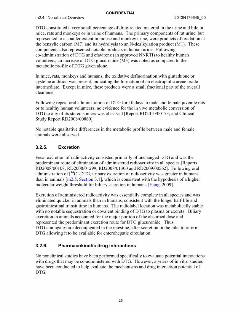
DTG constituted a very small percentage of drug-related material in the urine and bile in mice, rats and monkeys or in urine of humans. The primary components of rat urine, but represented to a smaller extent in mouse and monkey urine, were products of oxidation at the benzylic carbon (M7) and its hydrolysis to an N-dealkylation product (M1). These components also represented notable products in human urine. Following co-administration of DTG and efavirenz (an approved NNRTI) to healthy human volunteers, an increase of DTG glucuronide (M3) was noted as compared to the metabolic profile of DTG given alone.
In mice, rats, monkeys and humans, the oxidative defluorination with glutathione or cysteine addition was present, indicating the formation of an electrophilic arene oxide intermediate. Except in mice, these products were a small fractional part of the overall clearance.
Following repeat oral administration of DTG for 10 days to male and female juvenile rats or to healthy human volunteers, no evidence for the in vivo metabolic conversion of DTG to any of its stereoisomers was observed [Report RD2010/00173; and Clinical Study Report RD2008/00860].
No notable qualitative differences in the metabolic profile between male and female animals were observed.
3.2.5. Excretion
Fecal excretion of radioactivity consisted primarily of unchanged DTG and was the predominant route of elimination of administered radioactivity in all species [Reports RD2008/00108, RD2008/01299, RD2008/01300 and RD2009/00562]. Following oral administration of [14C]-DTG, urinary excretion of radioactivity was greater in humans than in animals [m2.5, Section 3.1], which is consistent with the hypothesis of a higher molecular weight threshold for biliary secretion in humans [Yang, 2009].
Excretion of administered radioactivity was essentially complete in all species and was eliminated quicker in animals than in humans, consistent with the longer half-life and gastrointestinal transit time in humans. The radiolabel location was metabolically stable with no notable sequestration or covalent binding of DTG to plasma or excreta. Biliary excretion in animals accounted for the major portion of the absorbed dose and represented the predominant excretion route for DTG glucuronide. Thus, DTG conjugates are deconjugated in the intestine, after secretion in the bile, to reform DTG allowing it to be available for enterohepatic circulation.
3.2.6. Pharmacokinetic drug interactions
No nonclinical studies have been performed specifically to evaluate potential interactions with drugs that may be co-administered with DTG. However, a series of in vitro studies have been conducted to help evaluate the mechanisms and drug interaction potential of DTG.

CONFIDENTIALm2.4. Nonclinical Overview 2013N179645_00
27
3.2.6.1. Potential effect of co-administered agents on DTG
In vitro and in vivo, DTG is primarily metabolized by UGT1A1 with a notable contribution from CYP3A4. UGT1A3 and 1A9 were minor glucuronidation pathways [Reports RD2008/00373 and RD2008/01339]. Therefore, drugs that are strong inducers of UGT1A1 or CYP3A4 may decrease DTG plasma concentrations. Although drugs that inhibit UGT1A1 and CYP3A4 may increase DTG plasma concentrations, based on the clinical interaction study with atazanavir, a potent UGT1A1 and CYP3A4 inhibitor, any increases in DTG concentrations are not expected to be clinically meaningful. Although DTG is a substrate for efflux transporters, no notable effect on DTG pharmacokinetics was observed in humans following co-administration with lopinavir/ritonavir, inhibitors of the efflux transporters P-gp and BCRP [m2.5, Section 3.3]. These data, together with the rapid absorption in humans, low to moderate pharmacokinetic variability and high intrinsic permeability suggests a low potential for drug interactions with BCRP and P-gp inhibitors that would result in clinically significant changes to DTG exposure.
3.2.6.2. Effect of DTG on co-administered agents
In vitro, DTG was noted to have little or no inductive effects on the human Pregnane X receptor (PXR) on CYP1A2, 2B6 or 3A4 mRNA (as determined by the increase in mRNA relative to vehicle control). DTG demonstrated little or no inhibition (IC50 values >30 M) in vitro on the transporters BCRP, bile salt export pump (BSEP), multidrug resistance protein (MRP) 2, MRP4, organic anion transporting polypeptide (OATP) 1B1, OATP1B3, organic cation transporter (OCT) 1 and P-gp, or the enzymes CYP1A2, 2A6, 2B6, 2C8, 2C9, 2C19, 2D6, 3A4, UGT1A1 or 2B7, demonstrating a low propensity to cause drug interactions through modulation of these systems. DTG glucuronide (M3) did not inhibit MRP2, thus, inhibition of biliary clearance of bilirubin glucuronides or glucuronide conjugates of co-administered drugs is not expected.
In vitro, DTG inhibited OAT1 and OAT3 with IC50 values of 2.12 M and 1.97 M,respectively; however, in vivo, no notable changes in plasma concentrations of the OAT substrates tenofovir [ING111604, m2.5, Section 3.3] or p-aminohipurate [ING114819, m2.5, Section 3.3] were observed in healthy subject Phase 1 studies.Multidrug resistance associated protein (MRP) 2 and MRP4 are anion transporters responsible for the transport of anions (e.g., tenofovir) from the renal tubule to the urine with MRP4 as the predominate transporter for tenofovir excretion. DTG did not inhibit MRP2 and weakly inhibited MRP4 (IC50 value of 84 M) with a 50-fold unbound Cmax value less than the Ki estimate. Furthermore, polymorphic MRP4 that decreasedtenofovir renal clearance by 15% also increased plasma tenofovir concentrations by 32% [Kiser, 2008], which was not observed in the drug interaction study assessing the impact of DTG on tenofovir PK. In addition, a physiological based pharmacokinetic (PBPK) mechanistic kidney model (Simcyp v,12 R1) developed for steady state concentrations of tenofovir (300 mg once daily) predicts that co-administration of DTG at 50 mg once daily would result in a minimal decrease in tenofovir renal clearance with no notable change in tenofovir exposure within the proximal tubule cells of the kidney [see m2.6.5, Table 15.1, Report 2013N171682]. Based on these collective data, no clinically significant interaction with tenofovir by DTG at the renal tubule is expected.

CONFIDENTIALm2.4. Nonclinical Overview 2013N179645_00
28
In vitro, DTG inhibited the basolateral renal organic cation transporter 2 (OCT2; IC50 =1.9 M) and the renal apical transporters, multidrug and toxin extrusion transporter (MATE) 1 (IC50 = 6.34 M) and MATE2-K (IC50 = 24.8 M) [see m2.6.5, Table 8.1, Report 2013N161621], which provides a mechanistic basis for the non-pathological mild serum creatinine increases observed in clinical studies. Because DTG inhibits OCT2, but only weakly OCT1, and both OCT1 and OCT2 are equally expressed in rat proximal tubules [Tahara, 2005], this effect on creatinine was not observed in rats. These in vitroresults indicate caution should be used due to the potential for a drug interaction in vivowhen DTG is co-administered with cationic compounds that have a narrow therapeutic index and in which a significant part of their clearance is by renal proximal tubule secretion by OCT2. DTG is contraindicated for co-administration with the OCT2 substrates dofetilide and pilsicainide because they possess narrow therapeutic indices that present the potential for toxicity due to higher exposure [m2.5, Table 2]. DTG has a low potential to affect the transport of MATE2-K substrates.
As a weak inhibitor of UGT1A1, DTG has the potential to interfere with the conjugation of bilirubin which could result in a mild increase in total or unconjugated bilirubin on prolonged treatment with DTG. Because bilirubin has low solubility and low permeability, it is transported to the UGT1A1 enzymatic site by glutathione-S-transferase. Since DTG has high permeability and does not rely on transport to the enzyme site, this favors rapid access by DTG to UGT1A1, although the affinity of bilirubin for UGT1A1 is higher than that of DTG.
3.3. ABC
3.3.1. Analytical methods and validation
In pharmacokinetic and toxicity studies, plasma ABC concentrations were measuredfollowing protein precipitation with trichloroacetic acid, ABC was separated using reversed phase HPLC with UV detection. For toxicity and human studies, the methods used for analysis were fully validated across each calibration range. All methods and limits of quantification were adequate with regard to specificity and sensitivity to support the kinetic analyses of ABC.
Determination of radiocarbon in biological samples was carried out by liquid scintillation counting. The profiling and identification of metabolites of ABC or comparator compounds were performed by HPLC with radiochemical and UV detection, or HPLC with mass spectrometric detection (LC-MS).
Immunoblotting experiments for metabolic bioactivation assessments were carried out following SDS-polyacrylamide gel electrophoresis of exhaustively extracted protein samples using anti-ABC antibodies as probes. The antibodies were raised in rabbits against ABC conjugated to keyhole limpet hemocyanin.

CONFIDENTIALm2.4. Nonclinical Overview 2013N179645_00
29
3.3.2. Absorption and pharmacokinetics
ABC has moderate to high plasma clearance, dose-proportional systemic exposure and high bioavailability.
3.3.2.1. Single dose
Intravenous: Following a single intravenous administration, ABC exhibited highplasma clearance (>70% liver plasma flow) in the mouse and monkey [Reports TEIN/94/0004, TEIN/94/0005 and TEIN/94/015]. The volume of distribution is moderate at approximately 1 L/kg. The terminal elimination half-life was rapid and ranged from 0.27 to 0.78 hours in mice and was 1.3 hours in monkeys.
Oral: ABC was rapidly absorbed in mice, rats and monkeys following oral administration, and the exposure was generally proportional to the dose. The bioavailability was greater than 76% in both mice and monkeys.
3.3.2.2. Repeat dose toxicokinetics
Oral: The repeat dose toxicokinetics of ABC were assessed as part of general toxicity,reproductive toxicity and juvenile toxicity studies. A comparison of values for systemic exposure to ABC (Cmax and AUC0-24) is presented in Table 5.
Repeated oral administration of ABC produced no consistent changes in pharmacokinetic parameters and in general, exposure was proportional to dose. No notable sex-related changes in systemic exposure were noted. Systemic exposure to ABC was similar after oral dosing to pregnant and non-pregnant rats and also similar between juvenile and mature rats.
3.3.3. Distribution
ABC is widely distributed to tissues including the cerebrospinal fluid and brain parenchyma. ABC crosses the placental barrier and is secreted into the milk of nursing animals. ABC has a low protein binding that has minimal influence on in vitro viral inhibition potency measurements. Although permeability is considered to be low, intestinal absorption is good.
3.3.3.1. Protein binding and blood cell association
The in vitro protein binding of ABC was low in mice (19%), monkeys (39%) and humans (40%) [Report TBZZ/93/0010].
3.3.3.2. Efflux-mediated transport and cell membrane permeability
In vitro, in Caco-2, parent MDCK and primary bovine brain endothelial cells [Reports RD1997/02021 and RD1998/00626], ABC was assessed as having low passivemembrane permeability but a potential for good intestinal absorption.

CONFIDENTIALm2.4. Nonclinical Overview 2013N179645_00
30
In vitro using overexpressed MDCK-II cell, literature reports have shown that ABC was a substrate for human P-glycoprotein (P-gp) [Shaik, 2007] and for the homologous murine Bcrp1 [Pan, 2007]. However, these researchers noted that ABC needed a concentration(500 M) in far excess of the normal therapeutic concentration to stimulate P-gp ATPaseactivity to a level of the positive control verapamil. The absolute bioavailability of ABC (83%) was high [Report RM1998/00065], indicating minimal impact of these transporters on intestinal absorption.
3.3.3.3. Tissue distribution
Following oral administration of [14C]-ABC, ABC-related material was rapidly absorbed and widely distributed to tissues [Reports RD1996/00065 and RD1997/04000] of mice and pregnant rats. Radioactivity was eliminated from most tissues by 48 hours post dose. Seven days post dose, radioactivity concentrations were low but quantifiable in some tissues including the esophagus, uveal tract, liver, outer ear and skin of mice. The binding to uveal tract of pigmented mice indicated ABC bound to melanin as commonly seen with basic drugs. Measurable concentrations of drug-related material were found in mouse brain and cerebrospinal fluid (CSF) at least 2 hours after dosing, with brain:blood and CSF:blood ratios of 0.09 to 0.11 and 0.36 to 0.51, respectively. After repeat oral dosing to monkeys the CSF to plasma ratio 1 hour after dosing ranged 0.14 to 0.26 and was constant with dose.
ABC-related material crosses the placenta of pregnant rats and rabbits with radioactivity concentrations in fetal tissue comparable with similar maternal tissues in the rat [Report RD1997/04000]. ABC was excreted into milk of nursing rats [Report RD1997/01909] and suggested that pre-weaned pups were exposed to ABC by nursing in the pre- and post-natal development study.
3.3.4. Metabolism
ABC is principally cleared via the hepatic route. The metabolite profiles are qualitatively similar in mice, monkeys and humans with adequate coverage for the circulating human metabolites in at least one nonclinical species. The two main plasma metabolites were a 5’-glucuronide (361W94) and a 5’-carboxylic (2269W93) acid. Carbovir (1144U88) and its precursor 139U91 were consistently observed as minor metabolic products, with plasma concentrations of 2.5% of the ABC concentrations in animals and humans.
3.3.4.1. In vitro biotransformation
Following in vitro incubation of ABC with mouse and rat liver S9 fractions, biotransformations of ABC, identified by LC-MS analysis, were all products of oxidation that were similar between mice and rats [Report TOZZ/95/0053].
In vitro studies [Report RD1997/04317] demonstrated that production of the 5’carboxylate metabolite was catalyzed by cytosolic alcohol dehydrogenase rather than by microsomal CYP450 enzymes.

CONFIDENTIALm2.4. Nonclinical Overview 2013N179645_00
31
In vitro, bioactivation of ABC appeared to lead to non-extractable residues in rat and human hepatocyte incubations [Report RD2000/02309]. This bioactivation and non-extractable residue formation appeared mediated through an aldehyde intermediate that was formed through the oxidative biotransformation by human and 22 ADH isozymes [Report RD2001/01777]. Following administration of ABC to rats, liver cytosol from control and ABC dosed rats were analyzed by SDS gel electrophoresis and immunoblotting using anti-ABC antibodies to identify protein targets for reactive metabolites of ABC in the rat [Reports RD2002/01292 and RD1999/00945]. A high association of ABC-related components with the low molecular weight (16 kD) translational inhibitor protein was suggested.
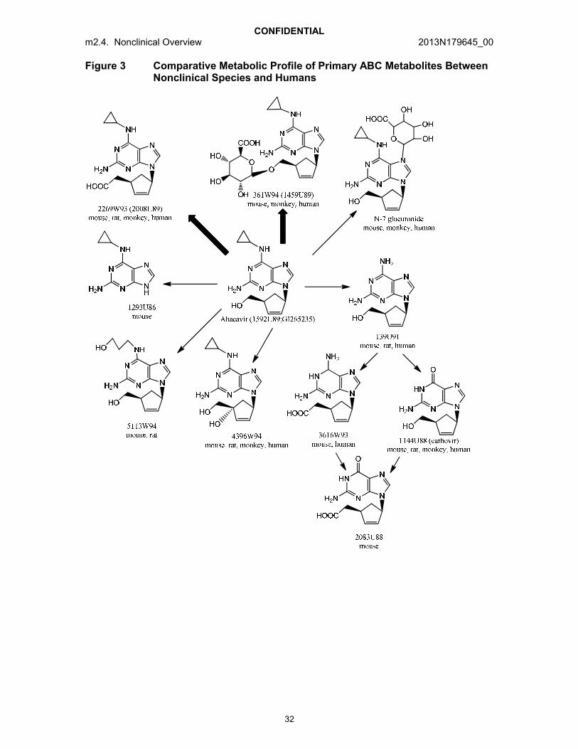
3.3.4.2. In vivo studies
A comparison of the primary characterized metabolites in animals and humans are presented in Figure 3. The stereochemistry was not explicitly determined and was assumed to be the same as parent ABC.
The two major metabolites of ABC in mice, monkeys and humans were 361W94, a 5’-ether glucuronide, and 2269W93, a 5’-carboxylic acid. These are designated with the bolded arrows in Figure 3. In rats, the 5’-carboxylate metabolite was the major biotransformation product whereas the 5’-glucuronide was present only in trace amounts (~2.2%). This metabolic difference contributed to the rationale for the selection of the mouse as the general toxicology species.
Plasma metabolic profile
After oral administration of ABC to mice and monkeys [Reports TEZA/91/0137, TEZA/91/0073, TTDR/92/0035 and TTDR/92/0036], the proportion of 5’-carboxylate concentrations in plasma of mice (20 to 30%) and monkeys (5 to 8%) relative to the systemic exposure of parent ABC remained the same across increasing doses and duration of dosing whereas the proportion of 5’-glucuronide in plasma of mice (25 to 42%) and monkeys (26 to 66%) relative to the systemic exposure of parent ABCincreased with increasing doses and duration of dosing. Concentrations of the metabolite carbovir (1144U88) in monkey plasma were very low representing approximately 1.2% of the parent ABC concentrations [Report TEZA/91/0137].
Conversion of ABC to its diastereomers in vitro in mouse or rat samples or in vivo in mouse, monkey or human samples was not observed [Report TOZZ/95/0053]. It is considered unlikely that metabolic generation of the enantiomer of ABC would occur because epimerization at two centers would need to occur without producing adiastereomer.
CONFIDENTIALm2.4. Nonclinical Overview 2013N179645_00
32
Figure 3 Comparative Metabolic Profile of Primary ABC Metabolites Between Nonclinical Species and Humans

CONFIDENTIALm2.4. Nonclinical Overview 2013N179645_00
33
Biotransformation
ABC was predominately metabolized and excreted in the urine as metabolic products. Approximately 10 to 13% of the dose in mice and monkeys and 2% of the dose in humans is excreted as unchanged ABC in urine. The major components in the urine from mice, monkeys and humans were the 5’-carboxylate (2269W93) and the 5’-glucuronide (361W94). Carbovir was recovered in urine at levels of <2% of dose in mice, rats, monkeys and humans. Fecal excretion was minimal and information on metabolites was not obtained.
The metabolic profiles between mice and monkeys, and humans are qualitatively similar with some quantitative differences. In general, ABC metabolism in humans was less extensive than in mice but more extensive than in monkeys.
3.3.5. Excretion
Excretion of ABC following oral administration was rapid with most excretion occurring within 24 hours post dose. Urine was the predominant route of excretion with 90% of the dose recovered in the urine of mice and 92% in urine of monkeys [Reports TEIN/94/0001and TEIN/94/0006]. This is similar to humans in which a mean of 83% of the dose was recovered in the urine. Mass balance was achieved with complete recovery of administered radioactivity. Feces represented a minor route of elimination.
3.3.6. Pharmacokinetic drug-drug interactions
3.3.6.1. In vitro drug interaction studies with ABC
Drug interaction studies conducted in vitro in human liver S9 incubations have shown that while ethanol (an ADH/ALDH substrate) inhibits the metabolism of ABC during co-incubation, ABC did not alter the metabolism of ethanol [Report RD1997/04351]. When ABC and amprenavir, a CYP3A4 inhibitor, were co-incubated in vitro in a human liver slice model, no evidence of inhibition of ABC metabolism was observed [Report RD1997/03644].
3.3.6.2. Potential effect of co-administered agents on ABC
ABC is metabolized to its two primary metabolites, a 5’-carboxylate (2269W93) and a 5’-glucuronide (361W94), by cytosolic alcohol dehydrogenase [Report RD2001/01777] and UGT2B7 [Report RD2000/02310], respectively. Ethanol increased the concentrations of ABC in humans [Study GM1998/00027], indicating that co-administered drugs which inhibit alcohol dehydrogenase may increase systemic ABCconcentrations. Because the CYP450 enzyme system does not metabolize ABC, ABCcan be administered with agents that are strong CYP450 inhibitors and are typically co-administered such as HIV protease inhibitors and antibacterials (e.g., clarithromycin).

CONFIDENTIALm2.4. Nonclinical Overview 2013N179645_00
34
3.3.6.3. Potential effect of ABC on co-administered agents
In vitro studies using human liver microsomal preparations showed no evidence for inhibition of CYP450 activity (CYP2D6, CYP2C9, CYP3A4) at clinically relevant concentrations (10.5 M to 100 mM) of ABC [Report RD1997/04317]. Studies in mice following repeat oral administration for 6 months suggested that ABC is unlikely to cause significant induction of CYP450 enzymes.
Based on the known metabolic pathways of ABC in humans and the lack of in vitro interactions between ABC and major human CYP450 enzymes, clinically relevant pharmacokinetic interactions are unlikely when ABC is co-administered with drugs that inhibit or are metabolized by CYP450 such as the HIV protease inhibitors.
3.4. 3TC
3.4.1. Analytical methods and validation
In pharmacokinetic and toxicity studies, plasma 3TC concentrations were measuredfollowing protein precipitation with trichloroacetic acid or solid phase extraction (for lower limits of quantification) which was then separated using reversed phase HPLC with ultraviolet (UV) absorbance detection for studies during early development and by tandem mass spectrometric (MS/MS) detection for later studies. All methods and limits of quantification were adequate with regard to specificity and sensitivity to support the kinetic analyses of 3TC.
Chiral analysis was performed on plasma or serum samples by collecting the eluate fraction of parent 3TC from a reversed phase C18 column and analyzing it with a chiral Cyclobond 1 Acetyl column and UV detection. This methodology was sufficiently sensitive to detect 2 ng of the enantiomer in the presence of 2 g/mL of 3TC.
Determination of radiocarbon in biological samples was carried out by liquid scintillation counting. The profiling and identification of metabolites of ABC or comparator compounds were performed by HPLC with radiochemical and UV detection, or HPLC with mass spectrometric detection (LC-MS).
3.4.2. Absorption and pharmacokinetics
3TC has moderate to high plasma clearance, dose-proportional systemic exposure and high bioavailability in animals and humans.
3.4.2.1. Single dose
Intravenous: Following a single intravenous administration, 3TC was rapidly cleared from the systemic circulation with a terminal elimination half-life in the rat and dog of approximately 1.2 to 1.7 hours. In rats, the renal clearance (3.3 mL/min) exceeded the glomerular filtration rate (approximately 1.8 mL/min), indicating that tubular secretion played a significant role in the elimination of 3TC. In dogs, metabolism was a more
CONFIDENTIALm2.4. Nonclinical Overview 2013N179645_00
35
significant contributor to the total clearance of 3TC with renal and metabolic clearance of approximately equal importance. In humans, 3TC was excreted primarily unchanged in the urine with tubular secretion playing a significant role [m2.5, Section 3.1]. The volume of distribution was moderate in rats and dogs and was equal to or greater than thetotal body water volume [Reports GDM/91/015, GDM/91/077, GDM/91/008 andGDM/91/080]. No notable sex-related differences in the pharmacokinetics were observed.
Oral: In rat and dog, 3TC was rapidly and extensively absorbed following oral administration with oral bioavailability ranging from 60% to 69% in the rat and from67% to 83% in the dog. At higher dose levels (600 to 1500 mg/kg), systemic exposures were linear with dose in the rat but the terminal phase half-life ranged 3.7 to 4.6 hours,indicating flip flop kinetics. In contrast, in the dog no notable change in half-life was observed but exposures generally increased less than proportionally at higher doses [Reports GDM/91/003 and GDM/91/036].
The pharmacokinetics of 3TC in human is more like the pharmacokinetics in rats than in dogs.
3.4.2.2. Repeat dose toxicokinetics
Oral: The repeat dose toxicokinetics of 3TC were assessed as part of general toxicity,reproductive toxicity and juvenile toxicity studies. A comparison of systemic exposure values (Cmax and AUC0-24) to 3TC is presented in Table 6.
Following repeat dose oral administration to the rat, the relationship between dose and concentration of 3TC in plasma was linear, and in general there was no change in systemic exposure over a dosing period of up to 6 months. Repeated administration of 3TC to the dog for up to 12 months caused an increase in systemic exposure with an increase in dose but the increase was less than proportional at higher doses. The systemic exposure to 3TC in juvenile rats (Day 16) was at least 2 times greater than the corresponding single dose adult values.
3.4.3. Distribution
The binding of 3TC to plasma proteins at therapeutic concentrations was low. After oral administration to rats, the distribution of 3TC was rapid and widespread throughout most tissues. 3TC crosses the placenta in pregnant rats and rabbits and is excreted into the milk of nursing rats.
3.4.3.1. Protein binding and blood cell association
The in vitro protein binding of 3TC was low and concentration-dependent. At clinical concentrations and above, binding was <17%. At 0.1 g/mL, protein binding ranged 35% to 50%. The low protein binding indicates that 3TC does not influence in vitro viral inhibition potency measurements. Binding to blood cellular components was moderate (40% to 56%) and similar across species in rats, dogs and humans [Reports GDM/91/010and GDM/91/011].

CONFIDENTIALm2.4. Nonclinical Overview 2013N179645_00
36
3.4.3.2. Efflux-mediated transport and cell membrane permeability
In vitro, 3TC was assessed as undergoing passive transcellular and paracellular diffusion across Caco-2 cell monolayers [Report UCP/92/028].
In vitro in Caco-2 and MDCK-MDR1 monolayers, 3TC was assessed as having a moderate permeability. 3TC also was shown to be a P-gp substrate but the low transport ratio indicates 3TC was not a good substrate and as a result is unlikely that P-gp would significantly affect its absorption [de Souza, 2009].
3.4.3.3. Tissue distribution
The pharmacokinetics of 3TC were determined with the chiral molecule, however, the program was initiated using the racemate, gsk001*, and the oral distribution profile of the racemate was studied by whole body autoradiography in the rat [ReportGDM/90/040].
Distribution of radiolabelled gsk002* was rapid and widespread throughout most tissues including the brain parenchyma with rapid clearance from tissues. There was no evidence of any sex-related difference in the distribution of gsk002*-related material nor was there evidence for any association with melanin in pigmented rats.
Following oral administration of [3H]-3TC to pregnant rats and rabbits, radioactivity crossed the placenta and was widely distributed in maternal tissues and in fetal tissues at low concentrations relative to maternal tissues. In rats, the pattern of distribution of radioactivity in maternal tissues was similar to that found in non-pregnant rats administered radiolabelled racemic gsk002*, indicating no impact of pregnancy or stereochemistry on the distribution of 3TC-related material. These studies indicate that during reproductive toxicity testing the fetuses received exposure to drug-related material [Reports GDM/91/050 and GDM/91/058].
Following oral administration of 3TC to lactating rats, concentrations of 3TC generally were higher in milk than in plasma. Thus, suckling pups would have been exposed to drug-related material during the peri- and post-natal toxicity studies [Report GDM/92/017].
3.4.4. Metabolism
3TC is cleared almost entirely by renal elimination of unmetabolized drug in the rat, with significant active tubular secretion. In the dog, renal and metabolic clearance are equally important. Two metabolites are present in dogs, the principal metabolite being the trans-sulphoxide metabolite and the minor metabolite being cytosine.
3.4.4.1. In vivo biotransformation
A comparison of the primary characterized metabolites in animals and humans are presented in Figure 4. The primary metabolic pathway is designated with the bolded arrow.
*新薬承認情報提供時に置き換え

CONFIDENTIALm2.4. Nonclinical Overview 2013N179645_00
37
Systemic (plasma or serum) metabolic profile
The systemic profile in rats following oral and intravenous administration show close agreement between plasma concentration of 3TC and total radioactivity, suggesting little systemic exposure to metabolites [Reports GDM/91/015 and GDM/91/077]. Following oral administration of radiolabelled 3TC to pregnant rabbits, a comparison of the serum AUC values indicated that 3TC accounted for 69% of the serum radiolabel [Report GDM/91/058] with a large portion attributed to tritiated water formed by intermolecular exchange. In dogs, 3TC accounted for 27% to 58% of the plasma radioactivity with the remaining radioactivity attributed to systemic exposure to metabolites [Reports GDM/91/008 and GDM/91/080]. Tritium exchange was small in these dog studies.
Chiral analysis of dog and human plasma or serum samples indicated no evidence for the presence of the (+)-enantiomer, gsk003*, confirming that no inversion of the single enantiomeric form occurs in vivo [Reports GDM/91/008 and WD1997/00356].
Biotransformation
3TC represented 90% of the urinary radioactivity in mice and rats and approximately 80% of the urinary radioactivity in rabbits [Reports GDM/92/045, GDM/91/014, GDM/91/015, GDM/91/077 and GDM/91/059]. In these 3 species, two additional products of metabolism were observed. In mice and rats one was identified as the trans-sulfoxide [Report GDM/94/123]. 3TC accounted for greater than 94% of the fecal radioactivity in rats and rabbits [Reports GDM/91/050, GDM/91/014, GDM/91/017 and GDM/94/076].
In dogs, 3TC represented 39% to 48% with two metabolites representing 41% to 57% of the urinary radioactivity [Report GDM/91/008]. The principal metabolite identified was the trans-sulphoxide metabolite of 3TC and the minor metabolites as cis-sulphoxide, cytosine and uracil [Reports GDM/91/080, GDM/94/123, NME/97/010 and NME/97/014].
No evidence of sex-related differences, pregnancy status, dose level, or between Han Wistar and AHA strains of rat in the metabolic profile of 3TC was observed.
*新薬承認情報提供時に置き換え

CONFIDENTIALm2.4. Nonclinical Overview 2013N179645_00
38
Figure 4 Comparative Metabolic Profile of 3TC Metabolites Between Nonclinical Species and Humans
3.4.5. Excretion
3TC was cleared almost entirely by renal elimination of unmetabolized drug in the rat, with active tubular secretion playing a significant role. In contrast, tubular secretion does not play a significant role in the elimination of 3TC in the dog, and renal and metabolic clearance are of equal importance.
Following oral and intravenous administration of [3H]-labelled 3TC to the mouse, rat, rabbit and dog, the majority of drug-related material was excreted in the urine, mainly as unchanged 3TC in the mouse, rat and rabbit [Reports GDM/92/045, GDM/91/014, GDM/91/015, GDM/91/077, GDM/91/059, GDM/91/008 and GDM/91/080] with most of the excretion occurring within the first 24 hours. The mechanism of the renal excretion of 3TC was investigated in the anesthetized rat model of renal clearance. Administration of trimethoprim or cimetidine significantly reduced the renal clearance of 3TC but not the renal cortex concentration, indicating that secretion in the renal proximal tubule by cationic transporter was involved.

CONFIDENTIALm2.4. Nonclinical Overview 2013N179645_00
39
3.4.6. Pharmacokinetic drug-drug interactions
3.4.6.1. In vitro drug interaction studies with 3TC
3TC inhibited the uptake of tetraethylammonium (TEA), a representative organic cation, into rat renal basolateral membrane vesicle (BLMV), but the inhibition was weaker than those of cationic drugs, cimetidine and trimethoprim.
3.4.6.2. In vivo drug interaction studies with 3TC
Following repeat oral administration of zidovudine and 3TC alone and in combination to B6C3F1 mice for 36 days, the pharmacokinetics of zidovudine was not notably altered by 3TC nor was the pharmacokinetics of 3TC notably altered by zidovudine [Report WPT/92/419].
Following repeat administration of 3TC (oral) and interferon (intraperitoneal) alone and in combination to B6C3F1 mice for 14 days, the pharmacokinetics of 3TC was not notably altered by interferon [Report WPT/93/572].
3.4.6.3. Potential effect of co-administered agents on 3TC
The transport of 3TC across Caco-2 cell monolayers was not affected by the presence of the nucleosides zidovudine, zalcitabine and didanosine, nor by the presence of acyclovir, probenecid, trimethoprim, sulfamethoxazole, ranitidine or cimetidine [Report UCP/92/028]. Using an isolated perfused rat kidney model, the elimination of 3TC was shown not to be affected by the presence of other nucleosides including zidovudine, although zalcitabine slightly reduced the secretion of 3TC.
Co-administration of 3TC with trimethoprim/sulfamethoxazole to HIV-infected patients demonstrated that 3TC AUC increased by 45% and renal clearance decreased by 35% as a consequence of competitive inhibition of OCT2 secretion of 3TC by trimethoprim. 3TC did not significantly alter the pharmacokinetic properties of either trimethoprim or sulfamethoxazole [Study UCP/94/043]. The magnitudes of these effects were not considered clinically significant but demonstrated that 3TC was actively secreted via the organic cationic transport system.
Results from these investigations indicate that no notable effect on 3TC absorption is expected and there is a low potential for interactions to affect 3TC disposition based on inhibition or induction of hepatic enzymes or transporters. Co-administration of agents that strongly inhibit OCT2 (such as trimethoprim) may result in increased systemic concentrations of 3TC, but based on the accumulated safety data any increase is unlikely to be clinically significant [see m2.5, Section 5].
3.4.6.4. Effect of 3TC on co-administered agents
In vitro incubation with pooled human liver microsomes, 3TC showed no evidence of inhibiting CYP3A4 catalyzed hydroxylation of midazolam at clinically relevant 3TC concentrations [Report GDM/94/124]. Western blotting of rat livers taken at the end

CONFIDENTIALm2.4. Nonclinical Overview 2013N179645_00
40
of a 90 day toxicity study indicated no significant changes in the expression levels of the P450 enzymes [Report GDM/94/125]. Based on these observations, the potential of 3TCto cause metabolic drug-drug interactions is low.

CONFIDENTIALm2.4. Nonclinical Overview 2013N179645_00
41
4. TOXICOLOGY
4.1. Presentation Order of Toxicology Summaries
Toxicology Summary Section
Toxicology for DTG, ABC and 3TC in combination Not applicable.
Toxicology of DTG alone Section 4.2
Toxicology of ABC alone Section 4.3
Toxicology of 3TC alone Section 4.4
Overall summary of all toxicology studies performed with DTG, ABC or 3TC
Section 5, Integrated Assessment of the Fixed Dose Combination Product
4.2. DTG
4.2.1. Choice of species
The species studied in the definitive toxicology evaluations (rats and monkeys) were selected on the basis of similarities in their pharmacokinetic and metabolic profiles to humans and extensive historical background data available for these species. The dog was not an appropriate species for toxicity testing with DTG due to intolerance (vomiting) observed after a single dose at 150 mg/kg. Exposure in this single dose TK study at 100 mg/kg/day was ~44 g.h/mL, which is approximately one-eighth that achieved in the monkey 14 day study at the high dose of 1000 mg/kg/day.
4.2.2. Single dose toxicity
Single dose oral acute toxicity studies have not been conducted in rats or monkeys with DTG; however, the potential for acute toxicity was assessed in repeat dose studies at the highest possible systemic exposure based on saturation of absorption (rat) or highest tolerable dose (monkey). No adverse clinical observations were noted following administration of DTG to rats at 1000 mg/kg/day in the 4 week toxicity study [ReportRD2008/01628]. DTG was not tolerated at doses 300 mg/kg/day in the 14 day monkey toxicity study and resulted in severe gastrointestinal intolerance leading to morbidity and mortality (Section 4.2.3.1).
A single dose TK study in dogs was conducted at doses up to 500 mg/kg. DTG was not tolerated and resulted in vomiting at doses 150 mg/kg [Report RD2009/00963].
CONFIDENTIALm2.4. Nonclinical Overview 2013N179645_00
42
4.2.3. Repeat dose toxicity
The toxicity of repeated oral gavage doses of DTG has been assessed in rats and monkeys in studies of up to 26 and 38 weeks, respectively [Reports RD2009/00410 and RD2009/00036].
Principal treatment-related effects of DTG in rats and monkeys were related to gastrointestinal toxicity. The NOAEL in the 26 week rat toxicity study was 50 mg/kg/day (Day 180 gender mean Cmax = 47 g/mL, AUC0-24 = 765 g.h/mL). Systemic exposure at the NOAEL is ~19X above the expected human exposure for a 50 mg QD dose. The NOAEL in the 38 week monkey toxicity study was 15 mg/kg/day (Day 270 gender mean Cmax = 5.1 g/mL, AUC0-24 = 39 g.h/mL). Systemic exposure (AUC) at the NOAEL is ~1X the expected human exposure for a 50 mg QD dose. However, it should be noted that the exposure margins at the NOAEL in the monkey are greater when compared on a mg/m2 basis (see Section 4.2.3.2).
Drug-related morbidity and mortality occurred in monkeys when DTG was administered at doses 50 mg/kg/day. Signs of GI effects (emesis, diarrhea) were observed at these doses. Body weight loss and the morbidity/mortality were considered secondary to profound dehydration due to GI intolerance as a result of local drug administration and not systemic toxicity.
The main findings from these studies are discussed in more detail below and summarized together with their effect and no effect doses in Table 1.
4.2.3.1. Treatment-related mortality/morbidity
In a 14 day study, one female monkey given 1000 mg/kg/day died on Day 13 after experiencing daily emesis and diarrhea [Report RD2007/01142]. This animal’s condition deteriorated over the dosing phase and the moribund condition was considered secondary to treatment-related effects on the digestive tract (emesis, diarrhea, ulcer in colon) and resultant significant changes in blood electrolytes. This animal’s systemic exposure (AUC0-24) on Day 1 was 277 g.h/mL. Gender mean Day 14 exposure (AUC0-24) at 1000 mg/kg/day was 360 g.h/mL, which corresponds to ~9X above the expected human exposure for a 50 mg QD dose.
In a 38 week monkey toxicity study, two males in the high dose group (50 mg/kg/day) died or were euthanized on Days 59/55 after signs of gastrointestinal intolerance which consisted of diarrhea and emesis and subsequent body weight loss [Report RD2009/00036]. These effects and appropriate safety metrics are described further in the discussion of gastrointestinal effects in Section 4.2.3.2.
4.2.3.2. Gastrointestinal effects
The primary finding from repeat dose toxicity studies with DTG up to 26 weeks in rats and 38 weeks in monkeys was gastrointestinal (GI) toxicity. In monkeys, the most sensitive species, GI toxicity was characterized primarily by vomiting, diarrhea and associated mortality as well as gastrointestinal lesions, and by gastric lesions in the rat.

CONFIDENTIALm2.4. Nonclinical Overview 2013N179645_00
43
In both species, these effects were observed at progressively lower doses with increased study duration. The GI toxicity is believed to be the result of local drug administration at the mucosal surface of the gut following oral dosing rather than systemic toxicity. The fact that affected animals had comparable exposures to animals at dose levels which were not affected is supportive of the conclusion that the GI toxicity is due to the larger local exposure in the GI tract in those dose groups. Therefore, mg/kg or mg/m2 metrics are appropriate determinates of safety cover for this toxicity because it is not based on systemic exposure. These estimates are provided below in addition to animal:human exposure comparisons based on AUC. Dermal and ocular irritancy studies in rabbits indicate DTG is a mild irritant, and GI toxicity may be a class effect of integrase inhibitors, as raltegravir (a marketed integrase inhibitor) caused irritation to GI mucosal surfaces in rodents [Merck, 2007]. A comparison of DTG animal to human exposure ratios (AUC0-24, mg/kg and mg/m2) in the definitive rat and monkey studies is presented in Table 3 and Table 4, respectively.
In rats, hemorrhage was observed in the lamina propria of the mucosa at 1000 mg/kg/day in the 4 week toxicity study and was reversible following a 4 week recovery period [Report RD2008/01628]. The NOAEL was 100 mg/kg/day. Exposure (end of study, gender mean) at 100 mg/kg/day was 752 g.h/mL, which corresponds to ~18X above the expected human exposure for a 50 mg QD dose. The NOAEL (100 mg/kg/day) is 100X the human mg/kg equivalent dose (based on 50 kg human) and 18X the human mg/m2
equivalent dose for a clinical dose of 50 mg QD. In the 26 week rat toxicity study, hemorrhage in the glandular stomach mucosa occurred in one male at the end of the 17 week dosing period and one male at the end of the 26 week dosing period in the 500 mg/kg/day group. No adverse findings were observed at the end of a 4 week recovery period. The NOAEL was 50 mg/kg/day. Exposure (end of study, gender mean) at 50 mg/kg/day was 765 g.h/mL, which corresponds to ~19X above the expected human exposure for a 50 mg QD dose. The NOAEL (50 mg/kg/day) is 50X the human mg/kg equivalent dose (based on 50 kg human) and 9X the human mg/m2 equivalent dose for a clinical dose of 50 mg QD.
Irritation of the gastrointestinal tract consisting of epithelial atrophy and mucosal hemorrhage in the stomach and lower GI tract (cecum, colon and/or rectum) was noted in monkeys given 300 mg/kg/day in the 14 day toxicity study. The NOAEL was 100 mg/kg/day. Exposure (end of study, gender mean) at 100 mg/kg/day was 190 g.h/mL which corresponds to ~5X above the expected human exposure for a 50 mg QD dose. The NOAEL (100 mg/kg/day) is 100X the human mg/kg equivalent dose (based on 50 kg human) and 35X the human mg/m2 equivalent dose for a clinical dose of 50 mg QD.
In the 4 week monkey toxicity study, histopathological changes of the GI tract occurred at 100 mg/kg/day and consisted of slight inflammatory cell infiltration in the lamina propria of the cecum, colon and rectum in both sexes; slight cell debris from the crypts of the cecum and colon in males; and atrophy of the mucosal epithelium of the cecum and colon [Report RD2008/00107]. This dose was associated with clinical signs of vomiting, diarrhea and body weight loss. The NOAEL was 50 mg/kg/day. Exposure (end of study, gender mean) at 50 mg/kg/day was 132 g.h/mL which corresponds to ~3X above the expected human exposure for a 50 mg QD dose. The NOAEL (50 mg/kg/day) is 50X the

CONFIDENTIALm2.4. Nonclinical Overview 2013N179645_00
44
human mg/kg equivalent dose (based on 50 kg human) and 18X the human mg/m2
equivalent dose for a clinical dose of 50 mg QD.
In the 38 week monkey toxicity study, the 50 mg/kg/day dose was reduced to 30 mg/kg/day on Day 70 for the remainder of the study due to GI intolerance. In the 17 week evaluation of the 38 week monkey study, slight mononuclear cell infiltration and hemorrhage in the lamina propria in the cecum and colon were noted in the animal that was euthanized on Day 55. Abnormal feces (observed through Day 131) associated with decreased food consumption and decreased body weight was noted in the 50/30 mg/kg/day group. At the end of the 38 week dosing period, one female in the 50/30 mg/kg/day group had adverse findings in the stomach consisting of multifocal mononuclear cell infiltration and slight hemorrhage in the lamina propria, very slight multifocal erosions and multifocal epithelial regeneration. At the end of a 4 week recovery period, multifocal mononuclear cell infiltration and very slight hemorrhage in the lamina propria and multifocal epithelial regeneration in the stomach were observed in one female. However, the changes in this animal were of lesser severity and there were no active erosions, suggesting recovery of changes upon cessation of treatment. Both animals with stomach lesions had diarrhea/vomiting prior to the dose reduction (50/30 mg/kg/day), but did not have clinical observations of toxicity following the dose reduction. Exposures at end of study for the two affected females were lower compared to the other animals in this dose group (AUC0-24 = 43.5 to 48.8 g.h/mL versus gender mean for 50/30 mg/kg/day group of 61.7 g.h/mL) and overlapped with exposures at 15 mg/kg/day (AUC0-24 range = 25.8 to 54.0 g.h/mL). This observation is consistent with a local GI toxicity as opposed to a systemic effect.
The NOAEL for the 38 week dosing period was 15 mg/kg/day (Day 270 gender mean AUC0-24 and Cmax of 39 g.h/mL and 5.1 g/mL, respectively), which corresponds to 0.9X and 2.1X the human AUC and Cmax exposure, respectively, for a 50 mg QD dose. The NOAEL for the 38 week dosing period (15 mg/kg/day) is 15X the human mg/kg equivalent dose (based on 50 kg human) and 5X the human mg/m2 equivalent dose for a 50 mg QD dose. The NOAEL for the 17 week interim evaluation was also 15 mg/kg/day; thus, there was not a decrease in the NOAEL from 17 weeks of dosing to 38 weeks of dosing.
Nonclinical evidence for GI toxicity with DTG (including vomiting, diarrhea and gastric/colonic erosions) did not translate into significant findings for DTG in double blinded randomized clinical trials, with a similar rate and nature of events reported for DTG compared to raltegravir and efavirenz/tenofovir/emtricitabine (Atripla)[see m2.5, Section 5.6.6.6].
4.2.3.3. Hepatic effects
Hepatocellular single cell necrosis and diffuse hepatocellular hypertrophy and/or vacuolation occurred in male monkeys given 1000 mg/kg/day in the 14 day study. Additional changes included transient ALT increases at 300 mg/kg/day, increased AST, bilirubin, GTP and triglycerides at 1000 mg/kg/day, and decreased total cholesterol at 1000 mg/kg/day. The NOAEL was 100 mg/kg/day. Exposure (end of study, gender mean) at 100 mg/kg/day was 190 g.h/mL which corresponds to ~5X above the expected

CONFIDENTIALm2.4. Nonclinical Overview 2013N179645_00
45
human exposure for a 50 mg QD dose. In the 38 week monkey toxicity study, liver findings were restricted to increased AST (2.5X) and bilirubin (2.8X) in the moribund animal in the 50 mg/kg/day group (euthanized on Day 55) [Report RD2009/00036]. The findings in the 38 week study were considered secondary to the moribund condition. Exposure (end of study, gender mean) at the NOAEL (15 mg/kg/day) was 39 g.h/mL which corresponds to ~1X the expected human exposure for a 50 mg QD dose. No treatment-related adverse effects on liver were observed in rats in studies up to 26 weeks.
Human subjects were carefully monitored for liver effects, and cumulative data to date suggests a hepatic safety profile for DTG that is comparable to raltegravir and efavirenz, the comparators used in the Phase III studies [see m2.5, Section 5.6.6.3].
4.2.3.4. Renal effects
In the 14 day rat study, there were statistically significant increases in urine specific gravity in males given 500 mg/kg/day and in females given 50 mg/kg/day [Report RD2007/01140]. Because no treatment-related microscopic findings were observed in the kidneys, the change was not considered toxicologically significant. In the 4 week rat study there was an increased incidence of urine protein and increased urine specific gravity in animals given 1000 mg/kg/day, however, there were no related changes in blood chemistry or microscopic findings, and none of these changes occurred in the rat 26 week study at up to 500 mg/kg/day (Day 180 AUC at 500 mg/kg/day = 1558 g.h/mLwhich corresponds to ~38X above the expected human exposure for a 50 mg QD dose).
Renal tubule dilatation occurred in monkeys given 1000 mg/kg/day in the 14 day study. BUN and creatinine were increased while serum sodium and chloride were decreased in these monkeys. In the 38 week monkey toxicity study, renal findings were restricted to increased BUN (12.5X) and creatinine (3.7X), and slight kidney dilatation of distal renal tubules and cellular and hyaline casts in the moribund animal in the 50 mg/kg/day group (euthanized on Day 55). These findings (in both the 14 day and 38 week monkey toxicity studies) were considered secondary to the moribund condition related to GI toxicity. The NOAEL in the 38 week toxicity study was 15 mg/kg/day. Exposure (end of study, gender mean) at 15 mg/kg/day was 39 g.h/mL which corresponds to ~1X the expected human exposure for a 50 mg QD dose.
Analyses of adverse events from the Renal Systems Organ Class in clinical studies do not suggest that DTG has an adverse effect on renal function [see m2.5, Section 5.6.6.4]. There was a low incidence of renal impairment or failure, and these events were more likely a consequence of underlying disease, co-morbid conditions and concurrent drugs, and were not thought to be related to DTG treatment.
Mild elevations of creatinine are expected for DTG. These are related to a likely benign effect on creatinine secretion with blockade of the OCT2 receptor, and do not progress on continued treatment with DTG. A higher incidence of dipstick proteinuria was noted in efavirenz controlled studies but not in a raltegravir controlled study. However, quantitative measures of proteinuria showed no difference between DTG and either efavirenz or raltegravir based combination antiretroviral therapy.

CONFIDENTIALm2.4. Nonclinical Overview 2013N179645_00
46
4.2.3.5. Bone marrow and lymph node effects
In the 14 day monkey study, hematologic changes included mild changes in reticulocye counts (males given 300 mg/kg/day) and red cell counts (females given 1000 mg/kg/day) that were statistically but not biologically significant. In males given 300 mg/kg/day, mean reticulocyte counts were increased during Week 1 and were minimally decreased in the absence of an effect on red cell mass parameters during Week 2. In females given 1000 mg/kg/day, a mild transient decrease in the RBC count (0.90X control mean in Week 1 and 0.97X control mean in Week 2) was associated with minimal changes in reticulocyte counts that included increases in 2 of 3 females during Week 2. These hematologic changes were not associated with a change in bone marrow nucleated cell count. There was microscopic evidence of gelatinous bone marrow and atrophy of the white pulp in the spleen in monkeys given 1000 mg/kg/day, and a decrease in the paracortical lymphocytes of the submandibular and/or mesenteric lymph nodes in monkeys given 300 mg/kg/day [Report RD2007/01142]. The NOAEL was 100 mg/kg/day. Exposure (end of study, gender mean) at 100 mg/kg/day was 190 g.h/mL which corresponds to ~5X above the expected human exposure for a 50 mg QD dose.
The hematology changes in the 14 day study were associated with moribundity, but were also relatively mild (RBC count ~0.91X in Week 1 and 0.88X in Week 4) and were associated with regenerative responses indicated by the increased reticulocyte counts in most animals. Importantly, these changes were not associated with changes in marrow total nucleated cell counts because the only decrease was observed in one male given 1000 mg/kg/day who had no change in red cell mass at any time point but a decrease in total WBC (~0.43X) in Week 2 that associated with a neutrophilic leukocytosis and increased fibrinogen in that animal. The peripheral regenerative response (reticulocytosis), combined with the lack of an effect on total nucleated cell counts in animals with red cell mass effects indicate the absence of an effect on erythropoiesis in the 14 day study. The microscopic diagnoses in bone marrow (gelatinous bone marrow) identified in two females and one male were only associated with a decreased total nucleated cell count in the male. Furthermore, this effect was limited to a non-tolerated dose and was not observed in studies of longer duration.
In the 4 week monkey study, decreased RBCs (0.91X) were observed in females given 100 mg/kg/day, with no correlating histopathology findings [Report RD2008/00107]. The NOAEL was 50 mg/kg/day. Exposure (end of study, gender mean) at 50 mg/kg/day was 132 g.h/mL which corresponds to ~3X above the expected human exposure for a 50 mg QD dose. No treatment-related adverse effects on bone marrow and lymph nodes were observed in non-moribund animals in the 9 month monkey toxicity study at doses 50/30 mg/kg/day [Report RD2009/00036].
No treatment-related adverse effects on bone marrow or lymph nodes were observed in rats in studies up to 26 weeks [Report RD2009/00410].
In summary, the hematology changes observed in monkey toxicity studies are confined to those dose groups that had evidence of GI illness (including GI atrophy and hemorrhage), body weight loss and/or anorexia, and are secondary rather than direct effects of DTG.

CONFIDENTIALm2.4. Nonclinical Overview 2013N179645_00
47
Bone marrow changes in the most affected animals are secondary to moribundity as well.The changes in the affected animals are consistent with inflammation, blood loss and protein/nutrient deficits caused by the gastrointestinal effects. There were no consistent hematology changes at doses below the NOAELs and there were no bone marrow effects in any animals, except those with gastrointestinal effects.
A review of hematology laboratory data from clinical trials revealed no signal for bone marrow or lymph node toxicity caused by DTG [see m2.5, Section 5.6.7].
4.2.4. Genetic toxicology
DTG did not cause gene mutations or chromosomal damage in two definitive in vitro tests (bacterial mutation assay and mouse lymphoma L5178Y cell assay), or in an in vivo oral rat micronucleus test [Report WD2007/00514, Report WD2007/00515, Report WD2007/00513]. Therefore, based on these data, DTG does not pose a genetic toxicity risk to humans.
4.2.5. Carcinogenicity
The carcinogenic potential of DTG was assessed in mice and rats following oral administration for 2 years [Report 2012N152419 and Report 2012N152418]. Based on recommendations from the FDA Executive Carcinogenicity Assessment Committee [FDA, 2010], the doses studied were 7.5, 25 or 500 mg/kg/day in CD-1 mice and 2, 10 or 50 mg/kg/day in Sprague Dawley rats, administered via oral gavage once daily in a vehicle of 0.5% HPMC and 0.1% Tween 80 (a water control group was also included). The high dose in each study was based on saturation of absorption and concern for GI effects over the course of 2 years. Dose spacing was based on AUC.
DTG was not carcinogenic to mice at doses up to 500 mg/kg/day or rats at doses up to 50 mg/kg/day following oral administration for 104 consecutive weeks. In both species, DTG administration had no effect on survival, there were no treatment-related clinical signs, and there were no neoplastic or non-neoplastic findings attributed to DTG.
The NOAEL for non-neoplastic findings after chronic oral administration was the high dose of 500 mg/kg/day for mice and 50 mg/kg/day for rats. When compared to the expected human exposure for a 50 mg QD dose, the systemic exposures were ~26X higher for mice and ~23X higher for rats.
4.2.6. Reproductive toxicology
There were no effects on fertility or early embryonic development in rats orally administered DTG at 1000 mg/kg/day in males or females [Report XD2009/00368]. The NOAEL was 1000 mg/kg/day, which corresponds to ~44X above the expectedhuman exposure for a 50 mg QD, based on gender averaged mean exposures achieved inthe 4 week rat toxicity study.
No adverse effects on fetal development were observed in pregnant rats orally administered DTG at 1000 mg/kg/day [Report XD2009/00367]. The NOAEL for

CONFIDENTIALm2.4. Nonclinical Overview 2013N179645_00
48
maternal and fetal toxicity was 1000 mg/kg/day, which corresponds to ~50X above the expected human exposure for a 50 mg QD dose.
In an embryofetal development study in rabbits, DTG was orally administered at 40, 200 or 1000 mg/kg/day to pregnant rabbits [Report XD2009/00366]. Suppressed body weight gain (13.6% on gestation Day 19), decreased food consumption (up to 53%) and scant or no feces/urine associated with the decreased food consumption were noted in the 1000 mg/kg/day dams. The NOAEL was 200 mg/kg/day for maternal general toxicity(~0.35X the expected human exposure for a 50 mg QD dose) and 1000 mg/kg/day for maternal reproductive function and embryofetal development (0.74X the expected human exposure for a 50 mg QD dose).
In summary, based on animal data, DTG is not anticipated to increase the risk of adverse developmental (or reproductive) outcomes in humans when used in accordance with dosing information in the product label [m1.14.1 (US) or m1.3 (EU)].
In a pre- and post-natal development study, DTG was administered to female rats at doses of 5, 50 or 1000 mg/kg/day from Day 6 of gestation to Day 20 of lactation [Report 2011N121663]. Suppressed body weight gain and decreased food consumption were noted in dams (F0) in the 1000 mg/kg/day group during the lactation period, which were associated with mild decreases in body weights in the offspring in the 1000 mg/kg/day group from pre-weaning until adolescence. There were no adverse effects on maternal pregnancy, parturition, lactation or offspring (F1) survival, behavioral or reproductive function. The NOAEL for maternal reproductive function was 1000 mg/kg/day (~43Xabove the expected human exposure for a 50 mg QD dose based on exposures achieved in female rats in the 4 week toxicity study). Due to the decreased body weights of the offspring observed at higher doses, the NOAEL for pre- and post-natal development of the offspring (F1) was 50 mg/kg/day. At this dose, the expected human exposure is ~32X above a 50 mg QD dose (extrapolated from gender mean exposures achieved in the rat 14 day toxicity study). Based on the fact that effects on offspring body weights were noted at doses where maternal toxicity was observed, and the presence of considerable safety margins expected at the proposed clinical doses, there is minimal risk for adverse effects on post-natal development in offspring of mothers receiving DTG.
DTG is excreted in the milk of lactating rats. Following oral administration (50 mg/kg) to lactating rats on Day 10 post partum, total radiocarbon concentrations in milk were up to 2-fold greater than those in maternal blood. The metabolite profile of milk indicated that parent DTG represented more than 95% of the total radiocarbon, consistent with the findings in plasma from female rats in an earlier study [Report 2012N132387]. These data suggest that F1 offspring in the pre- and post-natal toxicity study were exposed to the drug via the milk [Report 2011N121663]. Following oral administration of DTG(50 mg/kg) to pregnant rats on Day 18 post-conception, DTG-related material was found, by QWBA analysis, to be widely distributed to the fetuses over the 24 hour sampling period. These data indicate that DTG is able to cross the placental barrier [see above,Section 3.2.3.3 and m1.14.1 (US) or m1.3 (EU)].

CONFIDENTIALm2.4. Nonclinical Overview 2013N179645_00
49
4.2.7. Juvenile toxicology
A juvenile toxicity study in rats was conducted with DTG at oral doses of 0.5, 2 or 75 mg/kg/day from Days 4 to 66 post partum (pp) [Report CD2010/00023]. Two pre-weanling deaths were considered test article-related at 75 mg/kg/day. Over the pre-weaning treatment period (Days 4 to 21 pp), mean body weight gain was decreased(0.86X control mean gain) for males and females in the 75 mg/kg/day group and the decrease persisted throughout the entire study for females during the post-weaning period. There were no test article-related differences among the groups for the age at which offspring attained physical signs of sexual maturation (vaginal opening or balano-preputial skinfold separation). There were no changes considered related to DTGadministration in stage-dependent evaluation of spermatogenesis. There were no test article-related effects on T-cell-dependent antibody response (TDAR) measured on Day 67, and no effects on lymphocyte subsets (T-cells, both CD4 and CD8 subsets, and B-cells) and CD4 or CD8 T-cell receptor Vβ usage in peripheral blood. Therefore, the NOAEL in juvenile rats was 2 mg/kg/day (Day 32 pp gender mean AUC0-24 = 90 g.h/mL and Cmax = 7.6 g/mL). Clinical studies in pediatric patients conducted to date have not revealed any safety issues specific to this population [m2.5, Section 5.5].
4.2.8. Irritancy
Local tolerance studies have been performed for worker health and safety purposes. In vitro, DTG is slightly/mildly irritating to skin and ocular model systems. There was no indication of contact sensitization in a mouse local lymph node assay when DTG was administered topically.
4.2.9. Immunotoxicity
4.2.9.1. Immunotoxicity assessment in adults
Because the intended patient population is immunocompromised HIV-infected patients, a T-cell-dependent antibody response (TDAR) study was conducted in rats to evaluate immunotoxicity potential. Oral administration of DTG at doses up to 1000 mg/kg/day for 1 month had no effect on keyhole limpet hemocyanin (KLH) antibody titers in rats, thereby demonstrating no immunosuppressive effect of DTG on a TDAR (exposure ~44X above the expected human exposure for a 50 mg QD dose based on exposures achieved in the rat 4 week toxicity study) [Report RD2009/00751]. Furthermore, there were no signs of immunotoxicity from general toxicology study findings or clinical safetydata. Therefore, there is a negligible risk of immunotoxicity potential to adult patient populations treated with DTG.
4.2.9.2. Immunotoxicity assessment in juveniles
A concern for immunotoxicity potential was theorized for juveniles based on a publication demonstrating that two HIV integrase inhibitor compounds (p8 [5CITEP] and p10 [L-708,906]) have activity on recombination activating gene (RAG1/2) and therefore may affect T- and B-cell repertoire development [Melek, 2002]. To address the potential

CONFIDENTIALm2.4. Nonclinical Overview 2013N179645_00
50
effects of DTG on RAG1/2, immunotoxicity endpoints (TDAR, immunophenotyping and TCRVβ usage) were added to the definitive rat juvenile toxicity study (see above,Section 4.2.7). There were no test article-related effects on immunologic competence as measured by TDAR, and no effects on lymphocyte subset counts (T-cells, both CD4 and CD8 subsets, and B-cells) and CD4 or CD8 T-cell receptor Vβ usage in peripheral blood. Histopathology of immunologic organs (spleen, thymus, lymph nodes) and hematologyevaluation revealed no effects. The NOAEL for immunotoxicity endpoints was 75 mg/kg/day. These results provided a robust nonclinical assessment of potential developmental immunotoxicologic effects and suggest no unusual drug-specific risk of developmental immunotoxicity in juvenile animals.
4.2.10. Phototoxicity
In the absorption spectrum for DTG, the main peak in the UVA/UVB region is at 258 nm (outside the range of concern), and there are minor peaks at 310, 325 and 340 nm, with a tail extension to 395 nm [see Section 2.1.S.3, General Properties]. A whole body autoradiography (WBA) study in pigmented rats following a single oral administration of[14C]GSK1349572 (sodium salt) demonstrated wide tissue distribution of drug-related material without significant retention in skin or eyes.
No drug-related toxicity has been identified in the eye or skin during repeat dose oral toxicity studies of up to 6 months in the Sprague Dawley rat or 9 months in the cynomolgus monkey. Potential toxic effects on the eye and skin were assessed during these studies by ophthalmoscopy, macroscopic and microscopic examination. The Sponsor has conducted an extensive review of the clinical safety data and concludes that DTG is not phototoxic in human patients taking therapeutic doses for extended durations. The extent of exposure of DTG in the combined key Phase IIb and Phase III clinical studies is equal to approximately 1595.9 patient years. Out of a database of 1572 subjects (ISS safety population), only 2 subjects (82 and 55), both from the USA and both from Phase IIb Study ING112276, SPRING-1 who received DTG, reported photosensitivity as an adverse reaction. Both of the affected subjects were white males and the reactions were not thought related to DTG, which was continued in both cases. The time to onset of 688 days or 107 makes a relationship to DTG unlikely, especially as the sites were in areas that would have had exposure to sunlight. There were no reports of photosensitivity from any subjects treated with DTG in the Phase III program. Therefore, there is minimal risk for phototoxicity from oral treatment with DTG.

CONFIDENTIALm2.4. Nonclinical Overview 2013N179645_00
51
Table 1 Principal Toxicological Findings in Rats and Monkeys Following Oral Administration of DTG
Finding
Rat Monkey
Effect Dose (mg/kg/day)
No Effect Dose
(mg/kg/day)
Effect Dose (mg/kg/day)
No Effect Dose
(mg/kg/day)
Mortality/Morbidity:Adult animals: Death preceded by repeated emesis, diarrhea with significant weight lossJuvenile animals: Mortality preceded by decreased body weight gain
NO
75
NO
2
50
NA
30
NA
Clinical Observation: Emesis, diarrhea NO NO 50 10a
Body Weight Loss NO NO 50 15
Gastrointestinal Effects:Stomach: Gastric mucosal hemorrhage, mononuclear cell infiltration and/or multifocal epithelial regenerationStomach: Multifocal erosionsCecum, colon, rectum: Mucosal atrophy and/or hemorrhage
500
NONO
50
NONO
50/30
50/3050
15
1515
Hepatic Effects:Hepatocellular single cell necrosis and vacuolation with AST, and GTP elevations and/or increased bilirubin and triglyceridesALT elevations without corresponding anatomic pathology changesAST & bilirubin elevations secondary to moribundity
NO NO1000
300
50
300
100
15
Renal Effects Considered Associated with Moribund Condition: Renal tubule dilatation, increased BUN and CRE, and/or decreased serum sodium and chloride
NO NO 50 15
Bone Marrow and Lymphoid Changes Considered Associated with Moribund Condition and/or Stress: Gelatinous or hypocellular bone marrow, thymic atrophy, spleenic lymphoid atrophy, decreased lymphocytes of the submandibular and mesesenteric lymph nodes, adrenal hypertrophy/increased weight, decreased retics, platelets, increased fibrinogen and/or prolonged APTT
NO NO 300 100
Decrease in RBCs (females) NO NO 100 50
Findings Considered to be Associated with Malnutrition: Acinar cell atrophy in the pancreas and/or parotid gland
NO NO 100 50
Key:a = One male monkey in the 15 mg/kg/day group had non-adverse transient diarrhea with no effect on body weight
that recovered during the dosing period.ALT = Alanine aminotransferase. APTT = Activated partial thromboplastin time. AST = Aspartate aminotransferase. BUN = Blood urea nitrogen. BW = Body weight. CRE = Creatinine. FC = Food consumption. NA = Not Applicable. NO = Not observed. RBC = Red blood cell.

CONFIDENTIALm2.4. Nonclinical Overview 2013N179645_00
52
Table 2 Comparative Assessment of Mean Systemic Exposure Following Oral Administration of DTG
Species(Duration)
Dose (mg/kg/day)
Sex Cmax (g/mL) AUC0-24 (g.h/mL) Animal to Human AUC
Ratioa,b
(50 mg QD)Day 1 End of Study Day 1 End of Study
Ratc
(14 days)50 M 58.5 65.7 881 1040 25.4
F 75.4 95.6 1110 1610 39.4
150 M 82.7 74.1 994 1150 28.1F 83.3 106 1050 1740 42.5
500(NOAEL)
M 87.1 108 1360 1710 41.8F 117 124 1350 1950 47.7
Ratc
(4 weeks)2 M 3.5 4.7 39.3 53.0 1.3
F 4.6 7.8 60.0 81.7 2.0
10 M 15.2 23.7 220 274 6.7F 21.2 34.6 278 378 9.2
100(NOAEL)
M 43.7 49.2 693 722 17.7F 54.4 61.6 775 781 19.1
1000 M 95.1 119 1678 1837 44.9F 116 112 1615 1737 42.5
Rat(26 weeks)c
5 M 9.4 11.9 88.8 116 2.8F 12.0 20.1 138 290 7.1
50(NOAEL)
M 47.3 38.0 637 607 14.8F 56.5 56.6 731 922 22.5
500 M 95.3 85.1 1450 1338 32.7F 103.5 107 1450 1777 43.4
Monkeyc
(14 days)100
(NOAEL)M 21.3 24.0 172 192 4.7F 18.9 23.0 150 187 4.6
300 M 30.9 21.7 324 199 4.9F 17.5 23.3 142 271 6.6
1000 M 27.5 26.2 358 364 8.9F 20.9 30.3 237 354 8.7
Monkeyc
(4 weeks)25 M 8.7 13.9 60.3 108 2.6
F 11.2 15.4 67.9 83.9 2.1
50(NOAEL)
M 9.6 14.5 72.9 111 2.7F 10.7 20.4 68.7 153 3.7
100 M 12.1 16.0 99.0 148 3.6F 12.2 13.4 90.2 92.0 2.2
Monkey(38 weeks)d
3 M 2.9 3.0 15.2 18.9 0.46F 3.1 2.3 15.3 15.5 0.38
10 M 4.7 4.4 30.7 32.3 0.79F 6.1 5.1 34.5 37.7 0.92
15(NOAEL)
M 7.7 5.3 46.4 36.7 0.90F 5.5 4.8 30.6 40.9 1.0
50/30 M 9.0 7.5 62.9 61.7 1.5F 10.5 7.8 63.4 61.7 1.5

CONFIDENTIALm2.4. Nonclinical Overview 2013N179645_00
53
Comparative Assessment of Mean Systemic Exposure Following Oral Administration of DTG (Continued)
Species(Duration)
Dose (mg/kg/day)
Sex Cmax (g/mL) AUC0-24 (g.h/mL) Animal to Human AUC Ratioa,b
(50 mg QD)Day 1 End of Study
Day 1 End of Study
Mouse(14 day)e
10 M 16.4 14.9 188 218 5.3F 18.3 18.7 195 188 4.6
100 M 62.7 60.6 921 801 19.6F 62.0 73.4 980 1170 28.6
500 M 96.5 77.0 1140 1090 26.7F 93.2 90.7 1100 1190 29.1
1500(NOAEL)
M 106 104 1210 1240 30.3F 115 123 1520 1630 39.9
Mouse(13 week)e
10 M 16.0 18.5 211 257 6.3F 19.3 28.0 212 256 6.3
50 M 43.9 52.7 477 653 16.0F 53.7 62.6 528 740 18.1
500 M 77.3 82.1 923 1010 24.7F 88.3 109 1110 1300 31.8
1500(NOAEL)
M 109 103 1440 1320 32.3F 114 118 1420 1350 33.0
Rat (embryofetal development)
100 F 64.1 78.0 949 1252 30.6
300 F 74.0 82.2 1096 1409 34.4
1000(NOAEL)f
F 139 109 1841 2032 49.7
Rabbit (embryofetal development)
40 F 0.8 1.3 2.1 2.6 0.06
200g F 1.5 1.7 15.6 14.5 0.35
1000g F 2.3 2.1 36.8 30.1 0.74
Rat(juvenile)h
0.5 M 4.6 1.5 92.0 9.9 0.24F 4.7 2.4 86.5 27.1 0.66
2(NOAEL)
M 15.2 7.7 303 85.7 2.1F 16.4 7.5 316 93.3 2.3
75 M 88.0 69.9 1540 917 22.4F 85.4 77.4 1549 1044 25.5
Mouse(carcino-genicity)e,i
7.5 M 13.4 14.5 176 148 3.6F 20.3 16.6 235 157 3.8
25 M 40.8 27.4 579 327 8.0F 44.4 43.3 565 494 12.1
500(NOAEL)
M 77.7 71.9 1180 953 23.3F 94.7 94.5 1300 1210 29.6
Rat(carcino-genicity)j
2 M 7.3 7.7 101 100 2.4F 8.8 20.4 114 279 6.8
10 M 24.7 21.3 348 340 8.3F 34.3 40.4 501 731 17.9
50(NOAEL)
M 57.6 40.9 841 713 17.4F 87.0 68.5 1150 1140 27.9

CONFIDENTIALm2.4. Nonclinical Overview 2013N179645_00
54
Comparative Assessment of Mean Systemic Exposure Following Oral Administration of DTG (Continued)
Species(Duration)
Dose (mg/kg/day)
Sex Cmax (g/mL) AUC0-24 (g.h/mL) Animal to Human AUC Ratioa,b
(50 mg QD)Day 1 End of Study
Day 1 End of Study
Humanb 50 mg M/F 2.4 40.9 NA
Key: The systemic exposure margins within the main body text of m2.4 are presented as gender averaged means.a = Calculated for AUC based on end of treatment values.b = Based on exposure (Cmax and AUC0-t) achieved in the single dose pivotal bioequivalence study of DTG/ABC/3TC,
Study ING114580.c = Values are the mean of n=3 to 5.d = Values are the mean of n=7 to 9.e = Composite plasma toxicokinetic parameters from mice, n=3/sex/group/time point.f = The NOAEL was 1000 mg/kg/day for dams and embryofetal development.g = The NOAEL was 200 mg/kg/day for maternal general toxicity and 1000 mg/kg/day for maternal reproductive
function and embryofetal development.h = Composite plasma toxicokinetic parameters for juvenile rats were examined on Day 13 post partum (pp) and
Day 32 pp. Composite parameters were derived from mean plasma concentration data. n=3/time point/dose, withthe exception of the 8 hour time point on Day 13 pp following 75 mg/kg/day, for which n=4.
i = Toxicokinetics conducted on Day 26 and Day 182 instead of Day 1 and End of Study, respectively.j = Values are the mean of n=4/sex/group. Toxicokinetics conducted on Day 28 and Day 182 instead of Day 1 and
End of Study, respectively.QD = Once daily.Note: No observed adverse effect levels (NOAEL) are bolded. Values in parenthesis represent the range.

CONFIDENTIALm2.4. Nonclinical Overview 2013N179645_00
55
Table 3 Comparative Assessment of Mean Animal to Human Exposure Ratios (AUC, mg/kg and mg/M2) Following Oral Administration of DTG in the 4 and 26 Week Rat Toxicology Studies
Species(Duration)
Dose (mg/kg/day)
Sex Cmax
(g/mL)AUC0-24
(g.h/mL)Animal to Human Ratios Based on
AUC, mg/kg and mg/M2
(Gender Averaged Means)
Day 1 End of Study
Day 1 End of Study
AUC(50 mg QD)
mg/kg(50 mg
QD)
mg/M2
(50 mg QD)
Rat(4 weeks)
2 M 3.5 4.7 39.3 53.01.25 2 0.35
F 4.6 7.8 60.0 81.7
10 M 15.2 23.7 220 2746.1 10 1.8
F 21.2 34.6 278 378
100(NOAEL)
M 43.7 49.2 693 72214.1 100 17.6
F 54.4 61.6 775 781
1000 M 95.1 119 1678 183733.4 1000 176
F 116 112 1615 1737
Rat(26
weeks)
5 M 9.4 11.9 88.8 1163.8 5 0.88
F 12.0 20.1 138 290
50(NOAEL)
M 47.3 38.0 637 60714.3 50 8.8
F 56.5 56.6 731 922
500 M 95.3 85.1 1450 133831.6 500 88
F 103.5 107 1450 1777
Human 50 mg M/F 3.7 53.6 NA NA NA

CONFIDENTIALm2.4. Nonclinical Overview 2013N179645_00
56
Table 4 Comparative Assessment of Mean Animal to Human Exposure Ratios (AUC, mg/kg and mg/M2) Following Oral Administration of DTG in the 14 Day and 4 and 38 Week Monkey Toxicology Studies
Species(Duration)
Dose (mg/kg/day)
Sex Cmax
(g/mL)AUC0-24
(g.h/mL)Animal to Human Ratios Based on
AUC, mg/kg and mg/M2
(Gender Averaged Means)
Day 1 End of Study
Day 1 End of Study
AUC(50 mg
QD)
AUC(50 mg
BID)
mg/kg(50 mg
QD)
Monkey(14 days)
100(NOAEL)
M 21.3 24.0 172 1923.6 2.6 100
F 18.9 23.0 150 187
300 M 30.9 21.7 324 1994.4 3.1 300
F 17.5 23.3 142 271
1000 M 27.5 26.2 358 3646.7 4.8 1000
F 20.9 30.3 237 354
Monkey(4 weeks)
25 M 8.7 13.9 60.3 1081.8 1.3 25
F 11.2 15.4 67.9 83.9
50(NOAEL)
M 9.6 14.5 72.9 1112.5 1.8 50
F 10.7 20.4 68.7 153
100 M 12.1 16.0 99.0 1482.3 1.6 100
F 12.2 13.4 90.2 92.0
Monkey(38 weeks)
3 M 2.9 3.0 15.2 18.90.32 0.23 3
F 3.1 2.3 15.3 15.5
10 M 4.7 4.4 30.7 32.30.65 0.47 10
F 6.1 5.1 34.5 37.7
15(NOAEL)
M 7.7 5.3 46.4 36.70.72 0.52 15
F 5.5 4.8 30.6 40.9
50/30 M 9.0 7.5 62.9 61.71.2 0.82 50/30
F 10.5 7.8 63.4 61.7
Human 50 mg* M/F 3.7 53.6 NA NA NA
Key: Calculations are based on 50 kg human.For conversion of animal doses in mg/kg to dose in mg/m2 , multiply by Km (Km rat = 6; Km human = 34).* = Based on exposure (Cmax and AUC0-t) achieved in the single dose pivotal bioequivalence study of DTG/ABC/3TC,
Study ING114580.

CONFIDENTIALm2.4. Nonclinical Overview 2013N179645_00
57
4.3. ABC
4.3.1. Choice of species
In general, the similarities between the pharmacokinetics of ABC in man and the animal species used in the toxicity studies indicate that these species were appropriate for predicting the safety of the drug and its metabolites.
4.3.2. Single dose toxicity
The studies were designed to allow an estimation of the median lethal dose and maximum non-lethal dose, and the detection of any target organ toxicity following a 14 day observation period. Studies were conducted by the oral route of administration, since this is the intended route of administration in humans, and by the intravenous route to ensure maximum systemic exposure to ABC. The studies were conducted using CD-1 mice and CD rats [Reports TTEP/93/0018, TTEP/93/0019, TTEP/93/0016 and TTEP/93/0017].
ABC has a low order of acute oral toxicity in mice and rats. In mice, males were more severely affected than females, although in rats, females were the most sensitive. The median lethal doses were 1731.68 mg(succinate)/kg in male mice, greater than 1900 mg(succinate)/kg in female mice, and in excess of 2000 mg(succinate)/kg in male and female rats. The clinical signs exhibited by both species were similar (salivation, ptosis, decreased activity, laboured breathing and/or gasping, lacrimation, ataxia, cool to touch, high carriage, dehydration, body tremors and abnormally wide open eyes), but with the exception of male mice, these were only seen on the day of dosing. There was evidence of a reduction in body weight gain in mice, but not in rats. Macroscopic examinations revealed no treatment-related changes at necropsy.
The intravenous administration of ABC to mice and rats was limited to 260 mg(succinate)/kg by the solubility of the drug and the maximum practical dose volume. This dose was shown to be insufficient to cause death, but some of the signs seen during the single oral dose studies were observed at this dose and in mice at 195 mg(succinate)/kg. Therefore, the median lethal dose was greater than 260 mg(succinate)/kg in mice and rats of both sexes. Macroscopic examinations revealed no treatment-related changes at necropsy.
4.3.3. Repeat dose toxicity
Repeat dose toxicity studies with ABC were conducted in mice (CD-1), rats (CD or Han Wistar) and monkeys (cynomolgus).

CONFIDENTIALm2.4. Nonclinical Overview 2013N179645_00
58
4.3.3.1. General condition and mortality
Repeated oral administration of high doses of ABC was generally well tolerated at doses up to 330 mg(succinate)/kg/day for 6 months in mice, at up to 530 mg(sulfate)/kg/day for up to 3 months in rats and in monkeys at up to 300 mg(succinate)/kg/day for 12 months.
Clinical signs of toxicity were only noted at 420 mg(succinate)/kg/day in the 12 month monkey study, and although there were two deaths at this dose, they were both considered to be unrelated to treatment [Report RD1996/00310]. In 1 and 3 month studies in monkeys, up to 420 mg(succinate)/kg/day was well tolerated [Reports TTEP/94/0007 and TTEP/94/0047]. During the first 6 weeks of the 12 month study, there was sporadic emesis and failure to gain body weight at 420 mg/kg/day. Hunched posture, hypoactivity, decreased appetite and/or fecal alterations were also noted sporadically in these animals. Following a dose reduction to 300 mg/kg/day at Week 6, the drug was well tolerated with no alterations in clinical signs, body weight, ophthalmology or electrocardiographic findings, or gross pathology. Although the no effect level in the monkey was 50 mg(succinate)/kg/day, 300 mg/kg/day was considered to be the maximum tolerated dose for 12 months since treatment-related findings at this dose were mild.
4.3.3.2. Hematology effects
As with some other antiretroviral agents such as zidovudine [Ayers, 1996], administration of ABC to rats and monkeys was associated with minimal reductions in erythrocytic parameters. Increases in leucocyte counts (especially lymphocytes) were seen only in rats and reversed upon discontinuation of dosing. These minor changes in haematology parameters were only occasionally seen, and there was no evidence of an effect on bone marrow. In mice there was no effect on the hematology parameters at doses up to 1000 mg(succinate)/kg/day. Clinical chemistry analyses revealed only minor findings and included slightly increased cholesterol and triglyceride values, and a minimal increase in serum alanine aminotransferase and total protein at 330 mg/kg/day and above in studies of 1 to 6 months duration. These findings were probably associated with changes observed in the liver.
In the rat, there was no effect on red blood cell parameters at doses up to 530 mg(sulfate)/kg/day for up to 3 months [Report RD1997/03595]. However, minimal decreases (10%) in red blood cell parameters were seen in a dose range finding study at 500 mg(dihydrochloride)/kg/day. In a 30 day study, increases in total leucocyte, neutrophil, lymphocyte and monocyte counts, and a reduction in eosinophil count were observed at 135 mg(sulfate)/kg/day and above. Changes in erythroid parameters comprised only minimal reductions in red cell counts, seen only at 500 mg(dihydrochloride)/kg/day during a dose range finding study. All changes either completely reversed or showed signs of recovery after cessation of treatment.
In monkeys, hematologic changes were very minimal (approximately 15%) and included a slight decrease in red blood cell counts occasionally accompanied by a decrease in hemoglobin concentrations and hematocrit values [Report TTEP/94/0007].
CONFIDENTIALm2.4. Nonclinical Overview 2013N179645_00
59
4.3.3.3. Liver effects
The liver was a target organ in the mouse and rat treated with ABC. An increase in liver weight, evident microscopically as trace or mild hepatocellular hypertrophy, occurred in mice at doses of 330 mg(succinate)/kg/day and higher and was present in rats at 250 mg(dihydrochloride)/kg/day and above. In 1 and 3 month studies in the mouse, occasional individual cell necrosis was also seen at 330 (only 1 of 10 males in the 3 month study) and 1000 mg(succinate)/kg/day [Reports TTEP/95/0015 andTTEP/94/0035]. Pigment deposits were seen in the centrilobular hepatocytes and Kupffer cells in the 6 month mouse study and in Kupffer cells in 1 and 3 month rat studies [Reports RD1996/00245 and RD1997/03595]. These probably represented atreatment-induced increase in the endogenous pigment lipofuscin. Lipofuscin positive pigmentation has occasionally been observed in the liver as a non-specific response following administration of a number of unrelated compounds [Gopinath, 1987; Greaves,1990].
In order to further characterize the effects in the liver, ultrastructural examination of the liver by electron microscopy was conducted and revealed an increase in smooth endoplasmic reticulum content of the centrilobular hepatocytes in mice treated at 1000 mg(succinate)/kg/day for 3 months, but this was not seen in the 6 month study in which 330 mg(succinate)/kg/day was the highest dosage. In rats, increased amounts of rough endoplasmic reticulum (seen only in 1 of 4 rats evaluated) and glycogen (in 3 of 4 rats) were observed in the hepatocytes after treatment at 500 mg(dihydrochloride)/kg/day for 1 month.
There were a number of treatment-related serum biochemical changes in both species. In mice, increases in serum cholesterol, triglyceride and total protein concentrations and alanine aminotransferase activity were seen. In rats, there were increases in cholesterol, triglyceride and bile acid concentrations and alanine aminotransferase, amylase and lipase activities, and decreases in total protein and albumin concentrations. These findings generally occurred at the same doses as the microscopic liver changes, although triglycerides were also increased in a few females at 110 mg(succinate)/kg/day in the 6 month mouse study, and albumin concentrations were decreased in females at 135 mg(sulfate)/kg/day in the 3 month rat study.
In juvenile rats, similar changes were observed at 120 mg(base)/kg/day and above with no evidence of an age-dependent effect on response [Report RD1997/04060]. With the exception of hepatocellular hypertrophy in male mice treated at 1000 mg(succinate)/kg/day for 3 months, all of the above findings were either completely reversible or showed evidence of regression within a 4 week drug-free period.
Similar, but less pronounced, effects on the liver were seen in the cynomolgus monkey. In 1 and 3 month studies, liver weight was increased at 420 mg(succinate)/kg/day (females only in the 3 month study), but there were no corresponding structural or ultrastructural findings [Reports TTEP/94/0007 and TTEP/94/0047]. A mild increase in serum triglyceride concentrations at all doses (50, 140 and 420 mg(succinate)/kg/day) was seen only in the 1 month study. In the 12 month study, a dose of 420 mg(succinate)/kg/day resulted in adverse signs of toxicity (decreased appetite, hypoactivity) early in the dosing period and was reduced to 300 mg/kg/day at Week 6.

CONFIDENTIALm2.4. Nonclinical Overview 2013N179645_00
60
At the end of 6 months (interim kill) and 12 months of dosing, liver weight was slightly increased and slight centrilobular hepatocellular hypertrophy was seen microscopically at this dose. Ultrastructural analysis was conducted to further characterize the effects and showed slightly swollen mitochondria, a decrease in the amount of rough endoplasmic reticulum and an increase in the number of lysosomes and amount of pigmentation in the hepatocytes. Minimal increases in serum triglyceride concentrations and occasional transient mild increases in alanine aminotransferase at 140 or 300 mg/kg/day were also seen. Within 4 weeks of drug-free period all treatment-related findings had reversed.
In summary, the ultrastructural changes in rodents suggest that the liver findings were likely the result of an adaptive response, and therefore, their relevance to humans at therapeutic doses is questionable. The effect of ABC on hepatic enzyme activity was examined in mice at the end of the 6 month study, and slight increases in the activities of some cytochrome P450 enzymes were observed suggesting that ABC may be a weak enzyme inducer, and therefore the hepatic changes observed in rodents are believed to be associated with ABC-induced alterations in metabolic activity. In humans, liver microsomal enzyme systems are only minor routes of metabolism; therefore, these findings are unlikely to have implications for the therapeutic use of ABC. Cumulative data suggest a hepatic safety profile for DTG/ABC/3TC that is comparable to Atripla, RAL + ABC/3TC and DRV +RTV + ABC/3TC [m2.5, Section 5.6.6.3].
ABC AUC, Tmax and Cmax are increased in patients with mild hepatic impairment. Therefore, this fixed dose combination is not recommended in patients with moderate to severe hepatic impairment [m1.14.1 (US) or m1.3 (EU)].
4.3.3.4. Testes effects
Effects on the testes were only seen in rats, mainly in studies of short duration (30 days) and at doses of 135 mg(base)/kg/day and above. These consisted of a reduction in testicular size due to the loss of germ cells from the seminiferous tubules, which showed evidence of reversal following a treatment-free period. Except for minimal germ cell loss at 530 mg(sulfate)/kg/day during the 13 week rat study [Report RD1997/03595], no other treatment-related effects on the testes or epididymides were seen in studies of longer duration, or in mice or monkeys.
In 1 month oral studies, rats receiving ABC at a dose of 530 mg(sulfate)/kg/day showed slight to marked germ cell loss in some seminiferous tubules, with corresponding decreases in testis weight. Recovery from germ cell loss was evident in most males as an increase in the number of dividing spermatogonia and spermatocytes, and the presence of round spermatids once the treatment ended. It is likely that over a longer period reversal would become complete. In both studies, a very slight increase in the incidence of salivary gland duct degranulation was observed in males at 530 mg/kg/day at the terminal necropsy. Intact salivary glands and testes are known to exert a trophic influence on each other [Neuenschwander, 1990], and the salivary glands are known to show sexual dimorphism of the secretory ducts, the cells of which are hormone-dependent andtestosterone-sensitive [Kofoed, 1990]. Hormone levels were not measured in rats, but the observation of salivary gland duct degranulation suggests possible hormonal

CONFIDENTIALm2.4. Nonclinical Overview 2013N179645_00
61
involvement, as there is evidence that in rodents, sex steroid hormones have a specific role in the maintenance of their normal morphology and physiology.
In a pilot juvenile 31 day rat study, similar changes were noted [Report RD1998/00110]. Trace to mild vacuolar changes and relative immaturity in the testis were observed at 150 and 450 mg(base)/kg/day. These findings probably accounted for the decrease in testis weight at 450 mg/kg/day. No testicular changes were seen in the definitive 60 day juvenile rat study at doses up to 360 mg(base)/kg/day, and in the 13 week rat study only minimal (trace to mild) germ cell loss was seen at 530 mg(sulfate)/kg/day.
In a fertility and embryofetal development study in the rat, there was no effect on male reproductive function, pregnancy rates in females, male seminology, testes weights or male reproductive organ histology at up to 500 mg(sulfate)/kg/day [Report RD1997/04207].
The reduction in testicular weight occurred at systemic exposures (AUC) ~34X above the exposure in humans following a 600 mg dose (human AUC = 13.9 g.h/mL), based on exposure at 530 mg(salt)/kg/day in the 3 month toxicity study (end of study AUC = 473 g.h/mL). No treatment-related testicular changes were observed in rats at 35 mg(base)/kg/day, which corresponds to ~2X above the exposure in humans following a 600 mg dose (human AUC = 13.9 g.h/mL), based on exposure at 35 mg(salt)/kg/day in the 3 month toxicity study (end of study AUC = 33.9 g.h/mL). Since the testicular changes at 135 mg/kg/day were only minimal and appeared to be reversible, and because there were no effects on male rat reproductive function at up to 500 mg(sulfate)/kg/day and no changes in mice or monkeys, these findings are considered to be of limited clinical concern.
4.3.3.5. Other findings
Few additional changes were observed during the repeat dose studies. Minor changes in the kidneys were seen only in one rat 30 day study and were reversible. These changes comprised hyaline droplet formation in the proximal convoluted tubule epithelial cells of males only, and were observed at all doses tested (35 to 530 mg(base)/kg/day) at comparable incidences and severities (slight to moderate) [Report RD1997/04062]. Hyaline droplets of the same severity (moderate) were also seen in the kidneys of one control male. Hyaline droplets within the cytoplasm of the proximal convoluted tubule epithelial cells are known to develop spontaneously in male rats and are characterised biochemically as 2u-globulin. Humans do not excrete significant levels of proteins which migrate as 2u-globulin, and thus excretion or accumulation of this protein by male rats in toxicity studies is of little clinical significance [Hard, 1993]. Furthermore, the presence of hyaline droplets varies amongst normal untreated rats, and thus the non-dose-related slight increase in incidence noted is not of toxicological significance.

CONFIDENTIALm2.4. Nonclinical Overview 2013N179645_00
62
In one of the rat studies undertaken to qualify impurities of ABC sulphate [Report RD1998/00942], the cause of death was not evident for the 3 decedent animals. However, slight to moderate myocardial degeneration was present in two of these animals. This was considered as a possible contributory factor to death, and a relationship to treatment cannot be excluded. The findings were seen in animals receiving ABC with or without impurities, demonstrating that the impurity profile of the administered material did not play any part in these findings.
In previous rat studies, decedent animals had not shown any evidence of significant myocardial lesions, thus, the changes seen in the decedents from this study are of questionable relevance to treatment. However, very slight to slight myocardial degeneration has been noted at a low incidence in rats (predominantly males) receiving 530 mg(base)/kg/day that survived to the end of dosing in a number of the studies undertaken with ABC. In any one study, the findings were not considered unusual, but because similar findings occurred in more than one rat study with ABC, a link to treatment may be postulated. Spontaneous mild myocardial lesions are a common finding in aged rats, with a greater prevalence for males, and are also reported in younger animals less than 6 months of age [Greaves, 1992a]. In addition, the distribution of these lesions is indistinguishable from similar lesions that can be induced by a wide range of cardiovascular active drugs [Greaves, 1992a].
In conclusion, the low grade severity and low incidence of the changes seen in rats at the terminal kill, coupled with the potential for similar changes to occur spontaneously, make any relationship to treatment uncertain. However, when considered together with the more severe effects noted in decedents in one study, a possible treatment-related exacerbation of underlying spontaneous changes cannot be excluded. Irrespective of causality, these myocardial changes are not considered to represent any clinical significance due to their low grade severity, low incidence and occurrence after high doses of ABC (530 mg(base)/kg/day - ~32X above the systemic exposure in humans). In clinical studies with ABC, overall, the available data from observational cohorts and from randomised trials investigating the possible association between ABC and myocardial infarction are inconsistent, so can neither confirm nor refute a causal relationship.
4.3.4. Genetic toxicology
In vitro studies
ABC succinate did not cause mutations in a bacterial mutagenesis assay (Ames assay) at concentrations of up to 5000 g(succinate)/plate [Report TTEP/93/0034].
ABC succinate caused an increase in chromosomal aberrations in an in vitro chromosomal assay using cultured human whole blood lymphocytes under certain exposure conditions [Report UTX/95/126]. Exposure for 3 hours in the absence of S9 metabolic activation caused a significant increase at dose levels of 2800 g(base)/mL and higher in one of two assays; the increase could not be reproduced in the second assay. In the presence of S9 metabolic activation, the compound produced an increase in aberrations at dose levels of 2800 g(base)/mL and higher when cells were exposed to ABC for 3 hours. ABC succinate did not cause an increase in chromosome aberrations

CONFIDENTIALm2.4. Nonclinical Overview 2013N179645_00
63
during a 27 hour treatment in the absence of S9 metabolic activation. Exposing human lymphocytes continuously for 50 hours to ABC succinate in the absence of S9 metabolic activation produced a significant increase in aberrations at concentrations of 100 g(base)/mL and higher. In the L5178Y mouse lymphoma assay (tk +/- locus), ABCsuccinate did not produce a mutagenic response using a 3 hour exposure period in the presence or absence of S9 metabolic activation. However, an increase in mutant frequency in the absence of S9 was seen when a 24 hour exposure regime was used. The highest dose tested, 250 g(succinate)/mL, produced a 6-fold increase in mutant frequency compared to the control culture; relative total growth (survival) was 12% at this dose level.
The results of the in vitro genetic toxicology tests are typical of those seen with a range of other nucleoside analogues. The selectivity of ABC relies primarily on differences in substrate specificity for virally encoded reverse transcriptase versus mammalian cell polymerase enzymes. However, at the high concentrations used with in vitro genotoxicity assays, it is possible that this specificity is overwhelmed and some incorporation into mammalian DNA could occur, resulting in chromosome breakage events. In addition, it has been demonstrated that high concentrations of endogenous nucleosides such as thymidine, guanosine, adenosine and cytidine can induce genetic damage in cultured mammalian cells due to imbalances in intracellular pools [Kunz,1994]. The in vitro genotoxicity activity seen with ABC could be due to a combination of these effects. Thus, the relevance of these positive in vitro findings to the clinical situation is uncertain.
The lowest concentration at which positive findings were obtained in the in vitro mammalian cell assays was 100 g(base)/mL (in human lymphocyte cytogenetic assay).
In vivo studies
In an in vivo mouse bone marrow micronucleus test, male and female mice received ABC succinate orally at doses of 500, 750 and 1000 mg(succinate)/kg for 3 consecutive days. An elevation in the number of micronucleated polychromatic erythrocytes (MPCEs) was seen in males at doses of 750 and 1000 mg/kg, but this was only statistically significant at a dose of 1000 mg/kg [Report TTEP/95/0073].
On Day 2 of the 3 month toxicity study in mice, systemic exposure (AUC) at 1000 mg(succinate)/kg was 104.9 g.h/mL, which is approximately 8X higher than exposure achieved in humans (13.9 g.h/mL) at the therapeutic dose (600 mg/day). Cmax on the same occasion was 41.2 g/mL, which is approximately 10X higher than the Cmax concentration (4.0 g/mL) achieved in humans at 600 mg/day. The actual increase in MPCEs was small (2.3 times the control value), and similar activity is commonly associated with other nucleoside analogue drugs used in the treatment of AIDS [Phillips, 1991].
CONFIDENTIALm2.4. Nonclinical Overview 2013N179645_00
64
In an in vivo rat bone marrow micronucleus test, male Sprague Dawley rats received ABC sulfate orally at doses of 500, 1000 or 2000 mg/kg/day for 2 consecutive days. There was no effect on the percentage of micronucleated polychromatic erythrocytes at any of the doses tested [Report WD2004/01423]. Co-administration with 2000 mg/kg/day 3TC, in the same study, also failed to induce micronuclei.
4.3.5. Carcinogenicity
ABC sulfate was administered orally to male and female CD-1 mice for 2 years at doses of 55, 110 or 330 mg/kg/day. Systemic exposure (AUC) was approximately 3-, 5- and 21-fold above the therapeutic dose (600 mg/day) in humans, respectively. In mice, the percentage survival at Week 104 for males was 49%, 33%, 53% and 25%, and for females survival was 49%, 50%, 53% and 42%, relating to doses 0 (control), 55, 110 and 330 mg/kg/day, respectively [Report RD1998/00616]. In male and female mice, there was a treatment-related increase in the incidence of neoplasms that were considered to be statistically significant and/or biologically significant at a dose of 330 mg/kg/day. In males, these neoplasms consisted of squamous cell carcinoma of the preputial gland and hepatocellular adenoma of the liver. In females, an increase in the incidence of squamous cell carcinoma of the clitoral gland, hepatocellular adenoma of the liver and adenoma of the Harderian gland occurred. A direct structural counterpart for the rodent Harderian gland is not present in humans and therefore the clinical significance of this finding is unknown. The only biologically significant finding at a dose of 110 mg/kg/day was preputial gland carcinoma, seen in two males. Systemic exposure (AUC) at the dose level where tumors were absent in mice was 3X the exposure achieved in humans at the therapeutic dose (600 mg/day).
In mice, an increase in the incidence and severity of myocardial degeneration (cardiomyopathy) was seen in male and female mice dosed at 330 mg/kg/day. At this dose, the systemic exposure (AUC) in mice was 21X the exposure achieved in humans at the therapeutic dose (600 mg/day). The relevance of this finding to humans is not known. No signals have been identified in post-marketing experience to date.
ABC sulfate was administered orally to male and female Han Wistar rats for 2 years at doses of 30, 120 or 600 mg/kg/day. Systemic exposure (AUC) was approximately 1-,7- and 28-fold that of the therapeutic dose (600 mg/day) in humans, respectively. Increased mortality was seen in rats at a dose of 600 mg/kg/day, beginning at approximately Month 10 for males and Month 15 for females [Report RD1998/01696].
Statistically and biologically significant increases in the incidences of neoplasms were seen in males and females only at a dose of 600 mg/kg/day. In males, these neoplasms consisted of squamous cell carcinoma of the preputial gland, hepatocellular adenoma of the liver and papilloma of the urinary bladder. In females, there were increases in the incidences of squamous cell carcinoma of the clitoral gland, hepatocellular adenoma and carcinoma of the liver, papilloma and carcinoma of the urinary bladder, adenoma of thethyroid gland, and hemangiosarcoma of lymph nodes and subcutis. In rodents, spontaneous preputial and clitoral gland tumours are relatively rare and therefore their clinical significance is unknown. There are, however, no true counterparts of these glands in humans [Reznik, 1994]. Systemic exposure (AUC) at the dose level where

CONFIDENTIALm2.4. Nonclinical Overview 2013N179645_00
65
tumours were absent in rats was approximately 7X the exposure achieved in humans at the therapeutic dose (600 mg/day).
In rats, a treatment-related increase in the incidence of urothelial hyperplasia in the urinary bladder, and hydronephrosis, variable degrees of inflammation and renal tubular degeneration/regeneration in the kidney was observed, particularly in males dosed at 600 mg/kg/day. It is likely that these findings were related to the effects of urinary calculi (irritation, obstruction) which were also observed in this study. The high concentrations of ABC and ABC metabolites, which are predominantly eliminated in the urine (90%, 74% and 83% of the dose in mice, rats and humans, respectively), were also likely contributors to the occurrence of these changes. Although there are some quantitative species differences in the metabolites present in urine, the metabolite profiles in animals and humans are qualitatively similar. There was no increase in the incidence of similar or associated findings in the mouse.
Similar to findings observed in mice, an increased incidence and/or severity of myocardial degeneration was also seen in rats. In male rats, an increase in the severity of this finding occurred only at 600 mg/kg/day, and in females there was an increased incidence and severity at the 120 and 600 mg/kg/day doses. In previous studies in rats these heart findings were considered to be of equivocal significance due to the fact that spontaneous mild lesions are a common finding in aged rats, particularly in males [Greaves, 1992a]. The relevance of this finding to humans is not known. No signalshave been identified in post-marketing experience to date.
The clinical relevance of the non-neoplastic findings (which mostly occurred at 600 mg/kg/day and to a lesser extent at 120 mg/kg/day in rats) is uncertain. At 120 mg/kg/day, the systemic exposure (AUC) in rats was approximately 7 times the exposure achieved in humans at the therapeutic dose (600 mg/day).
4.3.6. Reproductive toxicology
Male rats were dosed for 70 consecutive days prior to and during an initial mating with treated females, a second mating with untreated, naïve females and until termination after 14 weeks of treatment. Females were dosed for 14 consecutive days prior to mating, throughout pairing and to Day 16 of pregnancy performance. Despite the effects on the testes seen histologically at 530 mg(sulfate)/kg/day during repeat dose studies, ABC sulfate had no effect on male reproductive function, pregnancy rates in females, male seminology, or male and female reproductive organ weights. There were no dominant lethal effects in males and there were no effects on the incidences of fetal variations or malformations [Report RD1997/04207].
Transient reductions in body weight and food consumption in males and gestational weight change in females were observed at 500 mg(sulfate)/kg/day. Liver weights were significantly increased in females at 500 mg/kg/day. Developmental toxicity was observed in treated females from the first mating at the maternally toxic dose of 500 mg/kg/day and consisted of an increased incidence of resorptions and decreased fetal body weights. The increase in resorptions (early fetal deaths) that was observed in treated females from the first mating to dosed males was not observed in untreated female
CONFIDENTIALm2.4. Nonclinical Overview 2013N179645_00
66
rats mated with the males in a second mating. Thus, this observation is not likely to be a male-mediated effect.
The mean number of implantations, and therefore the number of live fetuses, was significantly decreased at 160 mg/kg/day, but not at 500 mg/kg/day. An increase in resorptions was not seen at 160 mg/kg/day. The decrease in both parameters is likely to be due to the slight (but not statistically significant) decrease in the mean number of corpora lutea in the 160 mg/kg/day dose group. As the number of corpora lutea and implantation sites were not significantly decreased at 500 mg/kg/day, the decreases at 160 mg/kg/day are not considered to be treatment-related.
The NOAEL in the F1 fetuses was at least 160 mg/kg/day. There was no evidence of an increase in fetal malformations at up to 500 mg/kg/day, the highest dose administered.
Oral administration of ABC at a dose of 1000 mg(succinate)/kg resulted in maternal toxicity (depressed gestational weight gain and food consumption and increased liver weight) [Report RD1997/01057]. Developmental toxicity (depressed fetal body weight and crown rump length and increased incidences of fetal anasarca and skeletal malformations and variations) was observed at 1000 mg/kg/day. At a dose of 300 mg/kg/day, maternal effects were limited to increases in liver weight, and no evidence of developmental toxicity was observed (no cases of anasarca were observed at 300 mg/kg/day in 420 fetuses examined from 27 litters). An increased incidence of extra rib, a common fetal skeletal variation, was the only significant finding observed at 300 mg/kg/day. However, at a dose of 100 mg/kg/day, fetal anasarca was observed in 6 fetuses from 2 litters, and increased liver weight was observed in the dams. Pair-wise comparisons for fetal anasarca were statistically significant at 1000 mg/kg/day, but not at 100 mg/kg/day.
A review of mating records indicated no evidence that males used for breeding contributed to the finding of anasarca in this study. However, in the absence of anasarca at 300 mg(succinate)/kg/day, at which systemic exposure (AUC) to ABC sulfate has been shown to be approximately 4 times higher than at 100 mg/kg/day, the significance of the findings at 100 mg/kg/day is considered equivocal. In a preliminary rat study, 1500 mg(succinate)/kg/day caused very severe maternal toxicity and an increase in the incidence of fetal abnormalities. Malformations were limited to 8 fetuses from 3 litters with anasarca, 6 fetuses in 3 litters with cleft palate, 2 fetuses in 2 litters with micrognathia and 8 fetuses in 3 litters with short and curly tails. The only external variation was a kinked tail in one fetus from a dam dosed at 1500 mg/kg/day.
Administration of ABC succinate by gavage twice daily at total daily doses of 125, 350 or 700 mg/kg/day during major organogenesis resulted in maternal toxicity at doses of 350 and 700 mg/kg/day in New Zealand white rabbits [Report RD1997/01058]. Increased maternal mortality was observed at both doses (7 of 27 at each dose), and reduced body weight gain and food consumption were observed at a dose of 700 mg/kg/day. Four control rabbits also died and a single rabbit receiving 125 mg/kg/day died during the study. The large amount of dosing material administered (10 mL/kg twice daily) may have been a contributory factor in the deaths of these dams,
CONFIDENTIALm2.4. Nonclinical Overview 2013N179645_00
67
since mortality was observed in all groups including controls. However, the increased mortality at doses of 350 and 700 mg/kg/day was probably related to treatment.
There was no increase in the incidence of fetal malformations and variations at any of the doses tested, and no evidence of developmental toxicity was observed in this study[m1.14.1 (US) or m1.3 (EU)].
At a dose of 500 mg(sulfate)/kg/day, ABC was toxic to the F0 parental female rats, causing transient decreases in body weight during treatment, and was associated with an increase in stillbirths. In addition, reductions in body weight were recorded in both male and female F1 offspring during lactation and throughout the remainder of post-natal life, through mating to birth of the F2 litters [Report RD1997/04208].
No other adverse effects on the reproductive performance of the F0 parental females were observed, and the survival indices, reproductive performance of the F1 generation and survival of the F2 offspring were normal.
4.3.7. Mitochondrial toxicity
Studies with other NRTIs, such as zidovudine and 3TC, have produced evidence of mitochondrial toxicity (e.g., reductions in mitochondrial DNA content, changes in mitochondrial morphology) in the offspring of animals following in utero exposure. There are currently no nonclinical or clinical data assessing possible mitochondrial dysfunction following peri-natal exposure to ABC. However, the Antiviral Pregnancy Registry [APR, 2013] contains sufficient numbers of first trimester exposures to ABC inhumans to be able to detect at least a 2-fold increase in overall birth defects, and no such increase has been observed to date. For a more complete discussion of NRTI-related mitochondrial toxicity, refer to Section 4.4.7.
4.3.8. Juvenile toxicology
In a study in juvenile rats, brain weight (absolute and relative to body weight) was reduced in females at 120 mg(base)/kg/day, and in males and females at 360 mg/kg/day, and this remained apparent at 360 mg/kg/day after a recovery period. Mortalities and reductions in body weight and body weight gain were observed at 120 and/or 360 mg/kg/day [Report RD1997/04060]. The general appearance of the affected animals was similar to that of controls. Brain growth can be influenced in rats during the first 3 months of life by factors which restrict body growth; thus, in studies of 4 to 13 weeks duration normal brain weight may not be achieved [Greaves, 1990]. The histopathologic findings were consistent with reduced brain growth subsequent to malnutrition during the critical period of juvenile development. There was no morphologic evidence of a direct toxic effect of the drug on the developing brain.

CONFIDENTIALm2.4. Nonclinical Overview 2013N179645_00
68
The pattern of brain and body weight changes was characteristic of under-nutrition, such as might be induced by reduced suckling during the critical period of brain growth [West,1976; Dobbing, 1979; Greaves, 1992b; Okamura, 1994; Rosso, 1997]. Therefore, thiswas considered most likely to be an indirect change associated with reduced sucklingresulting from the marked reactions to treatment observed during the early period of treatment.
4.3.9. Hypersensitivity
The sensitisation potential of ABC was assessed in the guinea pig using the Magnusson and Kligman Maximisation Test. Based on the results of the study, ABC produced a 0% sensitisation rate (0/20 animals sensitised) and was classified as a non-sensitiser to guinea pig skin [Report WD1997/00540].
4.3.10. Phototoxicity
ABC does not absorb light in the 290 to 700 nm wavelength range. Despite the binding of drug-related material to melanin in pigmented species observed during the pharmacokinetic studies, no adverse effects were observed in eyes in toxicity studies in rats, mice and monkeys. Photosensitivity reactions have not been a clinical issue with ABC, for which there is significant clinical experience. Thus, nonclinical phototoxicity studies have not been performed with ABC.

CONFIDENTIALm2.4. Nonclinical Overview 2013N179645_00
69
Table 5 Comparative Assessment of Mean Systemic Exposure Following Oral Administration of ABC
Species(Duration)
Dose (mg/kg/day)
Sex Cmax
(g/mL)AUC0-24
(g.h/mL)Animal to
Human AUC Ratio
(600 mg)aSalt BaseFirst
SampleFinal
SampleFirst
SampleFinal
Sample
Rat(3 month)
NA 35 M 2.6 4.9 26.9 33.9 2.4F 3.8 5.4 19.8 25.3 1.8
NA 135 M 9.4 15.5 115 157 11.3F 9.7 13.6 92.7 131 9.4
NA 530 M 30.7 26.6 523 473 34.0F 24.1 28.4 298 424 30.5
Monkey(12 month)
50 35 M 2.06 2.56 18.1 22.1 1.6F 3.09 2.96 21.6 20.8 1.5
140 99 M 5.97 6.42 70.5 78.0 5.6F 8.76 12.4 70.5 78.6 5.7
300b 212 M 12.4 14.9 177 199 14.3F 12.3 16.4 149 143 10.3
Mouse(6 month)
55 39 M 2.3 2.9 7.2 6.8 0.49F 2.9 3.2 7.7 7.0 0.50
110 78 M 4.8 5.9 15.0 14.8 1.0F 6.1 5.6 19.3 16.2 1.2
330 234 M 19.4 12.4 99.8 47.2 3.4F 21.1 10.6 71.5 80.0 5.8
Rat(fertility andembryofetal
development)
60 51 M NA 6.6 NA 39.9 2.9F NA 4.8 NA 48.6 3.5
160 137 M NA 13.3 NA 123 8.8F NA 12.3 NA 118 8.5
500 427 M NA 24.7 NA 435 31.3F NA 26.0 NA 393 28.3
Rabbit(embryofetal development)
125 81 F NA 6.5 NA 15.1 1.1
350 227 F NA 10.8 NA 32.8 2.4
700 453 F NA 19.9 NA 102 7.3
Rat(juvenile)c
NA 40 M 6.0 1.9 ND ND NDF 4.9 2.1 ND ND ND
NA 120 M 14.0 5.7 ND ND NDF 14.5 5.1 ND ND ND
NA 360 M 35.1 20.0 ND ND NDF 31.7 11.8 ND ND ND
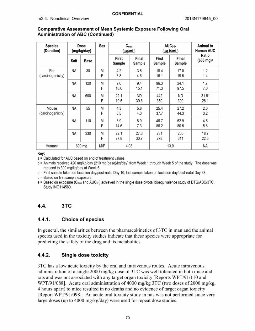
CONFIDENTIALm2.4. Nonclinical Overview 2013N179645_00
70
Comparative Assessment of Mean Systemic Exposure Following Oral Administration of ABC (Continued)
Species(Duration)
Dose (mg/kg/day)
Sex Cmax
(g/mL)AUC0-24
(g.h/mL)Animal to
Human AUC Ratio
(600 mg)aSalt BaseFirst
SampleFinal
SampleFirst
SampleFinal
Sample
Rat(carcinogenicity)
NA 30 M 4.2 3.8 18.4 17.0 1.2F 3.8 4.6 16.1 19.5 1.4
NA 120 M 9.6 9.4 96.3 24.1 1.7F 10.0 15.1 71.3 97.5 7.0
NA 600 M 22.1 ND 442 ND 31.8d
F 19.5 39.6 350 390 28.1
Mouse(carcinogenicity)
NA 55 M 4.3 5.8 25.4 27.2 2.0F 6.5 4.0 37.7 44.3 3.2
NA 110 M 8.9 8.9 46.7 62.9 4.5F 14.6 7.3 86.2 80.5 5.8
NA 330 M 22.1 27.3 231 260 18.7F 27.8 30.7 278 311 22.3
Humane 600 mg M/F 4.03 13.9 NA
Key:a = Calculated for AUC based on end of treatment values.b = Animals received 420 mg/kg/day (210 mg(base)/kg/day) from Week 1 through Week 5 of the study. The dose was
reduced to 300 mg/kg/day at Week 6.c = First sample taken on lactation day/post-natal Day 10; last sample taken on lactation day/post-natal Day 63.d = Based on first sample exposure.e = Based on exposure (Cmax and AUC0-t) achieved in the single dose pivotal bioequivalence study of DTG/ABC/3TC,
Study ING114580.
4.4. 3TC
4.4.1. Choice of species
In general, the similarities between the pharmacokinetics of 3TC in man and the animal species used in the toxicity studies indicate that these species were appropriate for predicting the safety of the drug and its metabolites.
4.4.2. Single dose toxicity
3TC has a low acute toxicity by the oral and intravenous routes. Acute intravenous administration of a single 2000 mg/kg dose of 3TC was well tolerated in both mice and rats and was not associated with any target organ toxicity [Reports WPT/91/110 andWPT/91/088]. Acute oral administration of 4000 mg/kg 3TC (two doses of 2000 mg/kg, 4 hours apart) to mice resulted in no deaths and no evidence of target organ toxicity[Report WPT/91/098]. An acute oral toxicity study in rats was not performed since very large doses (up to 4000 mg/kg/day) were used for repeat dose studies.

CONFIDENTIALm2.4. Nonclinical Overview 2013N179645_00
71
4.4.3. Repeat dose toxicity
The repeated administration of either 3TC or the racemate, gsk001*, was very well tolerated. Treatment-related effects were largely confined to the highest dose group following oral administration for up to 6 months in the rat (4000 mg/kg/day) and 12 months in the dog (3000 mg/kg/day for males; 2000 mg/kg/day for females) [Reports WPT/93/361 and WPT/92/407].
In rats, changes observed in clinical pathology parameters were minor and reversible. A chronic low grade irritation of the cecum/anus was seen with the highest dose of 3TC (4000 mg/kg/day). However, there was a clear no effect level for the cecal changes at the intermediate dose (850 mg/kg/day), which is approximately 142 times higher than the therapeutic dose (300 mg or 6 mg/kg for a 50 kg human) on a mg/kg basis. While approximately 35% of an orally administered dose of 3TC is unabsorbed and excreted in the feces of the rat, only 15 to 20% is unabsorbed in humans. Combining these safety margins, the finding in the cecum was not considered to represent a safety hazard for humans.
In a 3 month oral study in dogs, one female receiving a dose of 3000 mg/kg/day died during Week 5 and, over the following 2 weeks, 2 further animals from this group were killed for humane reasons [Report WPT/92/132]. While these deaths were considered to be treatment-related, the precise cause of death was not established and many of the effects seen in these animals were considered to reflect a general decline in condition after a prolonged period of inappetence. No deaths occurred in the 12 month dog study where the high dose in females was 2000 mg/kg/day.
In the chronic toxicity studies, hematological changes in the dog were more pronounced than in the rat. They consisted of mild to moderate decreases in erythrocyte, total leukocyte, neutrophil and lymphocyte counts, and increases in mean corpuscular volume (MCV), mean corpuscular haemoglobin (MCH) and mean activated partial thromboplastin time (APTT). White cell changes were restricted to the high dose group throughout, but minor red cell changes were evident in the low dose group (90 mg/kg/day) in the latter part of the 12 months study. All changes showed evidence of regression in recovery animals.
In the 12 month dog study, plasma folate and Vitamin B12 levels were measured, as reductions in these are a potential cause of macrocytic anaemia. Surprisingly, treatment-related increases in plasma folate and Vitamin B12 were seen, with values in the high dose males being approximately twice those of the control animals. The reason for this is unknown, but as both folate and Vitamin B12 are involved in nucleotide metabolism, it may be a biochemical response to the large doses of 3TC (a cytidine analogue). All changes showed evidence of regression in recovery animals.
*新薬承認情報提供時に置き換え

CONFIDENTIALm2.4. Nonclinical Overview 2013N179645_00
72
In dogs, excluding changes noted in 3 females that died or were euthanized in moribund condition in the 3 month study, histopathological findings were limited to slight centrilobular lipid deposition in the liver and moderate thymic atrophy in high dose animals (3000 mg/kg/day) in the 3 month study, and minimal hemosiderosis in the spleen in the high dose group of the 12 month study [Report WPT/92/407]. There was no alteration in bone marrow cytology. All changes ameliorated after a 4 week recovery period.
Preliminary studies in the marmoset were conducted on both the racemate, gsk001*, and 3TC using intravenous administration. gsk001* was administered at 600 mg/kg/day for 7 days and 3TC at doses up to 1200 mg/kg/day for 14 days. These studies revealed no unexpected toxicities and any findings were consistent with the responses seen in the rat and dog.
4.4.4. Genetic toxicology
In vitro studies
3TC was negative in the Ames assay at concentrations up to 5000 g/plate and in the fluctuation test using E. coli tester strains [Reports WPT/90/240 and WPT/93/246]. In an in vitro cytogenetic test in human peripheral lymphocytes, 3TC showed clastogenic activity at the highest concentrations evaluated in the absence of S9-mix (300 g/mL) and in the presence of S9-mix (2292.5 g/mL; 10 mM) [Report WPT/93/120]. In the mouse lymphoma assay, 3TC induced small increases in mutation frequency at concentrations of 1000 g/mL and above when a 24 hour exposure period was used[Report WPT/90/271], but was not mutagenic in the same test system at concentrations up to 5000 g/mL using a 3 hour treatment period. 3TC did not induce morphological transformation of mouse embryo cells at concentrations up to 320 g/mL in the absence of S9-mix or up to 5000 g/mL in the presence of S9-mix [Report UTX/92/018].
In vivo studies
Mitochondrial toxicity is considered to be a class effect of NRTIs; therefore, studiesinvestigating the potential effects of 3TC on mitochondrial DNA have been carried out in pregnant mice and their offspring.
B6C3F1/Tk+/- mice were administered daily intraperitoneal injections of 200 mg/kg/day 3TC from Days 1 to 8 of age. 3TC was found not to cause an increase in micronuclei, did not induce mutations in the Hprt gene and did not cause a significant increase in the Tk mutant frequency. Therefore, 3TC was not genotoxic in B6C3F1/Tk+/- mice [Von Tungeln, 2002]. For a more complete discussion of NRTI-related mitochondrial toxicity, refer to Section 4.4.7.
*新薬承認情報提供時に置き換え

CONFIDENTIALm2.4. Nonclinical Overview 2013N179645_00
73
When pregnant CD-1 mice were treated with 100 mg/kg/day 3TC from Day 12 to Day 18 of gestation there were no increases in the Hprt mutant frequency in male offspring on post-natal Days 13, 15 and 21 following treatment with 3TC compared with control [Torres, 2007]. A further study evaluated mutagenic effects in pregnant C57BL/6N mice treated with 120 mg/kg/day 3TC, from Day 12 to Day 18 of gestation, alone or in combination with zidovudine. No effects were detected in neonates from pregnant mice treated with 3TC alone, except for a weak increase in chromosomal damage measured on Day 1, and an increase in Tk mutant frequency in 5 week old female mice [Von Tungeln,2007].
No in vivo genotoxic activity was evident following oral doses of 3TC of up to 2000 mg/kg in either the rat bone marrow metaphase analysis assay, the rat bone marrow micronucleus test (3 daily doses) or the rat liver unscheduled DNA synthesis assay[Reports WPT/90/275, WPT/93/195 and WPT/90/408].
4.4.5. Carcinogenicity
3TC showed no evidence of carcinogenic potential in a 24 month study in mice receiving doses up to 2000 mg/kg/day in the diet (~12X above human exposure for 300 mg dose, 12.3 g.h/mL) [Report WPT/95/297]. A dose-related reduction in body weight gain was seen at all dose levels as were minor hematological changes. Histopathological examination showed a slightly higher incidence of histiocytic sarcomas in the 180 and 2000 mg/kg/day female groups. However, there was no trend with dose and the increases were only significant in one out of the two statistical methods employed. Furthermore, as the incidences were similar to that of historical controls, the occurrence of histiocytic sarcoma was considered to be incidental to treatment with 3TC. There was a slight but non-dose-related increase in the incidence and degree of mineralisation of the cortex of the kidney of all treated male mice. The femur of females treated with 600 or 2000 mg/kg/day showed a non-dose-related increase in bone deposition. Both of these findings were considered to be exacerbations of normal age-related changes in mice.
3TC showed no evidence of carcinogenic potential in a 24 month study in rats receiving doses up to 2000 mg/kg/day for males (~43X above human exposure for 300 mg dose, 12.3 g.h/mL) and 3000 mg/kg/day for females (~72X above human exposure for 300 mg dose, 12.3 g.h/mL) by dietary administration [Report WPT/95/298]. There was no effect of treatment on survival. Final body weight was 8 to 18% lower than that of the controls in groups treated with doses of 1000 mg/kg/day or more. There was no evidence of the expected decrease in leucocyte or erythrocyte counts in 3TC-treated groups at the end of the study. There was a slight increase in the incidence of endometrial epithelial tumors in females dosed at 3000 mg/kg/day. However, this was only slightly higher than historical control values but achieved statistical significance (p=0.017). Furthermore, there was no increase in the incidence of pre-neoplastic findings in the endometrium (e.g., proliferative changes) in the 3TC dosed groups. For these reasons the occurrence of endometrial tumors was considered to be unrelated to treatment. The only non-neoplastic finding of note was an increase in the incidence and severity of mineralization in the kidney, seen at doses of 1000 mg/kg/day and above, with accompanying suburothelial capillary dilatation in males at 2000 mg/kg/day. This was considered to be an exacerbation of a normal age-related change commonly seen in rat kidney.

CONFIDENTIALm2.4. Nonclinical Overview 2013N179645_00
74
4.4.6. Reproductive and developmental toxicity
In a rat fertility study, with the exception of a few minor changes at the high dose level, the overall reproductive performance of the F0 and F1 generation animals, and the development of the F1 and F2 generation, was unaffected by treatment with 3TC at doses up to 4000 mg/kg/day [Report WPT/93/210].
In oral organogenesis studies in rats, there were no treatment-related effects on gestation length and index, the incidence of pre- and post-implantation loss, the number of implantations and live fetuses, fetal sex ratio, fetal body weight or embryofetal development at doses up to 4000 mg/kg/day [Report WPT/91/305].
Rabbits were generally more sensitive to 3TC than rats and therefore lower doses had to be used for the rabbit organogenesis studies. In the main study, one intermediate dose (300 mg/kg/day) animal died on Day 17 (Day 1 = day of mating) [Report WPT/91/333]. One low dose (90 mg/kg/day), one intermediate dose (300 mg/kg/day) and 3 high dose (1000 mg/kg/day) animals aborted. A reduction in the amount of feces excreted, thin appearance and orange colored urine were noted in all groups, including the controls, although these findings were observed more frequently at the high dose level. Body weight gain was transiently reduced in the control, low and intermediate dose groups. At the high dose level, body weight loss and a reduction in food and water consumption were observed throughout the treatment period. There were no post mortem abnormalities attributable to 3TC in an animal found dead, the animals which aborted or the dams that were killed at term.
Fetal examination in the rabbit study revealed total in utero litter loss in 2 intermediatedose animals and in one high dose animal. Pre-implantation loss was slightly increased at all dose levels and, consequently, the mean number of viable fetuses per litter was decreased compared with the control group, although these findings were not dose-related. Since pre=implantation loss, as assessed here, may include very early post-implantation deaths which could coincide with the onset of dosing, this finding was considered to represent a possible treatment-related embryolethal effect. Post-implantation loss and the proportion of male fetuses were unaffected by treatment.
A further oral organogenesis study was performed to establish a no effect level for the increased incidence of pre-implantation loss seen in the previous study. Pre-implantation loss was slightly, but not significantly, increased at 40 and 90 mg/kg/day, with consequent reductions in the number of implantations and fetuses. The no effect level was 15 mg/kg/day.
At the lowest dose level at which the slight increase in pre-implantation loss was observed in rabbits (20 mg/kg BID), systemic exposure on Day 20 of pregnancy based on the Cmax and AUC (1.05 g/mL and 5.9 g/h/mL) was 0.48X the estimated exposure in humans at 300 mg. There was no evidence of any effect on pre-implantation loss in the rat at plasma Cmax concentrations approximately 32 times the anticipated human exposure for a 300 mg/day dose. Thus, while it is possible that the observed embryolethal effect in the rabbit is a species-specific phenomenon, if 3TC were administered to women during

CONFIDENTIALm2.4. Nonclinical Overview 2013N179645_00
75
early pregnancy, the possibility of a slight increase in the risk of embryonic loss cannot be discounted [m1.14.1 (US) or m1.3 (EU)].
No treatment-related external or visceral fetal abnormalities were apparent during organogenesis studies in rabbits following administration of 3TC at doses up to 1000 mg/kg/day. An indirect effect on fetal skeletal development (reduction in epiphysial ossification and increased incidence of unilateral supernumerary ribs) was observed at the highest dose only. In a subsequent study, skeletal examination revealed increases in the incidence of supernumerary ribs at doses up to 90 mg/kg/day. The occurrence of supernumerary ribs in the fetus has been shown in some species to be an indirect effect of maternal toxicity or stress [Beck, 1989; Branch, 1992], and appears to be a condition which is reversible post-natally and has no permanent structural effect on the skeleton [Wickramaratne, 1988]. This was not, therefore, regarded as a significant finding.
In a combined peri-/post-natal and juvenile toxicity study in rats, mated females received 3TC at 90, 450 or 2000 mg/kg twice daily from Day 17 of pregnancy until post-natal Day 22. Two male and female offspring from each dam received a single daily dose of 3TC orally at 90, 450 or 2000 mg/kg/day from Litter Day 3 until Litter Day 43. No treatment-related effects were observed on gestation length, gestation index, the number of offspring born, the proportion of male offspring, birth index, viability index or weaning/lactation index [Report WPT/93/165]. The majority of dams receiving 4000 mg/kg/day showed a swollen/reddened anus/rectum during the lactation period, which correlated with histological inflammatory changes at the ano-rectal junction. An increased incidence of swollen/reddened anus/rectum was also observed in some offspring receiving 2000 mg/kg/day, with similar histological changes to those seen in the dams. These changes, together with slight diffuse epithelial hyperplasia in the cecum of several dams and offspring at the high dose levels, were attributed to the prolonged exposure to large concentrations of unchanged 3TC in the gastrointestinal tract, resulting in typical responses to an irritant material. Similar cecal changes were also recorded in the 6 month oral toxicity study in the rat.
Hematological changes consistent with a mild macrocytic anaemia, as seen in the adult repeat dose rat studies, were noted in the juvenile rats treated with up to 2000 mg/kg/day 3TC. A statistically significant reduction in testis weight was observed in juveniles at this dose level. Histological examination of the testes revealed slight to moderate dilatation of the seminiferous tubules in a number of high dose males (8/25). There was no evidence of morphological abnormality other than slight compression of the germinal epithelium. None of the other toxicity studies conducted with 3TC using adult rats and dogs have revealed testicular effects. Similarly, no effects were noted in a fertility study in the rat. The toxicity would therefore appear specific to the developing/immature testis. The no effect level for the testicular effects was considered to be the intermediate dose level of 450 mg/kg/day (~80X above human exposure based on Cmax).
Pregnant CD-1 mice were administered 500 mg/kg BID 3TC orally from Day 10 of gestation to delivery. Offspring locomotion and nociceptive sensitivity were examined on post-natal Days 8, 14 and 28 after administration of two doses of GABAergic agonist muscimol. A 30 minute locomotor activity test and 60 second hot plate test were used.

CONFIDENTIALm2.4. Nonclinical Overview 2013N179645_00
76
Pre-natal 3TC exposure had a limited effect on locomotor activity development and noeffect on nociception [Ricceri, 2001].
3TC was administered orally to pregnant CD-1 mice at doses of 125, 250 or 500 mg/kg BID from Day 10 of gestation to delivery. Data on reproductive performance were collected and offspring were examined for a series of different somatic and behavioral end points. Learning and retention performances of a passive avoidance task on post-natal Day 20/21 were unaffected by 3TC treatment, while decreased habituation in an automated locomotor activity test was evident in male offspring exposed to 250 and 500 mg/kg [Calamandrei, 1999]. In another study in mice at the same doses, offspring behavior was examined on post-natal Day 35 in a 20 minute social interaction test. At adulthood, different behavioral endpoints were analyzed. Findings confirmed the low neurotoxicity of 3TC, however, some significant behavioural alterations were noted. These were a decrease in immobility in the open field test, an increase in the responsiveness to scopolamine shown by the 500 mg/kg 3TC treated mice in the open field and a longer escape latency in the first day of the reversal phase in the water maze. No significant changes in either pain sensitivity, social/affiliative or maternal behavior were found, although a higher occurrence of aggressive behavior toward foster pups was noted in both the 125 and 500 mg/kg females [Calamandrei, 2000].
4.4.7. Mitochondrial toxicity
Mitochondrial toxicity is a class effect of NRTIs [Lewis, 2005; Moyle, 2005]. Studies investigating the effects of in utero exposure to NRTIs, conducted in mice and monkeys, have focused on the potential to cause mitochondrial toxicity in the heart. There is relatively little data on the effects of 3TC alone; however, in one study, CD-1 mice were administered 75 mg/kg/day 3TC throughout gestation and for 28 days post-natally prior to cardiomyocyte examination for mitochondrial damage. There was a depletion in mtDNA copy number and an increase in mtDNA lesions in 3TC-treated 4 week old mice of both sexes; however, these parameters had recovered to control levels by 10 weeks of age. There were no significant mtDNA deletions or changes in cytochrome c oxidase(COX) staining intensity [Chan, 2007].
In order to assess the occurrence and persistence of cardiac changes, a study was conducted in which CD-1 mice were exposed in utero to 3TC (40 mg), zidovudine (80 mg) or 3TC plus zidovudine (40/80 mg) during Days 12 to 18 of gestation [Torres,2010]. Hearts from female mouse offspring were examined at 13 and 26 weeks post partum (pp) and ECG measurements indicated progressive thinning of the left ventricular posterior wall. The induction of cardiac effects by 3TC and zidovudine were additive compared with either drug alone.

CONFIDENTIALm2.4. Nonclinical Overview 2013N179645_00
77
Mitochondrial morphology and mtDNA content have also been investigated in the umbilical cord and cord blood of Erythrocebus patas monkeys exposed in utero to 24 mg/day 3TC during the last 4 weeks of gestation [Divi, 2007a]. Umbilical cord endothelial cells contained some swollen mitochondria with pale matrices and disorganised cristae. This was accompanied by a small (<35%) decrease in mtDNA content, which remained unaffected in cord blood leukocytes. In the same study, samples were evaluated from HIV-infected pregnant humans treated with 3TC (and/or other antiretrovirals): transplacental exposures induced similar mitochondrial damage in cord blood and umbilical cords from uninfected monkey infants and from human infants born to HIV-infected women.
Mitochondrial damage was also investigated in skeletal muscle from the offspring ofE. patas monkeys administered 3TC alone, or with zidovudine or stavudine, for the last 10 weeks of gestation, or for 10 weeks of gestation and 6 weeks after birth [Divi, 2007b]. Skeletal muscle mitochondrial compromise occurred at birth and was found to persist at 1 year of age, although the effect in monkeys which had been treated with 3TC alone wasmuch less extensive than in skeletal muscle from monkeys treated with the combinations.
The significance for humans of the nonclinical neurobehavioural and mitochondrial effects described above is uncertain. There are currently no clinical data assessing possible mitochondrial dysfunction following peri-natal exposure to 3TC alone, the limited available data being derived from infants exposed to 3TC combined with zidovudine. Evidence of mitochondrial dysfunction was reported in 8 of 1754 HIV uninfected children exposed peri-natally to NRTIs, with neurological symptoms prevalent among the affected individuals. Two of the 8 children, both of whom had been exposed peri-natally to 3TC and zidovudine, died at 11 and 13 months of age [Blanche, 1999]. Subsequently, the Peri-Natal Safety Review Working Group has systematically reviewed the cases of more than 20000 children born to HIV-infected mothers, among whom 1459 (6%) were exposed peri-natally to 3TC and zidovudine. Cases were reviewed for deaths at less than 5 years of age and evidence of mitochondrial disease among the living. The authors concluded that there was no indication that antiretroviral therapy was associated with deaths related to mitochondrial dysfunction [PSRWG, 2000]. In a further study in 1798 African infants exposed peri-natally to 3TC and zidovudine, there was no evidence of increased neurologic events after 18 months follow up [PETRA, 2002]. A review of the data from the Peadiatric AIDS Clinical Trials Group (PACTG) cohorts from protocols 219 and 219C concerning children peri-natally infected with HIV found a higher risk of possible mitochondrial dysfunction amongst children exposed to stavudine, 3TC, 3TC-stavudine combination and stavudine-didanosine combinations compared to other antiretrovirals. The authors deployed two sets of case definition criteria (Enquête Périnatal Française and the Mitochondrial Disease Classification) to identify children exhibiting clinical signs and symptoms consistent with possible mitochondrial dysfunction. Overall, 33.5% of 2931 HIV-infected children met at least one published case definition for ‘possible mitochondrial dysfunction’ and 16.4% met both sets of criteria. The case definitions were based on clinical signs since appropriate biochemical, histopathological, molecular and genetic data on which to confirm the diagnosis were not available, limiting the precision with which odds ratios could be calculated.

CONFIDENTIALm2.4. Nonclinical Overview 2013N179645_00
78
In addition, the Antiviral Pregnancy Registry [APR, 2013] contains sufficient numbers offirst trimester exposures to 3TC in humans to be able to detect at least a 1.5-fold increase in overall birth defects, and a 2-fold increase in defects in the more common classes,cardiovascular and genitourinary systems, but no increase associated with mitochondrial dysfunction has been observed to date.
4.4.8. Phototoxicity
3TC does not absorb light in the 290 to 700 nm wavelength range. Photosensitiviy reactions have not been a clinical issue with 3TC, for which there is significant clinical experience. Thus, nonclinical phototoxicity studies have not been performed on 3TC.

CONFIDENTIALm2.4. Nonclinical Overview 2013N179645_00
79
Table 6 Comparative Assessment of Mean Systemic Exposure Following Oral Administration of 3TC
Species(Duration)
Dose (mg/kg BID, Unless Stated)
Cmax (g/mL)a AUC0-t (g.h/mL) Animal to Human AUC Ratio
(300 mg)bEnd of Study End of Study
Rat(3 month)
45 4.7 20.9 3.4
300 30.9 145 23.5
2000 94.6 690 112
Rat(6 month)
90 11.8 69.3 11.3
425 31.7 133 21.6
2000 113 624 101
Dog(3 month)
45 21.5 66.2 10.8
260 89.8 367 59.7
1500 331 1582 257
Dog(12 month)
45 22.9 74.5 12.1
260 85.9 343 55.8
1500 (M) 213 872 142
1000 (F) 216 948 154
Rat(embryofetal development)
45 3.1 ND ND
300 23.8 ND ND
2000 66.9 ND ND
Rabbit(embryofetal development)
7.5 0.45 2.3 0.37
20 1.05 5.9 1.0
45 2.55 14.8 2.4
150 8.0 ND ND
500 68.4 ND ND
Rat(juvenile)
90 (males) 18.1 ND ND
90 (females) 21.1 ND ND
450 (males) 167 ND ND
450 (females) 176 ND ND
2000 (males) 406 ND ND
2000 (females) 472 ND ND
Mouse carcinogenicityc
180 mg/kg/day 1.1 16.4 1.3
600 mg/kg/day 3.4 47.6 3.9
2000 mg/kg/day 9.7 151 12.3

CONFIDENTIALm2.4. Nonclinical Overview 2013N179645_00
80
Comparative Assessment of Mean Systemic Exposure Following Oral Administration of 3TC (Continued)
Species(Duration)
Dose (mg/kg BID, Unless Stated)
Cmax (g/mL)a AUC0-t (g.h/mL) Animal to Human AUC Ratio
(300 mg)bEnd of Study End of Study
Rat carcinogenicityc 300 mg/kg/day 4.5 75 6.1
1000 mg/kg/day 15 242 19.7
2000 mg/kg/day 28 533 43.3
3000 mg/kg/day 38 880 71.5
Humand 300 mg/day 2.1 12.3 NA
Key:a = Approximate Cmax values are at either 1 or 2 hours post dose. Value presented is that obtained following the first
daily administration in the last week of the study, except for carcinogenicity studies where determinations weremade in Week 5.
b = In the case of twice daily dosing in animal toxicity studies, the AUCtau for a 12 hour dosing interval is doubledbefore calculating the approximate multiple of the AUCtau in man for a 24 hour dosing interval.
c = Carcinogenicity studies were dosed QD as opposed to other toxicity studies which were dosed BID.d = Based on exposure (Cmax and AUC0-t) achieved in the single dose pivotal bioequivalence study of DTG/ABC/3TC,
Study ING114580.
4.5. Impurities and Excipients
4.5.1. Combination drug product
For dolutegravir, there are no specified impurities in the proposed Drug Substance Specification that exceed the 0.15% w/w ICH qualification threshold [m3.2.S]. Abacavir and lamivudine are established active pharmaceutical ingredients (API) which have an extensive history of use in registered products since their introduction in the 1990’s. Specified impurities in the abacavir and lamivudine drug substance were previously considered qualified on the basis of the levels of the impurities present in batches used, and the profiles of impurities, which were tested in pivotal nonclinical toxicity studies. Abacavir and lamivudine drug substance are manufactured by an approved supplier using an approved route.
A genotoxic risk assessment of the dolutegravir drug substance showed there are no impurities of mutagenic concern at a level that would exceed the threshold of toxicological concern [TTC] as defined by the CHMP guidelines on the limits of genotoxic impurities (i.e., >1.5 g/day). A further genotoxic risk assessment was conducted to determine whether any additional items of concern would result from the combination of dolutegravir with abacavir and lamivudine. This risk assessment, in addition to forced degradation studies on the combination tablet [m3.2.P.8.3], did not result in any new impurities and the degradation profiles were comparable to TIVICAY (dolutegravir) and EPZICOM/KIVEXA (abacavir/lamivudine FDC). No additional risks were identified through an evaluation of the manufacturing process.

CONFIDENTIALm2.4. Nonclinical Overview 2013N179645_00
81
The excipients used in the formulation for DTG/ABC/3TC FDC are conventional and the amount per tablet falls within typical ranges used, with the exception of sodium starch glycolate, which is at a higher percentage than is typically utilised [see m3.2.P.2.2 for justification]. The specifications for the inactive ingredients comply with the United States Pharmacopeia/National Formulary (USP/USNF) and the European Pharmacopeia (Ph.Eur.). The film coating material is supplied by Colorconand comprises ingredients that are registered in at least one of the following:USP/USNF, US CFR, Food Chemicals Codex, Ph.Eur. or Japanese Pharmacopeia. All colorants used conform to EC Commission Regulation 231/2012.

CONFIDENTIALm2.4. Nonclinical Overview 2013N179645_00
82
5. INTEGRATED ASSESSMENT OF THE FIXED DOSE COMBINATION PRODUCT
The Sponsor has completed a comprehensive assessment of the nonclinical safety of DTG in support of treatment of patients with HIV. DTG has been evaluated in toxicity studies of up to 26 weeks in duration in Sprague Dawley rats, 38 weeks in cynomolgus monkeys and 13 weeks in Crl:CD-1 (ICR) BR mice. ABC and 3TC are both wellestablished products that have been in use worldwide for an extended period of time both individually and in combination (Kivexa). Both are routinely prescribed in clinical practice. The pharmacology, pharmacokinetics, toxicology and safety profile in clinical use of these two compounds are well known.
ICH Guidance M3 (R2) on Non-Clinical Safety Studies for the Conduct of Human Clinical Trials and Marketing Authorisation for Pharmaceuticals states that, “For most combinations which involve two late stage entities and for which there is adequate clinical experience with co-administration, combination toxicity studies would generally not be recommended to support clinical studies or marketing unless there is significant toxicological concern (e.g., similar target organ toxicity)” [ICH M3 (R2),2009]. In addition, according to Question 9 in the Questions and Answers documentwritten by the M3 (R2) Implementation Working Group for M3 (R2) Guideline “Non-Clinical Safety Studies for the Conduct of Human Clinical Trials and Marketing Authorization for Pharmaceuticals” regarding the scope of ICH M3 (R2), it is accepted that combination toxicity studies are not generally warranted for HIV products unless there is a specific cause for concern under clinically relevant conditions. An extensive review of the nonclinical data for DTG, ABC and 3TC has shown:
No anticipated adverse pharmacodynamic interactions and no anticipated impact on established safety margins as a consequence of pharmacokinetic interactions.
No nonclinical signals that might indicate the potential for additive or synergistic toxicity.
No increased risk with respect to effects on fertility.
No increased genotoxic or carcinogenic risk.
Based on this, together with data from the Phase 3 studies ING114467, ING113086 andING114915, the Sponsor considers that combination toxicity studies would provide no additional safety information and are not necessary to support the proposed marketing application for the DTG/ABC/3TC FDC STR.
A summary of the nonclinical information on which this conclusion is based is provided below:

CONFIDENTIALm2.4. Nonclinical Overview 2013N179645_00
83
5.1.1. Potential for pharmacodynamic interactions
The concomitant oral administration of DTG with ABC and 3TC is intended to decrease HIV viral load over the administration of any of the compounds alone. Thus,pharmacodynamic interaction is an intended component of the therapeutic activity of this combination. In vitro checkerboard experiments have shown that these agents are additive to synergistic in their activity; data from ING114467, ING113086 and ING114915 demonstrate that the combination is comparable to or superior to the currently approved regimen in patients.
5.1.2. Potential for pharmacokinetic interactions
Due to different routes of metabolism and elimination of the individual components in DTG/ABC/3TC FDC product, and the minimal effect of these agents on drug metabolizing enzymes or transporters, there is a low propensity for clinically significant drug interactions between DTG, ABC and 3TC when co-administered.
5.1.3. Potential for toxicologic interactions
Synergistic or additive toxicity is not expected from the co-administration of DTG with ABC and 3TC. While both ABC and DTG caused treatment-related hepatic effects, the findings in ABC-treated animals were believed to be adaptive changes related to metabolic enzyme induction, and with DTG the findings were observed only at doses that exceeded the maximum tolerated dose. There were no other common target organs of toxicities identified for the 3 compounds. Therefore, the potential for additive or synergistic toxicity at clinically relevant concentrations is considered low. No observable adverse effect levels (NOAELs) have been established for the 3 entities in general toxicity studies at exposure margins that are approximately at parity (DTG, ABC) or significantly higher (3TC) than expected clinical exposure.
5.1.3.1. Repeat dose toxicity studies
The primary finding from repeat dose toxicity studies up to 26 weeks in rats and 38 weeks in monkeys with DTG was GI toxicity. In monkeys, the most sensitive species, GI toxicity was characterized primarily by vomiting, diarrhea and associated mortality as well as gastrointestinal lesions, and by gastric lesions in the rat. In both species, these effects were observed at progressively lower doses with increased study duration. The GI toxicity is believed to be the result of local drug administration at the mucosal surface of the gut following oral dosing, rather than systemic toxicity. The fact that affected animals had comparable exposures to animals at dose levels which were not affected is supportive of the conclusion that the GI toxicity is due to the larger local exposure in the GI tract in those dose groups. Therefore, mg/kg or mg/m2 metrics are appropriate determinates of safety cover for this toxicity because it is not based on systemic exposure. The NOAEL for the 38 week monkey toxicity study (15 mg/kg/day) is 15X and 7.5X the human mg/kg equivalent dose (based on 50 kg human) and 5X the human mg/m2 equivalent dose for a 50 mg QD dose. GI toxicity in animals did not translate to an increased risk for clinical adverse events at DTG doses of 50 mg QD.

CONFIDENTIALm2.4. Nonclinical Overview 2013N179645_00
84
The target organs in repeat dose toxicity studies with ABC were the liver and testes. Liver findings included hepatocellular hypertrophy (mice, rats and monkeys) and individual cell necrosis (mice) with increases in cholesterol, triglycerides, total protein, bile acids and/or ALT across species. All findings were mild, reversible and showed evidence of regression except hepatocellular hypertrophy in male mice at 1000 mg(succinate)/kg/day for 3 months. ABC may be a weak enzyme inducer, and therefore hepatic changes are believed to be associated with ABC-induced alterations in metabolic activity rather than direct toxic effect on the liver. While liver alterations were also observed with DTG (hepatocellular single cell necrosis and diffuse hepatocellular hypertrophy, elevated liver enzymes), these effects only occurred in the monkey 14 day toxicity study at doses that were not tolerated and exposures ~5 to 9X above the anticipated clinical exposure for a 50 mg DTG dose given in combination with ABC and 3TC. Cumulative data suggest a hepatic safety profile for DTG/ABC/3TC that is comparable to Atripla, RAL + ABC/3TC and DRV +RTV + ABC/3TC.
ABC effects on the testes were only seen in rats, mainly in studies of short duration (30 days) and at dosages 135 mg(base)/kg/day. Findings included reduction in testicular size due to the loss of germ cells from the seminiferous tubules, which showed evidence of reversal following a treatment-free period.
The main treatment-related effects of 3TC in rats and dogs were hematological reductions in red cell counts. In dogs, these findings were associated with increased MCV and MCH, and reductions in total leukocyte, neutrophil and lymphocyte counts. A chronic low grade irritation of the cecum/anus was observed in the rat 6 month toxicity study with the highest dose of 3TC (4000 mg/kg/day). However, there was a clear no effect level for the cecal changes at the intermediate dose (850 mg/kg/day), which is approximately 150 times higher than the recommended therapeutic dose based on a mg/kg basis.
5.1.3.2. Genotoxicity and carcinogenicity
DTG was not genotoxic in a battery of in vitro tests and an in vivo rat micronucleus assay. ABC and 3TC were not mutagenic in bacterial tests, but like many other nucleoside analogues showed activity in in vitro mammalian tests such as the mouse lymphoma assay. However, an in vivo rat micronucleus test with ABC and 3TC in combination was negative.
Neither DTG nor 3TC was carcinogenic in 2 year rat or mouse carcinogenicity studies. However, ABC showed an increase in the incidence of malignant and non-malignant tumors in mice and rats. Malignant tumors occurred in the preputial gland of males and the clitoral gland of females of both species, and in rats in the thyroid gland of males and in the liver, urinary bladder, lymph nodes and the subcutis of females. The majority of these tumors occurred at the highest ABC dose of 330 mg/kg/day in mice and 600 mg/kg/day in rats. The exception was the preputial gland tumor which occurred at a dose of 110 mg/kg in mice. The systemic exposure at the no effect level in mice and rats was equivalent to 3 and 7 times the human systemic exposure during therapy.

CONFIDENTIALm2.4. Nonclinical Overview 2013N179645_00
85
While the carcinogenic potential of ABC in humans is unknown, the data suggest that a carcinogenic risk to humans is outweighed by the potential clinical benefit, and no increase in carcinogenic risk is anticipated with the DTG/ABC/3TC STR.
5.1.3.3. Fertility, pregnancy and lactation
DTG, ABC or 3TC had no effect on male or female fertility in animals.
DTG had no effect on embryofetal development in rats and rabbits. 3TC was not teratogenic in animal studies, but a small increase in early embryonic loss was seen when administered to pregnant rabbits at exposure levels comparable to those achieved in humans. However, there was no indication of this effect in the rat at exposure levels up to 35 times that in humans. ABC produced fetal malformations (increased incidences of fetal anasarca and skeletal malformations) and developmental toxicity (depressed fetal body weight and reduced crown rump length) were observed in rats at a dose which produced 35 times the human exposure, based on AUC. However, in the rabbit, no developmental toxicity and no increases in fetal malformations occurred at doses that produced 8.5 times the human exposure at the recommended dose based on AUC.
The safe use of DTG/ABC/3TC STR in human pregnancy has not been established. The effect of DTG on human pregnancy is unknown. In reproductive toxicity studies in animals, DTG was shown to cross the placenta. 3TC and ABC were associated with findings in animal reproductive toxicity studies. Therefore, administration of DTG/ABC/3TC STR in pregnancy should be considered only if the benefit to the mother outweighs the possible risk to the fetus.
It is recommended that HIV-infected women do not breast-feed their infants under any circumstances in order to avoid transmission of HIV. Furthermore, DTG and ABC are excreted in the milk of lactating rats, and 3TC is excreted in human milk at similar concentrations to those found in serum. It will therefore be recommended that mothers do not breast-feed their babies while receiving treatment with the DTG/ABC/3TC STR[m1.14.1 (US), Section 8.1].
CONFIDENTIALm2.4. Nonclinical Overview 2013N179645_00
86
6. OVERALL CONCLUSIONS
Extensive nonclinical development programmes of studies have been carried out with DTG, ABC and 3TC individually.
Data have shown that these agents (DTG, ABC and 3TC) are additive to synergistic in their activity when co-administered to patients.
In general, the systemic exposures at the NOAEL in animal toxicity studies are approximately at parity with (DTG, ABC), or significantly higher (3TC) than, the expected clinical exposures. The comparative biotransformation pathways between at least one animal species and humans for DTG, ABC and 3TC, individually, were similar and indicate that animals and humans were exposed to similar in vivo profiles with no disproportionate human metabolites observed for any of the drugs. The primary routes of elimination for the individual components, DTG, ABC and 3TC, in at least one animal species were consistent with those observed in humans. These data, taken together, support the selection of the toxicology species for the prediction and evaluation for the safe use of each drug and their metabolites in the proposed patient population when prescribed individually or in combination according to the proposed dosing regimen.
Based on the clinical experience with DTG, ABC and 3TC (alone and in combination), and the absence of nonclinical toxicology and pharmacokinetic signals that might indicate the potential for additive or synergistic interaction, the Sponsor considers that combination toxicity studies are not necessary to support the submissions of the DTG/ABC/3TC FDC STR.
The comprehensive nonclinical development package presented in this Nonclinical Overview, along with extensive marketing experience with ABC and 3TC, supports the proposed registration and safe use of DTG/ABC/3TC FDC in patients for the treatment of HIV.
CONFIDENTIALm2.4. Nonclinical Overview 2013N179645_00
87
7. LIST OF LITERATURE CITATIONS
Key Literature References
de Zwart L, Scholten M, Monbaliu JG, Annaert PP, Van Houdt JM, Van den Wyngaert Iet al. The ontogeny of drug metabolizing enzymes and transporters in the rat. Reproductive Toxicology, 2008;26:220-30.
Kiser JJ, Carten ML, Aquilante CL, Anderson PL, Wolfe P, King TM et al. The effect of lopinavir/ritonavir on the renal clearance of tenofovir in HIV-infected patients. Clinical Pharmacology & Therapeutics, 2008;83:265-72.
Kishi M, Emi Y, Sakaguchi M, Ikushiro S and Iyanagi T. Ontogenic isoform switching of UDP-glucuronosyltransferase Family 1 in rat liver. Biochemical and Biophysical Research Communications, 2008;377:815-9.
Melek M, Jones JM, O’Dea MH, Pais G, Burke TR Jr, Pommier Y et al. Effect of HIV integrase inhibitors on the RAG1/2 recombinase. Proceedings of the National Academy of Sciences, 2002;99:134-7.
Pommier Y, Johnson AA and Marchand C. Integrase inhibitors to treat HIV/AIDS. Nature Reviews Drug Discovery, 2005;4:236-48.
Saghir SA, Khan SA and McCoy AT. Ontogeny of mammalian metabolizing enzymes in humans and animals used in toxicological studies. Critical Reviews in Toxicology,2012;42:323-57.
Tahara H, Kusuhara H, Endou H, Koepsell H, Imaoka T, Fuse E et al. A species difference in the transport activities of H2 receptor antagonists by rat and human renal organic anion and cation transporters. The Journal of Pharmacology and Experimental Therapeutics, 2005;315:337-45.
Yang X, Gandhi YA, Duignan DB and Morris ME. Prediction of biliary excretion in rats and humans using molecular weight and quantitative structure-pharmacokinetic relationships. AAPS Journal, 2009;11:511-25.

CONFIDENTIALm2.4. Nonclinical Overview 2013N179645_00
88
References (available upon request)
APR (Antiretroviral Pregnancy Registry Report). 2013.
Ayers KM, Tucker WE Jr, Hajian G and De Miranda P. Nonclinical toxicology studies with zidovudine: Acute, subacute, and chronic toxicity in rodents, dogs, and monkeys. Fundamental and Applied Toxicology, 1996;32:129-39.
Beck SL. Prenatal ossification as an indicator of exposure to toxic agents. Teratology,1989;40:365-74.
Blanche S, Tardieu M, Rustin P, Slama A, Barret B, Firtion G et al. Persistentmitochondrial dysfunction and perinatal exposure to antiretroviral nucleoside analogues. Lancet, 1999;354:1084-9.
Branch S, Rogers JM, Setzer RW and Chernoff N. Supernumerary ribs (SNR): Isolated phenomenon or manifestation of rib field changes? Teratology, 1992;45:477.
Calamandrei G, Venerosi A, Branchi I, Valanzano A, Puopolo M and Alleva E. Neurobehavioural effects of prenatal lamivudine (3TC) exposure in preweaning mice. Neurotoxicology and Teratology, 1999;21:365-73.
Calamandrei G, Venerosi A, Branchi I, Valanzano A and Alleva E. Prenatal exposure to anti-HIV drugs. Long-term neurobehavioral effects of lamivudine (3TC) in CD-1 mice. Neurotoxicology and Teratology, 2000;22:369-79.
Chan SSL, Santos JH, Meyer JN, Mandavilli BS, Cook DL Jr, McCash CL et al. Mitochondrial toxicity in hearts of CD-1 mice following perinatal exposure to AZT, 3TC, or AZT/3TC in combination. Environmental and Molecular Mutagenesis,2007;48(3-4):190-200.
DeJesus E, Berger D, Markowitz M, Cohen C, Hawkins T, Ruane P et al. Antiviral activity, pharmacokinetics and dose response of the HIV-1 integrase inhibitor GS-9137 (JTK-303) in treatment-naive and treatment-experienced patients. Journal of Acquired Immune Deficiency Syndrome, 2006;43:1-5.
De Pablo C, Orden S, Esplugues JV and Alvarez A. Abacavir and didanosine induce leukocyte recruitment in mesenteric microvasculature: A compound-specific link with cardiovascular disease? Antiviral Therapy, 2010;15(Suppl. 4):A4-A5.
de Souza J, Benet LZ, Huang Y and Storpirtis S. Comparison of bidirectional lamivudine and zidovudine transport using MDCK, MDCK-MDR1, and Caco-2 cell monolayers. Journal of Pharmaceutical Sciences, 2009;98:4413-9.
Divi RL, Leonard SL, Kuo MM, Nagashima K, Thamire C, St. Claire MC et al. Transplacentally exposed human and monkey newborn infants show similar evidence of nucleoside reverse transcriptase inhibitor-induced mitochondrial toxicity. Environmental and Molecular Mutagenesis, 2007a;48:201-9.

CONFIDENTIALm2.4. Nonclinical Overview 2013N179645_00
89
Divi RL, Leonard SL, Walker BL, Kuo MM, Shockley ME, St. Claire MC et al. Erythrocebus patas monkeys offspring exposed perinatally to NRTIs sustain skeletal muscle mitochondrial compromise at birth and at 1 year of age. Toxicological Sciences,2007b;99:203-13.
Dobbing J and Sands J. Comparative aspects of the brain growth spurt. Early HumanDevelopment, 1979;3:79-83.
FDA 4 February 2010 correspondence: Special Protocol Assessment, Carcinogenicity Assessment Committee.
Gopinath C, Prentice DE and Lewis DJ. The liver. In: Atlas of Experimental Pathology. Lancaster:MTP Press Limited, 1987:43-60.
Greaves P. Histopathology of Preclinical Toxicity Studies. Interpretation and Relevance in Drug Safety Evaluation. Amsterdam:Elsevier, 1990:422-5.
Greaves P and Faccini JM. V. Cardiovascular system. In: Rat Histopathology: A Glossary for Use in Toxicity and Carcinogenicity Studies, 2nd edition. Amsterdam:Elsevier, 1992a:91-104.
Greaves P. XI. Nervous system and special sense organs. In: Rat Histopathology: A Glossary for Use in Toxicity and Carcinogenicity Studies, 2nd edition. Amsterdam:Elsevier, 1992b:258-65.
Grinsztejn B, Nguyen B-Y, Katlama C, Gatell JM, Lazzarin A, Vittecoq D et al. Safety and efficacy of the HIV-1 integrase inhibitor raltegravir (MK-0518) in treatment-experienced patients with multidrug-resistant virus: A Phase II randomised controlled trial. The Lancet, 2007;369:1261-9.
Hard GC, Rodgers IS, Baetcke KP, Richards WL, McGaughy RE, Valcovic LR. Hazard evaluation of chemicals that cause accumulation of 2u-globulin, hyaline droplet nephropathy, and tubule neoplasia in the kidneys of male rats. Environ Health Perspect1993; 99: 313-49.
Hazuda DJ, Miller MD, Nguyen BY and Zhao J. Resistance to the HIV-integrase inhibitor raltegravir: Analysis of Protocol 005, a Phase II study in patients with triple-class resistant HIV-1 infection. Abstract 8, XVI International HIV Drug Resistance Workshop. Antiviral Therapy, 2007;12:510.
Kofoed JA, Tumilasci OR, Curbelo HM, Fernandez Lemos SM, Arias NH andHoussay AB. Effect of testosterone on proteoglycans in the submandibular glands of the rat. Journal of Biological Buccale, 1990;18: 255-9.
Kohler JJ, Hosseini SH, Green EF, Abuin A, Laudaway RR and Lewis W. Absence of mitochondrial toxicity in hearts of transgenic mice treated with abacavir. Cardiovascular Toxicology, 2010;10:146-51.

CONFIDENTIALm2.4. Nonclinical Overview 2013N179645_00
90
Kunz BA, Kohalmi SE, Kunkel TA, Mathews CK, McIntosh EM and Reidy JA. Deoxyribonucleoside triphosphate levels: A critical factor in the maintenance of genetic stability. Mutation Research, 1994;318:1-64.
Lewis W. Nucleoside reverse transcriptase inhibitors, mitochondrial DNA and AIDS therapy. Antiviral Therapy, 2005;10(Suppl. 2):M13-27.
Li WS, Kwan YW and Leung GPH. Possible mechanisms underlying abacavir-related cardiovascular complications. Cardiovascular Research, 2010a;87(Suppl. 1):S56.
Li WS, Kwan YW and Leung GPH. Effects of abacavir on the vascular and platelet activities. Journal of Molecular and Cellular Cardiology, 2010b;48:5(Suppl. 1):S90-1.
Markowitz M, Morales-Ramirez JO, Nguyen BY, Kovacs CM, Steigbigel RT, Cooper DA et al. Antiretroviral activity, pharmacokinetics, and tolerability of MK-0518, a novel inhibitor of HIV-1 integrase, dosed as monotherapy for 10 days in treatment-naive HIV-1-infected individuals. Journal of Acquired Immune Deficiency Syndrome,2006;43:509-15.
McColl DJ, Fransen S, Gupta S, Parkin N, Margot N, Chuck S et al. Resistance and cross-resistance to first generation integrase inhibitors: Insights from a Phase II study of elvitegravir (GS-9137). Abstract 9, XVI International HIV Drug Resistance Workshop. Antiviral Therapy, 2007;12:S11.
Merck Research Laboratories. Raltegravir_ACM_BGPK_1.doc, Version 9.2, August 2007. FDA Advisory Committee, September 5, 2007. http://www.fda.gov/ohrms/dockets/ac/07/briefing/2007-4314b1-00-index.htm.
Mitsuya H, Weinhold KJ, Furman PA, St. Clair MH, Nusinoff-Lehrman S, Gallo RCet al. 3'-azido-3' deoxythmidine (BW-A509U): An antiviral agent that inhibits the infectivity and cytopathic effect of human T-lymphotropic virus type III/lymphoadenopathy-associated virus in vitro. Proceedings of the National Academy of Sciences USA, 1985;82:7096-100.
Mitsuya H and Broder S. Inhibition of the in vitro infectivity and cytopathic effect of human T-lymphotropic virus type III/lymphoadenopathy-associated virus (HTLVIII/LAV) by 2', 3'-dideoxynucleosides. Proceedings for the National Academy of Sciences USA, 1986;83:1911-5.
Moyle G. Mechanisms of HIV and nucleoside reverse transcriptase inhibitor injury to mitochondria. Antiviral Therapy, 2005;10(Suppl. 2):M47-52.
Neuenschwander SB and Elwell MR. Salivary glands. In: Boorman GA et al., editors. Pathology of the Fischer Rat. California:Academic Press Inc, 1990:31-42.
Okamura N, Kobayashi K, Akaike A and Yagi A. Protective effect of ginseng saponins against impaired brain growth in neonatal rats exposed to ethanol. Biological andPharmaceutical Bulletin, 1994;17:270-4.

CONFIDENTIALm2.4. Nonclinical Overview 2013N179645_00
91
Pan G, Giri N and Elmquist WF. Abcg2/Bcrp1 mediates the polarized transport of anti-retroviral nucleosides abacavir and zidovudine. Drug Metabolism and Disposition,2007;35:1165-73.
PETRA Study Team. Efficacy of three short-course regimens of zidovudine and lamivudine in preventing early and late transmission of HIV-1 from mother to child in Tanzania, South Africa and Uganda (PETRA study). Lancet, 2002;359:1178-86.
Phillips MD, Nascimbeni B, Tice RR and Shelby MD. Induction of micronuclei in mouse bone marrow cells: An evaluation of nucleoside analogues used in the treatment of AIDS. Environmental and Molecular Mutagenesis, 1991;18:168-83.
PSRWG (The Perinatal Safety Review Working Group). Nucleoside exposure in the children of HIV-infected women receiving antiretroviral drugs: Absence of clear evidence for mitochondrial disease in children who died before 5 years of age in five United States cohorts. JAIDS, 2000;25:261-8.
Reznik GK. Pathology of the clitoral and preputial glands. In: Mohr U, Dungworth DL and Capen CC, editors. Pathobiology of the Ageing Rat, Volume 2. Washington DC:ILSI Press, 1994:435-45.
Ricceri L, Venerosi A, Valanzano A, Sorace A and Alleva E. Prenatal AZT or 3TC and mouse development of locomotor activity and hot-plate responding upon administration of the GABAA receptor agonist muscimol. Psychopharmacology, 2001;153:434-42.
Rosso SB, Di Paolo OA, Evangelista de Duffard AM and Duffard R. Effects of 2,4-dichlorophenoxyacetic acid on central nervous system of developmental rats. Associated changes in ganglioside pattern. Brain Research, 1997;769:163-7.
Shaik N, Giri N, Pan G and Elmquist WF. P-glycoprotein-mediated active efflux of the anti-HIV1 nucleoside abacavir limits cellular accumulation and brain distribution. Drug Metabolism and Disposition, 2007;35:2076-85.
Torres SM, Walker DM, Carter MM, Cook Jr DL, McCash CL, Cordova EM et al. Mutagenicity of zidovudine, lamivudine, and abacavir following in vitro exposure ofhuman lymphoblastoid cells or in utero exposure of CD-1 mice to single agents or drug combinations. Environmental and Molecular Mutagenesis, 2007;48:224-38.
Torres SM, Divi RL, Walker DM, McCash CL, Carter MM, Campen MJ et al. In utero exposure of female CD-1 mice to AZT and/or 3TC: II. Persistence of functional alterations in cardiac tissue. Cardiovascular Toxicology, 2010;10:87-99.
Von Tungeln LS, Hamilton LP, Dobrovolsky VN, Bishop ME, Shaddock JG, Heflich RH et al. Frequency of Tk and Hprt lymphocyte mutants and bone marrow micronuclei in B6C3F1/Tk+/- mice treated neonatally with zidovudine and lamivudine. Carcinogenesis,2002;23:1427-32.
Von Tungeln LS, Williams LD, Doerge DR, Shaddock JG, McGarrity LJ, Morris SMet al. Transplacental drug transfer and frequency of Tk and Hprt lymphocyte mutants and

CONFIDENTIALm2.4. Nonclinical Overview 2013N179645_00
92
peripheral blood micronuclei in mice treated transplacentally with zidovudine and lamivudine. Environmental and Molecular Mutagenesis, 2007;48:258-69.
West CD and Kemper TL. The effect of a low protein diet on the anatomical development of the rat brain. Brain Research, 1976;107: 221-37.
Wickramaratne GA de S. The postnatal fate of supernumerary ribs in rat teratogenicity studies. Journal of Applied Toxicology, 1988;8:91-4.
Guidelines (not provided)
ICH Q3A (R2): Impurities in New Drug Substances. ICH Harmonised Tripartite Guideline. 25 October 2006.
ICH M4S Q&A (R4): CTD Safety (M4S) Questions and Answers (R4). 11 November 2003.
ICH Topic M3 (R2): Non-Clinical Safety Studies for the Conduct of Human Clinical Trials and Marketing Authorization for Pharmaceuticals. CPMP/ICH/286/95, June 2009.
CHMP/SWP/5199/02 (2006). European Medicines Agency (EMA).Guideline on the Limits of Genotox Impurities.


![Nonclinical Safety Assessment of Biotherapeutics 11... · o ICH S9 [Nonclinical evaluation for anticancer pharmaceuticals] o ICH M3(R2) [Guidance on nonclinical safety studies for](https://img.pdfslide.net/doc/110x75/5f733d0766c4e466827990be/nonclinical-safety-assessment-of-biotherapeutics-11-o-ich-s9-nonclinical-evaluation.jpg)