Embed Size (px)
Citation preview

Module 8
Identification of M. tuberculosis
1

Learning objectives
At the end of this module you will be able to:
evaluate growth characteristics for species identification; perform and interpret the PNB growth inhibition test; perform and interpret the niacin test; perform and interpret the nitrate reduction test; perform and interpret the catalase test
– heat-labile; Understand the principle of the immunochromatographic
test.
2
Content outline
• Definitive M. tuberculosis identification• Cultural tests:
– absence of growth on LJ containing PNB
• Biochemical tests:– niacin– nitrate reduction– catalase
• Immunochromatographic method
3

M. tuberculosis identification
Da eliminare?
PositiveAFB culture
PositiveAFB culture
Morphology
Phenotypic Approach
Biochemicaland growth tests
(Slow)
GenotypicApproach
DNA probes
DNA Sequencing
with amplification
w/o amplification
Molecular biology
Immunochromatography(fast)
4

Phenotypic approach: biochemical tests
Advantages:
• Inexpensive • Will identify members
of MTB complex• Gives reliable results
for some MOTT
Disadvantages:
•Labour-intensive•Long turn-around times•Technical expertise required• Limited to solid cultures
Immunochromatography: fast (<1 h), applicable to solid and liquid cultures
5

Genotypic approach
Advantages:
• Fast• Able to distinguish
between members of MTB complex
• Identifies NTM• Fewer biosafety
concerns
Disadvantages:
• Expensive• Requires dedicated
equipment• Requires technical
expertise• Traditional methods
still required for culture and DST
6

Tubercle bacilli identification chart without niacin test
7
Acid fast bacilli
Colonies visible
< 7 days
PigmentationYES
NO
10d – 8 wks
PNB
Heat stable
NO
catalase
Thermo labile
+
= tubercle bacilli+ few MOTT
= tubercle bacilli+ MOTT
= tubercle bacilli+ MOTT
NO tubercle bacilli= MOTT
-
= tubercle bacilli Nitrate reductase+
M.bovisM. africanum
-M.tuberculosis
M. africanum

Tubercle bacilli identification chart with niacin test
Acid fast bacilli
Colonies visible
< 7 days
PigmentationYES
NO
10d – 8 wks
PNB
niacin
+
= tubercle bacilli+ few MOTT
= tubercle bacilli+ MOTT
= tubercle bacilli+ MOTT
NO tubercle bacilli= MOTT
-
CatalaseM.bovisM. africanum
+ few MOTT
M.tuberculosisM. africanum
+
-thermolabile
M.bovisM. africanum
thermoresistantNO tubercle bacilli
= MOTT8

Phenotypic identification
Has to consider all the characteristics, including:
• Morphology (colonies/bacilli)
• Cultural tests
• Biochemical tests
9

Cultural tests
• Rate of growth
• Growth temperature
• Pigment production
• Colony morphology
• Selective inhibition (inoculation on PNB)
10

Rate of growth
• Rapid-grower: isolated in less than a week– not TB
• Slow-grower: usually 3 weeks, up to 6 weeks– could be TB
11

Growth temperature
• Incubation: 36 ± 1 °C
M. tuberculosis does not grow at lower or higher temperatures.
12

Pigment production
Non-chromogen TB
Chromogens non-TB
13

Appearance of colonies
M. tuberculosis
14
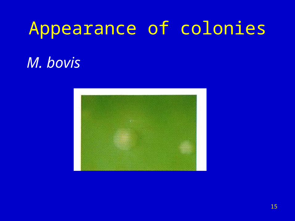
Appearance of colonies
M. bovis
15
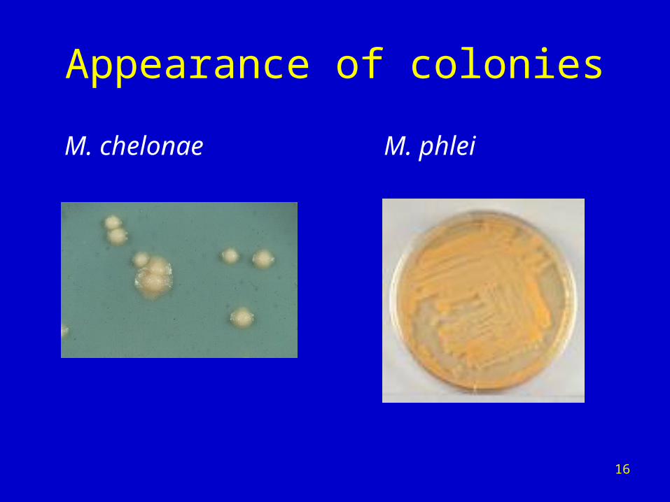
Appearance of colonies
M. chelonae M. phlei
16

ZN staining
Morphology “cord-like”
17

ZN staining
Morphology:
“striped”
“dispersed”
18

ZN staining
Morphology:
“sea urchin”
19

Growth on medium containing p-nitrobenzoic acid (PNB)
Procedure
• 1 LJ with PNB at 37 °C
• 1 LJ without PNB at 37 °C
Examine at 28 days
20

Growth on medium containing p-nitrobenzoic acid (PNB) – interpretation
• Abundant growth on both slopes: mycobacterial strain other than tubercle bacilli.
• Abundant growth on control tube and little or no growth on PNB medium: MTB complex strain.
• No growth on either slope: non-interpretable test, to be repeated.
M. tuberculosis fails to grow on PNB medium21

Biochemical tests• Niacin production• Nitrate reduction
• Catalase negative at 68 °C
Always use pure cultures, otherwise they will yield false results.
Test should be performed in the BSC – aerosols are produced.
22

Niacin production
• Metabolite accumulating in medium:– culture requirements– extraction.
• Differentiates M. tuberculosis from other species.
• Rarely positive in other mycobacteria.
23

Niacin test – procedure
24

Niacin test – interpretation
25

Always check expiry date of strips to avoid false-negatives!
26

Niacin test
Advantages:• Reliable• Easy to perform• Faster than PNB
Disadvantages:• Toxic reagents (banned)• Paper strips (expensive)
27

Nitrate reduction test
• M. tuberculosis reduces nitrates to nitrites.• Several species may reduce nitrates.• 4 hours.• Cultures tested:
– 4 weeks old– abundant growth.
28

Nitrate reduction test
Reagents • Preparation: see Annex 8.1.
Procedure
1. Add 0.2 ml sterile saline to a screw-cap tube.
2. Use a sterile loop/spade to emulsify 2 loopfuls/spadefuls of a 4-week-old culture in the saline.
3. Add 2 ml of the NaNO3 substrate.
4. Shake well and incubate upright in a 37 °C water bath for 3 hours, then remove.
29

Nitrate reduction test – controls
• Negative control: extract from an uninoculated tube of medium
• Positive control: extract from a culture of M. tuberculosis H37Rv.
30

• Negative: no colour. • Positive: red colour, varying from pink to very
deep crimson:
faint pink = +/-clear pink = 1+deep pink = 2+red = 3+deep red = 4+purplish red = 5+
Nitrate reduction test – results and interpretation
POSITIVE
31

Catalase test: principle
• Intracellular, soluble enzyme.
• Release of O2 and production of bubbles.
• Virtually all mycobacteria possess catalase enzymes, except for rare isoniazid-resistant mutants of M. tuberculosis and M. bovis.
32

Catalase test
• 68 °C test at pH7: strains of M. tuberculosis lose catalase activity.
33

Catalase test at 68°C, pH 7.0
• All mycobacteria produce catalase, usually thermoresistant but M.tuberculosis produces thermolabile catalase– 1 tube: incubation at 68 °C for 20 minutes– 1 tube: incubation at room temperature
for 20 minutes
– detection by H2O2
34

• If the unheated tube forms oxygen bubbles and the heated tube does not, the test strain produces heat-labile catalase and is likely to belong to the M. tuberculosis complex.
• If both heated and unheated tubes form oxygen bubbles, the test strain produces heat-stable catalase and is unlikely to belong to the M. tuberculosis complex.
• If neither the heated nor the unheated tube forms oxygen bubbles, the test has failed or the strain may be catalase-negative, such as rare isoniazid-resistant strains of M. tuberculosis. Repeat the test with a larger quantity of bacilli to eliminate the possibility of a false-negative test.
Catalase test at 68 °C, pH 7.0 –results and interpretation
35

Catalase tests – controls
68 °C tests
• Positive control, no bubble formation after heating: previously identified M. tuberculosis strain or an MTB reference strain from a culture collection.
• Negative control, bubble formation after heating: M. terrae grown on LJ.
36

• Alternative to molecular methods.
• Species ID by immunochromatographic method from positive culture (NOT from specimens).
• Rapid (15 minutes).
• User-friendly.
• Quality assured.
• Reproducible.
• ID on culture grown on liquid medium (protocol can be adapted to colony identification).
• Performance, cost, turn-around time compare well with other fast molecular methods.
Why the immunochromatographic test for low-income countries?
37

• Simple, rapid immunochromatographic method.• Detects MPB 64 Ag – predominant protein Ag, secreted
by MTB complex strains.• Simple lateral flow speciation test.• No sample processing/instrumentation required.• Storage and cryopreservation do not interfere with
detection.• Sensitivity of tubercle bacilli detection – 100%
What is the immunochromatographic test?
38

• The test strip consists of: - sample pad
– reagent pad– nitrocellulose membrane– absorbent pad.
• Antibodies are immobilized on nitrocellulose membrane as capture reagent (anti-MBP64) or control (anti-IgG) lines.
• Secondary Ab, which recognizes another epitope of Ag-MBP64 conjugated with colloidal gold particles, is used for Ag capture and detection on the membrane in a sandwich type.
How does the test work? (1)
39

• Negative, only one reddish-purple band appears in the control window.
• Positive, in addition to the control band, a clear distinguishable reddish-purple band also appears in the test window.
How does the test work? (2)
40
True and false exercise
1. M. tuberculosis can be identified only by the morphology of the colonies.
2. Expired reagents affect the results of identification tests.
3. The immunochromatographic test allows fast identification of TB complex from positive cultures.
41

Characteristics of M. tuberculosis
• AFB • Slow growth rate• Growth temperature 35–37 °C only • Typical morphology• No pigmentation
Additional• No growth on LJ medium containing p-nitrobenzoic acid• Niacin test: positive• Catalase test: negative at 68 °C • Nitrate test: positive
Module review: take-home messages
42

• What are the advantages and disadvantages of phenotypic and genotypic identification?
• Why should a complete set of standards be used for comparison during each nitrate reduction test?
• If all mycobacteria have the catalase enzyme, why is this test useful in species identification?
• List the culture tests for Mycobacteria species identification.
• What are the main characteristics that allow identification of M. tuberculosis?
Self-assessment
43