Embed Size (px)
Citation preview

Radiation protection in nuclear facilities
Module II

International Atomic Energy Agency, May 2015
v1.0
Background
In 1991, the General Conference (GC) in its resolution RES/552 requested the Director General to prepare 'a comprehensive proposal for education and training in both radiation protection and in nuclear safety' for consideration by the following GC in 1992. In 1992, the proposal was made by the Secretariat and after considering this proposal the General Conference requested the Director General to prepare a report on a possible programme of activities on education and training in radiological protection and nuclear safety in its resolution RES1584. In response to this request and as a first step, the Secretariat prepared a Standard Syllabus for the Post-graduate Educational Course in Radiation Protection. Subsequently, planning of specialised training courses and workshops in different areas of Standard Syllabus were also made. A similar approach was taken to develop basic professional training in nuclear safety. In January 1997, Programme Performance Assessment System (PPAS) recommended the preparation of a standard syllabus for nuclear safety based on Agency Safely Standard Series Documents and any other internationally accepted practices. A draft Standard Syllabus for Basic Professional Training Course in Nuclear Safety (BPTC) was prepared by a group of consultants in November 1997 and the syllabus was finalised in July 1998 in the second consultants meeting. The Basic Professional Training Course on Nuclear Safety was offered for the first time at the end of 1999, in English, in Saclay, France, in cooperation with Institut National des Sciences et Techniques Nucleaires/Commissariat a l'Energie Atomique (INSTN/CEA). In 2000, the course was offered in Spanish, in Brazil to Latin American countries and, in English, as a national training course in Romania, with six and four weeks duration, respectively. In 2001, the course was offered at Argonne National Laboratory in the USA for participants from Asian countries. In 2001 and 2002, the course was offered in Saclay, France for participants from Europe. Since then the BPTC has been used all over the world and part of it has been translated into various languages. In particular, it is held on a regular basis in Korea for the Asian region and in Argentina for the Latin American region. In 2015 the Basic Professional Training Course was updated to the current IAEA nuclear safety standards. The update includes a BPTC text book, BPTC e-book and 2 “train the trainers” packages, one package for a three month course and one package is for a one month course. The” train the trainers” packages include transparencies, questions and case studies to complement the BPTC. This material was prepared by the IAEA and co-funded by the European Union. Editorial Note
The update and the review of the BPTC was completed with the collaboration of the ICJT Nuclear Training Centre, Jožef Stefan Institute, Slovenia and IAEA technical experts.

Module II: Radiation protection in nuclear facilities
Page 3 of 97
CONTENTS
1 INTRODUCTION ................................................................. 5
1.1 Electromagnetic radiation.............................................. 5
1.2 Ionizing radiation ........................................................... 5
1.3 Natural and artificial sources of ionizing radiation ......... 7
1.4 Development of radiation science ................................. 8
2 INTERACTION OF RADIATION WITH MATTER ............. 11
2.1 Radiation, ionising radiation ........................................ 11
2.2 Interaction of charged particles with matter................. 12
2.3 Interaction of gamma rays with matter ........................ 13
2.4 Interaction of neutrons with matter .............................. 16
2.5 Exercise ...................................................................... 18
3 RADIATION DETECTION ................................................. 19
3.1 Physical basis of radiation detection ........................... 19
3.2 Gas detectors .............................................................. 20
Ionization chamber .......................................................... 24
Proportional counter ........................................................ 24
Geiger-Mueller counter .................................................... 24
3.3 Scintillation detector .................................................... 24
3.4 Thermoluminescent dosimeter (TLD) and optically stimulated luminescence dosimeter (OSLD) ............... 25
3.5 Neutron detection ........................................................ 26
BF3 proportional detector ................................................. 27
Compensated ionization chamber ................................... 27
Fission chamber .............................................................. 28
3.6 Questions .................................................................... 28
4 DOSIMETRIC QUANTITIES ............................................. 29
4.1 Absorbed dose (D) ...................................................... 29
Dose rate for point sources of radiation ........................... 30
Dose rate for other source geometries............................. 31
4.2 Equivalent dose (H) .................................................... 32
4.3 Effective dose (E) ........................................................ 33
4.4 Collective dose (Ec) ..................................................... 34
4.5 Exposure (X) ............................................................... 35
4.6 Exercises .................................................................... 35
5 BIOLOGICAL EFFECTS OF RADIATION ........................ 37
5.1 Basic cell biology ........................................................ 37
5.2 Radiation effects on cells ............................................ 39
5.3 Radiation effects on humans....................................... 42
Deterministic effects of radiation ...................................... 42
Stochastic effects of radiation .......................................... 44
5.4 Questions .................................................................... 46
6 EXTERNAL RADIATION EXPOSURE ............................. 47
6.1 Types of radiation exposure ........................................ 47
6.2 Time, distance, shielding............................................. 47
Time ................................................................................ 48
Distance .......................................................................... 48

Module II: Radiation protection in nuclear facilities
Page: 4 of 97
Shielding .......................................................................... 48
6.3 Shielding from gamma radiation .................................. 49
6.4 Shielding from beta radiation ....................................... 50
6.5 Shielding from neutron radiation ................................. 51
6.6 Exercises..................................................................... 51
7 INTERNAL RADIATION EXPOSURE ............................... 53
7.1 The pathways of radionuclides into the body .............. 53
7.2 Effective half-life, committed effective dose ................ 54
7.3 Dose coefficient, derived air concentration.................. 55
7.4 Exposure to radioactive noble gases .......................... 57
7.5 Protection against internal contamination.................... 58
7.6 Protective clothing and equipment .............................. 58
Respiratory protective equipment ..................................... 60
Fit test .............................................................................. 63
7.7 Exercises..................................................................... 64
8 RADIATION PROTECTION REGULATIONS ................... 65
8.1 The aim of radiation protection .................................... 65
8.2 International recommendations and standards ........... 66
ICRP recommendations ................................................... 66
IAEA Safety standards ..................................................... 67
8.3 Exercises and questions ............................................. 72
9 RADIATION PROTECTION IN NUCLEAR INSTALLATIONS .............................................................. 73
9.1 Aspects of radiation protection in the nuclear fuel cycle73
9.2 Important radionuclides for radiation protection .......... 74
Natural radionuclides ....................................................... 75
Fission products ............................................................... 76
Activation and corrosion products .................................... 77
Hot particles ..................................................................... 78
9.3 Questions .................................................................... 79
10 ENVIRONMENTAL MONITORING ................................... 80
10.1 Need for monitoring nuclear facilities .......................... 80
10.2 Exposure pathways to population ............................... 81
10.3 Objectives of monitoring .............................................. 83
10.4 Programmes for environmental monitoring ................. 84
10.5 Assessment of doses to members of the public .......... 85
10.6 Exposures of population from various stages of fuel cycle ............................................................................ 87
10.7 Natural background ..................................................... 89
10.8 Monitoring in emergency exposure situations ............. 90
Objectives of emergency monitoring ................................ 91
Source monitoring during the emergency ......................... 91
Environmental monitoring during the emergency ............. 92
Personal monitoring ......................................................... 94
10.9 Questions .................................................................... 95
11 REFERENCES .................................................................. 96
Module II: Radiation protection in nuclear facilities
Page 5 of 97
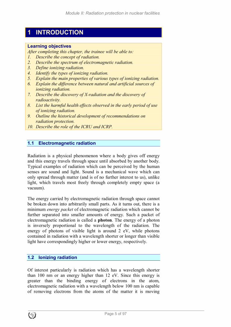
1 INTRODUCTION
Learning objectives
After completing this chapter, the trainee will be able to: 1. Describe the concept of radiation. 2. Describe the spectrum of electromagnetic radiation. 3. Define ionizing radiation. 4. Identify the types of ionizing radiation. 5. Explain the main properties of various types of ionizing radiation. 6. Explain the difference between natural and artificial sources of
ionizing radiation. 7. Describe the discovery of X-radiation and the discovery of
radioactivity. 8. List the harmful health effects observed in the early period of use
of ionizing radiation. 9. Outline the historical development of recommendations on
radiation protection. 10. Describe the role of the ICRU and ICRP.
1.1 Electromagnetic radiation
Radiation is a physical phenomenon where a body gives off energy
and this energy travels through space until absorbed by another body.
Typical examples of radiation which can be perceived by the human
senses are sound and light. Sound is a mechanical wave which can
only spread through matter (and is of no further interest to us), unlike
light, which travels most freely through completely empty space (a
vacuum).
The energy carried by electromagnetic radiation through space cannot
be broken down into arbitrarily small parts. As it turns out, there is a
minimum energy packet of electromagnetic radiation which cannot be
further separated into smaller amounts of energy. Such a packet of
electromagnetic radiation is called a photon. The energy of a photon
is inversely proportional to the wavelength of the radiation. The
energy of photons of visible light is around 2 eV, while photons
contained in radiation with a wavelength shorter or longer than visible
light have correspondingly higher or lower energy, respectively.
1.2 Ionizing radiation
Of interest particularly is radiation which has a wavelength shorter
than 100 nm or an energy higher than 12 eV. Since this energy is
greater than the binding energy of electrons in the atom,
electromagnetic radiation with a wavelength below 100 nm is capable
of removing electrons from the atoms of the matter it is moving

Module II: Radiation protection in nuclear facilities
Page: 6 of 97
through. Radiation passing through matter with a large enough photon
energy produces electron-ion pairs or ion pairs. This phenomenon is
called ionization and the associated radiation is ionizing radiation.
Within the electromagnetic spectrum (Figure 1.1), ionizing radiation
includes ultraviolet (partly), X and gamma radiation. Although these
types of radiation differ from each other in energy and origin, the
mechanisms of their interaction with matter are practically identical
and will from now on be collectively considered as gamma radiation,
because this is the type of electromagnetic radiation we will most
often deal with.
The radiation emitted by mobile phones or base stations has a wavelength of
above 10 cm. Since the photon energy of such radiation is a million times
smaller than the ionization energy, mobile phones clearly do not emit
ionizing radiation.
Figure 1.1: The electromagnetic radiation spectrum.
In addition to electromagnetic radiation with a large enough photon
energy, other forms of ionizing radiation include atom-sized particles
or of smaller mass, produced from radioactive decay, nuclear fission
and in accelerators. Their energies are of the order of magnitude of
MeV, which means they strongly ionize matter. It should be noted that
(thermal) neutrons with an energy of a few hundredths of and eV or
less also constitute ionizing radiation (on account of the nuclear
reactions they cause).
Module II: Radiation protection in nuclear facilities
Page 7 of 97
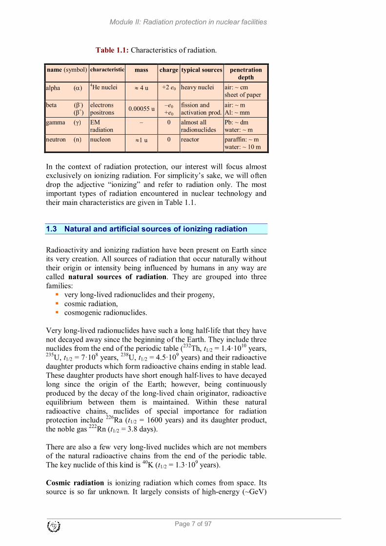
Table 1.1: Characteristics of radiation.
name (symbol) characteristic mass charge typical sources penetration
depth
alpha (α) 4He nuclei
≈ 4 u
+2 e0 heavy nuclei air: ~ cm
sheet of paper
beta (β-)
(β+)
electrons
positrons 0.00055 u
–e0
+e0
fission and
activation prod.
air: ~ m
Al: ~ mm
gamma (γ) EM
radiation
– 0 almost all
radionuclides
Pb: ~ dm
water: ~ m
neutron (n) nucleon ≈1 u 0 reactor paraffin: ~ m
water: ~ 10 m
In the context of radiation protection, our interest will focus almost
exclusively on ionizing radiation. For simplicity’s sake, we will often
drop the adjective “ionizing” and refer to radiation only. The most
important types of radiation encountered in nuclear technology and
their main characteristics are given in Table 1.1.
1.3 Natural and artificial sources of ionizing radiation
Radioactivity and ionizing radiation have been present on Earth since
its very creation. All sources of radiation that occur naturally without
their origin or intensity being influenced by humans in any way are
called natural sources of radiation. They are grouped into three
families:
� very long-lived radionuclides and their progeny,
� cosmic radiation,
� cosmogenic radionuclides.
Very long-lived radionuclides have such a long half-life that they have
not decayed away since the beginning of the Earth. They include three
nuclides from the end of the periodic table (232
Th, t1/2 = 1.4·1010
years, 235
U, t1/2 = 7·108 years,
238U, t1/2 = 4.5·10
9 years) and their radioactive
daughter products which form radioactive chains ending in stable lead.
These daughter products have short enough half-lives to have decayed
long since the origin of the Earth; however, being continuously
produced by the decay of the long-lived chain originator, radioactive
equilibrium between them is maintained. Within these natural
radioactive chains, nuclides of special importance for radiation
protection include 226
Ra (t1/2 = 1600 years) and its daughter product,
the noble gas 222
Rn (t1/2 = 3.8 days).
There are also a few very long-lived nuclides which are not members
of the natural radioactive chains from the end of the periodic table.
The key nuclide of this kind is 40
K (t1/2 = 1.3·109 years).
Cosmic radiation is ionizing radiation which comes from space. Its
source is so far unknown. It largely consists of high-energy (~GeV)

Module II: Radiation protection in nuclear facilities
Page: 8 of 97
protons that trigger nuclear reactions in the upper layers of the
atmosphere, producing other ionizing particles (gamma rays,
neutrons) which reach the Earth’s surface. Cosmic radiation makes a
significant contribution to natural background radiation or natural
human exposure to radiation. The intensity of cosmic radiation
increases with altitude; during a transatlantic flight we will receive an
additional dose comparable to a weekly dose of natural radiation on
ground.
Cosmic radiation is also the cause of the formation of cosmogenic
radionuclides. Though their half-lives are relatively short, they are
continuously formed by nuclear reactions which are triggered by
cosmic radiation in the atmosphere. The most important cosmogenic
nuclides are: 3H (t1/2 = 12.3 years),
7Be (t1/2 = 53 days) and
14C (t1/2 =
5760 years).
Artificial sources of radiation are sources made by man (i.e.
anthropogenic). They include:
� X-ray devices,
� particle accelerators,
� artificial radionuclides,
� nuclear reactors,
� materials with a technologically enhanced concentration of
natural radionuclides (TENORM),
In nuclear technology, the major sources of radiation are nuclear
reactors, which are simultaneously sources of artificial radionuclides.
1.4 Development of radiation science
Radiation has been part of the natural world ever since the birth of the
universe, just as humanity has always been exposed to various forms
of natural radiation throughout its evolution. We were not aware of
ionizing radiation, until two major discoveries were made in late 19th
century. First, on 8 November 1895, Wilhelm Conrad Röntgen
discovered penetrating rays which he called X-rays (nowadays they
are also called roentgen rays). Only a few months later (1 March
1896), Henry Becquerel discovered a phenomenon which was later
named radioactivity by Marie Curie.
Soon after both discoveries, X-rays in particular were found to have practical
value (e.g. to take pictures of the inside of the human body, see Figure 1.2),
so X-ray devices and radioactive substances started being used in physics,
chemistry and medicine. In those pioneering times, users were not aware that
excessive exposure to radiation has serious biological consequences. Nor did
they have any accurate instruments for measuring the intensity of radiation.
The earliest use of radiation was very soon followed by cases of
damage from intensive exposure to sources of radiation. In 1896, it
was found that exposure to X-rays may cause erythema (abnormal

Module II: Radiation protection in nuclear facilities
Page 9 of 97
skin redness), oedema (water accumulation in tissues) and epilation
(hair loss). In this period, X-ray tubes were calibrated simply on the
basis of the erythema caused on a hand exposed to X-radiation by the
operator.
Soon after, carcinogenic or cancer-inducing effects of radiation were also
observed. By 1911, 94 cases of tumours caused by X-radiation were reported,
50 of them among radiologists (doctors specialized in the use of radiation for
medical purposes). In 1922, an estimate was published that 100 radiologists
had died from radiation-induced cancer. Somewhat later, it was discovered that leukaemia (also called cancer of the blood or bone marrow) is much
more frequent in radiologists than other physicians. Marie Curie who worked
with radioactive materials died of leukaemia in 1934 aged 67 years.

Module II: Radiation protection in nuclear facilities
Page: 10 of 97
Figure 1.2: An X-ray of Mrs Röntgen’s hand, taken on 22 Dec. 1895,
less than two months after the discovery of X-rays.
Along with its obvious benefits, the use of radiation was thus
accompanied by incontrovertible proof of its harmful effects.
Relatively early on, this gave rise to the first recommendations for
reducing excessive exposure to radiation, which eventually developed
into a special scientific discipline – radiation protection.
The first limits on exposure to radiation were intended to prevent
acute effects such as skin lesions which occur after high exposure to
radiation. Later, limits were introduced to prevent more insidious
effects causing harm in the long term, such as cancer. Greater cancer
morbidity was first noted in miners working in mines containing
uranium ores and in female workers who used paintbrushes to apply
radium paint to dials of clocks and instruments from 1915 to 1930. To
be able to paint sharper numbers on the dial, they sharpened the tip of
the paintbrush with their lips, thereby taking in considerable quantities
of radium. However, the key group which allowed the long-term
effects of radiation to be quantitatively determined with greater
precision were the survivors of the atomic bombs at Hiroshima and
Nagasaki. It should be noted, however, that all the data obtained in
this way is based on individuals who were exposed to relatively high
radiation in a short amount of time. The greatest challenge of radiation
protection is how to use the data from high exposure in evaluating the
risk posed by accumulated low level exposure to radiation, which is
most commonly the case in occupationally exposed workers.
In the early period, there was a fair amount of confusion about the
quantities and units with which to measure exposure to radiation. For
this reason, the International Commission on Radiation Units &
Measurements (ICRU) was established in 1924. The first dosimetric
quantity, exposure, and its unit, the roentgen (R), were defined in
1928.
1928 also saw the establishment of the International Commission on
Radiological Protection (ICRP) which collects scientific data on the
effects of radiation and based on such data issues recommendations on
radiation protection.
Thus, in contrast to common belief, the health effects of ionizing
radiation are now very extensively studied and known, probably much
better than can be claimed for most other environmental substances
harmful to health.

Module II: Radiation protection in nuclear facilities
Page 11 of 97
2 INTERACTION OF RADIATION WITH MATTER
Learning objectives After completing this chapter, the trainee will be able to: 1. Explain the interaction of charged particles with matter. 2. Compare the range and shape of paths in matter for various
charged particles. 3. Describe the three most important types of interaction of gamma
radiation with matter. 4. Define the half-value layer and the tenth-value layer. 5. Use the equation for transmitted gamma ray flux as a function of
substance thickness and draw the relevant function. 6. Describe the interaction of neutrons with matter. 7. Describe the terms “fast neutron” and “thermal neutron”. 8. Describe the typical energy dependence of the neutron capture
cross section.
2.1 Radiation, ionising radiation
The term radiation is understood to include various fast particles (e.g.
electrons, alpha particles, neutrons) and electromagnetic radiation. As
regards the latter, nuclear technology is particularly concerned with
gamma and X-radiation. Energy packets of electromagnetic radiation
are called photons and they are treated in a similar way as other
(mass) particles.
Radiation particles have considerable energy, usually of the order of
magnitude of keV to MeV. When radiation passes through matter, the
particles collide with atoms or their nuclei and react with them. This
phenomenon is called the interaction of radiation with matter.
Radiation loses its energy by passing it on to atoms or nuclei in
matter. This energy transfer can happen by a number of different
physical processes which depend on the type and energy of the
radiation. As a rule, if the energy transferred is large enough, these
processes finish by stripping electrons away from the atom. An atom
which loses an electron of course becomes an ion. In this way, ions
and free electrons form along the path of charged particles. The ion-
electron pair is called the ion pair and the formation of ion pairs is
termed ionization. Hence we speak of ionizing radiation.
In the interaction of radiation with matter, the main dividing line is
between the interaction of charged particles and the interaction of
electrically neutral particles with matter. Electrically charged particles
(alpha and beta particles, fission fragments) interact with atoms or
electrons and nuclei by means of electrostatic force and directly cause
ionization in matter. In this case we speak of direct ionization.

Module II: Radiation protection in nuclear facilities
Page: 12 of 97
Electrically neutral particles (neutrons and gamma rays), on the other
hand, interact with atomic nuclei or electrons in various processes
causing the formation of charged particles (electrons and/or ions) with
a kinetic energy large enough for these secondary charged particles to
ionize matter. This is called indirect or secondary ionization.
2.2 Interaction of charged particles with matter
The most important charged particles in nuclear technology are alpha
particles, beta particles and fission fragments. Alpha particles and
fission fragments are considered heavy charged particles and beta
particles light charged particles. Electrically charged particles
interact with atoms or electrons and nuclei by means of electrostatic
force and cause direct ionization.
By forming ion pairs, the charged particles lose energy and slow
down. The intensity of interaction between charged particles and
matter is described by the following quantities:
� Specific ionization is the number of ion pairs per unit of path
length;
� Stopping power is the loss of energy per unit of path length.
Another important quantity is:
� Range – maximum depth a particle penetrates through matter.
Heavy charged particles (alpha particles, fission fragments) are
characterized by large specific ionization and stopping power and a
small range. The path of heavy particles is linear since they are
considerably heavier than electrons in matter. The range of an alpha
particle with 2 MeV energy is around 1 cm in air but only a few µm in
denser substances.
Figure 2.1: Path of an electron in matter.
range
inco
min
ge
lectr
on
end of path

Module II: Radiation protection in nuclear facilities
Page 13 of 97
Light charged particles (beta particles or electrons and positrons) have
much smaller specific ionization and stopping power than heavy
charged particles. Since their mass is identical to the mass of
electrons, they are deflected on colliding with electrons in atoms to the
same degree as the electrons. This is why their path follows a series of
angles or zig-zags. Their range is longer than for heavy charged
particles; a beta particle of 2 MeV energy has a range of around 8 m in
air and a few mm in metals.
2.3 Interaction of gamma rays with matter
Gamma rays are photons or packets (quanta) of electromagnetic
radiation with a very short wavelength. X-rays are similar in this
respect, except that γ rays originate from changes in atomic nuclei,
whereas X-rays originate from the electron cloud (and not the nucleus)
of an atom and generally have less energy than gamma rays. All other
properties of gamma and X radiation are the same, so the interaction
of both types of electromagnetic radiation with matter will be
discussed together.
Photons move in a straight line at constant speed (the speed of light).
As they travel though matter, they interact with atomic electrons and
in some cases also with atomic nuclei. The most important
interactions between gamma photons and atoms are:
� the photoelectric effect,
� Compton scattering,
� pair production.
Figure 2.2: The photoelectric effect.
The photoelectric effect leads to complete photon absorption. The
photoelectric effect involves a photon hitting one of the bound
electrons in an atom and passing all its energy to this electron. The
photon disappears as the electron is emitted by the atom with a kinetic
energy equal to the photon’s energy minus the electron’s binding
energy. This binding energy is usually considerably smaller than the
electron
incoming
photon γ
e-

Module II: Radiation protection in nuclear facilities
Page: 14 of 97
gamma ray energy, so the knocked-out electron has considerable
kinetic energy and travels on through matter like a beta particle would.
Compton scattering is a process in which a photon hits an electron in
an atom. Part of the photon’s energy is received by the struck electron
which consequently starts moving through matter like a beta particle,
while the remaining energy is emitted in the form of a photon. Its
energy is clearly smaller than that of the original photon, since the
overall energy is conserved. Momentum is also conserved, which is
why the electron and new photon fly off at defined angles.
Figure 2.3: Compton scattering.
Pair production occurs when a high energy photon approaches an
atomic nucleus and the strong electric field of the nucleus creates a
positron and an electron pair which fly off in opposite directions while
the photon disappears. The production of the electron-positron pair
requires 1.02 MeV, and the remaining energy is shared among the
electron and positron. Pair production is relevant only to photons with
high energy (>> 1.02 MeV).
Figure 2.4: Pair production.
Unlike charged particles, which continuously lose energy as they
incoming photon
e
e-
+

Module II: Radiation protection in nuclear facilities
Page 15 of 97
travel through matter, photons interact with atoms more rarely, but
when they do they lose all their energy (absorption) or a considerable
portion of it (scattering) at once. Consequently, the range of photons
cannot be defined like for charged particles; we can only predict the
probability of a photon scattering or being absorbed as it traverses a
certain thickness of matter. This means that the interaction of gamma
rays with matter is a random phenomenon. The probability of
interaction per unit path length is given by the linear attenuation
coefficient, µ.
Figure 2.5: Exponential attenuation of the transmitted flux of photons
passing through matter.
In mathematical terms, the process of photons travelling through
matter may be compared to radioactive decay which is likewise a
random process. Just as the number of radioactive nuclei diminishes
exponentially with time, the flux of monoenergetic photons, j, which
travel a certain distance without scattering or being absorbed also
reduces exponentially. In this context, the half-value layer, d1/2, is
defined as the thickness of a substance at which the flux of photons is
reduced to half its value (Figure 2.5).
The flux of transmitted photons passing through matter is described by
the equation:
2/120
d
d
jj−
= .
In addition to the half-value layer we also refer to the tenth-value
layer, d1/10, the thickness of a substance at which the flux of photons is
reduced by a factor of 10; after passing two tenth-value layers by a
factor of 100, after three tenth-value layers by a factor of 1000, etc.
j0
d1/2 2 d1/2 3 d1/2
j02
j04
j08
photo
n flu
x
substance thickness

Module II: Radiation protection in nuclear facilities
Page: 16 of 97
The exponential law of attenuation can be expressed through the
tenth-value thickness:
10/1100
d
d
jj−
= .
The half-value layer and tenth-value layer are related as follows:
d1/10 = 3.3 d1/2 .
2.4 Interaction of neutrons with matter
Neutrons are electrically neutral particles, so there is no electric
interaction with electrons or atomic nuclei. They interact with atomic
nuclei only by nuclear reactions via the nuclear force, which,
however, has a very short range. Since the nucleus takes up a very
small part of the atom’s volume (the radius of the nucleus is ten
thousand times smaller than the radius of the atom) neutrons travel
through matter as through almost empty space. Only now and then
neutrons happen to collide with atomic nuclei, when the neutron either
bounces off the nucleus (neutron scattering) or is absorbed by it,
causing a nuclear change. Owing to the random nature of neutron
interaction with matter, the intensity (flux) of neutrons attenuates
exponentially with the thickness of substance.
Neutrons released in nuclear fission or other nuclear reactions have
kinetic energy of the order of magnitude of MeV; they are called fast
neutrons. On colliding with the nuclei (scattering) they lose some of
their kinetic energy. This energy loss is the greatest when scattering
on light nuclei, which are present especially in the moderator. After
scattering, neutrons continue to travel through matter, but with less
kinetic energy than before colliding with a nucleus. This is why
neutrons are said to slow down by scattering. Consecutive scattering
gradually reduces the energy of neutrons until on average it is equal to
the kinetic energy of thermally moving atomic nuclei of matter. Such
neutrons are called thermal neutrons.
In contrast to scattering where a neutron slows down, absorption
involves a nucleus capturing a neutron. The most common absorption
reactions include radiative capture or the (n, γ) reaction, capture
reactions with emission of other particles such as (n, α) and (n, p)
reactions, and fission or (n, f) reaction.
In radiative capture or capture with emission of other particles the
neutron does not reappear after the reaction, hence these reactions
reduce the number of neutrons in matter. Conversely, a fission
reaction increases the number of neutrons.

Module II: Radiation protection in nuclear facilities
Page 17 of 97
The radiative capture reaction is the most frequent neutron absorption
reaction. For most nuclides, it may only occur with thermal neutrons.
In this reaction, a nucleus absorbs a neutron and the resulting excited
nucleus de-excites by emitting a gamma ray. With certain nuclides,
thermal neutrons can also induce (n, α) and (n, f) reactions, whereas as
a rule all other reactions are threshold reactions. This means that
neutrons require kinetic energy high enough (typically of the order of
magnitude of MeV) to induce such reactions.
The absorption cross section σa has three distinct regions depending on
neutron energy:
� 1/v region,
� resonance region,
� high energy region.
Figure 2.6: Cross section for radiative capture in 58
Fe.
As two examples, Figures 2.6 and 2.7 show the cross sections for
radiative capture in 58
Fe and for fission of 235
U. Both axes in the
figures have logarithmic scales.
Figure 2.7: Cross section for 235
U fission.
For the lowest energies, the absorption cross section is inversely

Module II: Radiation protection in nuclear facilities
Page: 18 of 97
proportional to neutron speed, hence this region is called the 1/v
region.
In the resonance region, the cross section value oscillates strongly.
The cross section value is extremely large for the neutron energies
which on collision give the nucleus exactly the energy it needs to be
excited into a particular excited state. We say that the neutron is in
resonance with the nucleus. For other energies, the cross section
values between individual resonances are much lower. The resonance
region usually corresponds to neutron energies from 1 eV to 100 keV.
Such neutrons are called epithermal neutrons.
For high energies (above 100 keV), individual resonances overlap to
such a degree that the end result is a once again smoother line of cross
section values which slowly falls as the energy increases. Neutrons in
this energy region are classed as fast neutrons.
2.5 Exercise
1. The half-value layer of a substance for photons of 1.5 MeV energy
is 1 cm. By how much is the photon flux reduced on passing
through 5 cm of this substance? And on passing through 10 cm of
the substance?

Module II: Radiation protection in nuclear facilities
Page 19 of 97
3 RADIATION DETECTION
Learning objectives After completing this chapter, the trainee will be able to: 1. Explain the terms direct and indirect ionising radiation and give
relevant examples of such radiation. 2. Describe the basic principle of detecting radiation with gas
detectors. 3. Explain and draw the current-voltage characteristic of gas
detectors. 4. Describe how an ionization chamber works. 5. Describe how a proportional detector works. 6. Describe how a Geiger-Mueller detector works. 7. Describe how a scintillation detector works. 8. Describe how TL and OSL dosimeters work. 9. Describe neutron detection with a BF3 proportional detector. 10. Describe neutron detection with a compensated ionization
chamber.
3.1 Physical basis of radiation detection
We are unable to detect ionizing radiation with our senses, but we can
detect and measure it with instruments, which are based on the
mechanisms by which radiation interacts with matter. The
consequences of the interaction of radiation with matter that can be
exploited for detection are:
� the production of free electrons and ions in matter (ionization),
� the production of excited atoms,
� the heating of matter,
� the occurrence of microscopic damage to matter,
� nuclear reactions in matter,
� bremsstrahlung (electromagnetic radiation which results from
the deceleration of charged – in particular β – particles in
matter).
The effect most frequently exploited for detection is ionization.
Regarding ionizing radiation, we distinguish two types of particles;
those that ionize matter directly and those that cause ionization
indirectly. The first group includes charged particles (e.g. electrons or
beta particles, protons, alpha particles and heavy ions) and the second
includes photons (gamma particles and X-radiation) and neutrons. As
regards photons, ionization is mostly caused by electrons resulting
from the photoelectric effect, Compton scattering or pair production.
As for neutrons, ionization is caused by the charged particles resulting
from nuclear reactions, e.g. α particles produced by the (n, α) reaction.
The device in which ionization is observed is called a detector. The
volume of matter used to collect the resulting charges is called the

Module II: Radiation protection in nuclear facilities
Page: 20 of 97
active volume of the detector and must be large enough to absorb a
substantial part of the energy of the radiation. This is why alpha
particle detectors can be much smaller than photon or neutron
detectors. Based on the type of matter undergoing ionization, the
following main types of detectors are distinguished:
� gas detectors,
� scintillation detectors,
� semiconductor detectors,
� other detectors.
In some detectors, the number of pulses measured is related to the
energy absorbed in the detector. Such a detector can be calibrated to
show the absorbed dose, which is why it is called a dosimeter.
3.2 Gas detectors
In principle, a gas detector is a condenser inside which radiation
ionizes a gas. Ion pairs are formed; negative particles (electrons)
accelerate towards the positive electrode (anode) and positive ones
(ions) towards the negative electrode (cathode). This produces an
electric current which is then additionally amplified and registered
(Figure 3.1).
Figure 3.1: The principle of gas detector operation.
The most common gas detector geometry is the coaxial detector
(Figure 3.2). Such a detector is shaped like a long cylindrical tube
which contains gas. A thin wire runs along its axis and is electrically
insulated from the tube wall. There is positive voltage on the wire,
hence the anode, whereas the wall is the cathode. Gamma radiation
interacts primarily with the tube wall, via photoelectric effect,
Compton scattering, or pair production. The secondary electrons from
these interactions travel inside the detector and ionize the gas.
However, if the radiation measured contains beta or alpha particles,
the tube should be designed to allow these particles to enter the

Module II: Radiation protection in nuclear facilities
Page 21 of 97
detector. This can be achieved with a very thin membrane at one side
of the tube, which is called the detector window.
Figure 3.2: Coaxial gas detector.
If a gas detector is exposed to a constant flux of ionizing particles and
the voltage, U, is changed, the current produced, I, also changes. The
dependence of current on voltage is called the current-voltage
characteristic of the detector (Figure 3.3); it may be divided into 6
distinct regions marked with Roman numerals from I to VI.
Figure 3.3: Current-voltage characteristic of a gas detector.
In the first region, at low voltage, the electric field in the detector is
weak and the ion pairs created often meet an oppositely charged
particle and recombine into electrically neutral atoms or molecules. If
the voltage, U, is higher, the movement of ions towards the electrodes

Module II: Radiation protection in nuclear facilities
Page: 22 of 97
is more direct, the probability of recombination falls and the current, I, rises. Nevertheless, in this region some of the created ion pairs still
recombine before reaching the electrodes, so the region is called the
recombination region. This region is not normally used for radiation
detection.
In region II the voltage is high enough for all of the released ion pairs
to reach the electrodes and as a result contribute to the current, I. This
means there is no recombination. Any additional increase in the
voltage does not increase the current, I. This constant current is
directly proportional to the number of ion pairs produced or to the
energy deposited in the detector by radiation. This voltage region is
called the ionization chamber region. The eponymous detector
operates within this region.
At higher voltage, U, the electric field in the counter is amplified
enough for the primary electrons released in the counter by radiation
to collide with gas molecules and ionize them. This produces new
(secondary) electrons which are also able to ionize further gas
molecules. This leads to an electron avalanche which reaches the wire
anode (Figure 3.4). In this way, each primary electron sets off an
avalanche of secondary electrons, which greatly increases the current,
I (region III in Figure 3.3). The resulting current changes with voltage,
but at a given voltage the factor of primary charge multiplication is
constant, which means that the resulting current is directly
proportional to the number of primary ion pairs. This voltage region is
called the proportional region, in which the proportional counter
operates.
Figure 3.4: Electron avalanche.
At voltages above the proportional region (region IV in Figure 8.3) the
avalanche multiplication increases the primary charge even more than
in the proportional region, but the multiplication factor is no longer
independent of the number of primary charges. Hence this region is
called the region of limited proportionality and is not normally used
for radiation detection.
At an even higher voltage, the electric field is so strong that
avalanches of electrons excite the internal electrons of gas atoms. The
ultraviolet rays produced by the transition of these atoms into the
ground state have enough energy to ionize other atoms throughout the

Module II: Radiation protection in nuclear facilities
Page 23 of 97
volume of the detector (Figure 8.5). This creates a very strong current;
however, it is independent of the number of primary ion pairs. The
current or pulse height does not change materially if the voltage on the
tube changes. This region, marked V in Figure 3.3, is called the
Geiger-Mueller plateau. The eponymous counter can only be used to
detect the presence of radiation and not to measure its energy.
Figure 3.5: Avalanche multiplication in a Geiger-Mueller detector.
If the voltage is higher than the Geiger-Mueller plateau (region V in
Figure 3.3), the gas in the detector is ionized just by virtue of the
strong electric field and collisions between molecules. Even with no
radiation present, this will create a strong current through the detector.
This region is called the breakdown region and usually destroys the
detector.
The dependence of the current-voltage characteristic of a detector on
the number of primary ion pairs (i.e. the energy deposited in the
detector by a particle) is shown in Figure 3.3 by the dotted curve
which represents the current-voltage characteristic for an increased
number of primary ion pairs released by one particle. A difference is
seen only at low voltages, in the operational region of the ionization
chamber and proportional counter. In these two regions the difference
is proportional to the difference in energy deposited by different
particles.
Figure 3.6: Pulse and current mode of operation.
The electric signal from a detector can be measured in two ways. In
pulse mode operation, we measure one event at a time, i.e. we
UV photon
UV photon
individualavalanches
e

Module II: Radiation protection in nuclear facilities
Page: 24 of 97
measure the change in detector voltage (pulse). The pulse height is
proportional to the charge. In the current mode, we measure the
average charge produced in the detector in a certain period of time,
i.e. the electric current through the detector (Figure 3.6).
Ionization chamber
The ionization chamber usually operates in the current mode at
voltages between 100 and 300 V. The filling gas is normally air. Since
the electric current produced is relatively weak (around 10-11
A), it
needs considerable amplification (around ten thousand-fold), which
should be linear. The ionization chamber is more suitable for
measuring strong radiation than for detecting individual events. The
uses of ionization chambers include measurement of the absorbed
dose and reactor power.
Proportional counter
Proportional counters operate at higher voltages than ionization
chambers, typically around 300 to 700 V. The output signal is directly
proportional to the energy deposited by incoming radiation. Due to
avalanche multiplication the electrical signals are about a thousand
times stronger than in ionization chambers. The filling gas is usually
argon, xenon or methane. Proportional counters operate almost
exclusively in the pulse mode.
Geiger-Mueller counter
Geiger-Mueller counters are filled with neon or argon and operate at
voltages around 1000 to 1200 V. Its typical feature is that the primary
ionization is multiplied by a factor from 104 to 10
8 and the resulting
electric signal is very strong (of the order of magnitude of 0.1 V) but
independent of the number of primary ion pairs. This means a Geiger-
Mueller counter cannot be used to measure radiation energy; it can
only be used to register individual ionization events. The signal
collection time or electron avalanche quenching time is a few hundred
microseconds. During the moment of primary ionization and the
moment of avalanche quenching the counter cannot count, so this time
is called the counter’s dead time, τ. Dead time is important at high
count rates or high radiation dose rates.
3.3 Scintillation detector
Some crystalline substances and organic materials (called scintillators)
are luminescent, which means they emit light after absorbing
radiation. The best-known example is a CRT TV screen which emits
visible light when hit by an electron beam from the cathode tube. This
effect is exploited by scintillation detectors. They consist of a
scintillator which is transparent to its own light, a photomultiplier and
supporting electronics (Figure 3.7). The most frequently used
scintillator is a sodium iodine crystal doped with thallium, denoted by
NaI(Tl).

Module II: Radiation protection in nuclear facilities
Page 25 of 97
Some of the energy of the absorbed radiation is emitted by the
scintillator as electromagnetic radiation or light. The more incoming
radiation is absorbed in the scintillator, the stronger the flash of light
that is produced. The resulting light flash is guided to the
photomultiplier where it is converted into an electric signal; its
amplification gives us a strong electric pulse which is proportional to
the energy of the light flash, i.e. to the energy deposited in the
scintillator by incoming radiation.
Figure 3.7: Scintillation detector.
A photomultiplier changes light pulses into electric pulses. The output
electric signal is directly proportional to the energy of incoming
radiation absorbed in the scintillator. A scintillation counter can
therefore measure the energy of incoming radiation. Its second
advantage is that the pulse develops in an extremely short time (µs), so
it can also be used in a strong radiation field. Finally, the scintillation
crystal absorbs gamma rays much more effectively than the thin gas in
gas detectors, so it is used chiefly to measure gamma radiation.
3.4 Thermoluminescent dosimeter (TLD) and optically stimulated luminescence dosimeter (OSLD)
To measure the quantities of radiation a human being has been
exposed to we use personal dosimeters. They measure the dose
received or the energy absorbed per unit mass of matter. In the past, a
photographic emulsion was used for this purpose, but today we most
frequently use a thermoluminescent dosimeter (TLD), an optically
stimulated luminescence dosimeter (OSLD) or an electronic
dosimeter.
In thermoluminescent substances (e.g. CaF2, LiF), radiation excites
electrons to higher energy levels just like in scintillators. However,
these electrons are not immediately restored to the ground state
(emitting light in the process) as in scintillators but remain captured in
what we call traps. The direct transition of an electron from a trap into
the ground state is not possible. This only happens when additional

Module II: Radiation protection in nuclear facilities
Page: 26 of 97
energy is supplied to them, which is done by heating. Luminescence
occurs when a photon is emitted during the transition into the ground
state (Figure 3.8).
Some dosimeters of more recent date are based on the optically
stimulated luminescence (OSL) effect. The principle of operation is
similar to the TLD, except that electron recovery from the trap to the
ground state (and light emission) is not stimulated by heating but by
light of selected wavelength. Al2O3 crystals can be used in OSL
dosimeters; their luminescence is stimulated with a green light laser
and the emitted light is blue.
Figure 3.8: The principle of thermoluminescent dosimeter (TLD)
operation.
TL and OSL detectors are passive; just a tablet with luminescent
material is irradiated and a reading is taken subsequently. After
reading and “erasing” (heating), the tablet is reused. Depending on its
composition, TLDs are used to establish the dose of X-rays, γ rays, β
rays and neutrons. OSL dosimeters enable multiple readings.
3.5 Neutron detection
Like photons, neutrons belong to the family of indirectly ionizing
particles. An important distinction is that photons react to a lesser or
greater extent with virtually all substances to form charged particles,
whereas neutrons form charged particles only in a relatively small
number of nuclear reactions.
To detect slow, i.e. thermal neutrons, the following nuclear reactions
are used:
10
B (n, α) 7Li
6Li (n, α)
3H
3He (n, p)
3H

Module II: Radiation protection in nuclear facilities
Page 27 of 97
235U (n, f) and
239Pu (n, f).
A substance containing the atoms of one of the targets in the above
reactions is added to a standard gas counter (e.g. ionization chamber
or proportional counter). Most commonly this substance is boron,
added either in gaseous form (e.g. as BF3 gas mixed with argon in the
counter) or by lining the interior walls of the counter with a thin layer
of a boron compound (e.g. boron carbide). On their way through the
detector, neutrons collide with 10
B nuclei, producing two positively
charged nuclei of lithium and helium, which share the released
reaction energy (2.79 MeV) among themselves. In most cases (94%) 7Li remains in an excited state, which lowers the kinetic energy of the
products to 2.31 MeV, but even this is easily detected. The resulting
electrical particles ionize the gas in the counter and this is registered in
the usual way.
BF3 proportional detector
As the name tells us, this is a proportional detector containing BF3
filling gas. This is the most common neutron detector. It operates in
the pulse mode but is not used to measure the energy of neutrons.
Instead, this mode enables a clear distinction between the signal of an
absorbed neutron and the signal of an absorbed γ ray (discrimination).
Compensated ionization chamber
This neutron detector effectively distinguishes neutron radiation
signals from gamma radiation signals. In principle, it involves two
chambers: the walls of the first are lined with boron to make it
sensitive to both neutrons and gamma rays, whereas the other is a
standard chamber and hence sensitive to gamma rays only. The
current in the first chamber results from both neutron and gamma
radiation, while that in the second chamber from gamma radiation
only, so the output signal (if compensation is properly performed) is
equal to the difference between the currents in the first and second
chamber and therefore proportional to the neutron flux only (Figure
3.9). The part of the compensated ionization chamber which is only
sensitive to gamma radiation is one of the rare examples of using a
detector in the recombination region.
10B lining
R ele trometerc
In+g
In+g
Ig
Ig
+

Module II: Radiation protection in nuclear facilities
Page: 28 of 97
Figure 3.9: Compensated ionization chamber.
Compensated ionization chambers are most frequently used in nuclear
reactors for their ability to measure a very wide range of neutron
fluxes, spanning up to ten orders of magnitude. Such detectors are said
to have a very large dynamic range.
Fission chamber
A fission chamber is an ionization chamber which has a window
rimmed with a thin foil of 235
U or 239
Pu. After absorbing thermal
neutrons, some nuclei of the fissile nuclide split; the resulting fission
products are heavily ionized and have considerable kinetic energy,
which is why the fission chamber generates a very strong and distinct
signal.
3.6 Questions
1. Why are some types of radiation called directly ionizing and
others indirectly ionizing?
2. How do we distinguish between the positive and negative charge
produced by ionization in a gas detector?
3. How do we measure the quantity of charge released?
4. Which gas detectors allow us to deduce the energy deposited by
radiation in the detector from the height of the voltage pulse?
5. What kind of measurements are scintillation detectors used for?
6. Describe the measurement procedure for a TL dosimeter.
7. What kind of information about neutrons does a BF3 detector give?
8. Why are BF3 detectors used in the proportional region of
operation?
9. Why does a compensated ionization chamber have two parts?

Module II: Radiation protection in nuclear facilities
Page 29 of 97
4 DOSIMETRIC QUANTITIES
Learning objectives
After completing this chapter, the trainee will be able to: 1. Define the absorbed dose, absorbed dose rate and associated
units. 2. Calculate the absorbed dose rate for cases of a point and line
source. 3. Define radiation weighting factors and give their values for
alpha, beta and gamma radiation and the range of values for neutrons.
4. Define the equivalent dose, equivalent dose rate and associated units.
5. Define tissue weighting factors. 6. Define the effective dose and its units. 7. Define the collective dose and its units. 8. Define exposure, exposure rate and associated units.
4.1 Absorbed dose (D)
In order to discuss the issues of radiation and radiation protection, we
need a suitable set of physical quantities and units to quantitatively
describe radiation fields and their effects.
In the context of nuclear physics, we have already met activity, a
physical quantity defined as the number of radioactive disintegrations
per unit time. Although the activity of a source is related to the
exposure to the radiation emitted by the source, the primary concern is
how much of this radiation actually reaches us. The situation can be
compared to a loudspeaker heard from a certain distance. If the
loudspeaker’s power is compared to source activity, we still need a
quantity which corresponds to the volume at the location of the
listener.
The basic dosimetric quantity is called the absorbed dose, D, defined
as the energy of radiation deposited (absorbed) in unit mass:
m
ED
∆∆
=
The unit for absorbed dose is the gray, in abbreviated form Gy, which
is defined as:
1 Gy = 1 J/kg
A target thus receives a dose of 1 Gy if 1 J of ionizing radiation is
deposited in 1 kg of matter. More frequently used and smaller units
are the mGy and µGy.

Module II: Radiation protection in nuclear facilities
Page: 30 of 97
1 mGy = 0.001 Gy = 10-3
Gy
1 µGy = 0.000001 Gy = 10-6
Gy
The old unit, which is often found especially in U.S. literature and
regulations, is the rad and is a hundred times smaller than the gray:
1 rad = 0.01 Gy
1 Gy = 100 rad
The absorbed dose rate, Ḋ, tells us how quickly the absorbed dose to
the target increases.
t
DD
∆∆
=&
The absorbed dose rate is measured in units of Gy/h, mGy/h etc.
Example:
A dosimeter is exposed to radiation. At 9:00, the absorbed dose is 3.7 mGy,
and at 16:00, it is 28.2 mGy. What is the dose rate?
Answer: ∆D = 28.2 mGy – 3.7 mGy = 24.5 mGy
∆t = 16 h – 9 h = 7 h
Ḋ = 24.5 mGy/(7 h) = 3.5 mGy/h
Dose rate for point sources of radiation
In case of a point* source of radiation with activity Ac, radiation
spreads from it isotropically, i.e. evenly in all directions. Let us
consider the simplest example, where a radionuclide emits one photon
with each decay. At distance r from the source, the photons spread
evenly over the surface of a sphere with radius r, so that S = 4πr2. At
this distance, the photon flux thus equals:
2π4 r
Ac=Φ
The dose rate is the product of the number of photons absorbed in the
target per unit time, and the energy they pass to the target on
absorption. The number of photons absorbed in the target is
proportional to the number of photons reaching the target, i.e. the
photon flux. This means the dose rate is proportional to the photon
flux:
2r
AcD ∝Φ∝&
* A source of radiation may be considered as a point source when its distance from
the target is more than 3 times its dimension.

Module II: Radiation protection in nuclear facilities
Page 31 of 97
The absorbed dose rate is directly proportional to the point source
activity and inversely proportional to the square of the distance
from the source. The proportionality constant is called the Γ
constant:
2r
AcD Γ=&
The gamma constant can be computed individually for each
radionuclide, but in practice we look it up in tables. It is normally
given for air. Its values for some of the more important radionuclides
are shown in Table 4.1. Since the gamma constant is directly
dependent on the mass absorption coefficient (which is approximately
12% greater for tissue than for air), the dose rates in tissue exceed
those in air by a factor of 1.12 as well.
Table 4.1: Gamma constants for certain nuclides.
nuclid
e
half-life Γ [Gy/h · m2/Bq]
16N 7.1 s 3.49·10
-13
24Na 15 h 4.59·10
-13
51Cr 27.7 days 5.45·10
-15
54Mn 312 days 1.1·10
-13
59Fe 44.5 days 1.57·10
-13
58Co 70.8 days 1.46·10
-13
60Co 5.27 years 3.25·10
-13
85Kr 10.7 years 4.74·10
-16
95Zr 64 days 1.1·10
-13
131I 8.04 days 6.71·10
-14
134Cs 2.06 years 2.37·10
-13
137Cs 30 years 7.56·10
-14
Dose rate for other source geometries
We also frequently deal with line sources of radiation, e.g. a pipe
carrying radioactive fluid. The dose rate falls inversely proportionally
to the distance from the line source:
d
acD
Γ=
π&
,
where ac is the specific activity (activity per unit of source length),
and d is the distance from the source.

Module II: Radiation protection in nuclear facilities
Page: 32 of 97
4.2 Equivalent dose (H)
Things would be simple if radiation effects were directly proportional
to the absorbed dose. Unfortunately, they are not. Besides the energy
deposited in the body by radiation, the degree of biological damage
also depends on the type of incoming radiation and sometimes on the
energy of the particles.
Biological effects increase with the stopping power of particles which
is also called linear energy transfer – LET. This means that particles
with a large LET (alpha particles, neutrons) cause more damage than
particles with a small LET (beta and gamma particles) at the same
absorbed dose.
Since there is no easy way of adequately expressing the dependence of
biological effects on linear energy transfer by a simple mathematical
formula, we need to introduce the radiation weighting factor wR
which accounts for the differences in the quality (effectiveness) of
various radiations.
To estimate the degree of biological damage, we introduce a new
dosimetric quantity, called the equivalent dose, H, defined as the
product of the absorbed dose and the radiation weighting factor wR:
H = wR · D
The unit for equivalent dose is called the sievert (Sv). This unit is
used to point out that we are dealing with equivalent and not absorbed
dose (the unit of the latter is Gy), although both can be expressed with
the same basic physical units:
1 Sv = 1 J/kg
Table 4.2: Radiation weighting factors (ICRP 2007).
type of
radiation
energy radiation
weighting
factor wR
alpha (α) all energies 20
beta (β) all energies 1
gamma (γ) all energies 1
neutrons (n) continuous function of
energy 2.5 ~ 20
The radiation weighting factor, wR, for beta and gamma radiation
equals 1 and its value for other types of radiation is greater than 1
(Table 4.2).

Module II: Radiation protection in nuclear facilities
Page 33 of 97
An equivalent dose of 1 Sv is a very large dose, so we often use
smaller units, mSv and µSv.
The old unit for equivalent dose, which is still encountered in
American literature and legislation, is the rem (roentgen equivalent
man):
1 rem = 0.01 Sv = 10 mSv
1 Sv = 100 rem
Example:
An organ with a mass of 70 g has absorbed 385 GeV of alpha radiation. What
is the equivalent dose received?
Answer: D = ∆E/∆m
= (385·109 · 1.6·10-19 J)/(70·10-3 kg) = 8.8·10-7 Gy = 0.88 µGy
H = D · wR = (0.88 · 20) µSv = 17.6 µSv
The equivalent dose rate, �� , tells us how quickly the equivalent dose
to the target increases:
RwDt
HH ⋅=
∆∆
= &&
The units used for equivalent dose rate are Sv/h, mSv/h and µSv/h.
Example:
What equivalent dose is received by a worker working for one year in a
laboratory where the equivalent dose rate is 1.5 µSv/h? Assume that there are
2000 working hours in a year.
Answer: H = Ḣ · t = 1.5 µSv/h · 2000 h = 3000 µSv = 3 mSv
4.3 Effective dose (E)
Quite clearly the biological consequences vary if a given equivalent
dose is received by the whole body or just by a part of the body or an
organ. In terms of biological effects, we also find that not all organs
are equally sensitive to radiation, i.e. the same equivalent dose
produces more harmful effects in some organs than in others.
This is why another quantity was introduced, the effective dose, E,
which basically expresses the share of whole body effects contributed
by a particular organ or tissue.
E = wT · H ,
where wT is the weighting factor for an organ or tissue (T). If
several organs are irradiated, each with its own dose, HT, the total
effective dose equals:

Module II: Radiation protection in nuclear facilities
Page: 34 of 97
∑=T
TTHwE
Table 4.3 indicates the values of the tissue weighting factor, wT. As
for equivalent dose, the unit for effective dose is the Sievert (Sv). It
should be emphasized that using the concept of effective dose only
makes sense at low doses (E << Sv) and that tissue weighting factor
values also apply to low doses only.
Table 4.3: Organ or tissue weighting factors (ICRP 2007).
tissue or organ tissue weighting
factor wT
breast 0.12
bone marrow 0.12
colon 0.12
lung 0.12
remainder 0.12
stomach 0.12
gonads 0.08
bladder 0.04
liver 0.04
oesophagus 0.04
thyroid 0.04
bone surfaces 0.01
brain 0.01
salivary glands 0.01
skin 0.01
TOTAL 1.00
4.4 Collective dose (Ec)
We also often want to indicate the overall effective dose received by a
group of people. This quantity is called the collective dose, Ec, and its
unit is the man·sievert (abbreviated to man·Sv). Its calculation is quite
straightforward as shown in the following example.
Example:
In a nuclear accident in a town with 2000 inhabitants, one half of the people
received a dose of 20 mSv and the other half 10 mSv. What is the collective
dose?
Answer: Ec = 1000 people · 20 mSv + 1000 people · 10 mSv = 30 man·Sv
If we generalize the above calculation, collective dose can be

Module II: Radiation protection in nuclear facilities
Page 35 of 97
expressed as:
∑=i
ii EnEc ,
where ni is the number of people that received dose Ei, or, dose E1,
was received by n1 people, dose E2 by n2 etc.
4.5 Exposure (X)†
Historically the oldest radiological quantity is exposure but it is no
longer used. It is defined by the ionization caused in air by X radiation
or gamma radiation:
m
QX
∆=
,
where Q is the sum of the electric charges of all ions of the same sign
released in a unit of mass, ∆m. The unit for exposure is As/kg air.
The relation to the old unit, the roentgen (R) is:
1 R = 2.58·10-4 As/kg
Inversely:
1 As/kg = 3876 R
Exposure rate, Ẋ, tells us how quickly the charge of released electrons
increases, in other words, what current flows from the air mass ∆m:
m
I
t
XX
∆=
∆∆
=&
The unit for exposure rate is A/kg air, but it is more common to use the old
units: R/s, R/h, mR/h etc.
4.6 Exercises
1. What is the absorbed dose in an organ with a volume of 40 cm3
and a density of 0.93 g/cm3 which absorbs 3·10
5 MeV radiation?
2. How many alpha particles of 6 MeV energy cause an absorbed
dose of 10 µGy in tissue with a mass of 70 g?
3. A point source of 137
Cs has an activity of 1200 MBq. What is the
absorbed dose rate in air 5 m away from the source?
4. Assess the activity of a 60
Co source for which the dose rate
measured at a distance of 3 m is 0.1 mSv/h.
5. Calculate the dose rate at 50 cm distance from a point source of:
† Exposure (X) and exposure rate (Ẋ) are no longer official units

Module II: Radiation protection in nuclear facilities
Page: 36 of 97
a) 2 GBq 51
Cr, b) 2 GBq 24
Na.
6. Somebody left a 370 MBq source of 24
Na in the laboratory by
mistake. An employee unaware of this source spent 8 hours at her
desk which is 2 m away from the source. Assess the dose she
received.
7. The activity of a 60
Co point source is 16 MBq. In how much time
is the annual effective dose, 5 mSv, reached at 1 m distance from
the source?
8. What is the equivalent dose of alpha particles if the absorbed
dose is 3.2 µGy?
9. What is the absorbed dose in an organ with a volume of 40 cm3
and a density of 0.93 g/cm3 which absorbs 3·10
5 MeV energy of
thermal neutrons?
10. A body part received 0.15 mGy radiation with a weighting factor
of 5 and 0.22 mGy radiation with a weighting factor of 10. What
is the total absorbed dose? What is the total equivalent dose?
11. After a scan with radioactive iodine, 131
I, the equivalent dose to
the thyroid was 61.5 mSv. What was the effective dose to the
patient?

Module II: Radiation protection in nuclear facilities
Page 37 of 97
5 BIOLOGICAL EFFECTS OF RADIATION
Learning objectives
After completing this chapter, the trainee will be able to: 1. Name the types of cells and list the main cell parts. 2. Name the main substances that form a cell and their rough
percentages. 3. Describe the cell division process. 4. Explain how radiation directly and indirectly affects the cell. 5. Describe the difference between the effects of different kinds of
radiation in cells. 6. Describe the time development of radiation damage in cells and
tissue. 7. Describe and distinguish between somatic and genetic effects. 8. Distinguish stochastic and deterministic effects. 9. List the deterministic effects of radiation. 10. Explain the concept of median lethal dose. 11. List the stochastic effects of radiation. 12. Clarify the risk of stochastic effects.
5.1 Basic cell biology
The basic building blocks of living organisms are cells. For a better
understanding of how ionizing radiation affects people, we will take a
brief look at the structure of cells and some vital processes that take
place in them.
The body of an adult person consists of around 4·1013
cells. Cells
differ from each other both in function and size. Most cells are
relatively small (around 10 µm), whereas nerve cells can be up to a
metre long. Cells fall roughly into two families: somatic cells and sex cells. Almost all cells in the body are somatic cells, and sex cells or
gametes are only relevant to reproduction. Gametes pass hereditary
information from one generation to the next.
The cell may be small in size but it is an extremely complex system. A
typical somatic cell is shown in Figure 5.1. The outer boundary of a
cell, called the cell membrane, both protects it from external factors
and also forms a link between the cell and its environment. Most of
the cell’s volume is filled with cytoplasm, a transparent mixture of
water, various molecules and electrolytes. Also floating in cytoplasm
are a number of structures called organelles, in which various
metabolic and other processes take place. The most important
organelle is the cell nucleus, which is the central element of the cell
and controls all its vital functions.
Cells are composed of ~70-85 % water, ~10-20 % proteins, ~10 %
carbohydrates (sugars) and ~2-3 % lipids (fats) and small quantities of

Module II: Radiation protection in nuclear facilities
Page: 38 of 97
inorganic substances.
People do not live with the same cells from birth to death; human cells
grow and regenerate. These processes are possible because cells
divide. Cell division is called mitosis. Precise cell reproduction is
ensured by the genetic material contained in the cell nucleus. During
the resting phase (when the cell is not dividing), the genetic material is
called chromatin. This is a complex of the intertwined strands of
deoxyribonucleic acid (DNA) macromolecules and certain proteins.
A DNA molecule has the characteristic shape of a double helix
(similar to an “infinite” ladder), formed by two long entwined chains
consisting of basic building blocks called nucleotides. The double
helix strings together pairs of nucleotides (one on either side of the
“rungs”) with one of the four organic bases: adenine (A) and thymine
(T), or guanine (G) and cytosine (C). In the resting phase, chromatin
controls the synthesis of proteins which give the cell its characteristic
features (shape and function).
Figure 5.1: Somatic cell.
During cell division chromatins can be observed under microscope as
chromosomes. They are spiralled, densely packed DNA. Human
beings have exactly 46 chromosomes in each somatic cell and exactly
23 chromosomes in each sex cell. Before cell division chromatins
(chromosomes) are doubled, and later on during mitosis distributed to
daughter cells. Therefore each new cell acquires one copy of each
chromosome. . This ensures the transmission of genetic information
about cell structure and functions to the next generation of cells.
The time between two cell divisions is called the cell cycle. Cell cycle
length varies according to the type of cell. Cells of the human intestine
divide approximately every 24 hours, while nerve and muscle cells
microvilli microfilaments
centriole
nucleus
ribosome
smooth endoplasmic reticulum
mitochondrion
rough endoplasmic reticulum
Golgi apparatus lysosomes
cell membrane

Module II: Radiation protection in nuclear facilities
Page 39 of 97
practically never.
5.2 Radiation effects on cells
No atom in a cell is free; all atoms are bound into molecules. Since
molecular bonds are formed by electrons, ionization results in the
breakdown of molecules or the formation of free radicals from
previously neutral molecules.
When ionization causes a complex organic molecule in one of the
organelles to split, this is a direct effect of ionizing radiation on the
cell.
Since cells to a large extent (70-85 %) consist of water, it is more
likely for radiation to ionize the water in the cell than to affect any of
the (vital) organic molecules. Water ionization involves several
chemical reactions called water radiolysis. Particularly important
products of radiolysis include free hydroxyl radicals, OH˙, and
hydrogen peroxide, H2O2. Both are strong oxidants which are able to
travel approximately 4 nm from their point of origin, where they have
a very aggressive effect on cell elements. This process is called an
indirect effect of ionizing radiation and its consequences are very
similar to those of the direct radiation effect. In forms of radiation that
rarely ionize matter (have a low LET), the indirect effect accounts for
around 70 % of damage; for high-LET radiation, this share is smaller
than the direct effects. It is important to note that both processes are
essentially chemical. The biological effects of radiation are thus
basically no different from the effects caused by various chemicals.
Radiation is just one of the ways in which harmful chemical
substances are formed in or enter the body (Figure 5.2).
Figure 5.2: Direct and indirect radiation effects on cells.
H20
OH
H20
H20H2O2
OH
direct effect of radiation on DNA
indirect effect

Module II: Radiation protection in nuclear facilities
Page: 40 of 97
The cell is such a complex system that, even at the cellular level, there
is no single mechanism through which radiation acts on the body.
Radiation effects depend on which of the cell organelles is affected
and what its role is in cell functioning. Although not all the details are
understood as yet, many studies suggest that the most sensitive part of
the cell is its nucleus, specifically the chromosomes. Of course,
radiation may also damage other organelles. If damage is caused to a
large molecular group, e.g. in the mitochondrion, the function of this
mitochondrion will very likely be impaired or fail entirely. However,
there are several mitochondria in most cells which can take over its
role. On the other hand, damage to the cell DNA affects the function
of the cell as a whole and may be transmitted to its progeny by
mitosis. DNA is therefore a critical target for ionising radiation.
As it turns out, many cells damaged by radiation continue to function.
During mitosis, however, a defective chromosome prevents them from
dividing properly and they die. This is called mitotic death. Radiation
effects are therefore most dramatic in organs with the most quickly
dividing cells (red marrow, large intestine epithelium, skin epithelium,
hair follicles). In contrast, tissues which are more static such as
muscles and bones are less sensitive to radiation. Also very sensitive
to radiation are embryos, especially in the first months after
conception, because their cells divide at a much faster rate than in
adults.
Figure 5.3: Survival of cells exposed to acute doses.
Parts of certain animal tissues can be removed and then grown in cell
colonies which multiply spontaneously by cell division. Exposing
such a colony to a short-term, acute dose of radiation enables very
precise measurement of cell death as a function of dose. Figure 3.3
shows the proportion of surviving cells as a function of the absorbed
dose. This figure very clearly indicates that radiation with high
stopping power or a high linear energy transfer (LET), e.g. alpha
radiation or neutron radiation, causes much greater biological damage
00.001
0.01
0.1
1
2 4 6 8 10 12
pro
port
ion
of su
rviv
ing c
ells

Module II: Radiation protection in nuclear facilities
Page 41 of 97
than radiation with a low LET such as beta and gamma radiation at the
same absorbed dose. This example illustrates why absorbed dose is
insufficient to describe biological effects and why we also need to use
the equivalent dose, which multiplies the absorbed dose by the
relevant radiation weighting factor. Figure 5.3 also shows that the
proportion of cells surviving irradiation with a high LET radiation
falls exponentially (the chart uses a logarithmic scale for the vertical
axis) with increasing dose, whereas in low-LET radiation, the
proportion of surviving cells decreases very slowly at lower doses and
only starts falling exponentially at high doses. Although the process of
cell death is not yet fully understood, this suggests that some kind of
repair mechanism exists in cells at low doses and low-LET radiation
which reduces the number of cells killed.
In certain cases, the damaged cells keep the ability to divide further.
As they divide, the change in their chromosomes is passed to the next
generations of cells. This phenomenon is called mutation. Just as the
mechanism of the effects of ionizing radiation on the cell is basically
no different from the mechanism that underlies the effects of other
environmental factors, mutations in the body are by no means caused
by radiation alone; they are so to speak a daily occurrence produced
by various external influences on cells.
Alternatively, mutations may cause pathological changes which are
manifested as the abnormal functioning of individual organs or as a
greatly increased rate of cell division in a tissue or organ. Such a
pathological change is called cancer.
In a way, it is paradoxical that radiation may cause cancer, while on the other
hand, radiation is also one of the most powerful tools for treating cancer. The
rapid division of cancerous cells makes them much more sensitive to
radiation than healthy tissue. Strong irradiation of the cancerous growth
destroys the cancerous cells, whereas the cells in the surrounding healthy
tissue largely survive. Of course, every effort is made to confine the beam of
radiation to the diseased tissue.
If any chromosome in the sex cells (in the sex glands – gonads) is
damaged, the mutation is transmitted to the offspring and it is only in
them – and/or their offspring – that certain pathological changes will
develop. This process is called genetic change.
Radiation damage to organisms consists of several phases of varied
duration:
� physical phase: ~10-12
s: ionization
� chemical phase: ~10-6
s: formation and diffusion of free radicals
~10-3
s: DNA damage
� biological phase: ~ seconds: DNA repair
~ hours: cell death
~ days: mutations, cell transformation,
chromosome aberrations
~ years: development of cancer, hereditary

Module II: Radiation protection in nuclear facilities
Page: 42 of 97
effects
5.3 Radiation effects on humans
The effects of radiation on humans can be classified by two criteria.
The first classification refers to the type of cells damaged or to the
person that suffers the effects of radiation. Symptoms which are a
consequence of damaged somatic cells and occur in the irradiated
person are called somatic effects. Symptoms which are a consequence
of damaged gonads and occur in the irradiated person’s offspring are
called genetic effects.
The second classification relates to the probability that the effect or
medical condition will occur. Effects as a consequence of radiation
which are certain to occur above a specific dose are called
deterministic effects. These effects are a consequence of irreparable
damage to cells and their death during unsuccessful division. Effects
which occur with a certain probability only in a specific percentage of
irradiated persons are called stochastic effects. These effects are a
consequence of accumulated mutations in irradiated cells.
It should be emphasized that the effects of radiation depend not only
on the equivalent or effective dose received, but also on how this dose
was received:
� Acute: a high dose in a short time interval.
� Chronic: prolonged irradiation with low doses.
� Local: exposure of a particular organ.
� Total: whole body irradiation.
Most information about the effects of radiation is drawn from cases of
acute irradiation with high doses (e.g. survivors of the Hiroshima and
Nagasaki bombings or accidents involving radiation sources).
However, what is most relevant for the practical application of
radiation protection principles, especially to occupationally exposed
workers, is multiple (chronic) low dose irradiation. Since we know
that repair mechanisms are at work at low doses, we may assume that
an assessment of harmful radiation effects based on data from acute
doses is a conservative assessment of the effects of chronic exposure
to the same dose.
Deterministic effects of radiation
As the name tells us, deterministic effects are effects which are certain
to occur following irradiation with a high enough dose. They will
manifest after a short delay, hours to weeks after irradiation. They are
also characterized by a dose threshold, i.e. the minimum dose, D0, at
which these effects occur. Below this threshold there are no radiation
effects, and above it their severity or intensity increases with the dose
received (Figure 5.4). Deterministic effects can also be
unambiguously linked to excessive exposure to radiation.

Module II: Radiation protection in nuclear facilities
Page 43 of 97
Deterministic effects include:
� erythema (skin redness),
� epilation (hair loss),
� reduced number of blood cells (resulting from bone marrow
damage),
� cataract,
� nausea, vomiting, diarrhoea,
� damage to children (deformation of organs, mental retardation),
� temporary or permanent sterility.
Figure 5.4: Deterministic effects of radiation.
The severity of deterministic effects as a function of received dose is
given in Table 5.1.
Table 5.1: Deterministic effects of acute whole-body irradiation.
Dose (Gy) Probable effect
0 – 0.5 No prominent effects, possible minor changes in blood
count.
0.6 – 1.2 Nausea and vomiting occur after the first day in 5-10 %
of the cases. No serious incidence of disease.
1.3 – 1.7 The first day is followed by nausea and vomiting, 25 %
cases display symptoms of radiation sickness. No fatal
cases.
1.8 – 2.2 In 50 % of the cases, nausea and vomiting occurs within
the first day. The symptoms of radiation sickness are
more prominent. No fatal cases are expected.
2.3 – 3.3 In 100 % of the cases, nausea and vomiting occurs within
the first day. Serious signs of radiation sickness appear.
3.4 – 5.0 Median lethal dose, LD50/30 – 50 % deaths in 30 days.
> 100 Radiation syndromes. 100 % deaths with a time delay

Module II: Radiation protection in nuclear facilities
Page: 44 of 97
from a few hours to a few months.
Protection against deterministic effects is relatively simple – by
ensuring that the doses to individual organs are lower than the
threshold dose, the risk of any pathological changes developing is
prevented.
Stochastic effects of radiation
Stochastic effects of radiation are effects which can only be predicted
to occur with a certain probability. In this case, a threshold dose, i.e. a
minimum dose below which there is no risk of these effects, cannot be
proved. A characteristic feature of stochastic effects is that the
probability of a medical condition occurring rises with increasing
dose, whereas the severity or intensity of an effect (in the case when it
occurs) is not related to dose. Furthermore, the medical condition
cannot be unequivocally linked to radiation exposure; all that may be
determined is a statistical increase in the medical condition beyond its
natural frequency or incidence due to other causes. Stochastic effects
of radiation include in particular:
� cancer,
� genetic effects.
Cancer is (after cardiovascular diseases) the second leading cause of
death. In the developed world, around 40 % of people contract cancer
and almost 20 % die from cancer. Cancer is not a single disease but a
group of around a hundred very similar diseases, all of which share
rapid, uncontrolled division of cells. Over 90 % of cancers are caused
by various chemicals which are called carcinogens. More than a
thousand carcinogens are known and most of them occur naturally in
the environment. Radiation is therefore just one – albeit not chemical
– of many carcinogens. Among the 86572 surviving victims of the
Hiroshima and Nagasaki bombings, 249 people died of leukaemia and
7578 of other types of cancer, but a comparison with other groups of
people shows that only 87 of the leukaemia cases and 334 of the cases
of other cancer types may be attributed to radiation exposure‡, while
the rest resulted from other, natural causes. This suggests that
radiation is not a conspicuously strong carcinogen.
The bulk of the data on additional incidence of cancer was obtained by
monitoring Hiroshima and Nagasaki survivors and from some
accidents involving sources of radiation. What is characteristic of all
these cases is that the individuals received relatively high radiation
doses. It was also found that, at high doses, the probability of
developing cancer is proportional to the dose received. For low doses,
the number of additional disease cases was too small to be statistically
identified. It was therefore assumed that even in low doses, the risk of
‡ UNSCEAR 2000 Report Vol. II: Sources and Effects of Ionizing Radiation
(http://www.unscear.org/docs/reports/gareport.pdf)

Module II: Radiation protection in nuclear facilities
Page 45 of 97
cancer is proportional to the dose received and that there is no “safe
dose” or threshold dose with no risk below that value. However, there
are many indications that certain repair mechanisms exist at low doses
which reduce this risk. Taking a conservative approach, however,
radiation protection regulations are based on the linear no-threshold hypothesis (dotted section of the curve in Figure 5.5).
Figure 5.5: Stochastic effects of radiation.
Leukaemia is the leading cancerous disease caused by radiation,
followed by breast, lung, thyroid and stomach cancer. For each of
these, the probability of occurrence can be determined and expressed
as the number of expected deaths per specific collective dose. The
total probability for any type of cancer is 5 % per 1 man·Sv.
Example:
Calculate the number of additional deaths from cancer in a population of
100,000 people exposed to an average dose of 10 mSv.
Answer: The collective dose is:
Ec = 100,000 people · 0.01 Sv = 1000 man·Sv
The expected additional number of cancer cases due to radiation is 5 % of the
collective dose, therefore 50 people. This number should be set against the
20,000 people in this population who will die of cancer from other causes.
What is also characteristic of all types of cancer is a latent period, i.e.
the time between radiation exposure and the onset of cancer. In
leukaemia the latent period is 5-15 years, and for other types of cancer
it is 10 years or more.
Genetic effects are another type of stochastic effect of radiation.
These are effects which are only displayed in the offspring of the
irradiated person. Being even less probable than cancer, there has been
so far no statistically proven increase of genetic effects in any group
of irradiated persons.

Module II: Radiation protection in nuclear facilities
Page: 46 of 97
5.4 Questions
1. What is the difference between the direct and indirect effects of
ionizing radiation in tissue cells?
2. What are the basic characteristics of deterministic and stochastic
effects of ionizing radiations?
3. What are the symptoms of radiation sickness and at roughly what
doses do changes occur in the body and on the skin?

Module II: Radiation protection in nuclear facilities
Page 47 of 97
6 EXTERNAL RADIATION EXPOSURE
Learning objectives
After completing this chapter, the trainee will be able to: 1. Describe the difference between external and internal exposure. 2. Give and describe examples of potential external exposure. 3. Describe methods of protection against external exposure. 4. Describe the use of time as a means of protection. 5. Describe the effect of distance on dose rate. 6. Describe how the type of source is taken into account. 7. Describe the influence of shielding on gamma radiation. 8. Describe the build-up of gamma radiation. 9. Calculate the effect of distance, time and shielding on gamma
radiation dose rate. 10. Describe shielding for sources of beta rays. 11. Explain the basic characteristics of neutron shields.
6.1 Types of radiation exposure
The main objective of radiation protection, as its name implies, is
protection against excessive exposure to radiation. This involves the
important step of calculating or assessing the received dose or dose
rate which depends on the configuration of the radiation source. The
methods of protection as well as the calculation of the dose received
very much depend on whether the source is located inside or outside
the human body and on the type of radiation emitted by the source.
When the source of radiation is located outside the body, this produces
external exposure. In this case, alpha radiation is harmless, because it
is already stopped by the dead outer layer of the skin. Similarly, beta
radiation is only harmful in some special cases (direct exposure to
unshielded sources, skin contamination, direct eye irradiation) and
safety is achieved just by wearing clothes and other protective items
(safety spectacles, gloves). Thus, when dealing with external sources
of radiation, it is most important to provide protection against gamma
and neutron radiation. Since – except in some special cases – a reactor
is a source of neutron radiation only during operation, the most
important consideration in practice is protection against gamma
radiation.
6.2 Time, distance, shielding
Exposure to radiation may in principle be reduced by three
parameters:
� time,
� distance,

Module II: Radiation protection in nuclear facilities
Page: 48 of 97
� shielding.
Time
The less time is spent in a field of radiation, the smaller is the dose
received. This time can be shortened, particularly with proper
preparation and practice outside the radiation area. This is to ensure
that the actual exposure time is the shortest time required to perform
the job and that the chances of a mistake (and the need to do the same
job again) are minimal. Examples of good practice for reducing time
are:
� precise planning and organisation of the work (breaking down
the tasks beforehand),
� meetings before the work is started (to plan and coordinate
activities),
� training on a mock-up,
� preparing tools in a clean environment,
� optimizing protective clothing,
� video surveillance,
� remote communication,
� delaying the beginning of work (decay of short-lived nuclides),
� ensuring appropriate working conditions (ventilation, lighting,
appropriate supply of air).
Distance
The radiation dose rate falls with increasing distance: for point sources
with the square of the distance, and for line sources proportionally
with distance. The distance from the source of radiation may be
increased by:
� using telescopic measuring instruments (teletectors),
� remote reading of instruments,
� using tongs, pincers, tweezers and other adapted tools,
� remote control of tools,
� using video surveillance,
� repairing portable parts in a hot machine shop,
� using remote communication,
� indicating hot spots and radiation levels,
� removing sources of radiation.
Shielding
Although distance and time can be put to good use as protection
against external radiation, shielding is a more reliable method of
reducing dose rate. In principle shielding alone could be used, but in
practice the scope of shielding depends on the possibilities (of the
location or equipment involved in a job), the actual effect on
collective dose (setting up a shield also results in radiation exposure),
as well as the ratio between its cost and the expected benefits (dose
reduction). Shielding consists of:
� portable lead blankets or screens,
� lead bricks,
� water (primary system, secondary side of steam generators),

Module II: Radiation protection in nuclear facilities
Page 49 of 97
� existing concrete walls, etc.
Due to gamma ray scattering and bremsstrahlung, shielding
calculations are not simple and an expert should be consulted on the
topic.
Example 1:
It is presumed that a worker repairing a valve will be irradiated with gamma
radiation at a rate of 1.5 mGy/h. How long can he work on the valve without
receiving an effective dose higher than 0.5 mSv?
Answer: We are dealing with gamma radiation, so the effective dose is identical to the
absorbed dose. The time in which a particular dose is reached equals:
t = D/Ḋ = 0.5 mGy/(1.5 mGy h-1) = 0.33 h = 20 min
Example 2:
One metre away from a point source of 60Co the dose rate is 1 mSv/h. What is
it at a distance of 10 cm?
Answer: If the distance is 10 times smaller, the dose rate is 100 times higher, thus 100
mSv/h. The problem is mathematically solved as follows:
Ḋ · r2 = Ḋ0 · r02 ⇒ Ḋ = Ḋ0 · (r0/r )
2 = 1 mSv/h · (10)2 = 100 mSv/h
The next sections describe how appropriate shields are determined for
gamma or beta radiation and for neutrons.
6.3 Shielding from gamma radiation
When discussing the interaction of gamma radiation with matter we
learned that the intensity j of monoenergetic gamma radiation falls
exponentially with shielding thickness:
2/120
d
d
jj−
= ,
where d1/2 is the half-value layer of the shielding material. The half-
value layer represents the thickness of a material for which the
intensity of monoenergetic gamma radiation is reduced by a factor of
2. Similarly, the tenth-value layer, d1/10 is the shielding thickness
which attenuates radiation by a factor of 10.

Module II: Radiation protection in nuclear facilities
Page: 50 of 97
Table 6.1: Conservative values of tenth-value layers for various
substances.
substance d1/10 [cm]
water 60
concrete 25
steel 10
lead 5.6
Since in addition to directly transmitted radiation Compton-scattered
photons also penetrate the shield, a slightly higher value of the tenth-
value layer is used to give a conservative assessment of shielding
effectiveness regardless of photon energy. Its values for radiation from
spent fuel elements are shown in Table 6.1.
If the shielding thickness equals the one tenth-value layer, radiation is
weakened by a factor of 10, at two such thicknesses by a factor of
10×10 = 100, at three thicknesses by 1000, etc.
Example:
On the surface of the spent fuel pit, the dose rate is 1 µSv/h. The fuel
elements are immersed 6 m below the water level. What would the dose rate
be at the same point if there was no water in the pit?
Answer: A 6-metre layer of water amounts to 10 tenth-value layers. Without a water shield the dose rate would thus be greater by a factor of 1010:
Ḣ = Ḣ0 · 1010 = 10-6 Sv/h · 1010 = 104 Sv/h
The examples given provide merely a rough assessment of shielding
effectiveness. For precise assessment or more complicated geometries,
calculation is best left to the experts.
6.4 Shielding from beta radiation
Beta emitters contribute to potential external exposure in two ways: by
the beta rays themselves, i.e. fast electrons (or positrons), and by the
bremsstrahlung produced by the stopping of electrons in matter. Beta
particles can be relatively easily stopped by using a shield with a
thickness larger than their range.
In the case of protected beta emitters, bremsstrahlung often poses the
only risk of external exposure. The share of bremsstrahlung is reduced
by using a substance with a low atomic number, Z, for the beta
radiation shield. In addition, the inner shield (designed to stop beta
particles) can be covered with an outer shield made of a material with
a high Z which additionally absorbs bremsstrahlung.

Module II: Radiation protection in nuclear facilities
Page 51 of 97
6.5 Shielding from neutron radiation
Like gamma radiation, neutron radiation is highly penetrating.
However, for several reasons, neutron shielding is rather more
complicated than photon shielding. While cross-sections for photons
vary evenly with energy and the atomic number of the substance,
cross-sections for neutrons vary quite unsystematically from element
to element and include complicated resonances depending on the
energy of the neutrons. Moreover, in a great many substances, the
values of cross-sections are better known for photons than for
neutrons.
Since neutrons only react with matter through nuclear reactions, the
effectiveness of a neutron shield is related to its neutron cross-section.
However, a substance with a large cross-section is not enough to
ensure good neutron shielding. Though scattering and absorption are
included in the total cross-section, neutrons only disappear by
absorption. On the other hand, nuclei normally have very small fast
neutron absorption cross-sections, which is why neutron shields work
on the principle of first slowing down the neutrons and then absorbing
them with a substance which has a large thermal neutron capture
cross-section. For radiative neutron capture, i.e. an (n, γ) reaction,
which releases high-energy gamma rays, the neutron shield must also
contain a substance which absorbs photons. In this respect, a more
favourable choice are neutron absorbers where the capture involves an
(n, α) reaction, e.g. boron.
Neutron shields consist of the following components:
� a material which slows down fast neutrons (by elastic scattering
on light nuclei), e.g. hydrogen in water, paraffin or concrete,
� a material which absorbs slow neutrons, e.g. boron, cadmium,
� a material which absorbs secondary γ rays, i.e. heavy elements,
e.g. steel or special concretes containing heavy elements.
6.6 Exercises
1. A dose rate of 1 mSv/h is measured at 1 m distance from a source
of γ radiation. At what distance from the source of radiation is its
contribution equal to the natural background, 0.1 µSv/h?
2. At a certain point, the dose rate is 200 mSv/h. What is the dose
rate after placing 15 cm thick shielding between the source of
radiation and the given point? The tenth-value layer of the
shielding material is 5 cm.
3. A source of 60
Co has an activity of 3.7·1010
Bq. What is the dose
rate at a distance of 2 m? What is the dose rate at the same
distance if a wall of 3 tenth-value layers is in between?
4. A pipe carrying a concentrate from an evaporator runs along the
wall of a long room. Six metres from the pipe, the dose rate is

Module II: Radiation protection in nuclear facilities
Page: 52 of 97
50 µGy/h. What is the dose rate at a distance of 1 m from the
pipe?

Module II: Radiation protection in nuclear facilities
Page 53 of 97
7 INTERNAL RADIATION EXPOSURE
Learning objectives
After completing this chapter, the trainee will be able to: 1. Explain what behaviour may lead to internal exposure. 2. Define the effective half-life for internal exposure. 3. Explain dose coefficients and derived concentrations. 4. Calculate the committed effective dose for an intake of radioactive
material into the body. 5. Describe exposure to radioactive noble gases. 6. Explain the methods of protection against internal exposure. 7. List basic protective clothes and their characteristics. 8. Explain the purpose of respiratory equipment. 9. Define the protection factor of respirators. 10. Calculate the required protection factor of respiratory equipment
for a known content of radionuclides in air. 11. Explain what a “fit test” means.
7.1 The pathways of radionuclides into the body
Internal exposure or internal contamination is caused by the presence
of radioactivity (radionuclides) in the body. Radionuclides can enter
the body in 4 ways:
� with food and drink (ingestion),
� by breathing (inhalation),
� through the skin,
� through open wounds.
In each of these cases, particular radionuclides follow separate
pathways, so the dose received may also depend on the mode of
intake. In contrast to external exposure, the greatest danger in internal
exposure is posed by alpha emitters and certain beta emitters.
The concentration of radionuclides in the body diminishes over time -
if there is no additional intake – due to their radioactive decay and
biological elimination from the body through the gastrointestinal tract,
urinary system and exhalation. Since the possibilities for artificially
increasing the elimination of radionuclides from the body are very
limited, the main objective of protection against internal exposure is to
prevent any internal exposure from taking place at all.
Preventing internal exposure starts by preventing contamination of the
environment in which people live and work. However, if a (working)
area is already contaminated, any work in the area is subject to special
measures. In the case of a particularly highly contaminated
environment or a high risk of internal contamination, personal
protective equipment (face masks, protective clothing) is used.
} (absorption)
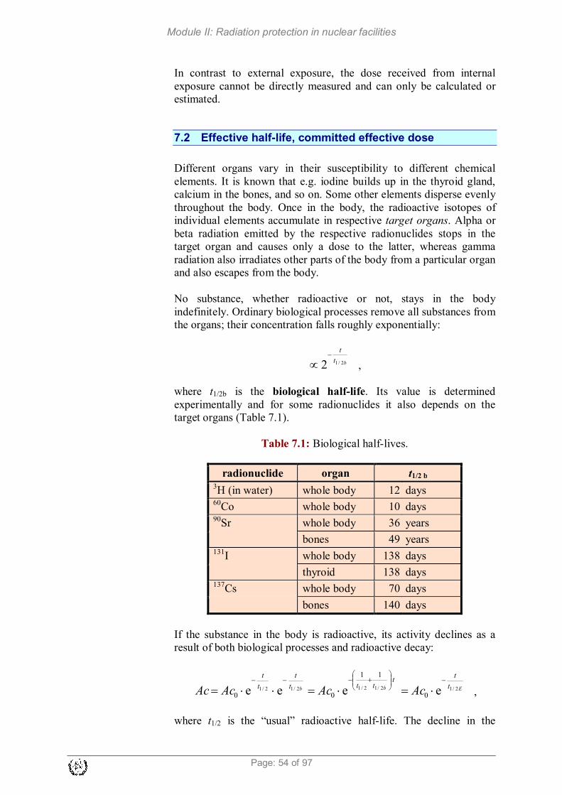
Module II: Radiation protection in nuclear facilities
Page: 54 of 97
In contrast to external exposure, the dose received from internal
exposure cannot be directly measured and can only be calculated or
estimated.
7.2 Effective half-life, committed effective dose
Different organs vary in their susceptibility to different chemical
elements. It is known that e.g. iodine builds up in the thyroid gland,
calcium in the bones, and so on. Some other elements disperse evenly
throughout the body. Once in the body, the radioactive isotopes of
individual elements accumulate in respective target organs. Alpha or
beta radiation emitted by the respective radionuclides stops in the
target organ and causes only a dose to the latter, whereas gamma
radiation also irradiates other parts of the body from a particular organ
and also escapes from the body.
No substance, whether radioactive or not, stays in the body
indefinitely. Ordinary biological processes remove all substances from
the organs; their concentration falls roughly exponentially:
bt
t
2/12−
∝ ,
where t1/2b is the biological half-life. Its value is determined
experimentally and for some radionuclides it also depends on the
target organs (Table 7.1).
Table 7.1: Biological half-lives.
radionuclide organ t1/2 b 3H (in water) whole body 12 days
60Co whole body 10 days
90Sr whole body 36 years
bones 49 years 131
I whole body 138 days
thyroid 138 days 137
Cs whole body 70 days
bones 140 days
If the substance in the body is radioactive, its activity declines as a
result of both biological processes and radioactive decay:
Ebb t
tt
ttt
t
t
t
AcAcAcAc 2/12/12/12/12/1 eeee 0
11
00
−
+−−−
⋅=⋅=⋅⋅= ,
where t1/2 is the “usual” radioactive half-life. The decline in the

Module II: Radiation protection in nuclear facilities
Page 55 of 97
activity of a radionuclide in the body is therefore defined by the
effective half-life, which is calculated by the following formulas:
b2/12/1E2/1
111
ttt+=
b2/12/1
b2/12/1
E2/1tt
ttt
+
⋅=
The effective half-life is therefore the time in which the activity of a
radionuclide in the body diminishes to half its initial value.
Example:
Calculate the effective half-life of 90Sr in bone. Its radioactive half-life is 28.6
years.
Answer:
years18
years49years6.28
years49years6.28
b2/12/1
b2/12/1
E2/1 =+
⋅=
+
⋅=
tt
ttt
Calculating the equivalent dose rate caused by a radionuclide in the
body is very complicated. It depends on its activity, mode of intake,
the type and energy of radiation, the chemical form of the substance
containing the radionuclide and the weighting factors of target organs.
However, the dose rate is rarely immediately relevant when dealing
with internal contamination. As a rule, radionuclides cannot be
artificially removed from the body, so the body remains exposed to
internal radiation until their decay and biological elimination – in
practice, this is approximately seven effective half-lives. When this
period exceeds life expectancy (this applies e.g. to strontium-90), the
irradiation period is held to be 50 years for adults and 70 years for
children. The dose received in this period is called the committed
effective dose. From the perspective of radiation protection, it
represents the most important dosimetric quantity of internal
exposure. It is always calculated and added to annual effective dose in
the year of intake of the radionuclide.
7.3 Dose coefficient, derived air concentration
Committed effective doses have been calculated for all radionuclides,
individually for breathing (inhalation) and swallowing (ingestion).
The quotient that links the committed effective dose and the intake of
activity is called the dose coefficient, h(g). The dose coefficient
values for some radionuclides are given in Table 7.2.

Module II: Radiation protection in nuclear facilities
Page: 56 of 97
Table 7.2: Dose coefficients h(g) for inhalation and ingestion.
Radionuclide Half-life h(g) [Sv/Bq]
inhalation ingestion 3H 12.3 years 1.8·10
-11 1.8·10
-11
51Cr 27.7 days 3.6·10
-11 3.8·10
-11
54Mn 312 days 1.5·10
-9 7.1·10
-10
59Fe 44.5 days 3.5·10
-9 1.8·10
-9
58Co 70.8 days 2.0·10
-9 7.4·10
-10
60Co 5.27 years 2.9·10
-8 3.4·10
-9
90Sr 29.1 years 1.5·10
-7 2.8·10
-8
131I 8.04 days 7.6·10
-9 2.2·10
-8
137Cs 30 years 4.8·10
-9 1.3·10
-8
239Pu 24100 years 4.7·10
-5 2.5·10
-7
Legislation lays down the dose limits, Em, which may be received by
workers or members of the public within a calendar year. Based on
these dose limits we can use dose coefficients to determine the annual
limit on intake (ALI) for a particular radionuclide:
(g)ALI m
h
E=
The derived air concentration (DAC) is defined as the concentration
of a given radionuclide in air (or in water or food – in this case it is
called the derived concentration) at which the annual limit on intake
for the radionuclide would be reached by inhalation (or drinking water
or by dietary intake).
When working with sources of radiation, the greatest challenge is to
prevent the intake of radionuclides into the body by inhalation, which
means that in practice, derived concentrations are of key importance.
They are calculated on the basis of the following premises:
� a worker works 2000 hours a year,
� the breathing rate is 1.2 m3 of air per hour.
The derived air concentration is thus:
a
ALIDAC
V= ,
where Va is the volume of air breathed by a worker during working
time and so Va = 1.2 m3/h · 2000 h = 2400 m
3.

Module II: Radiation protection in nuclear facilities
Page 57 of 97
Example:
Calculate the DAC for 131I in air if the dose limit is 20 mSv.
Answer:
3
319
3
a
m
a
Bq/m1100m2400BqSv106.7
Sv1020
)g(
ALIDAC =
⋅⋅
⋅=
⋅==
−−
−
Vh
E
V
The values of derived air concentrations for major radionuclides are
given in Table 7.3.
Table 7.3: Derived air concentrations for workplaces.
radionuclide DAC [Bq/m3]
3H (HTO) 460 000
60Co 290
90Sr 56
131I 1100
137Cs 1700
239Pu 0.18
7.4 Exposure to radioactive noble gases
Radionuclides which are the radioisotopes of noble gases do not form
chemical bonds with substances in the body, so they are not taken in
by breathing or food, at least not in a significant degree. If present in
air, they enter the lungs on inhalation and leave them on exhalation.
Any dose is chiefly due to external radiation from noble gases in the
ambient air. Since the received dose is independent of biological
processes, it is relatively easy to calculate. It is usually given as the
effective dose received in one day due to the presence of radioactive
noble gases in the air. Its values are shown in Table 7.4.
Table 7.4: Effective doses from exposure to noble gases.
Noble gas
radionuclide t1/2
Effective dose received in one day per unit
of concentration in air [Sv d-1
/Bq m-3
]
41
Ar 1.83 h 5.3·10-9
85
Kr 10.7 y 2.2·10-11
85m
Kr 4.48 h 5.9·10-10
133
Xe 5.24 d 1.2·10-10
135
Xe 9.1 h 9.6·10-10

Module II: Radiation protection in nuclear facilities
Page: 58 of 97
7.5 Protection against internal contamination
Protection against internal exposure is based on the principles of
defence in depth:
� Restriction of open sources of radiation: Sources of radiation
and processes which may cause radioactive contamination of
surfaces or air should be restricted to a reasonably small number
of designated areas.
� Control of areas containing sources of radiation: Areas where
work takes place should be suitably architecturally designed by
selecting surface materials that facilitate effective
decontamination and ensuring controlled outlets and good
ventilation with air filtration. Where necessary (the requirements
are laid down by legislation), work is carried out within a
controlled area. Access to this area is physically controlled,
only workers meeting certain requirements (personal protective
equipment, training, etc.) are allowed to work in this area and
appropriate equipment for personal contamination monitoring
and decontamination should be available at the exit.
� Preventing internal contamination by ingestion and
absorption: While working in radiation controlled areas it is
prohibited to eat, drink, smoke and work with open cuts or
wounds.
� Worker protection: Workers are required to use personal
protective equipment. Its purpose is to prevent skin
contamination (protective clothing) and – in cases of increased
air contamination – to restrict the intake of radioactive material
by inhalation (respiratory protective equipment).
7.6 Protective clothing and equipment
The purpose of protective clothing is to intercept the contamination
which would otherwise affect the worker’s skin or “normal” clothing,
which is why protective clothing is always presumed to be
contaminated. It is put on when entering the controlled area and taken
off on leaving it. The clothing must be designed so that it can be taken
off easily and without contaminating the skin or the clothes worn
underneath. Workers should therefore be first trained to properly put
on and take off their protective clothing.
In nuclear power plants, workers are also exposed to other industrial
hazards: heat, noise, possibility of falling objects, chemicals and so
on, which likewise call for special protective items.
Within the controlled area of a nuclear power plant, we always use
basic protective equipment which (depending on the facility)
consists of the following devices and clothes (Figure 7.1):
� a hard hat,

Module II: Radiation protection in nuclear facilities
Page 59 of 97
� underwear,
� cotton overalls,
� cotton gloves,
� protective shoes,
� a set of personal dosimeters (a daily – electronic and a monthly
– TLD/OSL dosimeter).
Figure 7.1: A worker wearing basic protective equipment (example).
Figure 7.2: A worker in Tyvek coveralls with a respirator.

Module II: Radiation protection in nuclear facilities
Page: 60 of 97
In areas where increased surface or air contamination is probable or
confirmed, one or several pieces of additional protective equipment
are also used according to the level of contamination:
� additional rubber gloves,
� shoe covers,
� Tyvek coveralls,
� special protective clothing,
� a respirator,
� a half mask,
� a mask,
� an air-supplied mask.
Table 7.5: Protective clothing in a controlled area (example).
Type of clothing washable single
use
for the body
cotton overalls yes
underwear yes
Tyvek yes
TOPSTRE yellow overalls yes
WET overalls yes
PE suit yes
COOLINE cooling jacket yes
for the head cotton cap yes
helmet yes
for the
hands
cotton gloves yes
rubber gloves yes
for the feet
protective shoes yes
low covers yes
high covers yes
Figure 7.2 shows an example of additional protective equipment
(Tyvek, respirator) and Table 7.5 gives the characteristics of various
typical kinds of equipment.
Respiratory protective equipment
The basic aim of any type of breathing apparatus or respirator is to
effectively reduce the inhalation of airborne substances which are
harmful to health into the lungs. Since breathing equipment to a
certain extent hampers natural breathing and/or the performance of
work operations, decisions on the use of breathing equipment should
be made in the knowledge that:
� Workers wearing a respirator carry out a given task 20 to 25 %
more slowly than they would without a respirator. In an area
where external radiation and air contamination are present, the
decision should comply with the ALARA principle to minimize

Module II: Radiation protection in nuclear facilities
Page 61 of 97
the sum of the external and internal dose.
� Working with breathing equipment increases the load on the
heart and lungs. For this reason all workers who are likely to use
respirators must undergo a special additional medical
examination.
Respiratory equipment falls into three main groups (Table 7.6):
� air-purifying respirators (air filtration),
� air-line respirators (with clean air supplied via a hose),
� self-contained breathing apparatus.
Table 7.6: Characteristics of respiratory equipment.
type shape principle of operation
PF
particles
only
PF
particles
and gases
air fi
ltra
tio
n half facepiece negative pressure during
inhalation
10
full facepiece negative pressure
during inhalation
50
full facepiece positive pressure 1000
air-
lin
e
half facepiece continuous air flow 1000
half facepiece flow during inhalation 10
full facepiece continuous air flow 2000
full facepiece flow during inhalation 50
full facepiece positive pressure, flow
during inhalation
2000
suit continuous air flow 2000
self
-co
nta
ined
bre
ath
ing
app
arat
us
full facepiece flow during inhalation 50
full facepiece positive pressure, flow
during inhalation
10000
full facepiece closed-circuit breathing 50
full facepiece closed-circuit breathing,
positive pressure
5000
The first group includes e.g. a filter half mask (Figure 7.3), the second
an air-line suit (Figure 7.4) and the third a mask with air supplied from
a cylinder (Figure 7.5).

Module II: Radiation protection in nuclear facilities
Page: 62 of 97
Figure 7.3: Filter half mask.
Figure 7.4: protective clothing (suit) with an air line.
Figure 7.5: Face mask with air supplied from a cylinder.
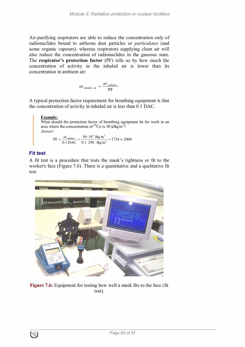
Module II: Radiation protection in nuclear facilities
Page 63 of 97
Air-purifying respirators are able to reduce the concentration only of
radionuclides bound to airborne dust particles or particulates (and
some organic vapours), whereas respirators supplying clean air will
also reduce the concentration of radionuclides in the gaseous state.
The respirator’s protection factor (PF) tells us by how much the
concentration of activity in the inhaled air is lower than its
concentration in ambient air:
PF
ambient
air inhaled
acac =
A typical protection factor requirement for breathing equipment is that
the concentration of activity in inhaled air is less than 0.1 DAC.
Example:
What should the protection factor of breathing equipment be for work in an area where the concentration of 60Co is 50 kBq/m3?
Answer:
20001724Bq/m2901.0
Bq/m1050
DAC1.0PF
3
33
ambient ≈=⋅
⋅==
ac
Fit test
A fit test is a procedure that tests the mask’s tightness or fit to the
worker's face (Figure 7.6). There is a quantitative and a qualitative fit
test.
Figure 7.6: Equipment for testing how well a mask fits to the face (fit
test).

Module II: Radiation protection in nuclear facilities
Page: 64 of 97
7.7 Exercises
1. Calculate the effective half-life for iodine 131
I. Its physical half-
life is 8.06 days and its biological half-life is 138 days.
2. Calculate the effective half-life for 137
Cs. Its physical half-life is
30 days and its biological half-life is 70 days.
3. What dose rate is a worker exposed to when working unprotected
in an atmosphere of 1 DAC of a given radionuclide?
4. Calculate the protection factor of respiratory equipment required
for an atmosphere with 5000 Bq/m3 of
131I. ALIinh = 2.6·10
6 Bq.
5. Calculate the protection factor of respiratory equipment required
for an atmosphere with 3000 Bq/m3 of
131I. ALIinh = 2.6·10
6 Bq,
Vinh = 2.4·103 m
3/year or 1.2 m
3/h.

Module II: Radiation protection in nuclear facilities
Page 65 of 97
8 RADIATION PROTECTION REGULATIONS
Learning objectives
After completing this chapter, the trainee will be able to: 1. Explain the aim of radiation protection. 2. Name the international organisations that issue recommendations
and guidance on radiation protection. 3. Explain the basic ICRP principles. 4. Explain the concept of ALARA. 5. Give the dose limitations laid down by ICRP. 6. Identify different work site classifications and respective
requirements. 7. Name the categories into which workers are classified. 8. Name the most important IAEA safety standard in the area of
radiation protection.
8.1 The aim of radiation protection
People enjoy great benefits from the application of X-radiation,
radionuclides and nuclear fuel in medicine, industry, research and
electricity generation. However, working with sources of radiation by
necessity implies a certain exposure of people to radiation, both
during normal use and in potential accidents which no human activity
is immune to. Under the assumption that any exposure of an
individual to radiation poses a certain risk, exposure is only justified
when the benefit derived from using a source is greater than this risk.
Thus the fundamental principle of radiation protection is to assess the
risks and benefits brought by the use of radiation. If the restrictions
imposed on the use of radiations are too mild, the risks are
unacceptably great; if the restrictions are too severe, the use of sources
becomes too complicated or too expensive, depriving us of the
benefits it could bring.
A simple example of assessing the positive and negative outcomes of
using radiation is found in health care: a patient receives (compared to
exposure in other activities) a relatively large dose, but the benefit of a
radiological scan or therapy for the patient is greater than the radiation
risk. Of course we should bear in mind that apart from the patients,
exposure also affects health workers, although in a lesser degree. So it
is also important to assess the benefit or risk brought by the use of
radiation to society at large. The situation with radiation in nuclear
power plants is similar. We know that nuclear power plants are very
efficient and environmentally friendly energy facilities, and yet their
operation unavoidably involves – relatively low, but nonetheless –
exposure of workers to radiation, minimal discharges of radioactive
substances, etc. This makes the question of benefits and risks a rather
Module II: Radiation protection in nuclear facilities
Page: 66 of 97
complex one which can only be answered by society as a whole. Even
in traffic where the risks are very obvious as well as easily proved,
there are rules that strike a balance between benefits and risks: no
doubt there would be fewer accidents if e.g. the speed limit on
motorways was 30 km/h, but the general assessment the risk with a
limit of 130 km/h is still acceptable and, at the same time, the benefit
of faster-moving traffic is much greater than the increased risk of
higher speeds.
8.2 International recommendations and standards
There are many international bodies and organisations around the
world that study the effects of radiation and issue recommendations on
radiation protection. They can be grouped into governmental and non-
governmental organisations, or as those issuing merely non-binding
recommendations and those international organisations which also
have legislative power over their member states.
Non-governmental organisations include the ICRP and ICRU, which
award membership to distinguished scientists based on their
professional accomplishments.
International organisations which are either partly or fully focused on
the issues of radiation protection and safety include the IAEA
(International Atomic Energy Agency), the UNSCEAR (United
Nations Scientific Committee on the Effects of Atomic Radiation), as
well as the European Commission (EC). The UNSCEAR mostly deals
with epidemiological studies of radiation effects and the IAEA issues
safety standards which are aligned with ICRP recommendations and
indirectly binding on member states (non-compliance leads e.g. to a
withdrawal of IAEA’s technical support). On the other hand, EU
directives are mandatory for member states.
ICRP recommendations
The ICRP (International Commission on Radiological Protection) is
an independent, non-governmental professional body operating since
1928 and its primary goal is to issue recommendations on radiation
protection.
As new findings about the effects of exposure to radiation came to
light over the years, the ICRP repeatedly lowered the recommended
annual dose limit (Figure 8.1).
Data on the increased number of cancerous diseases in atomic bomb
victims clearly showed that, at high doses, the probability of
developing radiation-induced cancer is proportional to the dose
received. At low doses, the probability is so small that radiation-
induced cancer cannot be statistically proved. However, there is also
no firm evidence that sufficiently low doses are harmless, so the ICRP

Module II: Radiation protection in nuclear facilities
Page 67 of 97
adopted the conservative hypothesis of the linear dependence of the probability of stochastic effects with no threshold (the linear no-
threshold model – LNT). This lead to a change in the overall concept
of radiation protection: rather than compliance with the highest
allowed doses, its goal is to reduce the probability of stochastic effects
in the entire population, whether workers or inhabitants, to the lowest
reasonable level.
Figure 8.1: The chronological development of the recommended
annual dose limit for exposed workers (since 1960 also for the public).
The ICRP system of limiting radiation exposure is based on three
principles:
� justification,
� optimization,
� dose limits.
Justification requires that sources of radiation be used only for those
activities which bring more benefit than harm due to exposure to
radiation.
Optimization requires that all exposures shall be kept as low as
reasonably achievable (ALARA), economic and social factors being
taken into account.
Dose limits are values of the effective dose or equivalent dose that
should not be exceeded.
Today, the ICRP has an outstanding international reputation and
authority. Despite having no legal force, its recommendations are
applied by most countries in their national legislations.
IAEA Safety standards
The findings of the UNSCEAR and the recommendations of
international expert bodies, notably the ICRP, are taken into account
in developing the IAEA safety standards.

Module II: Radiation protection in nuclear facilities
Page: 68 of 97
The preparation and review of the IAEA safety standards involves its
own Secretariat and four safety standards committees for safety in the
areas of nuclear safety (NUSSC), radiation safety (RASSC), the safety
of radioactive waste (WASSC) and the safe transport of radioactive
material (TRANSSC), and a Commission on Safety Standards (CSS)
which oversees the programme for their development.
The principal users of IAEA safety standards in the member states are
regulatory bodies and other relevant national authorities. All IAEA
member states may nominate experts for the IAEA safety standards
committees and may provide comments on draft standards. The CSS
membership is appointed by the IAEA Director General and includes
senior governmental officials having responsibility for establishing
national regulations. A management system has been established for
the processes of planning, developing, reviewing, revising and
establishing the IAEA safety standards. It articulates the mandate of
the IAEA, its vision on the future application of the IAEA safety
standards, policies and strategies, and corresponding functions and
responsibilities.
As indicated above, the ICRP is the main body which makes
recommendations with respect to radiological protection matters in
general, and dose limits in particular. For this reason, the IAEA has
embodied the ICRP recommendations (i.e., those in its publication
number 103 of 2007) in the latest publication of the Basic Safety
Standards (Radiation Protection and Safety of Radiation Sources:
International Basic Safety Standards, General Safety Requirements
Part 3 of 2014 [8], hereafter referred to as BSS).
The BSS lay down requirements for establishing governmental, legal
and regulatory frameworks for safety, specify basic obligations and
administrative requirements for practices, dose limits (in accordance
with the ICRP recommendations) and relevant radiation protection,
management, technical and safety verification requirements. They also
lay down detailed requirements for occupational exposure, public
exposure, emergency exposure situations (including interventions),
and also specify other exposures (potential, medical) to ionizing
radiation. BSS also identify the protection of the environment as an
issue necessitating assessment and the necessity of designing and
implementing security and radiation safety measures in an integrated
manner so that they do not conflict with each other.
Other publications of the IAEA safety standards series give more
detailed guidance on how the BSS requirements should be met in
particular situations. In this respect, Table 8.1 shows the current most
relevant IAEA safety standards which can be used for achieving an
appropriate level of safety and regulation of installations/facilities in
the nuclear fuel cycle.
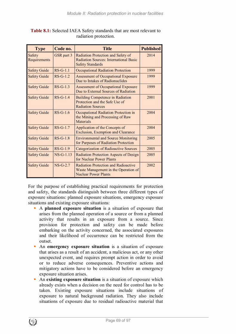
Module II: Radiation protection in nuclear facilities
Page 69 of 97
Table 8.1: Selected IAEA Safety standards that are most relevant to
radiation protection.
Type Code no. Title Published
Safety Requirements
GSR part 3 Radiation Protection and Safety of Radiation Sources: International Basic
Safety Standards
2014
Safety Guide RS-G-1.1 Occupational Radiation Protection 1999
Safety Guide RS-G-1.2 Assessment of Occupational Exposure
Due to Intakes of Radionuclides
1999
Safety Guide
RS-G-1.3
Assessment of Occupational Exposure
Due to External Sources of Radiation
1999
Safety Guide RS-G-1.4 Building Competence in Radiation
Protection and the Safe Use of
Radiation Sources
2001
Safety Guide RS-G-1.6
Occupational Radiation Protection in the Mining and Processing of Raw
Materials
2004
Safety Guide RS-G-1.7 Application of the Concepts of
Exclusion, Exemption and Clearance
2004
Safety Guide RS-G-1.8 Environmental and Source Monitoring
for Purposes of Radiation Protection
2005
Safety Guide RS-G-1.9 Categorization of Radioactive Sources 2005
Safety Guide NS-G-1.13 Radiation Protection Aspects of Design
for Nuclear Power Plants
2005
Safety Guide NS-G-2.7 Radiation Protection and Radioactive
Waste Management in the Operation of Nuclear Power Plants
2002
For the purpose of establishing practical requirements for protection
and safety, the standards distinguish between three different types of
exposure situations: planned exposure situations, emergency exposure
situations and existing exposure situations:
� A planned exposure situation is a situation of exposure that
arises from the planned operation of a source or from a planned
activity that results in an exposure from a source. Since
provision for protection and safety can be made before
embarking on the activity concerned, the associated exposures
and their likelihood of occurrence can be restricted from the
outset.
� An emergency exposure situation is a situation of exposure
that arises as a result of an accident, a malicious act, or any other
unexpected event, and requires prompt action in order to avoid
or to reduce adverse consequences. Preventive actions and
mitigatory actions have to be considered before an emergency
exposure situation arises.
� An existing exposure situation is a situation of exposure which
already exists when a decision on the need for control has to be
taken. Existing exposure situations include situations of
exposure to natural background radiation. They also include
situations of exposure due to residual radioactive material that

Module II: Radiation protection in nuclear facilities
Page: 70 of 97
derives from past practices that were not subject to regulatory
control or that remains after an emergency exposure situation.
For each of these exposure situations requirements applicable to two
categories of exposure are stated: for occupational exposures and for
public exposures. Requirements for medical exposures are stated only
for planned exposure situations.
For planned exposure situations, the following dose limits are set:
� For occupational exposure of workers over the age of 18
years, the dose limits are:
(a) An effective dose of 20 mSv per year averaged over five
consecutive years (100 mSv in 5 years), and of 50 mSv in any
single year;
(b) An equivalent dose to the lens of the eye of 20 mSv per year
averaged over 5 consecutive years (100 mSv in 5 years) and of
50 mSv in any single year;
(c) An equivalent dose to the extremities (hands and feet) or the
skin of 500 mSv in a year.
Additional restrictions apply to occupational exposure for a
female worker who has notified pregnancy or is breast-feeding.
� For occupational exposure of apprentices of 16 to 18 years of
age who are being trained for employment involving radiation
and for exposure of students of age 16 to 18 who use sources in
the course of their studies, the dose limits are:
(a) An effective dose of 6 mSv in a year;
(b) An equivalent dose to the lens of the eye of 20 mSv in a
year;
(c) An equivalent dose to the extremities (hands and feet) or the
skin of 150 mSv in a year.
� For public exposure, the dose limits are:
(a) An effective dose of 1 mSv in a year;
(b) In special circumstances, a higher value of effective dose in
a single year could apply, provided that the average effective
dose over five consecutive years does not exceed 1 mSv per
year;
(c) An equivalent dose to the lens of the eye of 15 mSv in a
year;
(d) An equivalent dose to the skin of 50 mSv in a year.
For emergency situations, the Safety Standards specify generic
criteria for acute doses for which protective actions and other response
actions are expected to be undertaken under every circumstance to
avoid or to minimize severe deterministic effects, and also sets
guidance levels for restricting the exposure of emergency workers.
In BSS a number of requirements for government, regulatory bodies,
registrants (registered users) and licensees, employers and workers are
stated. We list some requirements related to the responsibilities of
employers, registrants and licensees regarding protection of workers:

Module II: Radiation protection in nuclear facilities
Page 71 of 97
� Employers, registrants and licensees shall be responsible for the
protection of workers against occupational exposure.
Employers, registrants and licensees shall ensure that protection
and safety is optimized and that the dose limits for occupational
exposure are not exceeded.
� Employers, registrants and licensees shall establish and maintain
organizational, procedural and technical arrangements for the
designation of controlled and supervised areas, for local rules
and for monitoring of the workplace, in a radiation protection
programme for occupational exposure.
� Registrants and licensees, in cooperation with employers where
appropriate, shall establish, maintain and keep under review a
programme for workplace monitoring under the supervision of a
radiation protection officer or qualified expert.
� Employers, registrants and licensees shall be responsible for
making arrangements for the assessment and recording of
occupational exposure and for workers’ health surveillance.
� Employers, registrants and licensees shall provide workers with
adequate information, instruction and training for protection and
safety.
� Employers, registrants and licensees shall not offer benefits as
substitutes for measures for protection and safety.
� Employers, registrants and licensees shall make special
arrangements for female workers, as necessary, for protection of
the embryo or foetus and of breast-fed infants. Employers,
registrants and licensees shall make special arrangements for the
protection and safety of persons under 18 years of age who are
undergoing training.
The requirements for a legal and regulatory framework are supported
by optimised criteria for exemption of practices and sources and by
criteria for clearance of sources. The exemption level is a value
established by a regulatory body and expressed in terms of activity
concentration, total activity, dose rate or radiation energy, at or below
which a source of radiation need not be subject to some or all aspects
of regulatory control. The clearance level is a value established by a
regulatory body and expressed in terms of activity concentration, at or
below which regulatory control may be removed from a source of
radiation within a notified or authorized practice.
A general requirement related to all aspects of radiation protection and
safety is the requirement for a graded approach: the application of
the Standards shall be commensurate with the characteristics of the
practice or the source within a practice, and with the magnitude and
likelihood of exposures.

Module II: Radiation protection in nuclear facilities
Page: 72 of 97
8.3 Exercises and questions
1. What is the dose limit for the general public laid down by the
BSS?
2. What are the prescribed dose limits according to the rules for
occupationally exposed workers?
3. What does it mean if the activity of a nuclide is below the
prescribed exemption level?
4. A radiography source of 60
Co, is measured to have a dose rate of
4 mSv/h at a distance of five metres.
a) What is the source activity?
b) What is the dose rate at a distance of 2 m, at 0.5 m and at 10
cm?
c) How long does it take to exceed a dose of 5 mSv at 2 m
distance?
d) How long does it take to exceed the annual limit on the
equivalent dose to the skin for an occupationally exposed
worker at a distance of 10 cm?
e) How long does it take to exceed the annual dose limit for an
occupational radiation worker at a distance of 2 m?
5. A dose rate of 10 mSv/h is measured at 1 m distance from a
source of γ radiation. At what distance from the source of
radiation is the dose rate 10 µSv/h? At what distance should the
boundary of the controlled area be set?
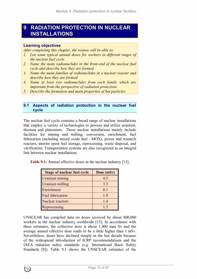
Module II: Radiation protection in nuclear facilities
Page 73 of 97
9 RADIATION PROTECTION IN NUCLEAR INSTALLATIONS
Learning objectives
After completing this chapter, the trainee will be able to: 1. List some typical annual doses for workers in different stages of
the nuclear fuel cycle. 2. Name the main radionuclides in the front-end of the nuclear fuel
cycle and describe how they are formed. 3. Name the main families of radionuclides in a nuclear reactor and
describe how they are formed. 4. Name at least two radionuclides from each family which are
important from the perspective of radiation protection. 5. Describe the formation and main properties of hot particles.
9.1 Aspects of radiation protection in the nuclear fuel cycle
The nuclear fuel cycle contains a broad range of nuclear installations
that employ a variety of technologies to process and utilize uranium,
thorium and plutonium. These nuclear installations mainly include
facilities for mining and milling, conversion, enrichment, fuel
fabrication (including mixed oxide fuel - MOX), power and research
reactors, interim spent fuel storage, reprocessing, waste disposal, and
vitrification. Transportation systems are also recognized as an integral
link between nuclear installations.
Table 9.1: Annual effective doses in the nuclear industry [15].
Stage of nuclear fuel cycle Dose (mSv)
Uranium mining 4.5
Uranium milling 3.3
Enrichment 0.1
Fuel fabrication 1.0
Nuclear reactors 1.4
Reprocessing 1.5
UNSCEAR has compiled data on doses received by about 800,000
workers in the nuclear industry worldwide [15]. In accordance with
these estimates, the collective dose is about 1,400 man Sv and the
average annual effective dose tends to be a little higher than 1 mSv.
Nevertheless, doses have declined steeply in the last decade because
of the widespread introduction of ICRP recommendations and the
IAEA radiation safety standards (e.g. International Basic Safety
Standards [8]). Table 9.1 shows the UNSCEAR estimates of the

Module II: Radiation protection in nuclear facilities
Page: 74 of 97
average annual effective doses in relation to different occupations in
the nuclear industry.
Criticality is one of the dominant safety hazards for non-reactor
facilities, because they employ a great diversity of technologies and
processes, and the materials of interest to radiation safety are more
dispersed throughout them. They may be used not only in bulk form
(fuel pellets, fuel elements, fuel rods, fuel assemblies, and so on), but
in distributed and mobile forms as well (e.g., different kinds of
solutions, slurries, gases, powders). This is in contrast to nuclear
reactors, where the bulk of the nuclear material is located in the
reactor core or fuel storage areas. As a result fissile materials may
accumulate in some parts of the equipment and may also escape from
the facility as a result of equipment leakage.
9.2 Important radionuclides for radiation protection
During the nuclear fuel cycle, different radionuclides are of major
concern for radiation protection. At the front end of the fuel cycle
(uranium/thorium mining and milling) only the natural radionuclides
from the uranium (or thorium) decay series present a radiation risk.
The resulting purified form of uranium concentrate is called
“yellowcake” which is used in the preparation of fuel for nuclear
reactors. It is denoted as U3O8 but is often actually ammonium
diuranate [(NH4)2U2O7].
As yellowcake does not contain appreciable amounts of uranium
progeny and uranium is very long-lived, the radiation risk is
substantially lower in the next stages of fuel manufacturing
(conversion to UF6, enrichment, fuel fabrication). For MOX fuel
fabrication, however, the radiation risk is substantially higher than for
uranium fuel, because plutonium has a higher specific activity and
radiotoxicity than uranium. At this stage, the main risk is from hot
particles, very small particles (smaller than normal dust particles)
containing actinides (uranium or plutonium) that may be inhaled.
Nuclear reactors are devices where the fission chain reaction takes
place. The reactor core is therefore the main source of radiation during
reactor operation. It is a very strong source of neutrons and fission
products. Fission products are very highly radioactive while in the
construction elements of the reactor neutrons produce additional
sources of radioactivity, the so-called activation or corrosion products.
Nuclear reactors can be divided into power reactors (nuclear power
plants) and research reactors. As power reactors generally have
significantly higher power than research reactors, the amount of
radioactivity produced is usually much larger than in the case of
research reactors. However, the radiation protection issues are at least
in principle similar for both types of reactors. Naturally, as NPPs are

Module II: Radiation protection in nuclear facilities
Page 75 of 97
much more complex installations than research reactors, the radiation
protection problems are also much more complex.
In the back end of the nuclear fuel cycle (reprocessing and/or waste
management, storage and disposal), all types of radionuclides (fission
products, activation products, actinides) are important for radiation
protection. As reprocessing or waste disposal usually takes place some
time after the waste has been removed from the reactor and power
plant systems, only the long-lived radionuclides (with half-lives
comparable or greater than the time since removal from the of reactor)
are of importance.
Natural radionuclides
Figure 9.1 shows the decay chain of 238
U. As uranium has a very long
half-life, its daughter products (progeny) tend to be in radioactive
equilibrium with uranium in soil or in ore. Radon-222, however, is an
isotope of the noble gas radon and hence it is not chemically bound to
soil but can easily diffuse to the surface.
234U
238U
2×10⁵ y 4×10
9 y
β -
α 234*Pa α
7 h
β -
230Th
234Th
8×10⁴ y 24 d
α
226Ra
1600 y
α
222Rn
4 d
α
210Po
214Po
218Po
138 d 162 µs 3 min
β - β -
α 210Bi α 214
Bi α5 d 20 min
β - β -
206Pb
210Pb
214Pb
stable 22 y 27 min

Module II: Radiation protection in nuclear facilities
Page: 76 of 97
Figure 9.1: The uranium-radium decay chain.
Radon and its short-lived daughter products are present in the air and
are easily inhaled. Radon as inert gas is also exhaled. Its progeny are
isotopes of polonium, bismuth and lead and they are chemically
attached to dust particles in the air. They are adsorbed in the human
respiratory system and remain there until they decay. In particular
both polonium isotopes that emit alpha particles produce a substantial
effective dose in the respiratory system. Radon therefore presents the
largest radiation risk from natural radioactivity and, in fact,
contributes one half of the average natural dose of population (1.2
mSv of 2.4 mSv total). A list of important radon daughter
radionuclides with their properties is given in Table 9.2.
Table 9.2: Radon and its short-lived progeny.
Nuclide t1/2 Eα [MeV] Eβ,max [MeV]
222
Rn 3.8 d 5.5 -
218
Po 3.1 min 6.0 -
214
Pb 27 min - 1.0
214
Bi 20 min 3.2
214
Po 164 µs 7.7 -
In uranium mines and milling facilities, the concentration of radon is
substantially higher than the average natural background, therefore
special measures need to be taken to protect the workers and the
population in the neighbourhood of these facilities. These measures
include ventilation and respiratory equipment.
Fission products
Fission products are a direct product of fission. They are produced
within the reactor fuel and have by far the greatest activity of all
radionuclides in the reactor or the nuclear power plant. During reactor
operation or immediately after its shutdown, the total activity of a
single fuel element is higher than 1018
Bq, a month after shutdown it is
around 1017
Bq and after 10 years in the spent fuel pit it is around
1016
Bq. Fortunately most fission products are elements which remain
chemically bound in uranium dioxide and are consequently not
released from the fuel pellet. In the context of nuclear safety, the pellet
is thus considered as the first barrier that prevents the dispersal of
radioactive materials.

Module II: Radiation protection in nuclear facilities
Page 77 of 97
Table 9.3: Major fission products with the main γ lines (they all emit
β particles as well, but β energies are only given for pure beta
emitters).
Nuclide t1/2 Eγ [MeV] Eβ,max [MeV]
85m
Kr 4.48 h 0.151
133
Xe 5.25 d 0.081
135
Xe 9.10 h 0.250
131
I 8.02 d 0.364; 0.637
137
Cs 30.2 y 0.662
90
Sr/90
Y 28.6 y - 2.3
3H 12.3 y - 0.02
It is particularly the noble gases (Kr and Xe isotopes; practically
entirely) which escape or diffuse out of the fuel pellet, as well as the
isotopes of volatile elements (I, Cs, Sr, tritium; the bulk of the
inventory). The pellet also partly releases the isotopes of Ba, Ce and
Ru/Rh. The radionuclides which escape from the pellet build up in the
empty space between the pellets and the fuel rod cladding, i.e. in the
gap and plenum. The cladding is the second barrier to the dispersal
of radioactive materials.
A PWR reactor core contains from 20,000 to 50,000 fuel rods and in a
very small fraction of them the cladding does not stay perfectly tight
throughout the service life. Some fission products (tritium and the
noble gases in particular) even diffuse through the cladding. For this
reason, a fraction of the fission products passes into the reactor
coolant. Table 9.3 gives the key fission products. 133
Xe has the
greatest activity of all.
Activation and corrosion products
Neutrons also activate other materials in the core or its immediate
environment: the coolant and the construction materials of the reactor.
Such radionuclides are called activation products. The coolant that
flows through the primary circuit (reactor, hot leg, steam generator,
cold leg, reactor pump) causes corrosion and erosion processes on the
walls of the primary circuit and on the construction elements of the
core. As a result, tiny particles of these materials pass into the coolant
and are activated as they travel through the core. Such radionuclides
are called corrosion products.

Module II: Radiation protection in nuclear facilities
Page: 78 of 97
Table 9.4: Major activation and corrosion products with the main γ
energies (they all emit β- as well).
Nuclide t1/2 formation Eγ [MeV] 16
N 7.1 s 16
O (n, p) 16
N 6.13; 7.12 41
Ar 1.83 h 40
Ar (n, γ) 41
Ar 1.29 51
Cr
27.7 d 50
Cr (n, γ) 51
Cr 0.32 54
Mn
312 d 54
Fe (n, p) 54
Mn 0.835 58
Co 70.8 d 58
Ni (n, p) 58
Co 0.811 59
Fe 44.5 d 58
Fe (n, γ) 59
Fe 1.099; 1.292 60
Co 5.27 y 59
Co (n, γ) 60
Co 1.332; 1.173
Except in some special cases (working in the proximity of the reactor
vessel, decommissioning) the most important activation products in
terms of radiation protection are the radionuclides spread by the
coolant through the primary circuit. These are either nuclides which
form directly in the coolant or activated materials of e.g. the reactor
vessel wall or core construction elements which enter the coolant due
to erosion and/or corrosion. In nature, such material is not much
different from the material entering the coolant by erosion-corrosion
from other parts of the primary circuit, except that the materials may
differ (the pressure vessel is made of steel and e.g. the steam generator
tubes of nickel alloy). Activation and corrosion products are therefore
discussed collectively. They typically spread throughout the entire
primary system via the coolant and eventually a part of them binds to
the inner walls of the pressure boundary. This process causes the
contamination of all primary system components.
Besides activation and corrosion products, fission products are also
released in the coolant. The pressure boundary of the primary circuit is
thus the third barrier to the dispersal of radioactive materials. Table
9.4 gives the major activation and corrosion products.
In this table also the gases nitrogen in argon are listed. 16
N is formed
by fast neutron reaction on oxygen in water molecules. It emits highly
penetrating gamma radiation and is the most important source of
radiation in the coolant during reactor operation. Argon is a
component of air (its fraction in air is about 1%) and it is mostly
formed in hollow spaces adjacent to the reactor vessel. In pool-type
research reactors, air is present dissolved in the coolant and 41
Ar
accounts for the largest part of the activity released.
Hot particles
The maintenance of pumps, valves and piping involves techniques
such as cutting, grinding and welding which may produce tiny
fragments that are carried away by the coolant. Often too small to be
retained by filters, they are activated as they pass though the core. A
similar thing happens to fragments resulting from the presence of

Module II: Radiation protection in nuclear facilities
Page 79 of 97
foreign bodies in the primary system, which travel in water and cause
damage to the walls and inner surfaces of primary system
components. This results in microscopically small solid particles with
high specific activity which are called hot particles.
Hot particles may contain a single radionuclide or a large number of
radionuclides. These nuclides can be fission, activation or corrosion
products. Exposure to hot particles is particularly significant in cases
of a hot particle landing on the bare skin or protective clothing of
workers, or their inhalation.
9.3 Questions
1. In what way do corrosion products form and what are their
characteristics?
2. How does 60
Co form in the primary system of a nuclear power
plant?
3. Name two main sources of hot particles and three of their
physical properties.

Module II: Radiation protection in nuclear facilities
Page: 80 of 97
10 ENVIRONMENTAL MONITORING
Learning objectives
After completing this chapter, the trainee will be able to: 1. Explain the need for environmental monitoring. 2. Explain the meaning of the term “exposure pathway”. 3. Describe the main external exposure pathways for members of the
public. 4. Describe the main internal exposure pathways for members of the
public. 5. Explain the meaning of the term “constraint” for public exposure. 6. Explain the primary objectives of monitoring. 7. Explain the basis of environmental monitoring programmes 8. List some monitored constituents of airborne and liquid
discharges. 9. Explain the terms representative person and critical group. 10. Explain the dose assessment approaches for members of the
public. 11. State the main exposure pathways for different stages of the fuel
cycle. 12. Compare doses from the natural background.
10.1 Need for monitoring nuclear facilities
In the current state of technology, the operation of nuclear and some
radiation facilities would not be possible without some radioactive
release to the environment. Almost all waste radioactive material is
collected, treated and stored in the form of High Level Waste (HLW)
or Low and Intermediate Level Waste (LILW), but waste processing
technology cannot retain all the radioactive content within the facility
boundaries, at least not at acceptable costs. Therefore small quantities
of low specific activity liquids and/or gases are discharged into the
environment. The consequence of these discharges is additional
exposure of the population, primarily in the vicinity of the facility.
During normal operation discharges are subject to statutory control:
they must be authorised and monitored. Regular monitoring of these
controlled discharges, both at the source of the discharge and in the
receiving environment, is essential in ensuring the protection of the
public and the environment. Monitoring is generally carried out by the
owners of nuclear and radiation facilities, and in some cases by
regulatory agencies.
In radiation and nuclear safety the meaning of noun “monitoring” is not
limited to the functions that have just been mentioned. According to
International Basic Safety Standards [8], monitoring is “the measurement of dose, dose rate or activity related to the assessment or control of exposure to radiation or radioactive substances, and the interpretation of the results.”
Depending on where the measurements are made, it could be classed as

Module II: Radiation protection in nuclear facilities
Page 81 of 97
individual (personal) monitoring, workplace monitoring, source monitoring and environmental monitoring. Depending on the purpose, it could be routine monitoring, task related monitoring and special monitoring.
During emergency situations releases are possible which highly
exceed values in normal operation. While these releases will probably
be uncontrolled, monitoring should be capable of assessing such
elevated dose rates and activities in the environment to support an
effective and timely emergency response.
10.2 Exposure pathways to the population
Controlled gaseous releases from nuclear facilities consist of a
mixture of gases, vapours and particulates containing radionuclides
which are usually discharged through the stack, although for small
facilities they may be made through discharge vents or working
hoods. Controlled liquid releases are typically contaminated water
discharged via pipelines into rivers, lakes or the sea.
Gaseous releases spread in the form of a radioactive cloud, but a part
of its radioactive content is also deposited on the ground and
vegetation. This effect, called fallout, is enhanced with precipitation.
Radionuclides from liquid discharge mix with water bodies and also in
groundwater. Irrigation spreads these radionuclides to soil on
cultivated land.
As a consequence of releases and radionuclide distribution in the
environment, humans are exposed to radiation through different
exposure pathways, which are presented in Figure 10.1.
Figure 10.1: The possible pathways of exposure for members of the
public as a result of discharges of radioactive material to the

Module II: Radiation protection in nuclear facilities
Page: 82 of 97
environment [16].
The main external exposure pathways [16] are:
a) Source of radiation → humans: direct exposure from a source of
ionizing radiation;
b) Source of radionuclides → atmosphere or water body → humans: exposure due to the plume of radionuclides in the atmosphere
(‘cloud shine’) or in water;
c) Source of radionuclides → atmosphere or water body → human skin: contact exposure from radionuclides on the skin;
d) Source of radionuclides → atmosphere or water body → soil or sediment or building surface or vegetation → human: exposure
from radionuclides deposited on the ground or on sediments (on
the shores of rivers, lakes or the sea) or building surfaces (walls,
roofs and floors) or vegetation (trees, bushes and grass).
The main internal exposure pathways [16] are:
a) Source of radionuclides → atmosphere → humans: inhalation of
radionuclides in the plume;
b) Source of radionuclides → atmosphere or water body → (soil or sediment) → vegetation and/or meat, milk, eggs or marine food → human: ingestion of radionuclides in food or beverages;
c) Source of tritium → atmosphere → humans: for tritium oxide in
the plume, by absorption through the skin;
d) Soil or sediment → humans: inhalation of resuspended
radionuclides.
The importance of the various exposure pathways depends on:
� The radiological properties of the material released (e.g. gamma,
beta or alpha emitters; physical half-life);
� The physical (e.g. gas, liquid or solid) and chemical (e.g.
organic or inorganic form, oxidation state, speciation, etc.)
properties of the material and its migration characteristics;
� The dispersal mechanism and factors affecting it (e.g. stack
height, meteorological conditions, etc.) and environmental
characteristics (e.g. climate, type of biota, agricultural
production, etc.);
� The locations, ages, diets and habits of the exposed individuals
or population.
As can be seen, the importance of each exposure pathway depends on
the source properties, and the environmental and population
characteristics. Therefore important exposure pathways must be
identified and evaluated for each nuclear facility.
For all nuclear and radiation facilities identification and evaluation of
exposure pathways must be done during the licensing process, when
limits for discharges and dose constraint for public exposure are also
laid down according to national legislation.

Module II: Radiation protection in nuclear facilities
Page 83 of 97
Simply put, the dose constraint is the dose limit for members of the
public related to a particular facility or source. Usually, dose
constraints for nuclear facilities are from 0.1 mSv to 0.3 mSv per year.
10.3 Objectives of monitoring
The main objective of monitoring is verifying compliance with the
requirements for protection and safety. As specified in IAEA Safety
Standards [16], the primary objectives of any monitoring
programme for protection of the public and the environment are
to:
a) Verify compliance with authorized discharge limits and any other
regulatory requirements concerning the impact on the public and
the environment due to the normal operation of a practice or a
source within a practice;
b) Provide information and data for dose assessment purposes, and to
assess the exposure or potential exposure of populations due to the
presence of radioactive materials or radiation fields in the
environment from the normal operation of a practice or a source
within a practice, and from accidents or past activities;
c) Check the conditions of operation and the adequacy of controls on
discharges from the source and to provide a warning of unusual or
unforeseen conditions and, where appropriate, to trigger a special
environmental monitoring programme.
In summary, monitoring should be regarded as an essential element of
the control of discharges to ensure protection of the public and the
environment. It is also an essential element in determining the actions
that should be taken to protect the public in intervention situations.
The following three types of monitoring are envisaged [16]:
1) Source monitoring, which is monitoring the activity of
Constraint [8]: A prospective and source-related value of individual dose (dose constraint) or risk (risk constraint) that is used in planned exposure situations as a parameter for the optimization of protection and safety for the source, and which serves as a boundary in defining the range of options in optimization. For public exposure, the dose constraint is a source-related value established or approved by the government or the regulatory body, with account taken of the doses from planned operations of all sources under control. The dose constraint for each particular source is intended, among other things, to ensure that the sum of doses from planned operations for all sources under control remains within the dose limit.

Module II: Radiation protection in nuclear facilities
Page: 84 of 97
radioactive materials being released to the environment, or of
external dose rates due to sources within a facility or activity;
2) Environmental monitoring, which is monitoring the external
dose rates due to sources in the environment and/or the
radionuclide concentrations in environmental media;
3) Individual monitoring, which is monitoring with equipment worn
by individuals, or measurements of the quantities of radioactive
materials in or on their bodies.
During normal operation of nuclear facilities, individual (personal)
monitoring outside the facility is rarely justified, but in emergency
situations it is required for emergency workers and also for public as
appropriate. Source and environmental monitoring are performed
routinely over the whole lifetime of the facility, and environmental
monitoring even before commissioning (for assessment of pre-
operational base-line conditions) and also after decommissioning of
the facility.
10.4 Programmes for environmental monitoring
Environmental monitoring is implemented through programmes which
must be based on the characteristics of the facility and an evaluation
of the exposure pathways. The scale of a monitoring programme
should be determined primarily by the significance of the expected
doses to the public. The programme should be designed such that
those radionuclides that are significant in terms of the dose received
by a representative person are monitored.
The frequency of monitoring and sampling should be determined by
the complexity of the environment, the significance of the doses to
members of the public and the properties of the radionuclides. For
example, if short lived 131
I is to be monitored, the frequency of
monitoring should be sufficient to detect it before it decays.
In the design of an environmental monitoring programme the
following characteristics of the environment in the vicinity of the
facility must be also taken into account:
(a) Prevailing wind direction;
(b) Meteorological variations;
(c) Current and future land use;
(d) Agricultural practices;
(e) Soil type and hydrological properties.
Relevant cultural, socioeconomic and demographic factors of the local
population should be also considered in the design of the
environmental monitoring programme.
Recommendations regarding the constituents and frequencies of
sampling/measurement of radionuclides for normal operation is given

Module II: Radiation protection in nuclear facilities
Page 85 of 97
in Table 10.1. More information on programmes for environmental
monitoring could be found in IAEA document SRS No. 64 [20].
Table 10.1: Environmentally monitored constituents and suggested
frequencies of sampling and measurement for discharges of
radionuclides to the environment [16].
Discharge Monitored constituents Frequency
Airborne External radiation
Gamma dose rate Continuously
Gamma dose — integrated Twice a year
Neutron dose rate (if neutron
radiation is foreseen)
Continuously
Neutron dose integrated (if
neutron radiation is foreseen)
Twice a year
Air, deposition
Air Continuous collection, weekly
to monthly measurement
Precipitattion Continuous collection, monthly
measurement
Deposition Continuous collection, monthly
measurement
Soil Once a year
Foodstuff and/or ingestion
Leafy vegetables Monthly during growing season
Other vegetables and fruits Selected samples, at harvest
Grain Selected samples, at harvest
Milk Monthly when cows are on
pasture
Meat Selected samples, twice a year
Drinking water and/or
groundwater
Twice a year
Terrestrial indicators Grass Monthly when cattle are on
pasture
Lichen, mosses, mushrooms Selected samples, once a year
Liquid Aquatic dispersion
Surface water Continuous sampling, monthly
measurement
Sediment Once a year
Aquatic foodstuffs Fish Selected samples, once a year
Shellfish Selected samples, once a year
Aquatic indicators Seaweeds, marine sponges Selected samples, once a year
Benthic animals Selected samples, once a year
10.5 Assessment of doses to members of the public
Since the dose to the public cannot be measured directly, it must be
estimated using environmental concentrations and appropriate habit
data. Therefore, for the purpose of protection of the public, it is
necessary to define a hypothetical exposed person to be used for

Module II: Radiation protection in nuclear facilities
Page: 86 of 97
determining compliance with the dose constraint. This is called the
representative person.
Current International Basic Safety Standards (BSS) [8] define a
representative person as “the individual receiving a dose that is representative of the most highly exposed individuals in the population”.
In the previous version of International Basic Safety Standards [21]
and in most national legislations dose constraints are related to
members of “critical group”, where the critical group is “A group of members of the public which is reasonably homogeneous with respect to its exposure for a given radiation source and given exposure pathway and is typical of individuals receiving the highest effective dose or equivalent dose (as applicable) by the given exposure pathway from the given source” [21].
The problem with this definition in environmental monitoring is that
neither is the critical group “critically” (i.e. badly or severely)
exposed, nor is the dose constraint defined for the group (it relates to
an individual). Another problem is that there are several critical
groups, each related to a particular exposure pathway. It is possible
that one group is critical for different (more than one) exposure
pathways, but this still is not sufficient argument to use that particular
group for comparison with dose constraint.
The concept of the critical group remains valid also in the current BSS
considering that the dose to a representative person is the
equivalent of, and replaces, the mean dose in the critical group.
However, the concept of the representative person is not related to a
particular exposure pathway and requires that all pathways are
accounted for. Simple addition of doses from different critical groups
is not possible, therefore groups should be also evaluated
regarding other (noncritical) exposure pathways to identify the
representative person.
Identification of critical groups for existing facilities, or a description
of critical groups for future facilities is demanding and requires
information about source characteristics and releases, environmental
characteristics, population distribution and habits (e.g. amount of
locally produced and consumed food). This information should be as
realistic as possible. With this information and the results of
measurements in the environment and on collected samples, it is
possible to assess doses to critical groups.
In normal operation dose assessment is relatively straightforward
when the external dose rate from a source or the concentration of
radionuclides in the environment (also in food and water) exceeds the
limits of detection for the measurement methods used and
consumption data are available. Internal doses are calculated by

Module II: Radiation protection in nuclear facilities
Page 87 of 97
multiplying the intake of radionuclides in air, water or food with the
appropriate dose coefficients, which are age specific. It is
recommended that doses are calculated for a 1-year-old infant, a 10-
year-old child, and an adult representing the three age categories 0 to
<6 years (infant), 6 to <16 years (child), and 16 to 70 years (adult).
When this is not the case, modelling must be used for dose estimation
and verification of (undetectable) concentration levels. For this
purpose additional data such as meteorological and hydrological
conditions, land utilisation and population habits, characteristics of
dwellings and occupancy should be available. If local data are not
available, regional or generic estimates can be used, but this should be
an exception, and not the rule.
More details on methods for the assessment of doses to members of
public can be found in the IAEA Safety Reports and Safety Guide
Assessment of Doses to the Public from Ingested Radionuclides, SRS
No. 14 [22], Generic Models for Use in Assessing the Impact of
Discharges of Radioactive Substances to the Environment, SRS No.
19 [23], and Radiological Environmental Impact Analysis for
Facilities and Activities, Draft Safety Guide DS427 [24] (this safety
guide will supersede Safety Guide NS-G-3.2 Dispersion of
Radioactive Material in Air and Water and Consideration of
Population Distribution in Site Evaluation for NPPs (2002)).
Various computer codes are available for dose assessment, but codes
and models should be verified and approved by the competent
authority. Models and codes are very useful in prospective dose
assessment, i.e. for doses that may be received in the future (e.g. next
year, or during the operation period of a facility, or during facility
decommissioning). This is also one of the most important parts of the
environmental impact assessment process, which must be carried out
during the stages of assessing the siting of a new nuclear programme
[25].
10.6 Exposure of the population from the various stages of the fuel cycle
The fuel cycle comprises three main stages: fuel fabrication (including
mining and milling), reactor operation and fuel reprocessing
(including waste processing). At each stage of the nuclear fuel cycle, a
variety of radionuclides are released in the form of liquids, gases, or
solid particles. The nature of the effluent depends on the particular
operation or process. Radionuclides discharged in all stages contribute
to the effective dose of the population in the vicinity of the facilities,
and also over a wider area. Long lived radionuclides are also
distributed globally, contributing to the effective dose of all mankind.
This contamination is not directly measurable globally and can be
estimated only through modelling.

Module II: Radiation protection in nuclear facilities
Page: 88 of 97
For all nuclear fuel cycle operations the local and regional exposures
are estimated by UNSCEAR to be about 0.9 man Sv per gigawatt-year
(GWy). The present world nuclear energy generation is about 250
GWy, so that the total collective dose from a year’s nuclear generation
is about 225 man-Sv. Generally individual doses are low, being
below 1 µSv in a year. However, certain individuals may receive
higher doses because of where they live and what they eat.
In Table 10.2 the highest annual doses to the public due to discharges
from the nuclear fuel cycle are listed. Also local and regional
collective doses for commercial fuel cycle facilities up to year 1997
are given.
The releases of radioactive materials from conversion, enrichment,
and fuel fabrication plants are generally small and consist mainly of
uranium series isotopes. Inhalation is the most important exposure
pathway.
For reactors, discharges of 14
C, radioactive particulates, 3H (airborne
and liquid), and noble gases radioisotopes are the main sources of
doses. In the past, 131
I was significant source of exposure, but now
releases are minimal. Ingestion is the most important exposure
pathway.
Doses from fuel reprocessing plants are mostly the consequence of
discharges of 14
C, 129
I, 137
Cs and 3H. In the past (before 1990),
137Cs
was the most dominant radionuclide contributing almost 90% to the
total collective dose (Table 10.2). The highest estimated doses were
the consequence of local seafood consumption containing actinides.
Also for other populations ingestion is the main exposure pathway.
Table 10.2: Annual doses due to discharges from the nuclear fuel
cycle. (Data compiled from [15] and [26]).
Stage of cycle Type of
effluent
Most exposed
persons
(mSv)
Local and
regional
collective dose
to year 1997
(man Sv)
Fuel fabrication Airborne
Liquid
0.01
0.01 900
Reactor
operation
Airborne
Liquid
0.001
0.004 2900
Fuel
reprocessing
Airborne
Liquid
0.05
0.14 4700

Module II: Radiation protection in nuclear facilities
Page 89 of 97
10.7 Natural background
As we have discussed in Chapter 1, there are a number of naturally
occurring radionuclides in the environment. Some of them are also
present in discharges from nuclear facilities (e.g. 3H,
14C, some
members of the uranium series), while others are specific for the
natural environment. Since natural radionuclides are present in air,
soil, water, food, and all other commodities, they contribute
significantly to public exposure. Inhalation of radon progeny, external
gamma exposure to radionuclides in the soil and ingestion of (almost
all natural) radionuclides are the main exposure pathways. In addition
to external gamma exposure (which is approximately 0.06 µSv/h),
people are also exposed to cosmic radiation. Exposure to cosmic
radiation increases with altitude from approximately 0.03 µSv/h at sea
level. For example, at 5 km it is 1 µSv/h and at 10 km it is 5 µSv/h.
Doses from the natural background are significantly higher than doses
from nuclear facilities and dose constraints. Average doses from
natural sources are presented in Table 10.3.
Table 10.3: Average radiation dose from natural sources [15].
Source
Worldwide average
annual effective
dose (mSv)
Typical range
(mSv)
External exposure
Cosmic rays
Terrestrial gamma rays
0.4
0.5
0.3 - 1.0a
0.3 - 0.6b
Internal exposure
Inhalation (mainly radon)
Ingestion
1.2
0.3
0.2 - 10c
0.2 - 0.8d
Total 2.4 1 - 10 a Range from sea level to high altitudes
b Depending on the radionuclide composition of soil and building materials
c Depending on indoor accumulation of radon gas
d Depending on the radionuclide composition of foods and drinking water
In addition to natural radionuclides and cosmic radiation,
contamination from past atmospheric tests of nuclear weapons (global fallout) is still present globally and is also considered as a part
of the natural background. The most important radionuclides from
testing in terms of human exposure are now 14
C, 90
Sr and 137
Cs.
Ingestion is the most important exposure pathway, with a global
average annual dose of 0.005 mSv (in 1963 the average annual dose
was 0.1 mSv!). Fallout from past nuclear accidents (Chernobyl and
Fukushima) is also considered as part of the natural background,
although it is generally much lower and more or less limited to the
northern hemisphere.

Module II: Radiation protection in nuclear facilities
Page: 90 of 97
For new facilities, assessment of local values of global contamination
is very important, since they are a part of the environmental baseline
conditions.
10.8 Monitoring in emergency exposure situations
During an emergency situation there is a possibility of the elevated
and uncontrolled release of radioactive material from a nuclear
facility. The result of these releases would be additional doses to the
population that will increase the likelihood of stochastic effects, or
even doses high enough to give rise to severe deterministic effects.
For some (smaller) releases doses will be insignificant and no special
action is justified, but for other (higher) releases, the doses will exceed
certain reference levels and protective and other response actions will
be justified to avoid or mitigate the incurrence of these doses.
Depending on the size, duration and characteristics of the release, the
anticipated and possible development of source status, weather
conditions and forecast, results of monitoring in the environment etc.,
the projected doses (or received doses in some circumstances) for the
population demand one or more of the following protective actions:
iodine thyroid blocking, evacuation, sheltering, prevention of
inadvertent ingestion, decontamination of individuals, relocation, and
restriction of consumption of food, milk and water. Generic criteria
for acute doses for which protective actions and other response actions
are expected to be taken under any circumstances to avoid or to
minimize severe deterministic effects, and generic criteria for
protective actions and other response actions to reduce the risk of
stochastic effects are given in [8]. More information on protective
actions and criteria that are used in emergency situations can be found
in Module 16, Emergency Preparedness and Response.
To make sure that all protective actions during the emergency are
implemented in time, emergency planning zones are defined around
facility in the emergency response preparatory stage. These zones are
defined according to the characteristics of required response in the
most hazardous emergency situation possible in the nuclear facility.
For nuclear power plant accidents, emergency planning zones are
defined as following:
1. Precautionary action zone (PAZ);
2. Urgent protective action planning zone (UPZ);
3. Extended planning distance (EPD); and
4. Ingestion and commodities planning distance (ICPD).
These zones encircle the nuclear power plant at approximately 3 to 5
km (PAZ), 15 to 30 km (UPZ), 50 to 100 km (EPD) and 100 to 300
km (ICPD). The PAZ is defined as the area where deterministic
effects are possible in the most hazardous emergency situations,

Module II: Radiation protection in nuclear facilities
Page 91 of 97
therefore protective actions (e.g. evacuation) should be implemented
before radioactive release from the power plant occurs. The UPZ is
defined where exposure of the public could exceed reference levels in
a short time (hours), and implementation of protective actions must
start before or shortly after the release. The EPD and ICPD are zones
where most protective actions are not implemented immediately, but
only after measurement data from the environment are available.
Protective actions in the PAZ an UPZ should be initiated before or
immediately after actual releases from the nuclear power plant start.
The conditions in the power plant (emergency classification) represent
the only initial information available. Later, during the development
of the accident data from source monitoring (source releases) might be
available (but not necessarily!), and later on, results of emergency
monitoring from the environment should also became available to
support initiation and implementation of effective protective actions.
For planning purposes, the emergency monitoring programme is
therefore divided into the following phases: the pre-release and early
phase (release), the post-release or intermediate phase, and the
recovery or remediation phase. Key data and requirements for
environmental monitoring evolve with time and the transition from
one phase to another.
Objectives of emergency monitoring
The specific objectives of emergency radiation monitoring in the
environment are [16]:
a) To provide accurate and timely data on the level and degree of
hazards resulting from a radiation emergency, in particular on
the levels of radiation and environmental contamination with
radionuclides;
b) To assist decision makers on the need to make interventions
and take protective actions;
c) To provide information for the protection of emergency
workers;
d) To provide information for the public on the degree of the
hazard;
e) To provide information needed to identify any people for
whom long term medical screening is warranted.
The emergency monitoring programme, which is a part of the
emergency management system, must be designed for a range of
possible emergencies. It must ensure that data all from instrumental
measurements, sample collection and sample analysis, dose
assessment and interpretation of results are available. For timely
transfer of these data a proper communication system must be
designed and established. The programme must also anticipate the
receipt of assistance from other organisations or states, if needed.
Source monitoring during an emergency
Source monitoring is the only source of credible information in the

Module II: Radiation protection in nuclear facilities
Page: 92 of 97
early phase of an emergency. Generally, it is mostly related to
atmospheric, and sometimes also to aquatic releases.
The primary purpose of source monitoring is to determine the
magnitude of the releases that might occur, that are occurring or that
have occurred. Data on releases and meteorological data (also weather
forecasts) serve as input to a computational model which can predict
the spread of released radioactivity and estimate doses to the
population. These predictions not only serve as the first orientation
regarding any required protective actions, but can also provide
valuable information for optimisation of the resources available for
environmental monitoring during the emergency.
Source monitoring provides valuable information if the release takes
place through a monitored path like the stack, or a discharge line.
However, during an emergency unmonitored release paths are also
possible. Therefore it is necessary to verify estimates from source
monitoring data and identify possible additional points of release. This
is especially important when release occurs as a consequence of some
unanticipated event, such as an explosion. The most efficient approach
in this case is to deploy properly trained and equipped personnel to
assess the conditions.
Instruments for source monitoring must be designed for monitoring
during an emergency. That means that they must have the appropriate
dynamic range and must work reliably even in extreme environmental
conditions (humidity, heat). Therefore a special accident monitoring
system is used in nuclear facilities (see Module 15 In-plant accident
management) which also provides information regarding source
releases.
Environmental monitoring during an emergency
During the release, post-release and recovery phase of an emergency
environmental monitoring is the most valuable source of data. It
enables credible predictions of doses to the population and should be
used as the decisive criterion for initiation and implementation of
protective actions. However, elaborate dose assessment requires
extensive monitoring data which is time consuming and so is not
possible and acceptable in emergency conditions. Therefore
Operational Intervention Levels (OILs) were developed, which serve
as simple measurable criteria for when a particular protective action is
justified. Examples of OILs for an accident in a light water nuclear
power plant are the simple dose rate measured one metre above
ground in the field, surface concentration of 137
Cs in the field, or an
activity concentration of 137
Cs in food. OILs were developed on the
assumption that the composition of release is similar irrespective of
the size of the release. Default values of OILs are used in the initial
phases of the emergency, and can be recalculated when accurate data
on releases is available. More about OILs can be found in Module 16,
Emergency preparedness and response.

Module II: Radiation protection in nuclear facilities
Page 93 of 97
In the release and post-release phase of an emergency, monitoring
should enable identification of areas where a radioactive cloud is
present and areas with significant contamination. These areas are
identified by elevated gamma dose rates (not true if the release
contains e.g. tritium or plutonium).
Monitoring in the early phase of an emergency
In the early phase of a severe accident involving airborne
contamination the priorities for environmental measurements and
sampling are as follows [16]:
1. Measurements of the external dose rate in air in defined zones
around the plant to determine whether the OILs may be
exceeded;
2. In-plume air sampling during the release to measure
radionuclide concentrations for evaluation of inhalation
hazards and revision of OILs;
3. After termination of the release, dose rate measurements over
the area to identify places where OILs for evacuation,
relocation, and restriction of food consumption are exceeded.
Field gamma spectrometry should also be performed to assess
the nuclide composition of ground contamination;
4. Specification of locations for continuous gamma measurement
can be made for assessing gamma doses over extended
periods;
5. Soil sampling and analysis for assessing ground deposition to
supplement field gamma spectrometry measurements.
6. Sampling of contaminated food, milk and water after the end
of release and plume passage enables a decision on food
restrictions and possible food disposal to be made.
Collection and assessing these data requires activation of
environmental monitoring teams. They are assembled and deployed to
populated areas in the pre-release phase of an event. It is very
important that teams are well trained and provided with necessary and
reliable measuring equipment, means of communication and reliable
maps. When support from other organisations or states is available, it
is necessary to ensure in advance that results from different teams are
compatible.
Measurements of external gamma dose and air sampling over large
areas can be done very efficiently with an appropriately equipped
plane or helicopter.
After plume passage samples of pasture, water, milk and other
foodstuffs should be collected and measurements should be made to
assess the need for restriction of their consumption. In nuclear
accidents milk is especially important because of its content of iodine
radioisotopes.

Module II: Radiation protection in nuclear facilities
Page: 94 of 97
Monitoring in the post-release phase of an emergency
In the post-release phase local levels of deposited contamination
should be evaluated in more detail. Field gamma spectrometry is the
most useful method for assessing the conditions, but is demanding and
time consuming. Data from gamma spectrometry should be
supplemented by measurements of soil samples.
An extensive programme of sampling and measurement of vegetables
and other locally grown produce, drinking water supplies and milk
from local dairies is needed for comparison with the OILs. The extent
and the nature of such sampling programmes will depend on the
extent and the scale of the release and the demographics of the
location in terms of local agricultural activities and the population
distribution.
The public should be promptly provided with results of environmental
monitoring or of other activities that directly involve them, their
homes, their communities or their workplaces, as well as with
interpretations of the results in terms of health risks and advice on
urgent precautionary protective actions, and other response actions
[16].
Personal monitoring
Personal (individual) monitoring of the population should be
conducted together with environmental monitoring to determine
whether decontamination or medical follow-up of people in the
emergency zones is warranted. Monitoring is necessary when
protective actions were delayed, i.e. evacuation was not finished
before the beginning of release, or it is not possible to confirm this.
Monitoring of the population includes beta/gamma surveying at
control points that must be established on the borders of the
emergency zones. Personal monitoring of the public may also include
measurements of internal contamination, either with a whole body
counter, or by measurement of radionuclides in excreta, or just by
non-spectrometric measurements of the concentration of 131
I in the
thyroid gland, or of 134
Cs and 137
Cs in the whole body.
Personal monitoring of the population is practised rarely and may be
appropriate only in condition of a severe emergency.
Personal monitoring of emergency workers includes measurement of
external dose and measurement of external and internal
contamination. Due to their tasks, the probability of incurring high
doses and contamination is elevated. They should be provided with
self-reading dosimeters and be informed about the site defined
emergency worker turnback dose (EWTD). Methods of external and
internal contamination assessment for emergency workers are the
same as described above.

Module II: Radiation protection in nuclear facilities
Page 95 of 97
Data on personal monitoring of the population and emergency
workers must be collected and evaluated, and should be used for
medical follow-up of exposed individuals.
10.9 Questions
1. The most important characteristic of monitoring equipment is its
sensitivity, since it will be used only for detection of low dose
rates and low activity concentrations. Is this true and, if not, why?
2. Explain possible routes of population exposure when radioactive
noble gas is discharged through the plant stack.
3. Explain possible routes of population exposure when radioactive
particulates are discharged through the stack.
4. List some constituents that must be monitored for airborne
discharges.
5. List some constituents that must be monitored for liquid
discharges.
6. Explain the meaning of the term “representative person”.
7. Explain the meaning of the term “critical group”.
8. Which radionuclide is the highest contributor to population dose
in the vicinity of a nuclear power plant?
9. Monitoring in the vicinity of a nuclear power plant has shown the
presence of tritium (3H). What can you conclude about the
operation of the power plant?
10. What are the specific objectives of emergency monitoring?
11. List the objectives of monitoring in different phases of an
emergency.

Module II: Radiation protection in nuclear facilities
Page: 96 of 97
11 REFERENCES
[1] CEMBER, H., JOHNSON, T, E., Introduction to Health
Physics, 4th ed., McGraw-Hill, New York (2009).
[2] GRUPEN, C., Introduction to Radiation Protection, Springer,
Berlin (2010).
[3] TURNER, J.E., Atoms, Radiation, and Radiation Protection, 2nd
ed., Wiley-VCH, Weinheim (2004).
[4] MARTIN, J.E., Physics for Radiation Protection, 2nd
ed., Wiley-
VCH, Weinheim (2006).
[5] KNOLL, G.F., Radiation Detection and Measurement, 4th
ed.,
Wiley&Sons, Hoboken (2010).
[6] STABIN, M.G., Radiation Protection and Dosimetry, Springer,
New York (2008).
[7] INTERNATIONAL COMMISSION ON RADIOLOGICAL
PROTECTION, 2007 Recommendations of the International
Commission on Radiological Protection, Publication No. 103,
Elsevier, Oxford (2007).
[8] EUROPEAN COMMISSION, FOOD AND AGRICULTURE
ORGANIZATION OF THE UNITED NATIONS,
INTERNATIONAL ATOMIC ENERGY AGENCY,
INTERNATIONAL LABOUR ORGANIZATION, OECD
NUCLEAR ENERGY AGENCY, PAN AMERICAN HEALTH
ORGANIZATION, UNITED NATIONS ENVIRONMENT
PROGRAMME, WORLD HEALTH ORGANIZATION,
Radiation protection and safety of radiation sources :
international basic safety standards, Safety Standards Series No.
GSR Part 3, IAEA, Vienna (2014).
[9] INTERNATIONAL ATOMIC ENERGY AGENCY,
Occupational Radiation Protection, Safety Standards Series No.
RS-G-1.1, IAEA, Vienna (1999).
[10] INTERNATIONAL ATOMIC ENERGY AGENCY,
Assessment of Occupational Exposure Due to Intakes of
Radionuclides, Safety Standards Series No. RS-G-1.2, IAEA,
Vienna (1999).
[11] INTERNATIONAL ATOMIC ENERGY AGENCY,
Assessment of Occupational Exposure Due to External Sources
of Radiation, Safety Standards Series No. RS-G-1.3, IAEA,
Vienna (1999).
[12] INTERNATIONAL ATOMIC ENERGY AGENCY, Building
Competence in Radiation Protection and the Safe Use of
Radiation Sources, Safety Standards Series No. RS-G-1.4,
IAEA, Vienna (2001).
[13] INTERNATIONAL ATOMIC ENERGY AGENCY,
Occupational Radiation Protection in the Mining and Processing
of Raw Materials, Safety Standards Series No. RS-G-1.6, IAEA,
Vienna (2004).
[14] INTERNATIONAL ATOMIC ENERGY AGENCY,
Application of the Concepts of Exclusion, Exemption and

Module II: Radiation protection in nuclear facilities
Page 97 of 97
Clearance, Safety Standards Series No. RS-G-1.7, IAEA,
Vienna (2004).
[15] UNITED NATIONS, Sources and Effects of Ionizing Radiation
(Report to the General Assembly), Scientific Committee on the
Effects of Atomic Radiation (UNSCEAR), UN, New York
(2000).
[16] INTERNATIONAL ATOMIC ENERGY AGENCY,
Environmental and Source Monitoring for Purposes of Radiation
Protection, Safety Standards Series No. RS-G-1.8, IAEA,
Vienna (2005).
[17] INTERNATIONAL ATOMIC ENERGY AGENCY,
Categorization of Radioactive Sources, Safety Standards Series
No. RS-G-1.9, IAEA, Vienna (2005).
[18] INTERNATIONAL ATOMIC ENERGY AGENCY, Radiation
Protection Aspects of Design for Nuclear Power Plants, Safety
Standards Series No. NS-G-1.13, IAEA, Vienna (2005).
[19] INTERNATIONAL ATOMIC ENERGY AGENCY, Radiation
Protection and Radioactive Waste Management in the Operation
of Nuclear Power Plants, Safety Standards Series No. NS-G-2.7,
IAEA, Vienna (2002).
[20] INTERNATIONAL ATOMIC ENERGY AGENCY,
Programmes and systems for source and environmental radiation
monitoring, Safety Report Series No. 64, IAEA, Vienna (2010).
[21] FOOD AND AGRICULTURE ORGANIZATION OF THE
UNITED NATIONS, INTERNATIONAL ATOMIC ENERGY
AGENCY, INTERNATIONAL LABOUR ORGANISATION,
OECD NUCLEAR ENERGY AGENCY, PAN AMERICAN
HEALTH ORGANIZATION, WORLD HEALTH
ORGANIZATION, International Basic Safety Standards for
Protection against Ionizing Radiation and for the Safety of
Radiation Sources, Safety Series No. 115, IAEA, Vienna (1996).
[22] INTERNATIONAL ATOMIC ENERGY AGENCY,
Assessment of Doses to the Public from Ingested Radionuclides,
Safety Reports Series No. 14, IAEA, Vienna (1999).
[23] INTERNATIONAL ATOMIC ENERGY AGENCY, Generic
Models for Use in Assessing the Impact of Discharges of
Radioactive Substances to the Environment, Safety Reports
Series No. 19, IAEA, Vienna (2001).
[24] INTERNATIONAL ATOMIC ENERGY AGENCY,
Radiological Environmental Impact Analysis for Facilities and
Activities, Draft Safety Guide DS427, IAEA, Vienna (2012).
[25] INTERNATIONAL ATOMIC ENERGY AGENCY, Managing
environmental impact assessment for construction and operation
in new nuclear power programmes. IAEA nuclear energy series
No. NG-T-3.11, IAEA, Vienna (2014).
[26] INTERNATIONAL ATOMIC ENERGY AGENCY, Radiation,
People and The Environment, IAEA, Vienna (2004).
The views expressed in this document do not necessarily reflect the views of the European Commission.