Embed Size (px)
Citation preview
10
Bann Khraisat
Wahab + Tamer + Bann
Mohammad Khatatbeh
1 | P a g e
Generation of Action Potential in Neural Cells:
- Our nervous system consists of neurons and supportive cells. A neuron is an electrically
excitable cell that receives, processes, and transmits information through electrical and
chemical signals. The neuron has three basic parts, the cell body (soma), dendrites
(increase surfaces area), and the axon (nerve fiber).
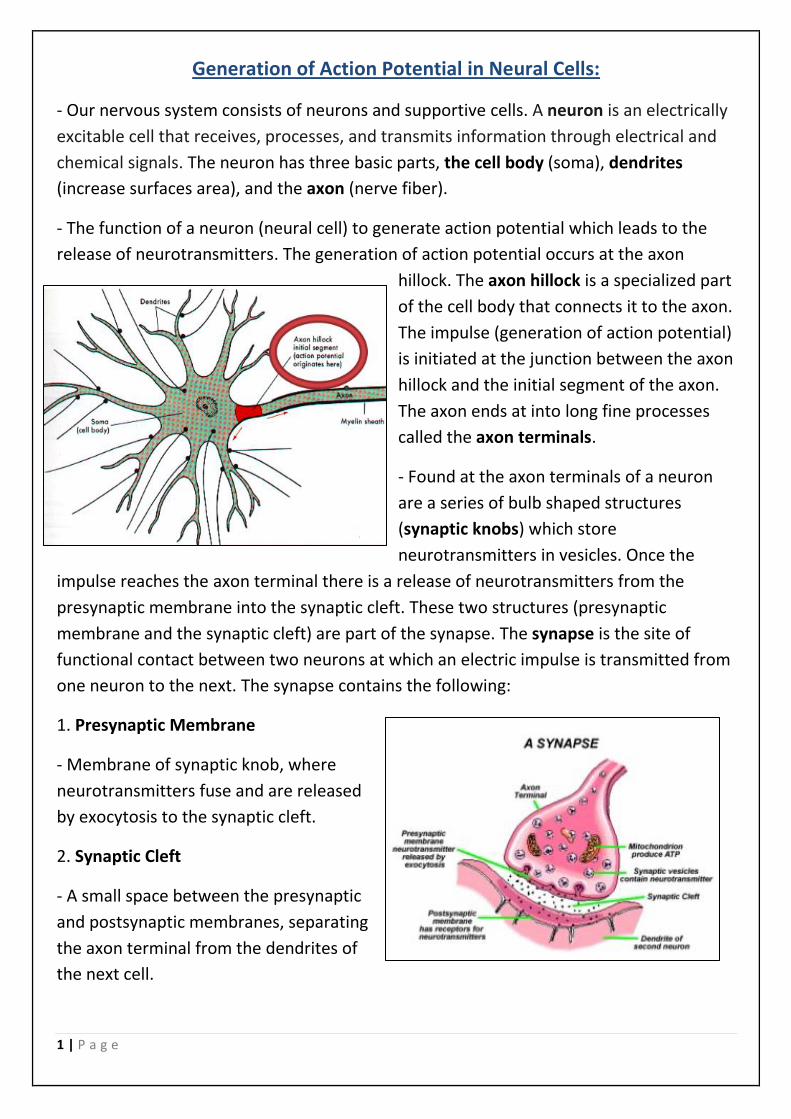
- The function of a neuron (neural cell) to generate action potential which leads to the
release of neurotransmitters. The generation of action potential occurs at the axon
hillock. The axon hillock is a specialized part
of the cell body that connects it to the axon.
The impulse (generation of action potential)
is initiated at the junction between the axon
hillock and the initial segment of the axon.
The axon ends at into long fine processes
called the axon terminals.
- Found at the axon terminals of a neuron
are a series of bulb shaped structures
(synaptic knobs) which store
neurotransmitters in vesicles. Once the
impulse reaches the axon terminal there is a release of neurotransmitters from the
presynaptic membrane into the synaptic cleft. These two structures (presynaptic
membrane and the synaptic cleft) are part of the synapse. The synapse is the site of
functional contact between two neurons at which an electric impulse is transmitted from
one neuron to the next. The synapse contains the following:
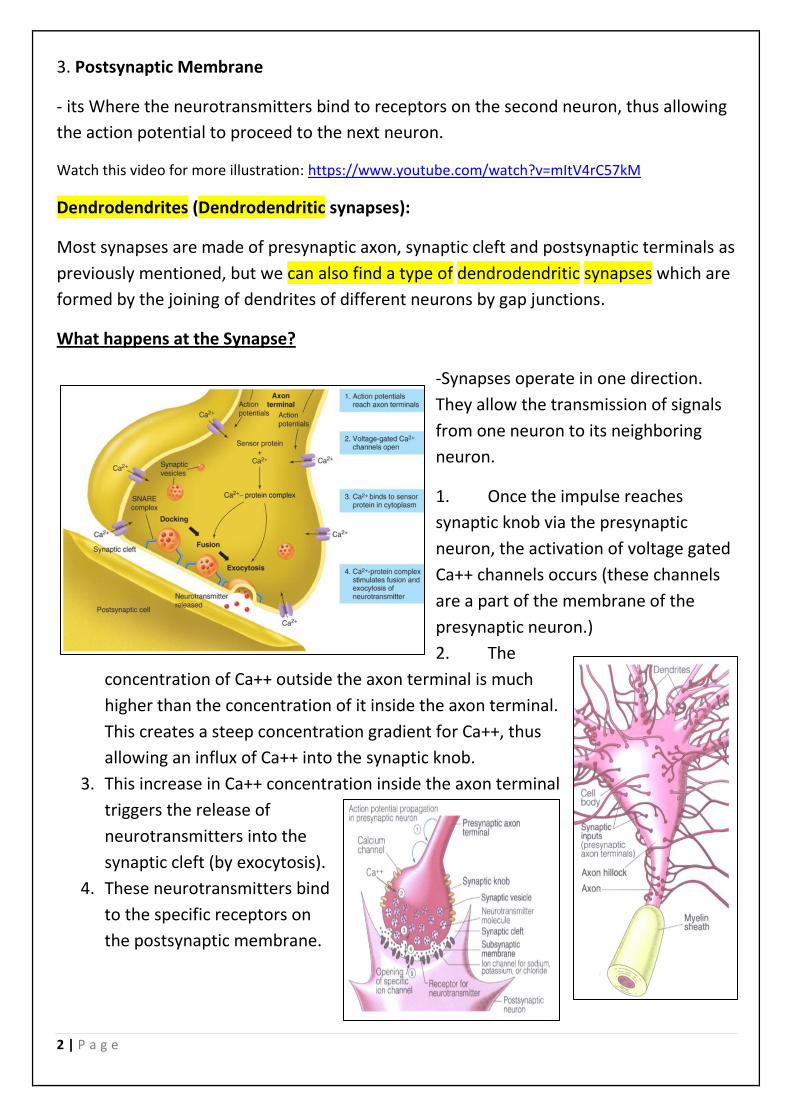
1. Presynaptic Membrane
- Membrane of synaptic knob, where
neurotransmitters fuse and are released
by exocytosis to the synaptic cleft.
2. Synaptic Cleft
- A small space between the presynaptic
and postsynaptic membranes, separating
the axon terminal from the dendrites of
the next cell.
2 | P a g e
3. Postsynaptic Membrane
- its Where the neurotransmitters bind to receptors on the second neuron, thus allowing
the action potential to proceed to the next neuron.
Watch this video for more illustration: https://www.youtube.com/watch?v=mItV4rC57kM
Dendrodendrites (Dendrodendritic synapses):
Most synapses are made of presynaptic axon, synaptic cleft and postsynaptic terminals as
previously mentioned, but we can also find a type of dendrodendritic synapses which are
formed by the joining of dendrites of different neurons by gap junctions.
What happens at the Synapse?
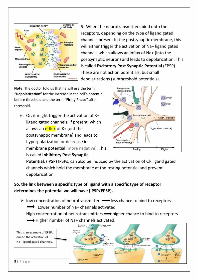
-Synapses operate in one direction.
They allow the transmission of signals
from one neuron to its neighboring
neuron.
1. Once the impulse reaches
synaptic knob via the presynaptic
neuron, the activation of voltage gated
Ca++ channels occurs (these channels
are a part of the membrane of the
presynaptic neuron.)
2. The
concentration of Ca++ outside the axon terminal is much
higher than the concentration of it inside the axon terminal.
This creates a steep concentration gradient for Ca++, thus
allowing an influx of Ca++ into the synaptic knob.
3. This increase in Ca++ concentration inside the axon terminal
triggers the release of
neurotransmitters into the
synaptic cleft (by exocytosis).
4. These neurotransmitters bind
to the specific receptors on
the postsynaptic membrane.
3 | P a g e
5. When the neurotransmitters bind onto the
receptors, depending on the type of ligand gated
channels present in the postsynaptic membrane, this
will either trigger the activation of Na+ ligand gated
channels which allows an influx of Na+ (into the
postsynaptic neuron) and leads to depolarization. This
is called Excitatory Post Synaptic Potential (EPSP).
These are not action potentials, but small
depolarizations (subthreshold potentials).
Note: The doctor told us that he will use the term
“Depolarization” for the increase in the cell’s potential
before threshold and the term “Firing Phase” after
threshold.
6. Or, it might trigger the activation of K+
ligand gated channels, if present, which
allows an efflux of K+ (out the
postsynaptic membrane) and leads to
hyperpolarization or decrease in
membrane potential (more negative). This
is called Inhibitory Post Synaptic
Potential. (IPSP) IPSPs, can also be induced by the activation of Cl- ligand gated
channels which hold the membrane at the resting potential and prevent
depolarization.
So, the link between a specific type of ligand with a specific type of receptor
determines the potential we will have (IPSP/EPSP).
➢ low concentration of neurotransmitters less chance to bind to receptors
Lower number of Na+ channels activated.
High concentration of neurotransmitters higher chance to bind to receptors
Higher number of Na+ channels activated.
This is an example of EPSP,
due to the activation of
Na+ ligand gated channels.
4 | P a g e
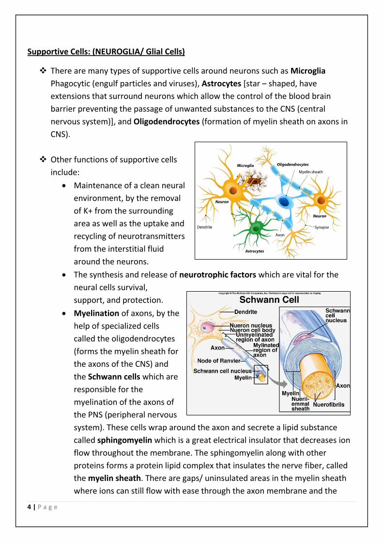
Supportive Cells: (NEUROGLIA/ Glial Cells)
❖ There are many types of supportive cells around neurons such as Microglia
Phagocytic (engulf particles and viruses), Astrocytes [star – shaped, have
extensions that surround neurons which allow the control of the blood brain
barrier preventing the passage of unwanted substances to the CNS (central
nervous system)], and Oligodendrocytes (formation of myelin sheath on axons in
CNS).
❖ Other functions of supportive cells
include:
• Maintenance of a clean neural
environment, by the removal
of K+ from the surrounding
area as well as the uptake and
recycling of neurotransmitters
from the interstitial fluid
around the neurons.
• The synthesis and release of neurotrophic factors which are vital for the
neural cells survival,
support, and protection.
• Myelination of axons, by the
help of specialized cells
called the oligodendrocytes
(forms the myelin sheath for
the axons of the CNS) and
the Schwann cells which are
responsible for the
myelination of the axons of
the PNS (peripheral nervous
system). These cells wrap around the axon and secrete a lipid substance
called sphingomyelin which is a great electrical insulator that decreases ion
flow throughout the membrane. The sphingomyelin along with other
proteins forms a protein lipid complex that insulates the nerve fiber, called
the myelin sheath. There are gaps/ uninsulated areas in the myelin sheath
where ions can still flow with ease through the axon membrane and the
5 | P a g e
intracellular fluid inside the axon. These gaps are called Nodes of Ranvier
and are used for transmission of impulses along a myelinated nerve
(generation of action potential).
❖ Many diseases are related to the destruction of the supportive cells rather than the
neural cells.
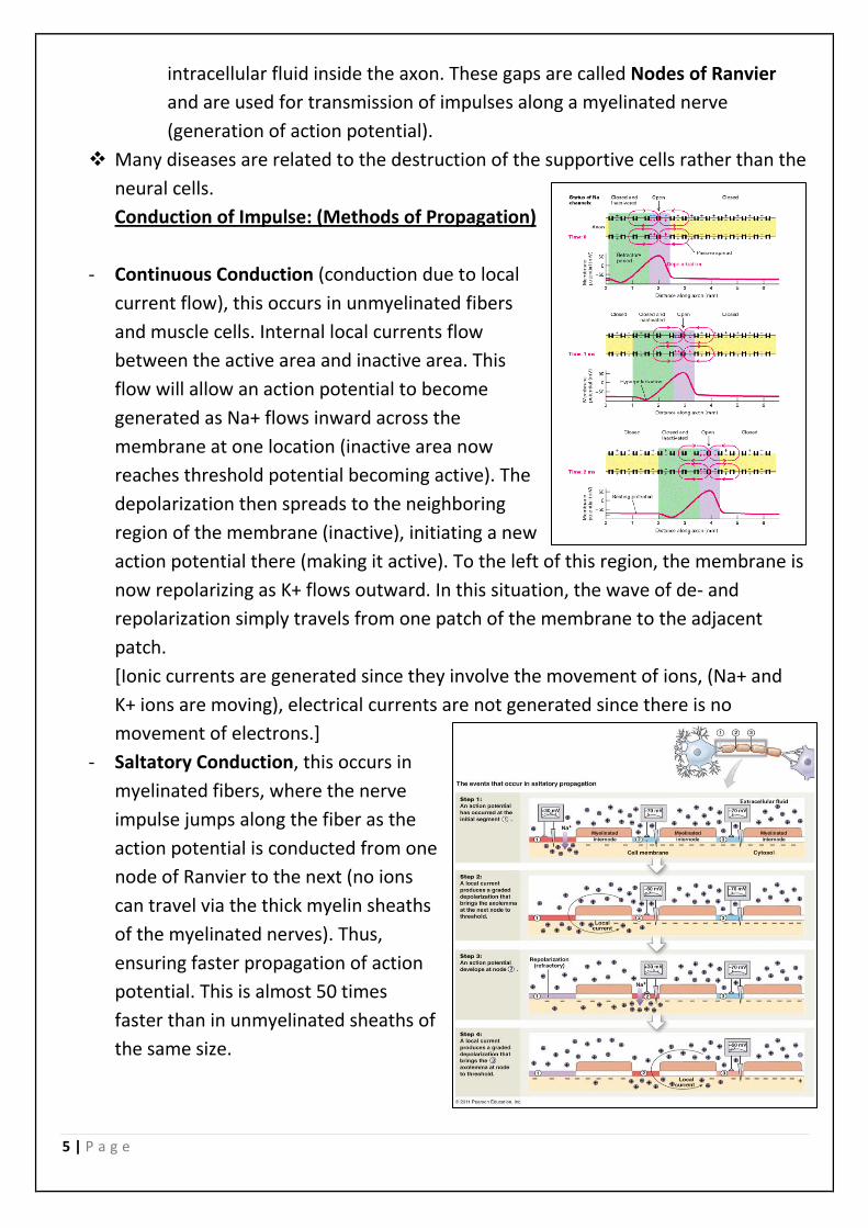
Conduction of Impulse: (Methods of Propagation)
- Continuous Conduction (conduction due to local
current flow), this occurs in unmyelinated fibers
and muscle cells. Internal local currents flow
between the active area and inactive area. This
flow will allow an action potential to become
generated as Na+ flows inward across the
membrane at one location (inactive area now
reaches threshold potential becoming active). The
depolarization then spreads to the neighboring
region of the membrane (inactive), initiating a new
action potential there (making it active). To the left of this region, the membrane is
now repolarizing as K+ flows outward. In this situation, the wave of de- and
repolarization simply travels from one patch of the membrane to the adjacent
patch.
[Ionic currents are generated since they involve the movement of ions, (Na+ and
K+ ions are moving), electrical currents are not generated since there is no
movement of electrons.]
- Saltatory Conduction, this occurs in
myelinated fibers, where the nerve
impulse jumps along the fiber as the
action potential is conducted from one
node of Ranvier to the next (no ions
can travel via the thick myelin sheaths
of the myelinated nerves). Thus,
ensuring faster propagation of action
potential. This is almost 50 times
faster than in unmyelinated sheaths of
the same size.
6 | P a g e
Factors influencing the rate of
conductance:
1- Myelination (If present, this increases the rate of conductance.)
2- Diameter of nerve fibers (Larger diameter, less resistance, faster
depolarization, higher velocity of conductance.)
Importance of Refractory Period:
o Ensures that the propagation of action potential happens in one direction
[Unidirectional, one-way]. For example, after action potential is propagated from
point A to point B in the picture below, point A starts repolarizing, preventing any
new action potential (relative refractory period). This assures the movement of the
action potential in one direction, allows the neuron to adjust briefly for the
propagation of the next stimulus, and limits the amount of action potentials sent
per minute.)
o If an impulse is initiated
at the cell body of a
neuron as well as at the
axon terminal
simultaneously, there will be no surpassing of the impulse due to the presence of
the refractory period.
For better understanding watch:
o 1.https://www.youtube.com/watch?v=Sa1wM750Rvs
o 2.https://www.youtube.com/watch?v=3SV1DpO7XvY
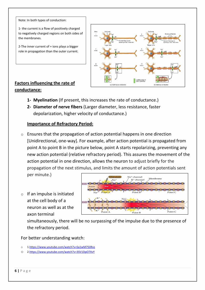
Note: In both types of conduction:
1- the current is a flow of positively charged
to negatively charged regions on both sides of
the membranes.
2-The inner current of + ions plays a bigger
role in propagation than the outer current.
7 | P a g e
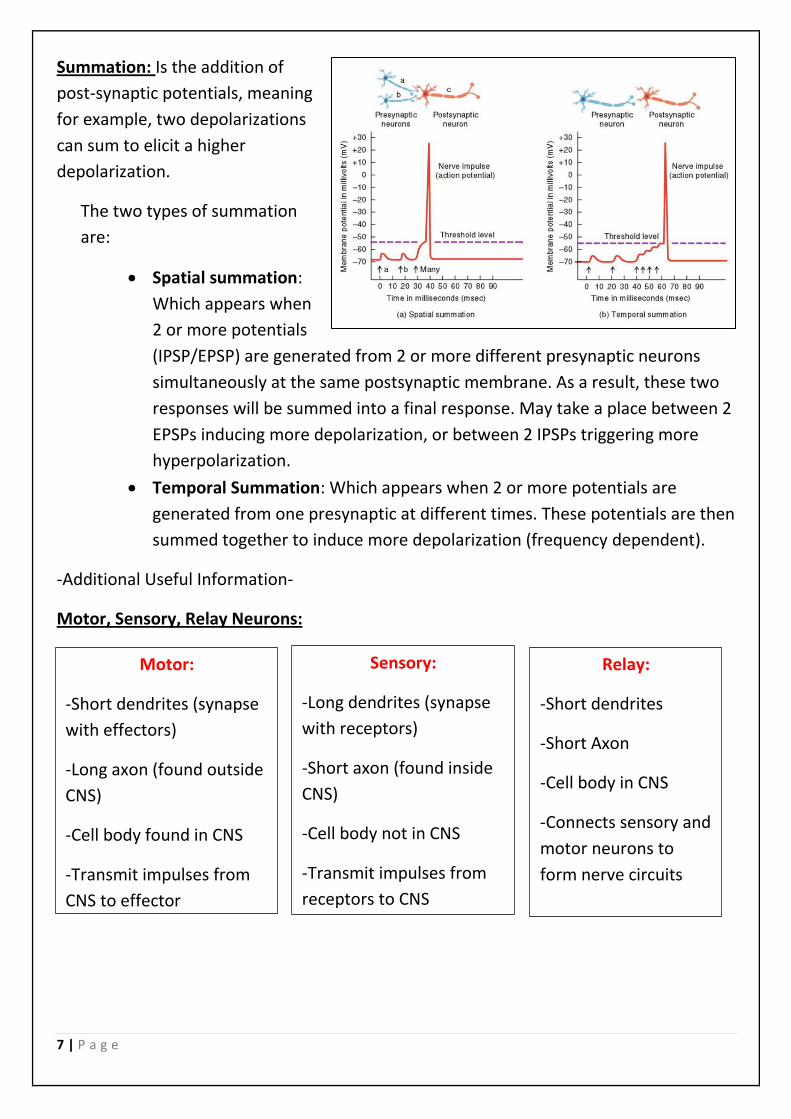
Summation: Is the addition of
post-synaptic potentials, meaning
for example, two depolarizations
can sum to elicit a higher
depolarization.
The two types of summation
are:
• Spatial summation:
Which appears when
2 or more potentials
(IPSP/EPSP) are generated from 2 or more different presynaptic neurons
simultaneously at the same postsynaptic membrane. As a result, these two
responses will be summed into a final response. May take a place between 2
EPSPs inducing more depolarization, or between 2 IPSPs triggering more
hyperpolarization.
• Temporal Summation: Which appears when 2 or more potentials are
generated from one presynaptic at different times. These potentials are then
summed together to induce more depolarization (frequency dependent).
-Additional Useful Information-
Motor, Sensory, Relay Neurons:
Motor:
-Short dendrites (synapse
with effectors)
-Long axon (found outside
CNS)
-Cell body found in CNS
-Transmit impulses from
CNS to effector
Sensory:
-Long dendrites (synapse
with receptors)
-Short axon (found inside
CNS)
-Cell body not in CNS
-Transmit impulses from
receptors to CNS
Relay:
-Short dendrites
-Short Axon
-Cell body in CNS
-Connects sensory and
motor neurons to
form nerve circuits
8 | P a g e
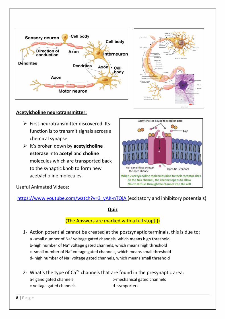
Acetylcholine neurotransmitter:
➢ First neurotransmitter discovered. Its
function is to transmit signals across a
chemical synapse.
➢ It’s broken down by acetylcholine
esterase into acetyl and choline
molecules which are transported back
to the synaptic knob to form new
acetylcholine molecules.
Useful Animated Videos:
https://www.youtube.com/watch?v=3_yAK-nTOjA (excitatory and inhibitory potentials)
Quiz
(The Answers are marked with a full stop[.])
1- Action potential cannot be created at the postsynaptic terminals, this is due to: a -small number of Na+ voltage gated channels, which means high threshold.
b-high number of Na+ voltage gated channels, which means high threshold
c- small number of Na+ voltage gated channels, which means small threshold
d- high number of Na+ voltage gated channels, which means small threshold
2- What's the type of Ca2+ channels that are found in the presynaptic area:
a-ligand gated channels b-mechanical gated channels
c-voltage gated channels. d- symporters
9 | P a g e
3- Acetylcholine is an excitatory transmitter, what happens if we increase its
concentration in the synaptic cleft?
a-more probability of binding with postsynaptic receptors causing an inhibitory potential
b-more probability of binding with postsynaptic receptors causing an excitatory potential
c- more probability of causing an action potential on the second neuron (postsynaptic neuron)
d- both b and c.
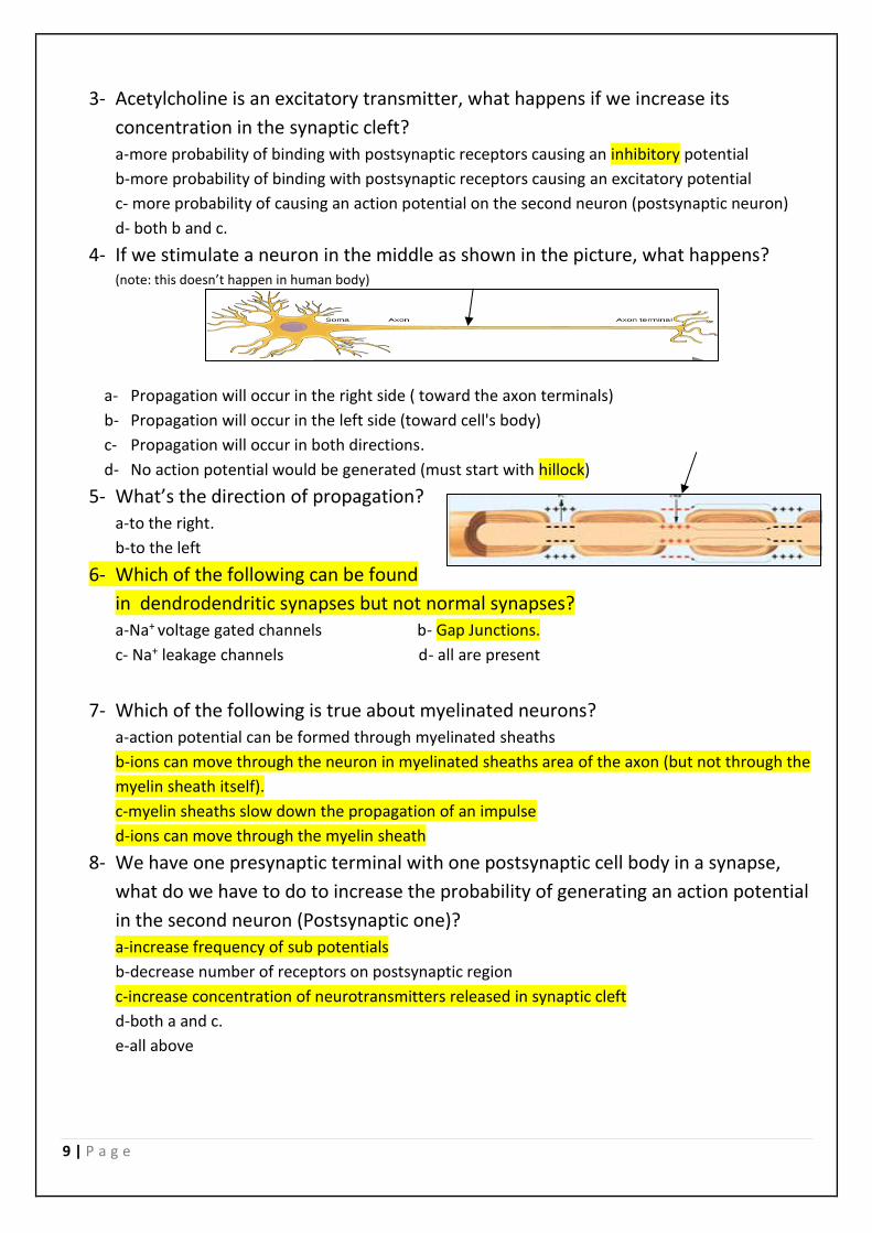
4- If we stimulate a neuron in the middle as shown in the picture, what happens? (note: this doesn’t happen in human body)
a- Propagation will occur in the right side ( toward the axon terminals)
b- Propagation will occur in the left side (toward cell's body)
c- Propagation will occur in both directions.
d- No action potential would be generated (must start with hillock)
5- What’s the direction of propagation?
a-to the right.
b-to the left
6- Which of the following can be found
in dendrodendritic synapses but not normal synapses?
a-Na+ voltage gated channels b- Gap Junctions.
c- Na+ leakage channels d- all are present
7- Which of the following is true about myelinated neurons?
a-action potential can be formed through myelinated sheaths
b-ions can move through the neuron in myelinated sheaths area of the axon (but not through the
myelin sheath itself).
c-myelin sheaths slow down the propagation of an impulse
d-ions can move through the myelin sheath
8- We have one presynaptic terminal with one postsynaptic cell body in a synapse,
what do we have to do to increase the probability of generating an action potential
in the second neuron (Postsynaptic one)?
a-increase frequency of sub potentials
b-decrease number of receptors on postsynaptic region
c-increase concentration of neurotransmitters released in synaptic cleft
d-both a and c.
e-all above
![Mohammad Azadi, Ph.D. - Semnan Universityprofs.semnan.ac.ir/FilesContainer/Professors/Mohammad Azadi... · Mohammad Azadi, Ph.D. sPage ]txep TpyT[Mohammad Azadi, Ph.D. Scientific](https://img.pdfslide.net/doc/110x75/5b6bec257f8b9a8d058de3ad/mohammad-azadi-phd-semnan-azadi-mohammad-azadi-phd-spage-txep-tpytmohammad.jpg)