Embed Size (px)
Citation preview
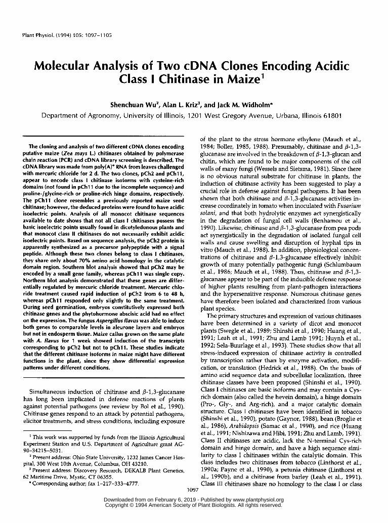
Plant Physiol. (1994) 105: 1097-1105
Molecular Analysis of Two cDNA Clones Encoding Acidic Class I Chitinase in Maize'
Shenchuan Wu', Alan 1. Kriz3, and Jack M. Widholm*
Department of Agronomy, University of Illinois, 1201 West Gregory Avenue, Urbana, lllinois 61 801
The cloning and analysis of two different cDNA clones encoding putative maize (Zea mays 1.) chitinases obtained by polymerase chain reaction (PCR) and cDNA library screening i s described. The cDNA library was made from poly(A)+ RNA from leaves challenged with mercuric chloride for 2 d. l h e two clones, pCh2 and pCh11, appear to encode class I chitinase isoforms with cysteine-rich domains (not found in pChl1 due to the incomplete sequence) and proline-lglycine-rich or proline-rich hinge domains, respedively. l h e pCh11 clone resembles a previously reported maize seed chitinase; however, the deduced proteins were found to have acidic isoeledric points. Analysis of all monocot chitinase sequences available to date shows that not all class I chitinases possess the basic isoelectric points usually found in dicotyledonous plants and that monocot class II chitinases do not necessarily exhibit acidic isoelectric points. Based on sequence analysis, the pCh2 protein i s apparently synthesized as a precursor polypeptide with a signal peptide. Although these two clones belong to class I chitinases, they share only about 70% amino acid homology in the catalytic domain region. Southern blot analysis showed that pCh2 may be encoded by a small gene family, whereas pChl1 was single copy. Northern blot analysis demonstrated that these genes are differ- entially regulated by mercuric chloride treatment. Mercuric chlo- ride treatment caused rapid induction of pCh2 from 6 to 48 h, whereas pCh11 responded only slightly to the same treatment. During seed germination, embryos constitutively expressed both chitinase genes and the phytohormone abscisic acid had no effed on the expression. The fungus Aspergillus flavus was able to induce both genes to comparable levels in aleurone layers and embryos but not in endosperm tissue. Maize callus grown on the same plate with A. flavus for 1 week showed induction of the transcripts corresponding to pCh2 but not to pChll. These studies indicate that the different chitinase isoforms in maize might have different fundions in the plant, since they show differential expression patterns under different conditions.
Simultaneous induction of chitinase and @-1,3-glucanase has long been implicated in defense reactions of plants against potential pathogens (see review by Bol et al., 1990). Chitinase genes respond to an attack by potential pathogens, elicitor treatments, and stress conditions, including exposure
' This work was supported by funds from the Illinois Agricultura1 Experiment Station and U.S. Department of Agriculture grant AG-
Present address: Ohio State University, 1232 James Cancer Hos-
Present address: Discovery Research, DEKALB Plant Genetics,
90-34215-5031.
pital, 300 West 10th Avenue, Columbus, OH 43210.
62 Maritime Drive, Mystic, CT 06355. * Corresponding author; fax 1-217-333-4777.
of the plant to the stress hormone ethylene (Mauch et al., 1984; Boller, 1985, 1988). Presumably, chitinase and @-1,3- glucanase are involved in the breakdown of @-1,3-glucan and chitin, which are found to be major components of the cell walls of many fungi (Wessels and Sietsma, 1981). Since there is no obvious natural substrate for chitinase in plants, the induction of chitinase activity has been suggested to play a crucial role in defense against fungal pathogens. It has been shown that both chitinase and @-1,3-glucanase activities in- crease coordinately in tomato when inoculated with Fusarium solani, and that both hydrolytic enzymes act synergistically in the degradation of fungal cell walls (Benhamou et al., 1990). Likewise, chitinase and @-l,S-glucanase from pea pods act synergistically in the degradation of isolated fungal cell walls and cause swelling and disruption of hyphal tips in vitro (Mauch et al., 1988). In addition, physiological concen- trations of chitinase and @-1,3-glucanase effectively inhibit growth of many potentially pathogenic fungi (Schlumbaum et al., 1986; Mauch et al., 1988). Thus, chitinase and @-1,3- glucanase appear to be part of the inducible defense response of higher plants resulting from plant-pathogen interactions and the hypersensitive response. Numerous chitinase genes have therefore been isolated and characterized from various plant species.
The primary structures and expression of various chitinases have been detennined in a variety of dicot and monocot plants (Swegle et al., 1989; Shinshi et al., 1990; Huang et al., 1991; Leah et al., 1991; Zhu and Lamb 1991; Huynh et al., 1992; Sela-Buurlage et al., 1993). These studies show that a11 stress-induced expression of chitinase activity is controlled by transcription rather than by enzyme activation, modifi- cation, or translation (Hedrick et al., 1988). On the basis of amino acid sequence data and subcellular localization, three chitinase classes have been proposed (Shinshi et al., 1990). Class I chitinases are basic isoforms and may contain a Cys- rich domain (also called the hevein domain), a hinge domain (Pro-, Gly-, and Arg-rich), and a major catalytic domain structure. Class I chitinases have been identified in tobacco (Shinshi et al., 1990), potato (Gaynor, 1988), bean (Broglie et al., 1986), Arabidopsis (Samac et al., 1990), and rice (Huang et al., 1991; Nishizawa and Hibi, 1991; Zhu and Lamb, 1991). Class I1 chitinases are acidic, lack the N-terminal Cys-rich domain and hinge domain, and have a high sequence simi- larity to class I chitinases within the catalytic domain. This class includes two chitinases from tobacco (Linthorst et al., 1990a; Payne et al., 1990), a petunia chitinase (Linthorst et al., 1990b), and a chitinase from barley (Leah et al., 1991). Class I11 chitinases share no homology to the class I or class
1097
www.plantphysiol.orgon February 6, 2019 - Published by Downloaded from Copyright © 1994 American Society of Plant Biologists. All rights reserved.

1098 Wu et al. Plant Physiol. Vol. 105, 1994
I1 enzymes, but are homologous to the acidic chitinases of cucumber (Bemasconi et al., 1987; Metraux et al., 1989), Arabidopsis (Samac et al., 1990), and tobacco (Lawton et al., 1992). However, there is an exception to the proposed clas- sification system: an acidic chitinase has been isolated from bean that has a Cys-rich domain (Margis-Pinheiro et al., 1991). This classification system was based on sequence information solely from tobacco and other dicotyledonous plants before any detailed studies of chitinases from mono- cots were available.
The antifungal activity of chitinase makes this protein an attractive candidate for overexpression to produce disease- resistant agriculturally important crop plants, as demon- strated by Broglie et al. (1991) with tobacco plants. Although the structure, expression, subcellular localization, and anti- funga1 activity of a number of plant chitinases have been studied, the pathogen specificity, biochemical properties, and roles of chitinases in plant defense remain to be further investigated. Previously, Huynh et al. (1992) cloned and determined the antifungal properties of two chitinases from maize (Zea mays L.) seeds and presented the amino acid sequence of a third chitinase, CHITD. In this paper, we report the isolation of two cDNA clones of putative class I chitinase isoforms from maize seedlings that had been treated with mercuric chloride. Although both are apparently class I chi- tinases, pCh2 belongs to a larger gene family than does pChl1, and their regulation pattems were dramatically dif- ferent in response to mercuric chloride and to challenge by the fungus Aspergillus flavus.
MATERIALS AND METHODS
Plant Materiais and Treatments
Maize (Zea mays L.) plants were grown in trays in a growth chamber with a 16-h photoperiod with 28OC days and 18OC nights. Seven-day-old seedlings of the maize inbred line Va26 were sprayed with a solution containing either 1 mg/mL ethephon, 1 mg/mL salicylic acid, or 0.2% mercuric chloride. Pots were then covered with plastic bags and seedlings were harvested between 6 and 48 h after the chemical treatment. Control plants did not receive any chemical treatment but were covered with plastic bags. Harvested tissues were frozen in liquid N1 and stored at -7OOC.
To obtain germinating maize embryos, the kemels were allowed to imbibe for 2 h in water either with or without 100 PM ABA and were then placed on filter paper saturated with the same solution in glass Petri dishes. Embryos were excised from the kemels after imbibition and after 1, 2, or 3 d and were immediately frozen in liquid N2 and stored at -7OOC.
Field-grown plants of the maize F, hybrid 873 X Mo17 were used for analysis of gene expression pattems in devel- oping kemels. Ears were inoculated with Aspergillus flavus 20 to 24 d after midsilking by using a modified pinboard inoculation technique (Calvert et al., 1978). In the center of the pin array was a 16-gauge hypodermic needle through which 5 mL of a spore suspension, consisting of 2 x 10⁵ conidia/mL, was injected through the husk. The inoculator was aligned with the ear axis and the pins were pushed through the husk and into the kemels. The inoculum was a
mixture of four isolates prepared from lyophilized cultures of A. flavus (Northern Regional Research Laboratory [Peoria, IL] isolates 6536,6539, and 6540 anda 1988 isolate from Illinois). The damaged controls were treated the same as inoculated ears except that no inoculum was included in the inoculator. Kemels were harvested at 9 to 23 d after inociilation and manual1 y separated into aleurone plus pericarp, endosperm (minus rnost of the aleurone), and embryos. A11 tissues were immedialtely frozen in liquid nitrogen and stored at -7OOC.
For in vitro studies, regenerable callus cultures were initi- ated froin immature embryos of Mo17 x LB31 2nd LB31 x Mo17, xrtaintained in the dark at 28OC on D medium (Duncan et al., 1985), and subcultured at 14-d intervals. Callus pieces (about 4 mm in diameter) were placed near the etlge of a 10- cm Petn dish and A. flavus spores were then inoculated in the center. Tissue was harvested after 1 week of incubation, before the fungus had grown over the callus, and frozen in liquid h12. Calli were then stored at -7OOC kefore RNA isolation.
Nucleic Acid lsolation and Cel Blot Analysis
Total IRNA was isolated from frozen tissue as previously described by using a guanidine-HC1 method (Bclanger and Kriz, 1989). For northem blot analysis, 10 Pg of total RNA was subjected to electrophoresis in formaldehyde-agarose gels and transferred to nylon membranes (Magnagraph, Mi- cron Separations, Inc., Westborough, MA). Isolation of maize genomic DNA from leaves of 7-d-old seedlings and Southem blot analysis were performed as described (Belangcr and f i z , 1989). For both RNA and DNA blots, the transfeired nucleic acids were UV linked to the membrane by using a IGtratalinker 1800 apparatus (Stratagene). The blots were hybridized over- night at 42OC using 50% formamide, 5X SSC, l x Denhardt's solution, 20 mM sodium phosphate (pH 6.8), 0.156 SDS, and 5% dexbran sulfate. The filters were washed with 2 X SSPE (360 mM NaCl, 20 mM sodium phosphate, 50 mM EDTA, pH 7.4) and 0.5% SDS once at room temperature and once at 68OC for 15 min each time. Then the filters were washed for another 15 min at 68OC with 0.2X SSPE and 0.1% SDS. The Tm for hybridization of the pChl l clone was calculated according to the following equation: Tm = 81.5OC - 16.6(log[Na']) + 0.41(% G + C) - 0.63(% formanude) (Sam- brook et al., 1989). This corresponds to hybridization condi- tions of Tm - 31 in 5X SSC, Tm - 34 in 2X SSPE (first wash), and Tm - 8 in 0.2X SSPE (final wash). fbsuming a decrease in Tm of l0C for 1% mismatch between C h l l and Ch2, the Tm of the hybrid is estimated at 4OoC, so the two clones would not be expected to cross-hybridize under these conditions. Experiments showed that there was less than 5% cross-hybridization between pCh2 and pChl1. The filters were exposed to x-ray film at -7OOC.
For use in hybridizations, the cloned cDNA fragments were isolated from the plasmid by EcoRI digestion, electrophoresis in a 0.8'% agarose gel, and use of the GeneClem I1 kit as recommended by the manufacturer (Bio 101, Inc., La Jolla, CA). The isolated fragment was labeled with [(Y-~'P]~ATP by using a commercial kit (Stratagene) employirtg random primers ilnd T7 DNA polymerase.
www.plantphysiol.orgon February 6, 2019 - Published by Downloaded from Copyright © 1994 American Society of Plant Biologists. All rights reserved.

Two Maize cDNA Clones Encoding Acidic Class I Chitinases 1 o99
PCR Amplification and Cloning
Amplification of 0.5 r g of genomic DNA was camed out in a 50-pL reaction mixture that contained Ta9 DNA polym- erase buffer supplemented with MgC12 to a final concentra- tion of 1.5 mM, 100 PM of each dNTP, 0.2 pg of each degenerate primer, and 2.5 units of Ta9 DNA polymerase (BRL). The mixture was cycled 35 times in a Perkin-Elmer Cetus DNA Thermal Cycler as follows: 94OC for 1 min; 37OC for 1 min; and 72OC for 2.5 min; with a final 72OC extension of 7 min. The primers were designed according to the con- served chitinase nucleotide sequences from barley (Leah et al., 1991) and tobacco (Shinshi et al., 1988). The upstream degenerate primer was AAA/GGGNTTT/CTAT/CACNTA, of which the 5’ nucleotide corresponds to nucleotide 207 in the sequence of barley chitinase; the downstream degenerate primer was TGGTTT/CTGGATGACN, of which the 3’ nu- cleotide corresponds to nucleotide 612 in the sequence of barley chitinase (Leah et al., 1991). Following amplification, PCR products were analyzed by gel electrophoresis.
Screening of a Maize Leaf cDNA Library
A X ZAP I1 cDNA library was made from poly(A)+ RNA isolated from maize seedlings 48 h after spraying with 0.2% HgC12. The procedure used was previously described by Belanger and Kriz (1989). Screening of the library with the cloned, PCR-amplified maize chitinase (TA98) fragment as radiolabeled probe was performed under the conditions used in the Southem blots described above. Fifteen plaques con- tinued to yield positive hybridization signals after the third round of screening, and four cDNA inserts of more than 1 kb were recovered from these clones as the recombinant pBluescript plasmid according to the manufacturer’s protocols.
DNA Sequencing
Both strands of the chitinase cDNA fragment in the Blue- script vector were sequenced using a combination of com- mercial vector-specific primers and custom-designed oligo- nucleotides. Dideoxynucleotide sequence analysis (Sanger et al., 1977) of denatured double-stranded DNA templates or single-stranded DNA prepared from M13 templates (Yan- isch-Perron et al., 1985) was performed with a modified T7 DNA polymerase (Sequenase; United States Biochemical Corp.). The DNA and deduced amino acid sequences were analyzed and assembled with the aid of MacVector computer software (IBI, New Haven, CT).
RESULTS
Amplification and lsolation of a Chitinase Cenomic Sequence Fragment
A strategy utilizing the PCR was employed to amplify and isolate maize chitinase cDNA clones. The amino acid se- quence of the barley protein (Leah et al., 1991) was compared with that of rice (Huang et al., 1991) to identify consensus sequences from which a pair of degenerate oligonucleotide primers were designed. Severa1 products were detected in the
PCR, which ranged from about 300 to 700 bp on ethidium bromide-stained agarose gels (data not shown). The DNA bands in the agarose gel corresponding to about 400 bp were purified and ligated to the dT-tailed EcoRV site of pBluescript SK. The resultant clones were sequenced and five were found to be homologous to chitinases from barley and rice. One of the PCR-derived clones (TA98) was then used to screen a maize cDNA library.
lsolation and Sequence Analysis of Two Class I Chitinase cDNA Clones
A X ZAP I1 cDNA library was screened with the 400-bp fragment of the TA98 chitinase clone. Approximately 250,000 plaques were screened at high stringency and 15 clones were hybridized to the PCR TA98 insert after three rounds of screening. Following plaque purification and in vivo excision of the pBluescript recombinant plasmid from the X ZAP 11, the cloned inserts were confirmed by Southem blot analysis. The four clones larger than 1 kb were sequenced, and two of the chitinase cDNA clones representing two different types of chitinases are described here.
The complete sequences of the two cDNA clones were determined. A full-length clone, designated pCh2, consisted of 1128 nucleotides with a 957-nucleotide open reading frame, which would encode a polypeptide of about 33.5 kD. This clone has 27 nucleotides in the 5‘ untranslated region and 144 nucleotides in the 3’ untranslated region. One pu- tative polyadenylation (AATAAA) signal is located at nucle- otides 1105 to 1110. The pChl l clone, which is not full length, since part of the 5’ end is lacking, consists of 1000 nucleotides with 173 nucleotides in the 3’ untranslated re- gion. Two putative polyadenylation (AATAAA) signals are located at nucleotides 944 to 949 and 965 to 970, respectively. The cDNAs have different stop codons, i.e. TAG for pCh2 and TAA for pChl l . These cDNAs share about 70% identity at the nucleotide sequence level.
Primary Structures of Maize Chitinases and Homology to Other Chitinases
The deduced amino acid sequences of pCh2 and pChll are presented and compared to those of other chitinase genes in Figure 1. Both chitinase clone open reading frames exhibit strong codon bias (89 and 87%, respectively) for G or C in their third codon position, as is common for other nuclear- encoded maize genes (Campbell and Gowri, 1990). The pCh2 polypeptide contains a hydrophobic putative signal peptide of 21 amino acids at the N terminus, as well as hevein and catalytic domains. The preprotein consists of 319 amino acids. The primary structure of the pChll gene product is not complete, since the clone is not a full-length cDNA, so the clone lacks a signal peptide and the Cys-rich domain, as expected for a class I chitinase, but it does have a hinge region that is Pro-rich. The polypeptide encoded by this clone resembles that from the seed chitinase gene CHITD reported by Huynh et al. (1992). The deduced amino acid sequences of pChll and CHITD exhibit more than 90% identity (Fig. 1). Since CHITD, a class I chitinase, and pChl l are so homologous, pChl l would also appear to encode a class I chitinase.
www.plantphysiol.orgon February 6, 2019 - Published by Downloaded from Copyright © 1994 American Society of Plant Biologists. All rights reserved.
1100 Wu et al. Plant Physiol. Vol. 105, 1994
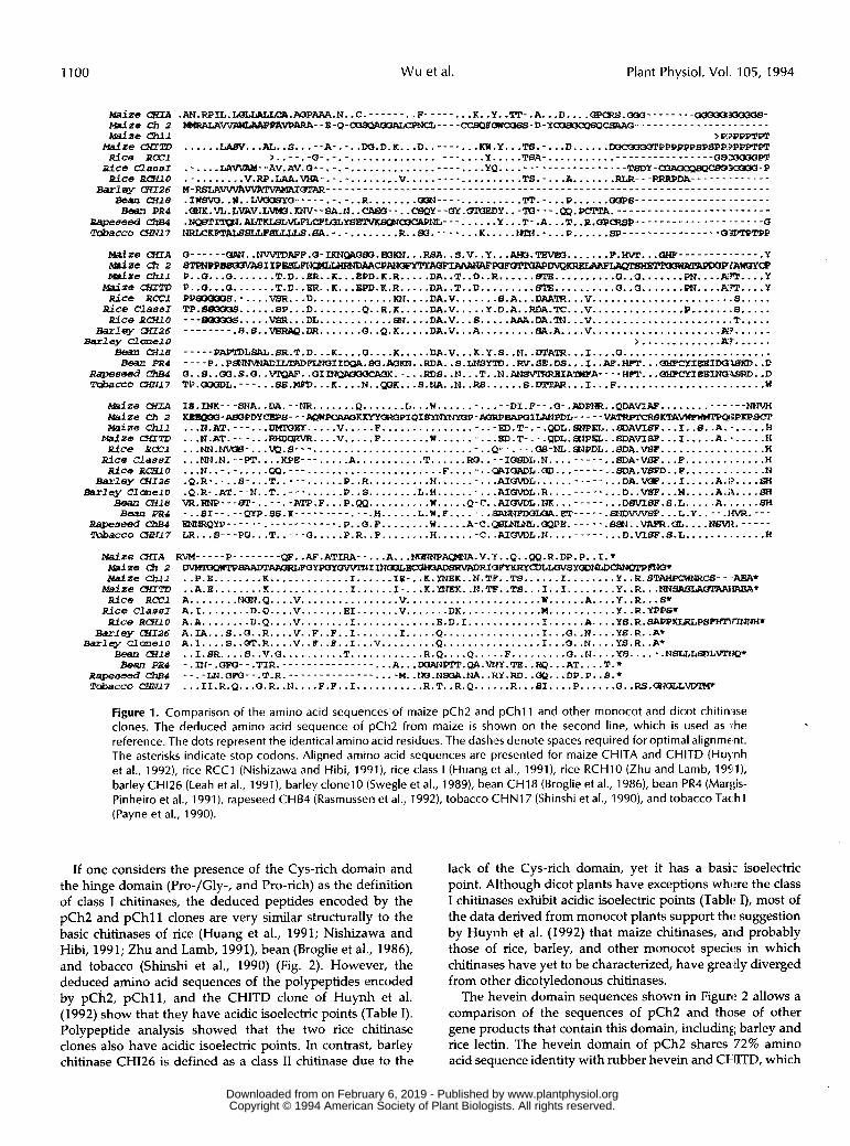
.AN.RPIL.I&LLALLCA.AoPAAR.IJ. .C. ........ F- ....... K. .Y. -"I'- .A. . .D.. . .-.M- ...... --y1ooo6- > R?PPPT€T
......LABV...AL.. S ..... A... .. W.D.K ... D. ........KW. Y. ..T8..... D...... M3CaK16PPPPPPSPSPP".PP. Qb" , . _- , -0- - - - _ - _ _ - _ _ - - - _ - _ _ _ _ _ - . ................... Y ..... TBA..... .......
.-....Um-- AV.AV.G....... ................. - _ - - YQ ..................... T8DY -Q3AacQ84cw33WUQ-P .......... V. Rp.LAA.vtIA..... ........ V ---.pRRpDA------.------ ................... TS.-...A-...... . . . . . . . . . . . . . . . . . . . . . . . . . . . . . . . . . . . . . . . . . . . . . . . . . . . . . . . . . . . . . . . . . . . . . . . . . . . . . . . .. .... aOPB---------------.------ .IWBM W..L.WCISYd........ R ........ ..... ..........'Tp..... P......
.~K.VL.L~V.LVM3.KNV--SA.N..CAGCI--..CBQY--(SY.(9mEDY..-~---.QP.PCTPA.-------------------------
.I...ALTF......... ...... Y...T..A...T..R.QP.P..................... Q ~ ~ K ~ ~ ~ S L L L L S .BA... . ........ R . . 8 0 . . . . . . . . K.... ..ZPN..... P......8P................ a"P
Q- _ - - - - ORN.. NV\PPDAPP.a-I ~~~BO.EOKN...RBA.. S.V .. Y ...AtK1.TBVEO....... ..................... Y 8 T P N P P ~ I I P E S L ~ ~ ~ ~ ~ A ~ I ~ ~ ~ F ~ ~ ~ ~ iAwwcP P .. Q...Q ....... T.D. .KR.. K...EPD.K.R.....DA..T..D..R......rvma..........Q..Q............m..... Y P .. Q...5 ....... T.D.. ER.. K...$PD.R.R.....DA..T..D.........GTE..........Q..Q.......prs....A..... Y PPB(1QMS.- . . . .V8R.. . D.. . . . . . . . . . . . K N . . . . DA.V ....... S.A...DAAZR...V ....................... S..... TP .880CX1S ...... BP...D ........ Q..R.R.....DA.V.....Y.D.A..RDA.TC...V...............P.......B..... ... ~ 5 . . . . .VSR... DL . . . . . . . . . . . . B B . . . . . DA.V ... 6. S. DA DA.^... V ....................... T..... - - - - . - - - .S.S ..VBRAQ.IlR....... Q..Q.K ..... DA.V ... A. . . . . . . . . B A . A. . . .V . . . . . . . . . . . . . . . . . . . . .A l? . . . . . .
> ............ .Av. ..... - - _ - - PAPI1)LSRL.SR.T.D ... K .... a....K.....DA.V ... K.Y.S..M. . D T A T R . . . I . . . . Q . . . . . . . . . . . . . . . . . . . . . . . .
0..8..CX3.S.O..~..OIINBKMOCPS#.-...RDS..N...T..N.~~IA~A----WPT...OHPCYIHEIN(448RD..D
~ ~ ~ ~ ~ - - E - Q - C a S Q A 0 3 A I X I P N C l t - - - - C C S Q P O N M B E I - D - Y ~ O - - - - - . - - - - - - - - - . - - - - - -
-.__ P . . P ~ I L ? ~ D F L N ( ~ I I ~ . ~ . ~ ~ . . R D A . . ~ . L N S ~ ' I D . . ~ . S ~ . D S . . . I . . ~ . H P P . . .
TP.OOoL.--- ... SB. MH)... R .... N. .. K...S.NA..N..RB......S......I...F........................ W
m w IB .INK--- SNA ..aA............ Q. ...... L...H............DI.P...Q..ADPNR..QDA......... m-JIBaPDYCBPf3- - -AQPIpcI\RaKllwOROPIQI~~F-AaRPBpsaILAIJPDL- - - - -vAIRpTcRBKT'AWWW?TPQ3PKPSC3'
_-- .__
... I?.AT.---...uMMET... .. V. .... F.... .. .............ED . T- ... QOL.8NPEL ... A.SF. .. I..S..A....... H ... N.AT .---...RHDOLRVR.... V ..... F........W...........gD.T....PDL...PEL..Q)AVISF...I.....A:..... H . . . N N . w v c I B - . . . W . S - - - ............................. 'P-- .--.QB-NL. ~ P D L . .8DA.VBp ................. W ... Mi.N.-- PT.... KPE--- ..... A ........... T......RO....ICISP)L.N......... .. $DA-V SF... F ............. H ... N..- .. .....CXI.......................... F....-..I3RIGADL.~...----- ..SDA.VSFD.. F ............. N
- -_ . . - ... DA.VQP...I ..... A.1? .... 6H .Q.R... .. 8....T...........R..R..........H..........AI.L.... .. .Q.R..AT...N .. T...........P..S........L.H..........AIUVD~.R......... ... D. .VSP. . .M.. .. .A.i\. .. .SI vR.RNp---~-..--.- ATP.F ... P.QQ .......... W.....Q.C..AI..NII........ .DsVIBF. 6. L. .A. t3H -..SI-- ... QYP.BS . O i - - - - - - - - - . - - . H... ... L.W.F ........BR"PDOLOA.............. L.Y..--.I~.--- ~ ~ Q ~ - - - - - - . - - - - - - - - - - - .P .. 5 . F ........ W.....A.C.(aQ...CIPPP.......BSPJ..VAPR.CIL....NSVIZ...... LR...S---PQ ... T ..... Q.....P.R..F........H.......C..AI(NDL.N......... D.VS8F.S.L H
.. ... .....
... ............. Msi2e CXTA RVM----- P........ QP..AF. ATIRA.. ... A.. .MJPJNP-.V.'Y..Q. .QQ.R.DP.P..I.* Maize (92 2 ~ B A A D T A P ~ R ~ ~ Y ~ Y ~ I ~ ? ~ ~ ~ ~ ~ ~ ~ Y ~ L ~ s Y ~ ~ ~ ~ ~ E a s f Z e Chll
M d Z e UfITD .. A.E. ....... K ............. I......I....K.YNEK..N.TIP..TS...I..I........Y..R... ..EReA' RiCe RCCl A .......... Q....V.................V.............................A....Y..R... S* Bice C l a a E I A.I. ....... D.Q .... V.. ..... E1 ....... V ....... DK.. ........... W.... ....... Y.. R.YPPSt RiCe R&lO A.A. ....... D.Q .... V ........ I. ............ E.D.I. ........... 1. ..... A....YS. R.SAPPKLRLPYPHm7T
WlqY (HI26 Barlsy C l O n e l O
.. P.E ........ K ............. I. ..... IE...K..BK .. N.TiP .. TS. ..... I........Y..R.BTAHPCWM1CS .. ,AEA*
A.IA ... S..Q..R....V..F..F..I.......I.....Q................I...a..N....YS.R.. A' A.1: .... S..d.R....V..B..R..I...V.........Q................I...O..N....YS.R.. A.
Bean (Hl .9 .. I.SR .... S..V.Q..........T............R.Q....Q.....F.........G..N....Y5...... NSLLL.L.1.. - pBJ -.nJ-.Qm-- =IR _ _ _ _ _ _ _ _ _ _ _ _ _ _ _ . A . . . . . . .DOA".QA.WJY.TE..RQ.. . AT....T.+ Repemed a p --.-LN.QPO--.P.R.-------------- .... M..LKjO.NE?3&.NA..RY.RD..OP...DP.P..S.* Tobecco cHN/7 ... 1I.R.Q ... O.R .. N .... F.P .. I...........R.T..R.Q......R...SI....P......Q..RS .CO.CILLVD.
Figure 1. Comparison of the amino acid sequences.of maize pCh2 and pChll and other monocot and dicot chitimse clones. The deduced amino acid sequence of pCh2 from maize is shsown on the second line, which is used as i:he reference. The dots represent the identical amino acid residues. The dashes denote spaces required for optimal alignment. The asterisks indicate stop codons. Aligned amino acid sequences are presented for maize CHITA and CHITD (Hu),nh et al., 1992), rice RCC1 (Nishizawa and Hibi, 1991), rice class I (Huang et al., 1991), rice RCHlO (Zhu and Lamb, 19Sll), barley CH126 (Leah et al., 1991), barley clonel0 (Swegle et al., 1989), bem CH18 (Broglie et al., 1986), bean PR4 (Mar1:is- Pinheiro et al., 1991), rapeseed CHB4 (Rasmussen et al., 1992), tobacco C H N 1 7 (Shinshi et al., 1990), and tobacco Tac.hl (Payne et al., 1990).
If one considers the presence of the Cys-rich domain and the hinge domain (Pro-/Gly-, and Pro-rich) as the definition of class I chitinases, the deduced peptides encoded by the pCh2 and pChl l clones are very similar structurally to the basic chitinases of rice (Huang et al., 1991; Nishizawa and Hibi, 1991; Zhu and Lamb, 1991), bean (Broglie et al., 1986), and tobacco (Shinshi et al., 1990) (Fig. 2). However, the deduced amino acid sequences of the polypeptides encoded by pCh2, pChl1, and the CHITD clone of Huynh et al. (1992) show that they have acidic isoelectric points (Table I). Polypeptide analysis showed that the two rice chitinase clones also have acidic isoelectric points. In contrast, barley chitinase CHI26 is defined as a class I1 chitinase due to the
lack of the Cys-rich domain, yet it has a basi,: isoelectric point. Although dicot plants have exceptions where the class I chitinases exhibit acidic isoelectric points (Tablrt I), most of the data derived from monocot plants support thrt suggestion by Huynh et al. (1992) that maize chitinases, arid probably those of rice, barley, and other monocot specirts in which chitinases have yet to be characterized, have grea tly diverged from other dicotyledonous chitinases.
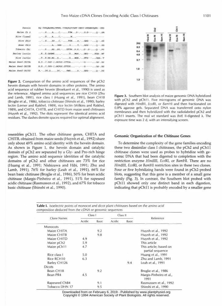
The hevein domain sequences shown in Figure 2 allows a comparison of the sequences of pCh2 and thase of other gene products that contain this domain, including; barley and rice lectiin. The hevein domain of pCh2 shares 72% amino acid seqiience identity with rubber hevein and CHITD, which
www.plantphysiol.orgon February 6, 2019 - Published by Downloaded from Copyright © 1994 American Society of Plant Biologists. All rights reserved.
Two Maize cDNA Clones Encoding Acidic Class I Chitinases 1101
Hevein
Maize Ch 3
Rice ClaaalRice CH10Bean CH 18
Tobacco ChiBarley Lectin
Kief lectinMaize Seed CHITAMaize Seed CHITBMaize Seed CHITD
EQ-roRQAGOKLCFNNL-CGSQYQYCGOT-DEYC-SPDHNOaSN- -CKD
. . - . . S . . . . A . . . . C . - . . . . F G W . . . 3 - . - . . G . O - - - . . . Q - - . S A
. . - . .S . . . .A . . . .C . - . . . . ..W. . . - - - - - - - - - - - . - - - - - - - - -
..-..S....AV...C.-....TOW....S.-..OAO---...Q--.SR
......... .A. . .GON- . . . . .G. . . . -T. - . -GPO- - - . . .Q- - .GG
. . - . . S . . . .AR. .SO. -. . .KPOW. . N . N . - . . O - . G - . . . . Q - - .FO
. .R..E.GSMffi. . . . . . . . . . .G. . .MM.-. .GKG- - - . -NOA- .WT
. . T . . K . N D . M X . . H . . - . . . . . 0 . . . M O R . - . . G T O - - -. . .GAS.-T
A.N..C.P1IF C.SKraY.GTTDA. . . - - - . - - - G - - - - - .
A.N..C.PHV-C.SKK3Y.GTTDE. . . - - - . - - - G - - - - - .
. . O P - - R S
..GP-.RS
= s iI 8 II uj m
A . - . . D G . D . . . . . D C . - . . . K W 3 . . . . . S . - . . G D G - - - . . .Q-- .DG
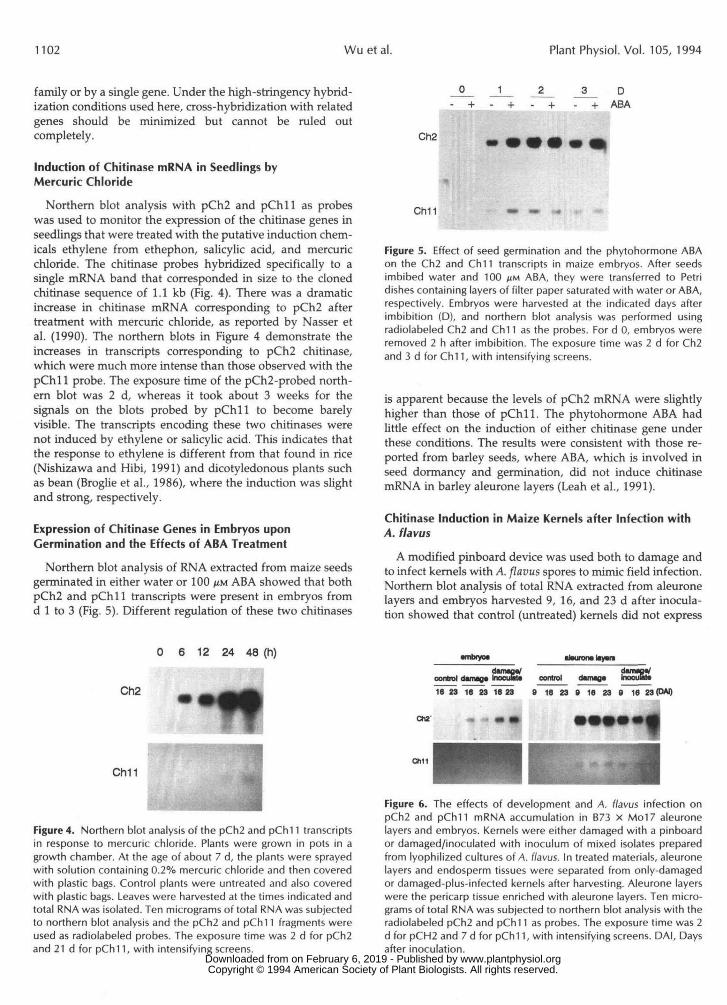
(kb)
8.5
6.44.8
3.7
1.9
:TC*4..
I
Figure 2. Comparison of the amino acid sequences of the pCh2hevein domain with hevein domains in other proteins. The aminoacid sequence of rubber hevein (Broekaert et al., 1990) is used asthe reference. Aligned amino acid sequences are rice CH 10 (Zhuand Lamb, 1991), rice class I (Huang et al., 1991), bean CH18(Broglie et al., 1986), tobacco chitinase (Shinshi et al., 1990), barleylectin (Lerner and Raikhel, 1989), rice lectin (Wilkins and Raikhel,1989), and CHITA, CHITB, and CHITD from maize seed chitinases(Huynh et al., 1992). The dots represent the identical amino acidresidues. The dashes denote spaces required for optimal alignment.
1.3
Figure 3. Southern blot analysis of maize genomic DNA hybridizedwith pCh2 and pCh11. Five micrograms of genomic DNA wasdigested with H/ndlll, EcoRI, or BamHI and then fractionated on0.8% agarose gels. Separated DNA was transferred onto nylonmembranes and then hybridized with the radiolabeled pCh2 andpChl! inserts. The mol wt standard was BstE ll-digested X. Theexposure time was 2 d, with an intensifying screen.
resembles pChll. The other chitinase genes, CHITA andCHITB, obtained from maize seeds (Huynh et al., 1992) shareonly about 49% amino acid identity with the hevein domain.As shown in Figure 1, the hevein domain and catalyticdomain of pCh2 are separated by a Gly- and Pro-rich hingeregion. The amino acid sequence identities of the catalyticdomains of pCh2 and other chitinases are 73% for rice(Huang et al., 1991; Nishizawa and Hibi, 1991; Zhu andLamb, 1991), 76% for barley (Leah et al., 1991), 66% forbean basic chitinase (Broglie et al., 1986), 50% for bean acidicchitinase (Margis-Pinheiro et al., 1991), 51% for rapeseedacidic chitinase (Rasmussen et al., 1992), and 67% for tobaccobasic chitinase (Shinshi et al., 1990).
Genomic Organization of the Chitinase Genes
To determine the complexity of the gene families encodingthese two dissimilar class I chitinases, the pCh2 and pChllchitinase clones were used as probes to hybridize with ge-nomic DNA that had been digested to completion with therestriction enzyme Hindlll, EcoRI, or BamHI. There are noHindlll, EcoRI, or BamHI restriction sites in these two clones.Four or five hybridizing bands were found in pCh2-probedblots, suggesting that this gene is a member of a small genefamily (Fig. 3). In contrast, the Southern blot probed withpChll showed only one distinct band in each digestion,indicating that pChll is probably encoded by a smaller gene
Table I. Isoelectric points of monocot and dicot plant chitinases based on the amino acidcomposition deduced from the cDNA or genomic sequences
Clone NamesClass I Class II
Acidic Basic AcidicReference
Basic
MonocotsMaize CHITAMaize CHITBMaize CHITDMaize pCh2Maize pChl 1
Rice class 1Rice RCH10Barley CH 126
DicotsBean CH 18Bean PR4
Rapeseed ChB4Tobacco DHN 17
4.95.94.7
5.06.3
4.5
9.29.8
9.2
9.19.5
Huynh et al., 1992Huynh et al., 1992Huynh et al., 1992This articleThis article; based on
partial sequenceHuang et al., 1991Zhu and Lamb, 1991
9.4 Leah et al., 1991
Broglie et al., 1986Margis-Pinheiro et al.,
1991Rasmussen et al., 1992Shinshi et al., 1990
www.plantphysiol.orgon February 6, 2019 - Published by Downloaded from Copyright © 1994 American Society of Plant Biologists. All rights reserved.
1102 Wu et al. Plant Physiol. Vol. 105, 1994
family or by a single gene. Under the high-stringency hybrid-ization conditions used here, cross-hybridization with relatedgenes should be minimized but cannot be ruled outcompletely.
Induction of Chitinase mRNA in Seedlings byMercuric Chloride
Northern blot analysis with pCh2 and pChll as probeswas used to monitor the expression of the chitinase genes inseedlings that were treated with the putative induction chem-icals ethylene from ethephon, salicylic acid, and mercuricchloride. The chitinase probes hybridized specifically to asingle mRNA band that corresponded in size to the clonedchitinase sequence of 1.1 kb (Fig. 4). There was a dramaticincrease in chitinase mRNA corresponding to pCh2 aftertreatment with mercuric chloride, as reported by Nasser etal. (1990). The northern blots in Figure 4 demonstrate theincreases in transcripts corresponding to pCh2 chitinase,which were much more intense than those observed with thepChll probe. The exposure time of the pCh2-probed north-ern blot was 2 d, whereas it took about 3 weeks for thesignals on the blots probed by pChll to become barelyvisible. The transcripts encoding these two chitinases werenot induced by ethylene or salicylic acid. This indicates thatthe response to ethylene is different from that found in rice(Nishizawa and Hibi, 1991) and dicotyledonous plants suchas bean (Broglie et al., 1986), where the induction was slightand strong, respectively.
Expression of Chitinase Genes in Embryos uponGermination and the Effects of ABA Treatment
Northern blot analysis of RNA extracted from maize seedsgerminated in either water or 100 /HM ABA showed that bothpCh2 and pChll transcripts were present in embryos fromd 1 to 3 (Fig. 5). Different regulation of these two chitinases
JL_ D
+ ABA
Ch2
Ch11
Figure 5. Effect of seed germination and the phytohormone ABAon the Ch2 and Ch 11 transcripts in maize embryos. After seedsimbibed water and 100 ^M ABA, they were transferred to Petridishes containing layers of filter paper saturated with water or ABA,respectively. Embryos were harvested at the indicated days afterimbibition (D), and northern blot analysis was performed usingradiolabeled Ch2 and Chl 1 as the probes. For d 0, embryos wereremoved 2 h after imbibition. The exposure time was 2 d for Ch2and 3 d forCh11, with intensifying screens.
is apparent because the levels of pCh2 mRNA were slightlyhigher than those of pChll. The phytohormone ABA hadlittle effect on the induction of either chitinase gene underthese conditions. The results were consistent with those re-ported from barley seeds, where ABA, which is involved inseed dormancy and germination, did not induce chitinasemRNA in barley aleurone layers (Leah et al., 1991).
Chitinase Induction in Maize Kernels after Infection withA. flav us
A modified pinboard device was used both to damage andto infect kernels with A. flavus spores to mimic field infection.Northern blot analysis of total RNA extracted from aleuronelayers and embryos harvested 9, 16, and 23 d after inocula-tion showed that control (untreated) kernels did not express
0 6 12 24 48 (h)
Ch2 ftCh11
Figure 4. Northern blot analysis of the pCh2 and pCh! 1 transcriptsin response to mercuric chloride. Plants were grown in pots in agrowth chamber. At the age of about 7 d, the plants were sprayedwith solution containing 0.2% mercuric chloride and then coveredwith plastic bags. Control plants were untreated and also coveredwith plastic bags. Leaves were harvested at the times indicated andtotal RNA was isolated. Ten micrograms of total RNA was subjectedto northern blot analysis and the pCh2 and pCh! 1 fragments wereused as radiolabeled probes. The exposure time was 2 d for pCh2and 21 d for pChl 1, with intensifying screens.
•mbfyot ataman* liywt
control dwiwgtcontrol dimiQ« ____ ____ ____16 23 16 23 1623 8 16 23 9 16 23 9 16 23 (DM)
•—1CM'
Will
Figure 6. The effects of development and A. flavus infection onpCh2 and pCh11 mRNA accumulation in B73 x Mo17 aleuronelayers and embryos. Kernels were either damaged with a pinboardor damaged/inoculated with inoculum of mixed isolates preparedfrom lyophilized cultures of A. flavus. In treated materials, aleuronelayers and endosperm tissues were separated from only-damagedor damaged-plus-infected kernels after harvesting. Aleurone layerswere the pericarp tissue enriched with aleurone layers. Ten micro-grams of total RNA was subjected to northern blot analysis with theradiolabeled pCh2 and pChl 1 as probes. The exposure time was 2d for pCH2 and 7 d for pCh! 1, with intensifying screens. DAI, Daysafter inoculation.
www.plantphysiol.orgon February 6, 2019 - Published by Downloaded from Copyright © 1994 American Society of Plant Biologists. All rights reserved.
Two Maize cDNA Clones Encoding Acidic Class I Chitinases 1103
either of the chitinase genes corresponding to pCh2 and, pChl 1 (Fig. 6). The damaged and infected kernels did contain' chitinase mRNA in the aleurone layers and embryos, with
the majority being present in the aleurone layers. Levels ofpCh2 mRNA were higher than those of pChll mRNA inboth aleurone layers and embryos. Only low levels of tran-scripts of both chitinases were detected in embryos. Further-more, neither chitinase mRNA was found to be induced inendosperm tissues by damage or infection by the fungus orin damaged kernels within 24 h (data not shown).
Chitinase Induction in Maize Callus after Challenge withA. flavus
Regenerable maize calli derived from the hybrids Mol7 XLB31 and LB31 X Mol7 were challenged with A. flavus inagar plates containing growth medium. After 1 week of co-incubation without actual contact between the fungus andcallus, RNA was isolated from the callus and subjected tonorthern blot analysis. There was a dramatic induction ofpCh2 mRNA by fungal co-incubation in both genotypestested (Fig. 7). Induction of pChll mRNA was not detectedin these experiments. Untreated control callus did not havemeasurable levels of the transcripts corresponding to eitherCh2 or Ch 11. These results further indicate the possibledifferent roles of the different chitinases in the plant-defensereactions, as suggested by Mauch and Staehelin (1989) foracidic and basic chitinases.
DISCUSSION
The present study describes the isolation and primarycharacterization of two cDNA clones from maize leaves,pCh2 and pChll, with sequences similar to those of otherchitinases. Based on the nucleotide and deduced amino acid
Ch11
Figure 7. Induction of pCh2 and pChl 1 mRNA in maize callus co-cultured with A. flavus. Control callus did not receive any treatment(—). The treated callus tissue (+) was co-cultured with fungal hyphaeproduced from spores placed in the center of the plates of callusgrowth medium and was removed after 7 d, before the hyphaecontacted the callus. Each lane contains 10 Mg of RNA, which wastransferred to a nylon membrane and was probed with the pCh2and pChll inserts. The exposure time was 2 d for pCh2 and 7 dfor pChl 1, with intensifying screens.
sequences of the full-length pCh2 clone and the partialpChll clone, they both apparently encode class I chitinaseswith the following features: (a) a highly hydrophobic signalpeptide of 21 amino acids; (b) a Cys-rich domain (also calledthe hevein domain) of 40 amino acids (missing from thepartial pChll clone); (c) a hinge domain of 24 amino acidswith a Pro- and Gly-rich region; and (d) a catalytic domain.The pChl 1 clone is similar, but not identical, to one of threechitinase genes (CHITD) described in maize seeds (Huynh etal., 1992), which has a Pro-rich hinge domain (Fig. 1). Basedon the available deduced amino acid sequences, pChll andCHITD share 90% identity (Fig. 1), indicating that they aremembers of a gene family but are not different alleles of thesame gene. It has been shown that the maize seed chitinases,CHITA and CHITB, are significantly different in their bio-chemical and in vitro antifungal activity properties, eventhough they show 87% amino acid sequence homology(Huynh et al., 1992). Huynh et al. (1992) concluded thatmaize chitinases are more divergent than other plant chiti-nases. The sequences of the chitinase genes we obtained frommaize leaves also support a similar observation, because only61% identity was found in the amino acid sequences of thepCh2 and pChll catalytic domains (Fig. 1). The homologybetween pCh2 and rice chitinases is much higher than amongmaize chitinases, i.e. between 71 and 74% (Fig. 1). Thecomparison of the hevein domain within the maize chitinasegene family in Figure 2 also shows greater divergence. Al-though the chitinase genes pCh2 and CHITD demonstratesignificant homology to rubber hevein, the other maize chi-tinase genes, such as CHITA and CHITB, share less identityin the hevein domains (Fig. 2).
Huynh et al. (1992) showed that lack of chitin-bindingdomains, i.e. the hevein domain, did not influence the anti-fungal activities of either CHITA or CHITB. However, theantifungal activities of the proteins encoded by pCh2 andCHITD remain to be determined, even though they bothhave high homology in the chitin-binding sequence (Fig. 2).It is also true that class II chitinase, which lacks both thehevein and hinge domains, can effectively digest chitin (forinstance, barley chitinase pCHI26, as shown by Leah et al.,1991).
Although almost all dicot class I chitinases have basicisoelectric points, there are two reported exceptions whereacidic chitinases have Cys-rich domains in their N-terminalregions: in bean (Margis-Pinheiro et al., 1991) and in garlic(van Damme et al., 1993). Likewise, some monocot class Ichitinases do not always have basic isoelectric points, as inthe case of dicot chitinases. Therefore, we can conclude thatmonocot class I chitinase does not necessarily have a basicisoelectric point, as suggested for dicot chitinases from se-quence information (Shinshi et al., 1990). However, it is notknown if some monocot chitinases are different from theirdicot counterparts or if our examples are exceptions. Aschitinase nucleotide and protein sequences from monocotplants accumulate, this question should be answered. On theother hand, in studies of a tobacco 0-1,3-glucanase gene(Shinshi et al., 1988), it has been found that some sequencesnecessary for targeting the proteins to the vacuole are locatedat the C terminus. Recent studies of tobacco pathogen-relatedproteins in transgenic plants confirmed that the vacuolar www.plantphysiol.orgon February 6, 2019 - Published by Downloaded from
Copyright © 1994 American Society of Plant Biologists. All rights reserved.
1104 Wu et al. Plant Physiol. Vol. 105, 1994
pathogenesis-related proteins might have the targeting infor- mation in a short C-terminal propeptide (Melchers et al., 1993). This peptide is then removed during or after transport into the plant vacuole, indicating its importance in this proc- ess. Although there is no homology between the C termini of the pCh2 and pChl l chitinase genes and the C terminus of the tobacco /3-1,3-glucanase gene, there is a 14-amino acid C-terminal extension in C h l l (Fig. l), which could be im- portant for this process (Neuhaus et al., 1991). Since these two types of maize chitinases show different expression pattems, as demonstrated by their responses to chemical treatments and expression in germinating seeds, there may be differences in the mechanisms controlling the regulation of gene expression.
The differences of the maize chitinase genes are also re- flected in the genomic organization of these genes. Southem blots probed with pCh2 and pChl1 showed that more bands are present with pCh2 than with pChl1 (Fig. 3), indicating a larger gene family for pCh2 than for pChl l . This result indicates that the differences in gene numbers for pCh2 and pChl l may possibly explain why the expression level of pCh2 was much higher than that of pChl l in response to different stresses. Restriction maps show that at least three other cDNA clones isolated in the course of this study belong to the pCh2 family (data not shown). Rescreening the cDNA library made from mercuric chloride-treated leaf mRNA to identify the full-length clone with homology to pChl l was unsuccessful even under low-stringency screening condi- tions, indicating that the expression level of this clone is very low under those conditions. However, A. flavus-infected aleu- rone layers and germinating embryos showed a greater abun- dance of pCh l l mRNA than did leaves (Figs. 5 and 6).
The possibility of different roles for pCh2 and pChl l chitinases in vivo might be indicated by the different expres- sion levels observed when plants were treated with mercuric chloride (Fig. 4). Mercuric chloride has been shown to induce a set of pathogenesis-related proteins in maize, including chitinase and /3-1,3-glucanase (Nasser et al., 1990). Northem blot analysis of RNA isolated from maize seedlings treated with mercuric chloride showed a strong and linear induction of pCh2 mRNA, but only a slight induction of pChl1 mRNA. The transcripts encoding these two chitinase clones were not induced by the other chemical treatments, such as ethylene and salicylic acid (data. not shown), showing that the re- sponses are different when compared to rice (Nishizawa and Hibi, 1991) and dicotyledonous plant chitinases, such as that from bean (Broglie et al., 1986). Ethylene was shown to be a weak and a potent inducer of rice and bean chitinase expres- sion, respectively. The differences observed here and those from rice and bean may indicate that different sets of genes are involved in plant defense-related reactions. Thus, the induction of maize chitinases is different from that of dicot plants, and this also contrasts with the results where ethe- phon and salicylic acid were able to induce rice chitinase mRNA (Nishizawa and Hibi, 1991).
In the experiments conducted by Huynh et al. (1992), where up to 5 r g of poly(A)+ RNA was used in northem blots, the accumulation of mRNA for one of the chitinase clones (CHITA) was demonstrated in maize seeds, roots, and shoots. However, when only 1 pg of poly(A)+ RNA was used,
only seeds that had imbibed showed visible hybridization, which is consistent with our data obtained from 8;erminating embryos (Fig. 5). This indicates that maize embiyos consti- tutively express both pCh2 and pChl1 chitinase 'genes with- out environmental stimuli. The differential regulation and expression of pCh2 and pChl l suggest that each might have some specific role under each condition.
Inocullation of developing kemels with A. flazws resulted in a large induction of chitinases in both aleuronr? layers and embryos (Fig. 6), with the pCh2 mRNA levels being much higher t han those of pChl1. These genes were not induced in endosperm tissues (data not shown). Infection of kemels with furigi utilized physical damage of tissue prior to inocu- lation, but northem blot analysis of RNA from ltemels that were only damaged revealed no chitinase transclipts after 2, 4, 6, and 24 h of treatment (data not shown). This might indicate that wounding alone did not induce the chitinase genes, olr that 24 h of wounding was not long enough to induce the defense reaction. Since these experiinents were conducted in the field, it is possible that natural infection caused the induction of chitinase mRNAs in the damaged- only kernels after 9 d. Embryos were found to have far less of both chitinase mRNAs than aleurone layers (Fig. 6). The lower level of chitinase induction in embryos mi3ht indicate that fungal infection of kemels could occur throu,gh the silks, thus bypassing the aleurone layer.
The evidence indicates that maize and probably other cereal chitinase genes are more divergent than those from other plants that have been described, although the gene products need to be tested in vitro for their antifungal activity. Further study of this divergence and its importance are especial1 y needed because information conceminl; cereal chi- tinases ie very limited. Individual chitinases may each play a particular role in vivo under a given condition, iis indicated by the olbservations: (a) the pCh2 multiple-gene family and the pChl l single-gene family suggest different roles in the defense-related process; and (b) these two genm are both induced in the event of fungal infection but not when chal- lenged by mercuric chloride, nor are they induct.d in callus exposed to the fungus, and the genes are expressed in a tissue-specific manner. More study is needed to determine the exact role of the different maize chitinases.
ACKNOWLEDCMENTS
We wish to thank Dr. D. White of the Department of Plant Pathology at the University of Illinois for providing control and treated field-grown ears and Dr. Aree Waranyuwat for providing fungi-treated callus for our study.
Received November 22, 1993; accepted March 13, 1994. Copyright Clearance Center: 0032-0889/94/105/1097/09. The GenBank accession numbers for nucleotide sequeiices of pCh2
and pChll are L00973 and L16798, respectively.
LITERATURE CITED
Belanger DC, Kriz AL (1989) Molecular characterization of the major inaize embryo globulin encoded by the Glbl gene. Plant Physiol91: 636-643
Benhamou N, Joosten MHAJ, De Wit PJGM (1990) Subcellular localization of chitinase and of its potential substrate in tomato
www.plantphysiol.orgon February 6, 2019 - Published by Downloaded from Copyright © 1994 American Society of Plant Biologists. All rights reserved.
Two Maize cDNA Clones Encoding Acidic Class I Chitinases 1105
root tissues infected by Fusarium oxysporum f . sp. radicis-lycopersici. Plant Physiol92: 1108-1120
Bernasconi P, Locher R, Pilet PE, Jolles J, Jolles P (1987) Purification and N terminal amino-acid sequence of a basic lysozyme from Parthenocissus quinquifolia cultured in vitro. Biochim Biophys Acta
Bol JF, Linthorst HJM, Cornelissen BJC (1990) Plant pathogenesis- related proteins induced by virus infection. Annu Rev Photopathol
Boller T (1985) Induction of hydrolases as a defense reaction against pathogens. In JL Key, T Kosuge, eds, Cellular and Molecular Biology of Plant Stress. Alan R. Liss, New York, pp 247-262
Boller T (1988) Ethylene and the regulation of antifungal hydrolases in plants. Oxf Surv Plant Mo1 Cell Biol 5 145-174
Broekaert WF, Lee H-I, Kush A, Chua N-H, Raikhel N (1990) Wounding induced accumulation of mRNA containing a hevein sequence in laticifers of rubber tree (Hevea brasiliensis). Proc Natl Acad Sci USA 87: 7633-7637
Broglie K, Chet I, Holliday M, Cressman R, Biddle P, Knowlton S, Mauvais CJ, Broglie R (1991) Transgenic plants with enhanced resistance to the fungal pathogen Rhizoctonia solani. Science 254
Broglie KE, Gaynor JJ, Broglie RM (1986) Ethylene-regulated gene expression: molecular cloning of the genes encoding an endochi- tinase from Phaseolus vulgaris. Proc Natl Acad Sci USA 8 3
Calvert OH, Lillehoj EB, Kwolek WF, Zuber MS (1978) Aflatoxin B1 and G1 production in developing maize kemels from mixed inocula of Aspergillus flavus and A. parusiticus. Phytopathology 68:
Campbell WH, Gowri G (1990) Codon usage in higher plants, green algae, and cyanobacteria. Plant Physiol 92: 1-11
Duncan DR, Williams ME, Zehr BE, Widholm JM 61985) The production of callus capable of plant regeneration from immature embryos of numerous Zeu mays genotypes. Planta 165 322-331
Gaynor JJ (1988) Primary structure of an endochitinase mRNA from Solanum tuberosum. Nucleic Acids Res 1 6 5210
Hedrick SA, Bell JH, Boller T, Lamb CJ (1988) Chitinase cDNA cloning and mRNA induction by fungal elicitor, wounding, and infection. Plant Physiol86: 182-186
Huang J-K, Wen L, Swegle M, Tran J-C, Thin TH, Naylor HM, Muthukrishnan S, Reeck GR (1991) Nucleotide sequence of a rice genomic clone that encodes a class I endochitinase. Plant Mo1 Bioll6 479-480
Huynh QK, Hironaka CM, Levine EB, Smith CE, Borgmeyer JR, Shah DM (1992) Antifungal proteins from plants: purification, molecular cloning, and antifungal properties of chitinases from maize seed. J Biol Chem 267: 6635-6640
Lawton K, Ward E, Payne G, Moyer M, Ryals J (1992) Acidic and basic class I1 chitinase mRNA accumulation in response to TMV infection of tobacco. Plant Mo1 Bioll9 735-743
Leah R, Tommerup H, Svendsen I, Mundy J (1991) Biochemical and molecular characterization of three barley seed proteins with antifungal properties. J Biol Chem 266: 1564-1573
Lerner DR, Raikhel NV (1989) Cloning and characterization of root specific barley lectin. Plant Physiol 91: 124-129
Linthorst JHM, van Loon LC, van Rossum CMA, Mayer A, Bol JF, van Roekel JSC Meulenhoff EJS, Cornelissen BJC (1990a) Iso- lation of complementary DNA clones encoding pathogenesis-re- lated proteins P and Q, two acidic chitinases from tobacco. Proc Natl Acad Sci USA 87: 98-102
Linthorst JHM, van Loon LC, van Rossum CMA, Mayer A, Bol JF, van Roekel JSC, Meulenhoff EJS, Cornelissen BJC (1990b) Analysis of acidic and basic chitinases from tobacco and petunia and their constitutive expression in transgenic tobacco. Mo1 Plant Microbe Interact 3 252-258
Margis-Pinheiro M, Metz-Boutique MH, Awade A, de Tapia M, le Ret M, Burkard G (1991) Isolation of a complementary DNA encoding the bean PR4 chitinase: an acidic enzyme with an amino- terminus cysteine-rich domain. Plant Mo1 Biol 17: 243-253
Mauch F, Hadwiger LA, Boller T (1984) Ethylene: symptom, not signal for the induction of chitinase and @-1,3-glucanase in pea pods by pathogens and elicitors. Plant Physiol76 607-611
915: 254-260
28: 113-138
1194-1197
6820-6824
501-506
Mauch F, Mauch-Mani B, Boller T (1988) Antifungal hydrolases in pea tissue. 11. Inhibition of fungal growth by combination of chitinase and 8-1,3glucanase. Plant Physiol88: 936-942
Mauch F, Staehelin LA (1989) Functional implications of the sub- cellular localization of ethylene-induced chitinase and P-1,3-glu- canase in bean leaves. Plant Cell 1: 447-457
Melchers LS, Sela-Buurlage MB, Vloemans SA, Woloshuk CP (1993) Extracellular targeting of the vacuolar proteins AP24, chi- tinase and @-1,3glucanase in transgenic plants. Plant Mo1 Biol 21:
Metraux JP, Burkhart W, Moyer M, Dincher S, Middlesteadt W, Williams S, Payne G, Carnes M, Ryals J (1989) Isolation of a complementary DNA encoding a chitinase with structural homol- ogy to a bifunctional lysozyme/chitinase. Proc Natl Acad Sci USA
Nasser W, de Tapia M, Burkard G (1990) Maize pathogenesis- related proteins: characterization and cellular distribution of 1,3- P-glucanases and chitinase induced by brome mosaic virus infec- tion or mercuric chloride treatment. Physiol Mo1 Plant Pathol 36
Neuhaus J-M, Sticher L, Meins F, Boller T (1991) A short C- terminal sequence is necessary and sufficient for the targeting of chitinases to the plant vacuole. Proc Natl Acad Sci USA 88:
Nishizawa Y, Hibi T (1991) Rice chitinase gene: cDNA cloning and stress induced expression. Plant Sci 76: 211-218
Payne G, Ahl P, Moger M, Harper A, Beck J, Meins F, Ryals J (1990) Isolation of complementary DNA clones encoding patho- genesis-related proteins P and Q, two acidic chitinases from to- bacco. Proc Natl Acad Sci USA 87: 98-102
Rasmussen U, Bojsen K, Collinge DB (1992) Cloning and charac- terization of a pathogen-induced chitinase in Brussica napus. Plant Mo1 Biol 2 0 277-287
Samac DA, Hironaka CM, Yallaly PE, Shah DM (1990) Isolation and characterization of the genes encoding basic and acidic chiti- nase in Arubidopsis thaliana. Plant Physiol93 907-914
Sambrook J, Fritsch EF, Maniatis T (1989) Molecular Cloning: A Laboratory Manual. Cold Spring Harbor Laboratory Press, Cold Spring Harbor, NY
Sanger F, Nicklen S, Coulson AR (1977) DNA sequencing with chain terminating inhibitors. Proc Natl Acad Sci USA 7 4
Schlumbaum A, Mauch F, Vogeli U, Boller T (1986) Plant chiti- nases are potent inhibitors of fungal growth. Nature 324 365-367
Sela-Buurlage MB, Ponstein AS, Bres-Vloemans SA, Melchers LS, van den Elzen PJM, Cornelissen BJC (1993) Only specific tobacco (Nicotiana tabacum) chitinase and ~-1,3-glucanase exhibit antifun- gal activity. Plant Physiol 101: 857-863
Shinshi H, Neuhaus J-M, Ryals J, Meins F (1990) Structure of a tobacco endochitinase gene: evidence that different chitinase genes can arise by transposition of sequences encoding cysteine-rich domain. Plant Mo1 Biol 14 357-368
Shinshi H, Wenzler H, Neuhaus JM, Felix G, Hofsteenge J, Meins F (1988) Evidence for N- and C-terminal processing of a plant defense-related enzyme: primary structure of tobacco prepro-0- 1,3-glucanase. Proc Natl Acad Sci USA 85: 5541-5545
Swegle M, Huang J-K, Lee G, Muthukrishnan S (1989) ldentifica- tion of an endochitinase cDNA clone from barley aleurone cells. Plant Mo1 Biol 1 2 403-412
van Damme EJM, Willems P, Torrekens S, van Leuven F, Peumans WJ (1993) Garlic (Allium sutivum) chitinases: characterization and molecular cloning. Physiol Plant 87: 177-186
Wessels JGH, Sietsma JH (1981) Funga1 cell walls: a survey. In W Tanner, FA Loewus, eds, Encyclopedia of Plant Physiology, New Series, Vol 138. Springer-Verlag, Berlin, pp 352-394
Wilkins TA, Raikhel NV (1989) Expression of rice lectin is govemed by two temporally and spatially regulated mRNAs in developed embryos. Plant Cell 1: 541-549
Yanisch-Perron C, Vieira J, Messing J (1985) Improved M15 phage cloning vectors and host strains: nucleotide sequences of the M13 mp18 and pUC19 vector. Gene 3 3 103-119
Zhu Q, Lamb CJ (1991) Isolation and characterization of a rice gene encoding a basic chitinase. Mo1 Gen Genet 226 289-296
583-593
86 896-900
1-14
10362-10366
5463-5467
www.plantphysiol.orgon February 6, 2019 - Published by Downloaded from Copyright © 1994 American Society of Plant Biologists. All rights reserved.