Embed Size (px)
Citation preview

Mycobiology 31(2): 74-80 (2003)Copyright © 2003 by The Korean Society of Mycology
-
s-es
es
tThe
byellsh,
-),io-iledout21
ry
s andric
es of
-
r-
Molecular and Morphological Characterization of Green Mold, Trichoderma spp.isolated from Oyster Mushrooms
In-Young Choi�, Seung-Beom Hong1 and Mahesh C. Yadav2
Jeollabuk-do Agricultural Research and Extension Services, Iksan 570-704, Korea1Korean Agricultural Culture Collection, National Institute of Agricultural Biotechnology, Suwon 441-707, Korea2National Research Centre for Mushroom, Solan-173213, H.P., India
(Received February 3, 2003)
Isolates of Trichoderma spp. collected from Pleurotus ostreatus and P. eryngii beds, which included loosened substrate com-pactness and development of green colour, were grouped into three species. The occurrence of different species of Tricho-derma was as T. cf. virens (70.8%), T. longibrachiatum (16.7%) and T. harzianum (12.5%). The conidia of Trichoderma spp.were ellipsoidal, obovoid and phialides were bowling pins, lageniform and the length of phialides was 3.5~10.0 × 1.3~3.3µµµµm.Phialides of T. cf. virens and T. harzianum were tending clustered, but it was solitary disposition in T. longibrachiatum. T.cf. virens was characterized by predominantly effuse conidiation, sparingly branched, and fertile to the apex and it was penicillate type. RAPD analysis could detect variability amongst three different species of Trichoderma using two newly designedURP-primers. However, intra-specific variation could not be detected in all the isolates except for rDNA sequence data clasified Trichoderma isolates into three distinct groups representing three species. The profiles of rDNA sequences of isolatrepresenting a species showed high similarity in T. cf. virens and T. harzianum. However, there was a variation in rDNAsequences of isolates representing T. longibrachiatum. The results of present study reveals that molecular techniques of RAPDand rDNA sequencing can greatly aid in classification based on morphology and precise identification of fast evolving speciof Trichoderma.
KEYWORDS: Pleurotus spp., RAPD, rDNA sequencing, Trichoderma spp.
Oyster mushrooms are the most commercially grownmushroom, being cultivated on over 60% of mushroomfarms, and had the highest income from mushroom inKorea (Oh et al., 1999). Since 1986 when artificial culti-vation techniques were developed using rice straw, cottonwaste and sawdust, commercial production of oystermushroom has been seriously affected by green mold epi-demics. Amongst oyster mushrooms, Pleurotus ostreatusis the most popular cultivated mushroom in Korea (Bae etal., 1996; Kang et al., 2001). In the Agaricus bisporus,the seriousness of the disease is indicated by yield lossesup to 30~100% experienced in 1995 in Clester county ofPennsylvania (Samuels et al., 2002). The most seriousoutbreak of Trichoderma species on mushroom crops wascaused by biotype Th-2 of T. harzianum, in Ireland in1985-86 and resulted in loss of about 3~4 million poundsin mushroom industries in UK and Ireland (Fletcher,1990).
So far, the main diseases reported in the oyster mush-rooms are green mold caused by Trichoderma, black-grayvelvet caused by Trichrus spiralis, fire mold caused byNeurospora sp., brown spot of Pseudomonas tolaassi, andvirus in Korea (Kim et al., 1995, 2000).
Trichoderma is a genus of filamentous Deuteromycetesand its habitats includes forest and agricultural soil and
living plant. Some species of Trichoderma have been usedto control other fungi under biological control of plandiseases, and to produce enzymes (Samuels, 1996). genus Trichoderma causes disease in oyster mushroom competition for nutriments and lysis of the mushroom cby secretion of hydrolytic enzymes (Goltapeh and Dnae2000).
The first serious attempts to morphologically distinguish Trichoderma species was made by Rifai (1969who divided them into nine taxa on the basis of conidphore branching and conidia shape. The most detamorphological studies of the anamorphs were carried by Bissett (1984, 1991a, b), who distinguished about texa in sect. Pachybasium and seven in sect. Longibrachi-atum. Recently, other taxonomic methods supplementato morphology showed a great diversity of Trichoderma.Samuels (1994, 2002) has used isoenzyme profiles ataxonomic technique. ITS and 5.8S rDNA sequences afingerprinting techniques have revealed the intra-generelationships amongst species of Trichoderma (Fujimoriand Okuda, 1994; Kim et al., 2000).
The purpose of this study was to reduce damagcaused by the green mold disease by quick diagnosisspecies of Trichoderma involved and to establish phylogenetic relationship amongst Trichoderma spp. isolated fromPleurotus spp. by morphological and molecular characteistics. We report about occurrence of Trichoderma spe-*Corresponding author <E-mail: [email protected]>
74

Molecular and Morphological Characterization of Green Mold, Trichoderma spp. 75
andofi-
size
se
res-
d
ra-ewonsr
by
ts
for
%mf
ed),so-Rmn-lf,
s-
e-ed-
pro-hip
cies, morphological characteristics studied using scanningelectron microscope and the phylogenetic analysis revealedby RAPD analysis and rDNA sequencing.
Materials and Methods
Fungal isolates. Twenty one isolates of Trichodermaspp. were isolated from Pleurotus ostreatus and P. eryngiicultivation beds having base materials viz; rice straw, cot-ton waste and sawdust from oyster farms. Three standardisolates used for identification were obtained from KACC(Korean Agricultural Collection, National Institute of Ag-ricultural Science and Technology, Suwon, Korea) (Table 1).
Morphological observations. For the isolation of Tri-choderma spp. from the infected samples collected fromdifferent oyster mushroom farms, fast growing myceliawith green colour and lower mycelial density than mush-room mycelia were picked up with help of pins from thesymptom part of the beds. These isolates were cultured onMEA (malt extract agar, difco) and PDA (potato dextroseagar, difco) and were incubated in normal light for 2~3days at 25oC. For morphological characterization of Tri-choderma spp., observations on morphology of conidio-phores, phialides and conidia were made using scanning
electron microscope. The data include the degree nature of aggregation of conidiophores, the pattern branching of the conidiophores, the disposition of phalides, the shape and size of phialides, the shape andof conidia and type of hyphae.
DNA extraction. Genomic DNAs from different iso-lates of Trichoderma spp. were extracted following themethod of Zolan and Pukkila (1986). Trichoderma iso-lates were grown in shaking cultures of potato dextrobrowth (PDB) for two weeks at 25oC. After harvestingmycelial from different isolates, the mycelial mats wefreeze-dried. Mycelial mats were grinded with micro-petle for DNA extraction microcentrifuge tubes (1.5 ml) andincubated in 400µl lysis buffer[3% SDS, 50 mM EDTA,50 mM Tris-HCl (pH 7.2), 1% 2-mercaptoethanol] at 68oCfor 1 hour. After that, the mixture was gently extractewith chloroform : isoamylalcohol (24 : 1) containing 5%phenol and centrifuged at 12,000 rpm at room tempeture for 1 min. The supernatant was transferred to a ntube and equal volume of absolute EtOH in the solutiand centrifuged for pelleting the DNA. The pellet wawashed with 70% EtOH and dissolved in TE buffe(10 mM Tris-HCl pH 8.0, 1 mM EDTA).
PCR Amplification and rDNA sequencing.RAPD analysis: The primers for RAPD amplificationwere 20-mer URP-primers which were synthesized KACC. PCR amplification was performed in a 50µl reac-tion mixture containing 1 pmol of both primers, 2.0 uniof Taq DNA polymerase (Promega), 0.1 mM dNTP, 5µl10×buffer and 100 ng template DNA. The parameters PCR amplification were 94oC for 4 min; 35 cycles of 95oCfor 1 min, 55oC for 1 min, 72oC for 2 min and final exten-sion 72oC for 8 min. PCR product was analyzed on 1.2agarose gel. UPGMA dendrogram was derived froRAPD profiles of genomic DNA in the 24 isolates oTrichoderma spp. with two URP-primers. The similaritycoefficient (F) was calculated as the fraction of sharfragments between pairs of isolates. F = 2Nxy/(Nx + Nywhere Nxy is the number of PCR products shared by ilates X and Y, while Nx and Ny are total number of PCproducts in isolates X and Y, respectively. A dendrograwas constructed with NTSYS-pc (ver. 2.0) using the uweighted pair-group method with arithmetic mean (Roh1993).rDNA sequencing: Amplified PCR products wereligated into pGEM T vector (promega co.) and was tranformed with E. coli DH5α. The sequencing of rDNA wasdone using dideoxy chain termination method. The squences of rDNA were compared with already publishdata of Trichoderma spp. and selected ITS regions sequences. ITS sequences data was aligned with clustal gram and analysis was done for phylogenetic relations
Table 1. List of Trichoderma spp. selected from oyster mush-room media and sawdust compost in this study
Isolateno.
Species name HostCompostbase
Origin
1 T. cf. virens P. ostreatus Rice straw Iksan2 T. cf. virens P. ostreatus Cotton waste Wanju3 T. cf. virens P. ostreatus Cotton waste Iksan4 T. cf. virens P. ostreatus Rice straw Iksan5 T. cf. virens P. ostreatus Rice straw Iksan6 T. cf. virens P. ostreatus Cotton waste Wanju7 T. cf. virens P. ostreatus Rice straw Wanju8 T. cf. virens P. ostreatus Rice straw Wanju9 T. cf. virens P. ostreatus Rice straw Wanju
10 T. cf. virens P. ostreatus Rice straw Wanju11 T. cf. virens P. ostreatus Cotton waste Wanju12 T. cf. virens P. ostreatus Rice straw Wanju13 T. cf. virens P. ostreatus Rice straw Wanju14 T. cf. virens P. ostreatus Rice straw Wanju15 T. cf. virens P. ostreatus Rice straw Wanju16 T. cf. virens P. eryngii Sawdust Iksan17 T. harzianum P. ostreatusSawdust Ikasn18 T. harzianum P. eryngii Sawdust Iksan19 T. longibrachiatum P. ostreatusSawdust Iksan20 T. longibrachiatum P. ostreatusSawdust Iksan21 T. longibrachiatum P. eryngii Sawdust Iksan22 T. longibrachiatum P. eryngii Sawdust KACCa
23 T. harzianum P. eryngii Sawdust KACC24 T. cf. virens P. eryngii Sawdust KACC
aKACC (Korean Agricultural Culture Collection, National Institute ofAgricultural Science and Technology, Suwon, Korea).
76 Choi et al.
toeadnce
D)
by molecular evolutionary genetic analysis (MEGA, ver-sion 1.02, The Pennsylvania State University) and soft-ware (Kumar et al., 1993). The distance was measured byKimura’s 2-parameter and phylogenetic tree was obtainedfrom PCR products and ITS sequences of Trichodermaspp. Phylogenetic relationships were inferred by theneighbor-joining method (Saitou and Nei, 1987).
Results
Symptoms and occurrence. Symptoms of Trichodermainfection in Pleurotus beds were characteristic white my-celia which initially produces a denser compact (well-net)
mycelia than that of Pleurotus. White mycelia of Tricho-derma had many aerial mycelia and it gradually turns green colour. Later on, the mycelia had deep green sprin substrates of mushroom beds (Fig. 1). The occurreof different species of Trichoderma on Pleurotus beds wasT. cf. virens (70.8%), T. longibrachiatum (16.7%) and T.harzianum (12.5%). T. longibrachiatum and T. harzianum
Fig. 1. Symptoms of Trichoderma infection on the beds of Pleurotus ostreatus (A and B) rice straw based substrates, (C and cotton waste based substrates.
Table 3. Morphological characteristics of Trichoderma spp. observed by SEM (25oC, PDA, 3 days)
Morphologicalcharacteristics
T. cf. virens T. longibrachiatum T. harzianum
Conidiashapecoloursize (µm)L : W
ellipsodal, obovoiddark green1.5~4.0 × 1.0~2.53 : 2
ellipsodal, obovoiddilute green1.0~4.8 × 1.0~3.13 : 2
ellipsodal, subglobosepale green1.3~3.3 × 1.3~2.53 : 2
PhialidesshapedispositionAcropleurogenoussize (µm)L : W
bowling pin, lageniformtending clusteredfew3.5~10.0 × 1.7~3.03 : 1
bowling pin, lageniformsolitarycommon4.3~9.6 × 1.3~2.75 : 3
bowling pin, lageniformtending clustereda few4.4~7.2 × 2.6~3.35 : 3
Hyphaetypewidth (µm)
penicillate2.0~6.0
tree branches1.5~4.1
tree branches1.5~6.0
Table 2. Occurrence of Trichoderma spp. selected from mush-room beds
Species T. cf. virens T. longibrachiatum T. harzianum
Ratio (%) 70.8 16.7 12.5

Molecular and Morphological Characterization of Green Mold, Trichoderma spp. 77
ip-o-th
thaldly
re
aesadr ofec-
mber
idi
were mainly isolated from sawdust based substrate and T.cf. virens was isolated from the rice straw and cottonwaste substrates (Table 2).
Morphological characteristics. Differences in morpho-logical characteristics of Trichoderma spp. are summa-rized in Table 3 and Fig. 2. The conidia were ellipsoidaland obovoid in T. cf. virens and T. longibrachiatum. How-ever, it was ellipsoidal and subglobose in T. harzianum.The phialides were bowling pin, lageniform and thelength was 3.5~10.0 × 1.3~3.3µm. Phialides of T. cf.virens and T. harzianum were tending clustered, but T.longibrachiatum was solitary disposition. Especially, T.cf. virens was characterized by predominant conidiation,many divided branches, gathering all finger to top, andfertile to the apex. That was penicillate type. Conidio-phores of T. cf. virens were smoothly bend, gather and notspread to top. Conidia broadly rounded to obovoid, bothends broadly rounded or with the base narrower. Phi-alides were hung like banana in the conidiophore, baseand apex were more narrow than middle. T. longibrachia-tum had conidiophores with long and flexuous, a few sidebranches and smoothly. Phialides was single, long, slightly
wider in the middle than at the base. Conidia were ellsoidal to obovoid, smooth and dilute green. Conidiphores of T. harzianum were spread to the top and smooor rounded, wide near the base. Phialides of T. harzianumwere arising mostly in crowded but had a angle wiconidiophore, and had whorls of 2~6 on the terminbranches. Conidia subglobose to ellipsoidal, apex broarounded, base more narrowly rounded.
PCR fingerprinting of Trichoderma. ReproducibleRAPD profiles of genomic DNAs of different Tricho-derma spp. are presented in Fig. 3. Both the primers weuseful in classifying Trichoderma spp. and the sizes of thePCR products ranged between 100 bp to 4.0 kb withtotal of 6~13 bands in each RAPD profile. The isolathad a total of 435 bands with two primers, primer 1 h168 bands and primer 2 had 267. The average numbebands with primer 1 and primer 2 was 7 and 11, resptively. T. longibrachiatum had 6~8 bands with primer 1and had less bands than the other species, but the nuof bands with the primer 2 was less in T. longibrachia-tum and T. harzianum than those of T. cf. virens. Theintra-species bands of T. cf. virens, T. harzianum and T.
Fig. 2. Morphological characteristics of Trichoderma spp. observed by SEM. (A-C) Conidiophores, phialides and conarespectively, of T. cf. virens. (D-F) Conidiophoers, phialides and conidia respectively, of T. longibrachiatum. (G-I)Conidiophores, phialides and conidia respectively, of T. harzianum.

78 Choi et al.
es
tes
pe
s
ofand a
rgeper
s
longibrachiatum were similar in both the primers. TheUPGMA dendrogram based on RAPD profiles of ge-
nomic DNA divided the Trichoderma spp. into threegroups, namely T. cf. virens in group I, T. harzianum ingroup III and T. longibrachiatum III (Fig. 4). The isolatesof each Trichoderma spp. had about 90% similarity.Between T. cf. virens and T. harzianum there was about70% similarity, but the similarity between these speciand T. longibrachiatum was only about 40%.
Genetic relationship. Genetic relationship among dif-ferent species of Trichoderma based on ribosomal DNAsequencing profiles is shown in Fig. 5. The 24 isolatested were divided into three groups. T. cf. virensincluded in group I, T. harzianum and T. longibrachiatumincluded in group II and III, respectively. The first grouincluded T. cf. virens isolates from rice straw, cotton wastand sawdust substrates. Seventeen isolates of T. cf. virenshad 100% of DNA similarity. Also, 3 isolates of T. har-zianum from sawdust had 100% of DNA similarity awell. T. cf. virens and T. harzianum had very close phylo-genetic relationship. The third group which comprised T. longibrachiatum isolates had more genetic distance thother groups. However, the isolates within subgroup hahigh DNA sequences similarity.
Discussion
Green mold disease caused by Trichoderma spp. is a seri-ous problem of oyster mushroom in Korea. It causes laeconomic losses to the mushroom growers. In the pa
Fig. 3. Representative RAPD fingerprints using primer 1 andprimer 2 of 24 isolates of Trichoderma spp. Lanes1~16 and 24 : T. cf. virens; Lanes 17~18 and 23 : T.longibrachiatum; Lanes 19~22 : T. harzianum; M :1 kb ladder marker (promega).
Fig. 4. UPGMA dendrogram derived from RAPD profiles ofgenomic DNA in the 24 isolates of Trichoderma spp.with two URP-primers
Fig. 5. UPGMA dendrogram derived from sequencing profileof ribosomal DNA in the 24 isolates of Trichodermaspp.
Molecular and Morphological Characterization of Green Mold, Trichoderma spp. 79
nyex
desand
i-hchn,
es
asor
ithalex
nithin
hesi-re
nom-ousndt
n-ed.e
yesr
tely
ting
Alu-
or-ts.o
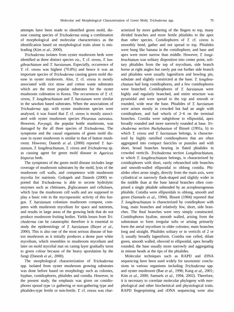
attempts have been made to identified green mold, dis-ease causing species of Trichoderma using a combinationof morphological and molecular characteristics as theidentification based on morphological traits alone is mis-leading (Kim et al., 2000).
Trichoderma isolates from oyster mushroom beds wereidentified as three distinct species viz., T. cf. virens, T. lon-gibrachiatum and T. harzianum. Especially, occurrence ofT. cf. virens was highest (70.8%) and hence it was animportant species of Trichoderma causing green mold dis-ease in oyster mushroom. Also, T. cf. virens is mostlyassociated with rice straw and cotton waste substrateswhich are the most popular substrates for the oystermushroom cultivation in Korea. The occurrences of T. cf.virens, T. longibrachiatum and T. harzianum were similarin the sawdust based substrates. When the associations ofTrichoderma spp. with oyster mushroom species wereanalysed, it was found that T. cf. virens is mostly associ-ated with oyster mushroom species Pleurotus ostreatus.However, P.eryngii, the popular bottle mushroom, wasdamaged by the all three species of Trichoderma. Thesymptoms and the causal organisms of green mold dis-ease in oyster mushroom is similar to that of button mush-room. However, Danesh et al. (2000) reported T. har-zianum, T. longibrachiatum, T. virens and Trichoderma sp.as causing agent for green mold disease in Agaricusbisporus beds.
The symptoms of the green mold disease includes largecoverage of mushroom substrates by the mold, lysis of themushroom cell walls, and competence with mushroommycelia for nutrients. Goltapeh and Danesh (2000) re-ported that Trichoderma is able to secrete hydrolyticenzymes such as chitinases, β-glucanases and cellulases,which lyse the mushroom cell walls and are supposed toplay a basic role in the mycoparasitic activity of this fun-gus. T. harzianum colonizes mushroom compost, com-petes with mushroom mycelium for space and nutrients,and results in large areas of the growing beds that do notproduce mushroom fruiting bodies. Yields losses from Tri-choderma can be catastrophic therefore it is essential tostudy the epidemiology of T. harzianum (Bayer et al.,2000). This is also one of the most serious disease of but-ton mushroom as it initially produces a dense pure whitemycelium, which resembles to mushroom mycelium andlater on mold mycelial mat on casing layer gradually turnsto green colour because of the heavy sporulation by thefungi (Danesh et al., 2000).
The morphological characterization of Trichodermaspp. isolated from oyster mushroom growing substrateswas done before based on morphology such as colonies,hyphae, conidiophores, phialides and conidia. However, inthe present study, the emphasis was given on conidio-phores spread type i.e gathering or non-gathering type andphialides-type fertile or non-fertile. T. cf. virens was char-
acterized by more gathering of the fingers to top, madivided branches and more fertile phialides to the apthan other species. Conidiophores of T. cf. virens issmoothly bend, gather and not spread to top. Phialiwere hung like banana in the conidiophore, and base apex were more narrow than middle. However, T. longi-brachiatum was solitary disposition into center point, soltary phialides from the top of mycelium, side brancborne at right angles but rarely put out further side branand phialides were usually lageniform and bowling pisubulate and slightly constricted at the base. T. longibra-chiatum had long conidiophores, and a few conidiophorwere branched. Conidiophores of T. harzianum werehighly and regularly branched, and entire structure wpyramidal and were spread to the top and smooth rounded, wide near the base. Phialides of T. harzianumwere arisen mostly in crowded but had an angle wconidiophore, and had whorls of 2~6 on the terminbranches. Conidia were subglobose to ellipsoidal, apbroadly rounded and more narrowly rounded at base. Tri-choderma section Pachybasium of Bissett (1991a, b) towhich T. virens and T. harzianum belongs, is character-ized by highly ramified conidiosphores that are ofteaggregated into compact fascicles or pustules and wshort, broad branches bearing in flated phialides crowded verticils. Trichoderma section Longibrachiatum,to which T. longibrachiatum belongs, is characterized byconidiophores with short, rarely rebranched side brancand smooth-walled ellipsoidal to oblong conidia. Phalides often arose singly, directly from the main axis, wecylindrical or narrowly flask-shaped and slightly wider ithe middle than at the base lateral branches often cprised a single phialide subtended by an acropleurogenphialide. Conidia were ellipsoidals to oblong, smooth agreen (Samuels et al., 1994). Bissett (1984) reported thaT. longibrachiatum is characterized by conidiophore withlong, main branches and relatively few, short, side braches. The final branches were very simply constructConidiophores hyaline, smooth walled, arising from thsubstratum to form irregular tufts or arising primarilform the aerial mycelium in older colonies; main branchlong and straight. Phialides solitary or in verticils of 2 o3; usually broadly lageniform. Conidia one celled, dilugreen, smooth walled, obovoid to ellipsoidal, apex broadrounded, the base usually more narrowly and aggregain minute heads at the tips of the phialides.
Molecular techniques such as RAPD and rDNsequencing have been used widely for taxonomic concsions in various organisms including Trichoderma spp.and oyster mushroom (Bae et al., 1996; Kang et al., 2001;Kim et al., 2000; Samuels et al., 1994, 2002). Therefore,it is necessary to correlate molecular phylogeny with mphological and other biochemical and physiological traiRAPD fingerprinting and rDNA sequencing were als

80 Choi et al.
a-n
mc-l
ion
.,ngin-
he-
0.
ces.
o-tate
A.
t-
R.
i-
cal
O.i-
A
y-
used to classify T. harzianum strains that were antagonis-tic to the commercial production of mushroom (Muth-umeenakshi et al., 1994), Trichoderma species and theAscomycetes Hypocrea jecorina (Kubicek and Harman,1998). We observed genetic diversity within a collectionof Trichoderma spp. isolates based on RAPD patterns.Both the primers were useful in classifying and groupingof Trichoderma isolates. The intra-species bands of T. cf.virens, T. harzianum and T. longibrachiatum were similarin both the primer. T. longibrachiatum had 6~8 bands withprimer 1 and had less bands than the other species, but thenumber of bands with the primer 2 was less in T. longi-brachiatum and T. harzianum than those of T. cf. virens.RAPD fingerprints using the URP-primers were helpfulin classification and grouping of Trichoderma isolatesand were in agreement with morphological observations.UPGMA dendrogram based on RAPD profiles of ge-nomic DNA had high similarity within group but eachgroup had long distance. The analysis of rDNA sequenc-ing of Trichoderma spp. reveals the genetic variation inisolates representing T. cf. virens and T. harzianum. How-ever, intra-species isolates has 100% DNA base sequencesimilarity. T. longibrachiatum isolates had more geneticdistance than other groups. However, the isolates withinsubgroup had a high DNA sequences similarity. Also, T.cf. virens and T. virens are spread type of conidiophores.The shape and size of conidia and phialids are similar toeach other. The analysis of rDNA sequencing reveals dif-ference in rDNA base sequences of T. cf. virens and T.virens. However, T. cf. virens has more similarity with T.harzianum in rDNA sequences. Therefore, we have namedthe new species of T. virens as T. cf. virens.
Acknowledgments
This work was supported by the Post-doctoral FellowshipProgram of Korea Science & Engineering Foundation(KOSEF).
References
Bae, S. C., Seong, K. Y., Lee, S. W., Go, S. J., Eun, M. Y. andRhee, I. K. 1996. Phylogenetic relationships among Pleurotusspecies inferred from sequence data of PCR amplified ITSregion in ribosomal DNA. Korean J. Plant Pathol. 24: 155-165.
Bayer, D. M., Wuest, P. J. and Kremser, J. J. 2000. Evaluation ofepidemilolgical factors and mushroom substrate characteristicsinfluencing the occurrence and development of Trichodermagreen mold. Science and Cultivation of Edible fungi. 633-640.
Bissett, J. 1984. A revision of the genus Trichoderma. I. SectionLongibrachiatum sect. nov. Can. J. Bot. 62: 924-931.
_____. 1991a. A revision of the genus Trichoderma. II. Infrage-neric classification. Can. J. Bot. 69: 2357-2372.
_____. 1991b. A revision of the genus Trichoderma. III. SectionPachybasium. Can. J. Bot. 69: 2373-2417.
Danesh, Y. R., Goltapeh, E. M. and Rohani, H. 2000. Identifiction of Trichoderma species causing green mold in buttomushroom farms, distribution and their relative abundance. Sci-ence and Cultivation of Edible fungi. 653-659.
Fletcher, J. T. 1990. Trichoderma and Penicillium diseases ofAgaricus bisporus. A Literature Review for The HorticulturalDevelopment Council. London. ADAS.
Fujimori, F. and Okuda, T. 1994. Application of the randoamplified polymorphic DNA using the polymerase chain reation for efficient elimination of duplicate strains in microbiascreening. . Fungi. J. Antibiot. (Tokyo) 47: 173-182.
Goltapeh, E. M. and Danesh, Y. R. 2000. Studies on interactbetween Trichoderma species and Agaricus bisporus myce-lium. Science and Cultivation of Edible Fungi. 661-666
Kang, H. W., Park, D. S., Park, Y. J., You, C. H., Lee, B. MEun, M. Y. and Go, S. J. 2001. Genomic differentiation amooyster mushroom cultivars released in Korea by URP-PCR fgerprinting. Mycobiology 29: 85-89.
Kim, J. W., Kwon, S. I. and Kang, H. J. 1995. Studies on tPathogenic Pseudomonas causing bacterial disease of cultivated mushroom in Korea. Korean J. Plant Pathol. 11: 353-360.
Kim, K. Y., Lee, G. J., Ha, M. G., Lee, T. H. and Lee, J. D. 200Intrageneric relationships of Trichoderma based on internaltranscribed spacers and 5.8S rDNA nucleotide sequenMycobiology 28: 11-16.
Kubicek, C. P. and Harman, G. E. 1998. Trichoderma and Glio-cladium Vol. 1. Taylor and Francis Ltd. London. 1-94.
Kumar, S., Tamura, K. and Nei, M. 1993. MEGA: molecular evlutionary genetics analysis, version 1.0. The Pennsylvania SUniversity, University Park.
Muthumeenakshi, S., Mills, P. R., Brown, A. E. and Seaby, D. 1994. Intraspecific molecular variation among Trichodermaharzianum isolates colonizing mushroom compost in the Briish Isles. Microbiology 140: 769-777.
Oh, S. J., Chun, C. S., Park, J. S., Kim, H. K. and Fermor. T.1999. Studies on the effect of vinyl mulching on Pleurotusostreatus cultivation. Kor. J. Mycol. 27: 107-111.
Rifai, M. A. 1969. A revision of the genus Trichoderma. Mycol.Pap. 116: 1-56.
Rohlf, F. J. 1993. NTSYS-pc, numerical taxonomy and multivarate analysis system. State Univ. of New York, Stony Brook.
Samuels, G. J., Petrini, O. and Manguin, S. 1994. Morphologiand macromolecular characterization of Hypocrea schweinitziiand its Trichoderma anamorph. Mycologia 86: 421-435.
_____. 1996. Trichoderma: a review of biology and systematicsof the genus. Mycol. Res. 100: 923-935.
_____, Dodd, S. L., Gams, W., Castlebury, L. A. and Petrini, 2002. Trichoderma species associated with the green mold epdemic of commercially grown Agaricus bisporus. Mycologia94: 146-170.
Saitou, N. and Nei, M. 1987. The neighbor-joining method: new method for reconstructing phylogenetic trees. Mol. Bio.Evol. 24: 189-204.
Zolan, M. E. and Pukkila, P. J. 1986. Inheritance of DNA methlation in Coprinus cinereus. Mol. Cell. Biol. 6: 185-200.