Embed Size (px)
Citation preview

Molecular Aspects of MembraneTrafficking in Paramecium
Helmut Plattner and Roland KissmehlDepartment of Biology, University of Konstanz, D-78457 Konstanz, Germany
Results achieved in the molecular biology of Paramecium have shed new light
on its elaborate membrane trafficking system. Paramecium disposes not only of
the standard routes (endoplasmic reticulum ! Golgi ! lysosomes or secretory
vesicles; endo- and phagosomes ! lysosomes/digesting vacuoles), but also of
some unique features, e.g. and elaborate phagocytic route with the cytoproct and
membrane recycling to the cytopharynx, as well as the osmoregulatory system
with multiple membrane fusion sites. Exocytosis sites for trichocysts (dense-core
secretory vesicles), parasomal sacs (coated pits), and terminal cisternae (early
endosomes) display additional regularly arranged predetermined fusion/fission
sites, which now can be discussed on a molecular basis. Considering the regular,
repetitive arrangements of membrane components, availability of mutants for
complementation studies, sensitivity to gene silencing, and so on, Paramecium
continues to be a valuable model system for analyzing membrane interactions.
This review intends to set a new baseline for ongoing work along these lines.
KEY WORDS: Ciliates, Membranes, Membrane traffic, Membrane fusion,
Paramecium. � 2003 Elsevier Inc.
I. Introduction
Membrane traYcking has manyfold facets. It includes budding of vesicles
(‘‘fission’’), in contrast to vesicle docking and membrane fusion, and,
furthermore, intracellular transport, including membrane recycling
(Kirchhausen, 2000). A plethora of details is known, particularly from
yeast and mammalian systems, although many important aspects still remain
International Review of Cytology, Vol. 232 185 Copyright 2003, Elsevier Inc.0074-7696/03 $35.00 All rights reserved.

186 PLATTNER AND KISSMEHL
to be elucidated, such as the simple sounding question: which molecules form
the pore during membrane fusion? Or the more intriguing question: how are
specific pathways of membrane traYcking predetermined?
Over the years, ciliated protozoa, such as Paramecium and Tetrahymena,
have served as model systems for many aspects of cell biology (Plattner, 2002;
Turkewitz et al., 2002), although molecular information just on membrane
traYcking now lags behind other systems. Therefore, frequently only a
cursory discussion of certain aspects has been possible in the past, although
some specific advantages (outlined in the following paragraph) of these
ciliated protozoa make them most appropriate to address some specific
questions.
For several reasons, this review concentrates on Paramecium as a model
system: (i) This cell disposes of a regular ‘‘design’’ (Plattner, 2002); (ii) this
marks several routes not so overt in other ciliated protozoa; (iii) many more
analyses on membrane traYc have been executed with this cell than in other
ciliates; (iv) an international Paramecium genomics project (Dessen et al.,
2001; Speeling et al., 2002) has delivered new molecular details; (v) while the
basic machinery for membrane-to-membrane interactions, i.e., docking and
fusion, can now safely be assumed to be the same in Paramecium as in
‘‘higher’’ eukaryotes, additional proteins, not previously known from other
cells, have been found; (vi) Paramecium is also attractive to study molecular
diversification inside one cell; and (vii) reliable molecular information on this
ciliated protozoan becomes increasingly available. The newly started Tetra-
hymena genome project (Turkewitz et al., 2002) may allow a stimulating
comparison with basic findings from the rapidly progressing Paramecium
project, which steadily develops in the direction of proteomics.
Membrane traYcking can encompass transport, docking, fusion, and
fission. The latter is a process forming two compartments from one.
Membrane fusion appears much more complicated and requires recognition
of the partners to be fused, i.e., specific docking, before final fusion. The
diVerent pathways of membrane traYcking occurring in a Paramecium cell
are outlined in Fig. 1. Molecular details known so far are summarized in
Table I.
For diVerent types of membrane fusion in higher eukaryotes, it is now well
established that the molecular machinery, including N-ethylmaleimide-
sensitive factor (NSF)/soluble NSF attachment proteins (SNAP)/SNAP re-
ceptor (SNARE) proteins, governs membrane-to-membrane interactions by
forming pin-like mechanical links between adjacent membranes to clamp
them together and, thus, to prepare them for fusion (Sollner et al., 1993a,b;
Rizo and Sudhof, 1998; Jahn and Sudhof, 1999; Jahn and Grubmuller, 2002;
Martin, 2002; Mayer, 2002). NSF in a structure-bound form is an ATPase
that acts as a SNARE-specific chaperone (Whiteheart et al., 2001). It thus
allows to bring SNAREs in an appropriate position, i.e., to form pins

FIG. 1 Scheme of the main membrane traYcking routes in Paramecium, including diVerent routes of vesicular transport, vesicle docking, and fusion, as
well as vesicle fission. Omitted are minor routes, such as some aspects of the endo/phago/lysosomal system, and aspects of the biogenesis of organelles,
such as alveolar sacs. For further comments, see text.

TABLE I
Gene Products, Relevant for Membrane Trafficking in Paramecium, for which Genes Are Known with EMBL Data Bank Accession Numbers Indicateda
Gene product Trafficking steps Availability (accession #)
Adaptor proteins
AP1 TGN ! lysosomal budding Partial sequences, confidential (available soon)
AP2 Endocytosis Confidential (available soon)
AP3 Endosomes PT006K15R
AP4 TGN/endosomes/lysosomes PT017M05R
ARFb/SAR (monomeric
G-protein)
Golgi, etc. (brefeldin A target) Several sequences, confidential (available soon)
Calcineurin (PP2B) Endocytosis
A-SUc (catalytic) AF014922, AJF67906 and confidential sequences (available soon)
B-SU (regulatory) AJ554047, AJ554048
Calmodulin Different steps M34540, further sequences confidential (available soon)
Clathrin, heavy chain Different steps, including endocytosis
and lysosome budding
PT008K05R, PT014I15U
Coatamer proteins ER ! Golgi, intra-Golgi
a-COP PT017G21U
Other COPs Confidential sequences (available soon)
Regulators of monomeric Different steps of trafficking
G-proteins
GAP (guanine nucleotide
activation proteins)
AF129515, several confidential sequences
GEF (guanine nucleotide
exchange factors)
Several sequences, confidential (available soon)
188
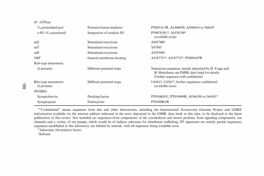
Hþ-ATPase
V0 proteolipid part Potential fusion mediator PT001A13R, AL446850, AJ566616 to 566619
a-SU (V0 associated) Integration of catalytic SU PT007I19U*, AJ538330*
(available soon)
nd2 Stimulated exocytosis AJ437480
nd7 Stimulated exocytosis Y07803
nd9 Stimulated exocytosis AJ293945
NSF General membrane docking AJ347751*, AJ347752*, PT009A07R
Rab-type monomeric
G-proteins Different potential steps Numerous sequences, mainly depostied by D. Fraga and
B. Hinrichsen; see EMBL data bank for details.
Further sequences still confidential
Rho-type monomeric
G-proteins
Different potential steps U03615, U03617, further sequences confidential
(available soon)
SNAREs
Synaptobrevin Docking/fusion PT018K05U, PT018009R, AJ566288 to 566301*
Synaptojanin Endocytosis PT020M01R
a‘‘Confidential’’ means sequences from this and other laboratories, including the International Paramecium Genome Project and GDRE
(information available via the internet address indicated in the text), deposited in the EMBL data bank at this time, to be disclosed at the latest
publication of this review. Not included are sequences from components of the cytoskeleton and motor proteins, from signaling components, ion
channels and a variety of ion pumps, which would be of indirect relevance for membrane traYcking. PT signatures are mainly partial sequences;
sequences established in this laboratory are labeled by asterisk, with all sequences being available soon.bAdenosine ribosylation factor.cSubunit.
189

190 PLATTNER AND KISSMEHL
between adjacent membranes. NSF is an AAA-type ATPase (Neuwald et al.,
1999), which is released upon ATP hydrolysis (Littleton et al., 2001). Once
established, SNARE pins are resistant to disruption by NSF and the SNAP
protein, type aSNAP (Weber et al., 2000). SNAREs are considered relevant
actors not only in docking, but, by some authors, also in membrane fusion
(Nickel et al., 1999). Alternatively work has assigned this function to secre-
tory carrier membrane proteins (SCAMPS) (Brand and Castle, 1993; Liu
et al., 2002). Some time ago, there was indirect evidence of the relevance of
the lipoprotein basepiece, V0 (cf. Wilkens, 2001), of a H þ-ATPase in neuro-
transmitter release (Morel et al., 1991). Latest developments envisage this
molecular component as a fusogen when present without the Hþ-pumping
catalytic part so that matching pairs in adjacent membranes could closely
approach each other and serve this function (Mayer, 2001; Peters et al.,
2001). Also, in yeast, V0 and SNAREs are connected via a Vtc protein
(Muller et al., 2002).
At this time, actual fusogenic proteins are not yet established with any
precision in most systems and, therefore, are still a matter of intense debate
(Zimmerberg, 2001; Jahn and Grubmuller, 2002). Nevertheless, the involve-
ment of NSF, SNAPs, and SNAREs in mediating membrane-to-membrane
contacts is widely accepted. All models operate with molecules arranged side
by side, in both of the membranes to interact. In the case of SNAREs, they
are designated as v or t type, e.g., during exocytosis, depending on whether
they occur on the vesicle membrane or on the the cell membrane as the target
membrane (Sollner et al., 1993a,b; Sollner and Rothman, 1994). Synaptobre-
vin and cellubrevin are v-SNAREs, SNAP-25 and syntaxin are t-SNAREs.
Monomeric ‘‘small’’ GTP-binding proteins (G-proteins) also participate in
vesicle docking (Rothman and Sollner, 1997; Takai et al., 2001).
Exocytosis sites in Paramecium are ultrastructurally clearly defined micro-
domains (Plattner, 1987, 2002; Vayssie et al., 2000). A concept envisaging the
microdomain arrangement of SNAREs at exocytosis sites slowly emerges
also in mammalian systems (Chamberlain et al., 2001; Lang et al., 2001).
While synaptotagmin is an established Ca2þ sensor during stimulated exo-
cytosis (Sugita et al., 2002; Tucker and Chapman, 2002), it also participates
in some internal membrane fusion processes that it modulates (Grimberg
et al., 2003).
When vesicles pinch oV, a molecular machinery that is rather diVerent
from the NSF/SNAP/SNARE complex is engaged (Section IV,B,1). DiVer-
ent ‘‘coat protein complexes’’ help collect the proper components in vesicles
to be formed (Nickel and Wieland, 1997; Kirchhausen, 2000; Malkus et al.,
2002; Yang et al., 2002; Gundelfinger et al., 2003). Pinching oV is mediated
by the monomeric G-protein, dynamin to which diVerent auxiliary proteins
are coassembled. These aspects are discussed in more detail in the respective
sections.

MEMBRANE TRAFFIC IN PARAMECIUM 191
II. Paramecium: A Cell with Elaborate Routes of IntenseMembrane Traffic
A. Value as a Model System
Functional and (ultra)structural features of membrane traYcking and some
of the membrane-to-membrane interaction sites in Paramecium have been
analyzed thoroughly (Allen, 1988; Gortz, 1988; Plattner, 1993) before mo-
lecular biology became available as a tool for this organism. Some aspects,
like the organization of trichocyst docking sites, are so prominent that some
time ago, in connection with classical genetic (Beisson et al., 1976) and
dynamic ultrastructural studies (Knoll et al., 1991), this system delivered
novel hints to important determinants for vesicle docking and membrane
fusion. It then served to establish new models of these basic phenomena
(Plattner, 1987, 2002; Plattner and Kissmehl, 2003). Another aspect of mem-
brane traYcking with impact on work with ‘‘higher’’ eukaryotes dealt with
the digestive cycle (Allen and Fok, 1993a, 2000). This work also revealed
novel aspects, such as phagosome acidification by dedicated organelles, the
acidosomes (Allen and Fok, 1983). Nonetheless, molecular details governing
membrane interactions have remained elusive with this system until recently,
while a number of laboratories analyzing mainly yeast and mammalian cells
achieved great progress at the molecular level.
B. Recent Molecular Approaches to the Study ofMembrane Trafficking
Until recently, information on molecular details was available for Parame-
cium only to a limited extent (Kung et al., 2000), although some additional
molecules previously unknown from mammalian cells had been identified as
determinants of some aspects of membrane traYcking (Vayssie et al., 2000).
On the basis of an indexed genomic library with�60,000 macronuclear DNA
clones (Keller and Cohen, 2000), the group of Jean Cohen (CNRS, Gif-sur-
Yvette) initiated an international Paramecium genome project (Dessen et al.,
2001), which delivers a steadily increasing number of sequences, including
some relevant for basic aspects of membrane traYcking (Table I). It allows us
to design appropriate primers and to clone the respective genes.
Cloning of the Paramecium NSF gene, in collaboration with the Cohen
group, was a pivotal step (Froissard et al., 2002; Kissmehl et al., 2002).
Because NSF is a chaperone for SNAREs and because these interact
with additional components, this now opens the door to many other com-
ponents of the docking/fusion machinery. Such work allows us to predict

192 PLATTNER AND KISSMEHL
immunogenic sites and, with the monospecific antibodies (ABs) thus
produced, we can proceed to the proteomics aspect.
(Ultra)structural localization studies depend greatly on technical achieve-
ments. For NSF, this is particularly intriguing because it is bound only
transiently to sites of membrane interaction and then released. For LM
fluorescence, we have therefore carefully permeabilized cells in the presence
of NEM and the nonhydrolyzable ATP analogue, ATP-g-S (which keeps
NSF in place) under conditions allowing the cells to survive before they were
exposed to anti-NSF ABs (Kissmehl et al., 2002). Gene silencing involved
macronuclear injection of a number of open reading frames (ORFs) of the
NSF gene, followed by single cell processing for EM analysis (Froissard et al.,
2002).
We now see that in many molecules of interest, sequence homologies may
be due to widely scattered amino acids rather than to large motifs. Therefore,
in retrospect, we now understand why, using ABs available from other
systems, previous attempts to identify proteins relevant to membrane
traYcking have consistently failed. With exceptions, this also requires ad-
equate scepticism toward localization studies using ABs against heterologous
proteins in a variety of ciliated protozoa, unless specificity has been docu-
mented with particular care. Molecular biology has turned out to be a
mandatory prerequisite for any further functional and structural work.
C. Established Membrane Trafficking Routes
Cotranslational sequestration of proteins in the rough endoplasmic reticu-
lum (ER) for transport to the Golgi complex and subsequent delivery to
lysosomes or to ‘‘clear secretory vesicles’’ for constitutive (nonstimulated)
exocytosis belong to the standard repertoire of eukaryotic cells, including
Paramecium (Allen, 1988; Gortz, 1988) and other protists (Plattner, 1993). In
addition, these cells also produce dense-core secretory vesicles (‘‘tricho-
cysts’’) for stimulated exocytosis (Adoutte, 1988; Plattner et al., 1993). A
rather intriguing vesicle traYcking system is represented by the digestive
cycle, including the formation of phagosomes (‘‘food vacuoles’’) and their
further processing (Fok and Allen, 1993; Allen and Fok, 2000). Finally, the
osmoregulatory system, normally present in two copies, involves membrane
traYcking ‘‘on the spot,’’ i.e., repeated multiple membrane fusions and
fissions without any large-scale movements (Allen, 2000; Allen and Naitoh,
2002).
Many of these membrane-to-membrane interaction sites are predeter-
mined structurally in Paramecium. This is true for the sites of phagosome
formation in the ‘‘cytostome’’/‘‘cytopharynx’’ area (Allen, 1988; Fok and
Allen, 1993; Allen and Fok, 2000), the release site for spent phagosomes at

MEMBRANE TRAFFIC IN PARAMECIUM 193
the ‘‘cytoproct’’ (Allen and Wolf, 1974), for the reversible attachment sites
of radial canals to the two ‘‘contractile vacuoles’’ and for their outlets at the
cell surface (Tominaga et al., 1998), as well as for the constitutive exo/
endocytosis sites (‘‘parasomal sacs’’) located close to ciliary origins (Allen
et al., 1992), as well as for the docking/release sites of trichcocysts (Pape and
Plattner, 1995; Pouphile et al., 1986; Vayssie et al., 2000).
III. Standard Route: Endoplasmic Reticulum to Golgi Complexand Further On
A wealth of molecular details is known on membrane traYcking in higher
eukaryotes, including integral and membrane-associated proteins, as well as
temporary coats for specific budding processes (Kirchhausen, 2000). Only
circumstantial evidence is available in Paramecium for this obvious route.
The presence of a 61-kDa protein with immunoreactivity with ABs against
mammalian calreticulin-ABs in the ER of Paramecium (Plattner et al.,
1997b) suggests a Ca2þ- and glycosylation-dependent chaperone function,
based on data from other cells (Leach et al., 2002; Schrag et al., 2003). The
same holds for a calnexin homolog and for some disulfide isomerases whose
genes have been cloned (D. Geissinger, R. Kissmehl, H. Plattner, AJ567915,
AJ567916). Glycosylphosphatidylinositol (GPI)-anchored variant surface
antigens (svAGs) are abundantly synthesized in Paramecium (Capdeville,
2000), a process known to take place also in the ER (Ferguson, 1999).
Other prominent ER products comprise precursor proteins of trichocyst
content, the pretrichynins (Gautier et al., 1994, 1996), not to speak of any
other components generally derived from ER.
DiVerent proteins are probably transported to the Golgi complex by
vesicles coated with coatamer proteins (COPs) as molecular filters, notably
‘‘coat protein complex II,’’ COPII (Nickel and Wieland, 1997; Malkus et al.,
2002). Although basic details are not known in Paramecium, the occurrence
of COPs is quite certain because of the EM appearance of such coats (see
later) and because at least one fragmentary gene sequence of one type of COP
is known (Table I).
A Paramecium cell contains a large number, probably several hundred, of
Golgi fields, each of small size (Esteve, 1972; Allen, 1988). The same holds
true of Tetrahymena (Kurz and Tiedtke, 1993). This may explain why the
rather simple task to identify these organelles by specific fluorescent labeling
has not been accomplished to any satisfactory extent. Using ABs against the
Golgi-specific (clathrin-)adaptor protein type AP1 (Traub et al., 1995), we
saw numerous punctate internal labeling sites, which disappeared with
brefeldin A, although at relatively high concentration (M. Momayezi and

194 PLATTNER AND KISSMEHL
H. Plattner, unpublished observations). This agent causes Golgi breakdown
(Nebenfuhr et al., 2002) by interfering with binding of the monomeric
G-protein, type ARF, which normally mediates vesicle delivery. Partial
sequences of ARF are available from Paramecium (Table I). In addition, in
mammalian cells, AP1 is engaged in vesicle traYc from the trans-Golgi
network (TGN) to endocytotic vesicles and back to the TGN (Hinners and
Tooze, 2003). COPI-type proteins would be additional markers (Nickel and
Wieland, 1997; Yang et al., 2002), whereas ‘‘standard’’ Golgi markers gave
no signal (unpublished observation). For AP1 and COPs, see Table I. Defin-
itely more molecular work is needed to identify the Golgi complex on the
light microscope level in future studies on membrane dynamics. This is also
mandatory to understand the budding of primary lysosomes in Paramecium
on the basis of the respective auxiliary molecules, such as AP1 and COPI.
Considering detailed knowledge available from other eukaryotes (Urbe et al.,
1997; Avran and Castle, 1998), much remains to be analyzed with ciliates.
Some of the trichocyst content components are glycosylated, probably
including core and peripheral glycosylation (Luthe et al., 1986; Allen et al.,
1988; Glas-Albrecht et al., 1990). For the ‘‘mesh-like sheath,’’ a structure
connecting the paracrystalline secretory matrix with the membrane in mature
trichocysts (Adoutte, 1988), passage through the Golgi complex has been
documented more clearly. A monoclonal AB, recognizing a 56/57-kDa com-
ponent of the mesh-like sheath, also labels the Golgi complex (Momayezi
et al., 1993). Finally, ABs against pretrichynins also produce Golgi labeling
(Garreau De Loubresse, 1993). The same work, as well as that by Allen et al.
(1989), shows some vesicles, budding oV the Golgi complex which display a
smooth cytoplasmic coat, probably of the coatamer type. Such Golgi vesicles
do not bind ABs against mammalian COPs (which does not exclude such
identity) and side-by-side bristle-coated vesicles are found (Allen and Fok,
1993b, 2000), probably for the transport of lysosomal enzymes. However,
neither budding of COP nor of clathrin-coated vesicles has been analyzed in
any detail in Paramecium as yet. Not only for a COP subunit, but also for the
clathrin heavy chain, sequences are available from Paramecium (Table I) that
should enable one for more detailed work.
In other eukaryotes, lysosomal enzymes may be delivered by clathrin-
coated vesicle budding from the TGN (Urbe et al., 1997; Avran and Castle,
1998). According to the ultrastructural details mentioned, one may assume
the same for ciliates. In Tetrahymena, lysosomal enzymes analyzed so far are
glycosylated (Taniguchi et al., 1985), although they lack a mannose-6-phos-
phate signal (Banno et al., 1993). (Note that not all mammalian lysosomal
enzymes carry this tag.)
The occurrence of small G-proteins in Paramecium has been derived from
Western overlay studies with radioactive GTP-g-S (Peterson, 1991), but their
contribution to any precise step of vesicle traYcking has not been suYciently

MEMBRANE TRAFFIC IN PARAMECIUM 195
analyzed up to now. Table I summarizes preliminary partial cloning of
di Verent types of monomeric G-proteins in Paramecium. In addition to the
work already published (Fraga and Hinrichsen, 1994), these authors have
deposited additional sequences of a variety of monomeric G-proteins (Table
I). This aspect would be particularly interesting regarding the interplay
between G-proteins and SNAREs at membrane interaction sites, as
‘‘throttles and dampers’’ (Rothman and Sollner, 1997). The di Verent G-
proteins are understood as facilitating membrane interactions, while specifi-
city may arise more from SNAREs (Avery et al., 1999; McNew et al., 2000;
Scales et al., 2000; Xue and Zhang, 2002). For their function in vivo, they
require auxiliary proteins of the type guanine nucleotide-activating proteins
(GAP) and guanine nucleotide exchange factors (GEF), as they also emerge
in the Paramecium genome (Table I).
How do Golgi-derived vesicles fuse to form the elaborate trichocyst struc-
tures (Adoutte, 1988; Garreau De Loubresse, 1993)? Considering the com-
plex, polar structure of a trichocyst, with a ‘‘tip’’ and a ‘‘body’’ part, it would
be interesting to know how the specific components of the secretory contents
and the specific membrane components are put together, probably by specific
vesicle fusion processes. This also occurs in higher eukaryotes (Tooze et al.,
2001), where it depends on the Ca2þ sensor synaptotagmin (type III in mast
cells; Grimberg et al., 2003), a gene product not known from Paramecium as
yet. Some glycoproteins may be sorted out when trichocyst contents leave the
Golgi complex (Allen et al., 1989). The matrix of the trichocyst body contains
paracrystalline trichynin proteins derived from pretrichynins by proteolytic
processing (Gautier et al., 1994, 1996). While some components are glycosy-
lated (Glas-Albrecht et al., 1990), di Verential lectin-binding sites are con-
centrated in di Verent domains within a trichocyst (Luthe et al., 1986; Allen
et al., 1988), the tip and contains secretory lectins (Haacke-Bell and Plattner,
1987). In total, these studies support a Golgi passage.
Normal trichocysts are not formed in the ‘‘trichless’’ mutant (Pollack,
1974) due to the absence of posttranslational cleavage of pretrichynins,
which are then released in the untriggered mode (Gautier et al., 1994). This
would be a good model for analyzing the cross-talk between contents and
organelle surface with regard to the choice of the export route—the consti-
tutive and the stimulated pathway, respectively (see Section IV,A). A normal
trichocyst also contains an inherent polarity signal on its membrane surface,
as it normally undergoes saltatory transport along microtubules, in plus !minus direction, with the tip first (Aufderheide, 1977; Plattner et al., 1982;
Glas-Albrecht et al., 1991). In contrast, dense-core secretory vesicles (‘‘chro-
maYn granules’’) isolated from bovine adrenal medullae, after injection into
Paramecium cells, all accumulate at the plus end of microtubules, i.e., the
direction they go in the cells of origin (Glas-Albrecht et al., 1990). Some
mutants cannot deliver their trichocysts, often with aberrant ultrastructure,

196 PLATTNER AND KISSMEHL
to the cell surface and in some other mutants the organellar polarity is
disturbed, resulting in nonextrudable trichocysts at the cell membrane
(Pouphile et al., 1986). Similar mutants defective on diVerent levels of the
secretory pathway have been obtained from Tetrahymena (Sauer and Kelly,
1995; Melia et al., 1998). With these cells it was also possible to show that
secretory content sorting precedes condensation into a mature form
(Turkewitz et al., 1991; Bowman and Turkewitz, 2001).
Another aspect of vesicle traYcking along the Golgi route is the delivery of
vsAGs to the cell membrane, as we found in ultrathin section and freeze-
fracture immuno gold-labeling studies (Flotenmeyer et al., 1999). After
cloning NSF genes in Paramecium (Kissmehl et al., 2002) in our analysis of
the ultrastructural eVects of NSF gene silencing, we found circumstantial
evidence of involvement of NSF-based molecular machinery on the ER and
Golgi level. We found (i) inflated ER cisternae, as if products of ongoing
synthetic activity could no longer be delivered further on, (ii) accumulation
of small vesicles, labeled with ABs against NSF, amidst ER-rich domains,
and (iii) fragmentation or even absence of Golgi areas (Kissmehl et al., 2002).
In addition to NSF, another ATPase, p97, in conjunction with additional
proteins, serves to establish contacts between internal membrane systems
in higher eukaryotes (Uchiyama et al., 2002). No information on this is
available from ciliates.
IV. Membrane Trafficking and Vesicle Movement
A. Exocytosis
1. Constitutive Exocytosis
In Paramecium, this involves delivery of vsAGs to the cell membrane, prob-
ably at the same sites where parasomal sacs are formed during endocytosis
(Capdeville et al., 1993; Flotenmeyer et al., 1999). The fact that these sites
almost always display the coated pit/coated vesicle aspect, rather than show-
ing delivery of smooth vesicles for constitutive exocytosis, may be explained
easily by the longevity of the former, e.g., in metazoans (Marsh and
McMahon, 1999; Sankaranarayanan and Ryan, 2000; Rappoport and
Simon, 2003). NSF gene silencing (Kissmehl et al., 2002) resulted in a more
or less aberrant ultrastructural appearance of these sites, as well as of the
nearby early endosomes (‘‘terminal cisternae’’; see later).
Another constitutive exocytotic process is the release of pretrichynins by
the ‘‘trichless’’ mutant, which cannot transform them by proteolytic cleavage
to paracristalline assemblies (Garreau De Loubresse, 1993). This is also

MEMBRANE TRAFFIC IN PARAMECIUM 197
assumed for corresponding mutants in Tetrahymena (Bowman and
Turkewitz, 2001). Also in Tetrahymena, an experimentally inhibited para-
crystalline arrangement of mucocyst contents was found not to alter mem-
brane tra Ycking (Chilcoat et al., 1996), as the organelles still find their
preformed sites for stimulated exocytosis. The molecular determinants to
divert stimulated from constitutive exocytosis have to be searched in the
secretory organelle membrane components, not only in mammalian systems,
but also in ciliates (Section IV,A,2). In mammalian cells, both pathways are
deviated according to Kelly’s ‘‘selection and exclusion’’ hypothesis for
the delivery of secretory products into the stimulated and the constitutive
pathway, respectively (Kelly, 1985; Urbe et al., 1997).
The ongoing release of lysosomal contents by constitutive exocytosis has
been analyzed most thoroughly in Tetrahymena (Tiedtke et al., 1993). It
appears to be Ca2þ dependent. The sites of release and the final function
are under debate (Florin-Christensen et al., 1990). Remarkably, some
enzymes, such as b-hexosaminidase, are released only partially into the
medium, while they are retained partially on the cell surface (Kiy et al., 1993).
As in other systems, NSF-based processes may be involved not only in
stimulated (Section IV,A,2), but also in constitutive exocytosis (Avran and
Castle, 1998; Gerst, 1999). This has not yet been analyzed with ciliates and it
has to remain open, therefore, for a variety of sites where constitutive
exocytosis occurs in Paramecium. This concerns the two release sites of the
contractile vacuoles and that of spent phagosomes (‘‘cytoproct’’). For the
following reasons, they can be most safely assumed to use NSF and, there-
fore, by implication, also SNARE proteins. These sites are stained intensely
by anti-NSF ABs when NSF detachment is inhibited by blocking its ATPase
activity with ATP-g-S (Kissmehl et al., 2002). From ciliates, no information
is available on synaptotagmin involvement in lysosome traYcking, which is
in contrast to mast cells, where it (negatively) modulates lysosome exocytosis
(Baram et al., 1999). The cytoproct is also distinctly labeled by ABs against
some common sequences of annexins (Knochel et al., 1996). These are ABs
diVerent from those that label trichocyst docking sites. This finding suggests
the involvement of annexins in cytoproct positioning and/or functioning.
2. Stimulated Exocytotic Pathway
Trichocyst docking sites alternate with cilia along longitudinal rows (Allen,
1988). At the emergence of both these organelles, the layer of flat cortical
calcium stores (‘‘alveolar sacs’’), which are tightly attached to the cell mem-
brane, is interrupted. Within the cell membrane, trichocyst docking sites are
delineated by a double row of freeze-fracture particles (proteins) arranged as
a ‘‘parenthesis’’ before and as a �300-nm-wide ‘‘ring’’ after trichocyst
docking, respectively (Plattner et al., 1993; Vayssie et al., 2000). Docking of
198 PLATTNER AND KISSMEHL
a trichocyst normally entails the assembly of approximately nine ‘‘rosette’’
particles (proteins) in the center of a ring, i.e., directly in the fusogenic zone
(Beisson et al., 1976; Pape and Plattner, 1990), as well as of ‘‘connecting
material’’ between the two membranes (Beisson et al., 1980; Plattner et al.,
1980). In most strains, the presence of a rosette is an infallible indication of
exocytotic membrane fusion competence (Pouphile et al., 1986). In fact, a
nondischarge mutant of P. caudatum, which unexpectedly possesses rosettes
(Watanabe and Haga, 1996), was found to perform ‘‘silent’’ membrane
fusion without any visible trichocyst release due to a mutated Ca2þ-binding
component in the secretory contents (Klauke et al., 1998). The only true
exception known is strain nd12, which, when cultivated at a nonpermissive
temperature, possesses rosettes in the absence of any fusion capacity
(Mohamed et al., 2002). Rosette assembly is also required for mucocyst
secretion in Tetrahymena (Orias et al., 1983).
What is the molecular identity of trichocyst docking and fusion machinery,
including connecting material, particularly rosette protein particles? In this
context, a variety of sequences are available from Paramecium (Table I),
notably for the following gene products: NSF, synapto-/cellubrevin, calmo-
dulin (CaM), monomeric G-proteins, and several ND gene products identi-
fied by rescue experiments and complementation cloning with nondischarge
mutants (see later). Considering the many hours-long lifetime of a trichocyst
docking site (Plattner et al., 1993), localization by anti-NSF ABs is not
feasible because NSF is removed rapidly from adjacent membranes after
establishing the SNARE interaction (Section II). Unfortunately, ABs against
Paramecium SNAREs are also not available as yet. However, it could be
shown that the assembly of rosettes, which may require only minutes (Pape
and Plattner, 1990; Plattner et al., 1993; Klauke and Plattner, 2000), clearly
requires SNAREs for the following reasons. In the nd9 mutant, a �2-h-long
temperature shift from nonpermissive to permissive conditions (28�C !
18�C) normally causes the assembly of rosettes and induction of exocytosis
capacity. NSF gene silencing applied during a 28�C ! 18
�C transfer inhibits
rosette assembly and functional recovery (Froissard et al., 2002).
Another component of trichocyst docking sites is the Ca2þ-binding pro-
tein, CaM. We localized CaM by ABs against CaM from Paramecium
precisely at docking sites (Momayezi et al., 1986), which is particularly
concise in isolated cell surface complexes (‘‘cortices’’). Conversely,
Kerboeuf et al. (1993) induced rosette assembly in a cam-mutant, containing
mutated CaM, by transfection with the wild-type CaM gene. This was
paralleled by acquirement of exocytosis competence. The role of CaM in
establishing and/or maintaining trichocyst docking/exocytosis sites in a func-
tional state is supported by the inhibitory eVect of ‘‘anti-CaM drugs,’’ which,
in Paramecium, cause the detachment of trichocysts (Klauke and Plattner,
2000). CaM was found to participate in yeast vacuole fusion (Peters and

MEMBRANE TRAFFIC IN PARAMECIUM 199
Mayer, 1998) and in dense-core vesicle exocytosis in mammlians. In this
context, CaM may account for a Ca2þ-dependent priming of exocytosis, as
it binds to a variety of components of the docking/fusion machinery, such as
the v-SNARE, synaptobrevin (Quetglas et al., 2002), rab-type monomeric G-
protein (Coppola et al., 1999), and the putative Ca2þ sensor, synaptotagmin
(Tucker and Chapman, 2002). Thus, Ca2þ not only mediates exocytotic
membrane fusion, but it also primes docking sites (at a preceding stage;
Von Rueden and Neher, 1993). Although synaptotagmin—the Ca2þ sensor
to be expected also in ciliates—has so far remained undetected in Parame-
cium, we presume its occurrence based on a similar requirement of exocytotic
membrane fusion for local Ca2þ increase (Plattner and Klauke, 2001). This is
supported by acceleration of the exo–endocytosis cycle by increasing the
extracellular Ca2þ concentration (Plattner et al., 1997b), as it also occurs,
for instance, in neuronal cells (Palfrey and Artalejo, 1998). Trichocyst exo-
cytosis, which, just like in any other eukaryote, requires a Ca2þ signal
(Erxleben et al., 1997; Plattner and Klauke, 2001), undoubtedly requires a
Ca2þ sensor, but CaM is less likely to act as a sensor than synaptotagmin
(Sugita et al., 2002). The actual Ca2þ sensor in ciliates definitely has to be
identified.
Currently, we assume that the following components contribute to forma-
tion of the ‘‘connecting material’’ described previously: (i) CaM is one of the
components; (ii) we conclude from indirect evidence obtained in our NSF
studies that SNAREs are additional ones; (iii) furthermore, additional pro-
teins, including monomeric G-proteins may contribute at least transiently;
(iv) a synaptotagmin-like Ca2þ sensor appears mandatory; and (v) additional
components are the ND7 (Skouri and Cohen, 1997) and ND9 gene products
(Froissard et al., 2001) and possibly some proteins interacting with ABs
against common sequences from mammalian annexins (Knochel et al., 1996).
In a scrutinized discussion on mechanisms of exocytotic membrane fusion
in Paramecium (Plattner and Kissmehl, 2003), we tried to evaluate the
potential relevance of two of the currently most discussed candidates for
fusion pore formation: V0 part of the Hþ-ATPase and SNAREs, respectively
(Section II). For the availability of V0 genes and the a subunit, see Table I.
Based on structural details of trichocyst exocytosis sites and their ultrastruc-
tural transformation during membrane fusion (Knoll et al., 1991), we found
some arguments in favor of V0, but unambiguous clarification again requires
unequivocal identification by molecular biology. Conversely, is it mere coin-
cidence that approximately nine rosette particles occur at an active trichocyst
exocytosis site and that up to nine active fusion complexes in the sea urchin
egg are calculated to be required for optimal operation of an exocytotic event
(Vogel et al., 1996)? Meanwhile, molecular data on Hþ-ATPase components
accumulate steadily and, in conjunction with ABs against its subunits and
against SNAREs, may finally allow us to analyze their relevance for

200 PLATTNER AND KISSMEHL
membrane fusion during trichocyst exocytosis. Consider that no decision has
been reached so far in any other system where dense-core secretory organelle
docking/fusion sites are structurally much less distinct.
B. Endocytosis, Phagocytosis, and Lysosomal System
1. Endocytosis
This is considered separate from phagocytosis (see Section IV,B,2) and we
diVerentiate between exocytosis-coupled endocytosis and constitutive endo-
cytosis via parasomal sacs. Considering the very detailed molecular infor-
mations on exo–endocytosis coupling in mammalian cells (Gundelfinger
et al., 2003), amazingly little is known in Paramecium.
All work with Paramecium agrees that trichocyst ‘‘ghosts’’ are resealed,
fragmented, and internalized without a clathrin coat (Allen and Fok,
1984; Plattner et al., 1985). According to quenched-flow/freeze-fracture an-
alysis, all these steps are accelerated by an increasing extracellular Ca2þ
concentration (Plattner et al., 1997a). Right after resealing, a ‘‘ring þ ros-
ette’’ configuration is transformed into a ‘‘filled ring’’ and then to a ‘‘paren-
thesis’’ (see Section IV,A,2) as ghosts detach (Knoll et al., 1991; Plattner
et al., 1997b). Ghosts are removed completely, depending on the strain
analyzed, normally with t1/2 �3 to 9 min (Plattner et al., 1985). This step is
also accelerated with an increasing extracellular Ca2þ concentration during
stimulation (Plattner et al., 1997a), as it also occurs, for instance, in neurons
(Palfrey and Artalejo, 1998) where it accelerates the internalization of
SNAREs by endocytosis (Sankaranarayanan and Ryan, 2001). Trichocyst
docking sites become available again for the insertion of new trichocysts,
depending on organelle biogenesis, at a much slower rate (Plattner et al.,
1993).
Vesicles arising from exocytosis-coupled endocytosis are of the smooth
type (Plattner et al., 1985) and travel far into the cell, even close to the Golgi,
complex as shown by peroxidase labeling (Allen and Fok, 1980, 1984), but
recycling could not be shown. After a limited extent of exocytosis, the marker
goes to secondary lysosomes (Allen and Fok, 1984). Massive exocytosis
induction in the presence of a fluid phase marker entails delivery to digesting
vacuoles (Luthe et al., 1986).
Parasomal sacs are established sites of ongoing endocytosis in Paramecium
and in Tetrahymena, as shown by the fluid phase marker horseradish perox-
idase (Allen and Fok, 1993a; Allen et al., 1992) and by the application of
cationic ferritin (Nilsson and VanDeurs, 1983), respectively. After endocy-
totic uptake, these markers reach flat compartments that have not been
identified so far. Although parasomal sacs display a bristle coat, its identity
MEMBRANE TRAFFIC IN PARAMECIUM 201
with clathrin should now finally be shown because the gene of the heavy
chain is now known (Table I). As to physiological functions of these sites,
GPI-anchored vsAGs are pressed into these sites, probably by their mere
abundance, and thus subjected to endocytosis and delivery to early endo-
somes (Allen et al., 1992; Allen and Fok, 1993; Fok and Allen, 1993), called
‘‘terminal cisternae,’’ and food vacuoles, as derived from immunogold-label-
ing experiments with Paramecium (Flotenmeyer et al., 1999). This transport
to digesting vacuoles may serve a permanent turnover (Capdeville et al.,
1993).
In higher eukaryotes, coated vesicle formation requires an established set
of proteins. Altogether, the molecular machinery required for endocytosis
via coated pits is widely di Verent from that for exocytosis. For the availabil-
ity of gene sequences, see Table I. Depending on the type of endocytosis
vesicle, this may include clathrin and its adaptor protein, AP2, dynamin,
synaptojanin, and so on (Hinshaw, 2000; Sorkin, 2000), but not SNARE
assemblies (Holroyd et al., 2002). There are di Verent types of endocytosis
vesicles and not all components are necessarily occurring at the same time,
but dynamin is common to most of them (Conner and Schmid, 2003). In fact,
dynamin does show up in Paramecium (AJ386320, AF357193, AL448655), in
addition to the following indirect hints to its occurrence. Dynamin is depho-
sphorylated during endocytosis by the protein phosphatase 2B (PP2B, calci-
neurin, CaN) (Marks and McMahon, 1998). PP2B has a catalytic CaM-
binding A subunit and a Ca2þ-binding regulatory B subunit (Klee et al.,
1998). CaN has been isolated from Paramecium, and ABs prepared against it
(Kissmehl et al., 1997) strongly labeled parasomal sacs on the EM level
(Momayezi et al., 2000). Meanwhile, two genes of the A subunit has been
cloned in the laboratory of R. D. Hinrichsen (AF 014922), and the B subunit
has been cloned also in our laboratory (Table I). No other components are
identified in Paramecium. This also holds for endosomes themselves.
Actin-binding proteins are also recruited to endocytosis sites (Marsh and
McMahon, 1999; Qualman and Kessels, 2002). In a late stage of coated
vesicle release from the cell membrane, F-actin assembly, between the plasma
membrane and the pinched-oV endocytotic vesicle, emerges in mammalian
cells (Merrifield et al., 2002; Tse et al., 2003). Although F-actin occurs in the
Paramecium cortex, its precise localization remains to be elucidated (see
Section IV,B,2).
Membrane piecemeals from digesting vacuoles, after defecation, are re-
trieved from the cytoproct (Allen and Wolf, 1974; Allen and Fok, 1993a,
2000). Discoidal vesicles thus formed serve recycling to the nascent food
vacuole. It is not understood whether labeling of the cytoproct by ABs against
some common sequences of annexins (Knochel et al., 1996) is to be seen in
connection with the exocytotic and/or with the endocytotic activities taking
place at this site or merely in conjunction with positioning of the cytoproct.

202 PLATTNER AND KISSMEHL
ABs against Paramecium NSF also label the cytoproct when the dissociation
of NSF is inhibited (Kissmehl et al., 2002). This indicates the involvement of a
SNARE machinery, probably in the exocytosis of the spent food vacuole.
2. Phagocytosis and Lysosomal System
Food bacteria are engulfed by ciliates at the cytopharynx, by formation of a
nascent food vacuole that is acidified to kill the ‘‘prey,’’ before the digesting
(food) vacuole migrates by cyclosis through the cell. F-actin contributes to
phagosome formation (see later) just as in mammalian cells (Zhang et al.,
2002; Tse et al., 2003). Concerning Paramecium, we are ignorant about any
involvement of rho-type monomeric G-proteins known in other cells
(Chimini and Chavrier, 2000), but corresponding sequences are present
(Table I). The subsequent pathway involves delivery and retrieval of
lysosomal enzymes (Allen and Fok, 1993a, 2000; Fok and Allen, 1993).
In contrast to these scrutinized ultrastructural and functional analyses of
the phagolysosomal cycle in Paramecium, molecular data are scant—in
striking contrast to the wealth of information in mammalian cells (Chimini
and Chavrier, 2000; Garin et al., 2001). We know nothing about the occur-
rence of the standard immuno marker for both, late endosomes and lyso-
somes, Lamp-2 (Clague, 1998), in ciliates. Again, although ABs against NSF
also label the site of phagosome formation in Paramecium (Kissmehl et al.,
2002), it is not known precisely which process it serves, i.e., phagosome
fission, fusion with discoidal vesicles and/or acidosomes. From work with
other cells, one may assume that only membrane fusions, but not fission
processes, e.g., during phagosome formation, would require a NSF/SNAP/
SNARE machinery (Hinshaw, 2000; Sorkin, 2000). However, other work
assumes that NSF would participate in phagosome formation (Coppolino
et al., 2001), and phagosomal membranes isolated from mouse macrophages
were found to contain such components (Garin et al., 2001). Any role of NSF
in phagosome formation, if any, remains to be elucidated, but its occurrence
may equally well reflect the multiple membrane-to-membrane interactions
occurring in the life of a phagosome.
Before a nascent food vacuole is pinched oV, two events take place:
(i) F-actin is assembled (Allen and Fok, 1983; Kersken et al., 1986) and
(ii) a number of acidosomes line up around the surface of a nascent food
vacuole with which, after pinching oV, they fuse rapidly (Allen and Fok,
1983). Acidosomes are small vesicles, so far of undetermined origin, that
transport Hþ-ATPase molecules to nascent phagosomes. For sequences
available, see Table 1. Whether an equivalent pathway also occurs in mam-
malian cells is not ascertained. Also, the signal for the rapid, multiple fusion
of acidosomes with a food vacuole remains to be elucidated. Also unknown is
the involvement, along the digestive cycle, of V0 subunits of the Hþ-ATPase

MEMBRANE TRAFFIC IN PARAMECIUM 203
in the respective fusion processes. If V0 subunits would ever act as a fusogen,
this would most likely occur here, where it abounds so much (Fok et al.,
2002), particularly as this function has been derived from analyses with yeast
vacuoles (Section II). The binding of CaM to phagosomal membranes along
their pathway (Momayezi et al., 1986) suggests a role in membrane inter-
action, as it is required for such interaction and final fusion in yeast vacuoles
(Peters and Mayer, 1998). Accordingly, CaM and Ca2þ/CaM-binding pro-
teins are encountered on the surface of phagocytic vacuoles of T. thermophila
(Gonda et al., 2000).
As outlined in Section IV,A,1, materials ingested by smooth vesicles
formed during exocytosis-coupled endocytosis, as well as by coated vesicles,
can also be delivered to the phagolysosomal system. In this case, a signal
indicating the ‘‘empty’’ state has to mediate this transport route. This is
opposite to the findings during ‘‘frustrated exocytosis,’’ where a signal indi-
cating the ‘‘filled’’ state allows the reattachment of trichocysts previously
detached without preceding contents release (Section IV,C,1).
NSF gene-silencing experiments have resulted in the inhibition of phago-
cytotic vacuole formation, decreasing cell size, and finally in cell death
(Froissard et al., 2002; Kissmehl et al., 2002). Immunofluorescence labeling
by anti-NSF ABs was visible most clearly at the beginning and at the end of
the digestive cycle, i.e., at the cytopharynx and the cytoproct, respectively.
However, at the EM level, the increased delivery of NSF into bona fide
lysosomes was recognized (Kissmehl et al., 2002). It is diYcult to judge
whether this simply indicates the disposal of aged NSF molecules in the
absence of any further biosynthesis.
Further aspects of membrane traYcking along the phagosomal route, to
be elucidated on a molecular scale, are the fusion with lysosomes and the
retrieval of lysosomal enzymes at a later stage (Allen and Fok, 1993a, 2000;
Fok and Allen, 1993). Ciliates oVer the advantage of precise timing of the
phagocytic cycle, i.e., one can isolate defined phagosomal stages, as estab-
lished for Tetrahymena (Vosskuhler and Tiedtke, 1993). In Tetrahymena,
some of the spots of two-dimensional electrophoresis gels have been partially
microsequenced (Maicher and Tiedtke, 1999)—two of them were novel.
Along the phagocytic pathway of T. thermophila diVerent monomeric
G-proteins are attached sequentially (Meyer et al., 1998). This may reflect
selective vesicle interaction steps.
Very strikingly, NSF gene silencing increased the number of autophago-
somes considerably (Kissmehl et al., 2002), although their formation begins
with closure of a wrapping membrane compartment (ER), i.e., by a mem-
brane fusion process. Our data are compatible with findings in Saccharo-
myces cerevisiae (Ishihara et al., 2001), where this step does not require NSF/
SNARE machinery, in contrast to fusion of autophagosomes with the vacu-
ole (lysosome). Inhibition of this NSF-dependent step may be one reason of

204 PLATTNER AND KISSMEHL
the accumulation of autophagosomes in Paramecium after NSF gene silen-
cing. The primary reason may be their increased formation due to starvation,
as NSF gene silencing shuts down food delivery via phagocytosis (Froissard
et al., 2002). Accordingly, in Tetrahymena, starvation induces autophagy
(Nillson and VanDeurs, 1983).
To summarize, with none of the manifold membrane interactions occur-
ring during the digestive cycle is the molecular basis of membrane interaction
and fusion known in any satisfactory detail. However, one may reasonably
assume the following details. (i) F-actin, which coats nascent and later food
vacuoles (Allen and Fok, 1983; Kersken et al., 1986), may participate in or
even mediate cyclosis, and it would have to be organized in a loose form to
allow organelle interactions. F-actin contributes to phagosome formation,
just like in mammalian cells (Zhang et al., 2002; Tse et al., 2003). (ii) CaM
and a NSF-based machinery are probably involved in these membrane
interactions. (iii) Apart form SNAREs, V0 would be available as a candidate
for fusion pore formation.
F-actin is generally assumed to be required to drive cyclosis, also in
Paramecium (Sikora, 1981). Several actin genes have been cloned in Parame-
cium by Dıaz-Ramos et al. (1998) and in our laboratory. Data available
previously have been corrected and supplemented by additional sequences
(R. Kissmehl, E. Wagner, J. Mansfeld, and H. Plattner, unpublished obser-
vations). The localization and function of the respective gene products have
not yet been completed.
C. Long-Range Vesicle Movements
1. Detachment of Mature Trichocysts from the Cell Surface
The application of ‘‘anti-CaM drugs’’ or of increased Mg2þ concentrations in
the medium can detach exocytosis-competent trichocysts from the cell mem-
brane (Klauke and Plattner, 2000). The requirement of CaM for establishing
trichocyst docking sites has been commented in Section IV,A,2. It is not
known whether the enrichment of CaN at and close to docking sites
(Momayezi et al., 2000) is relevant directly for exocytosis or exocytosis-
coupled endocytosis or indirectly, e.g., for governing Ca2þ dynamics. Experi-
mentally detached trichocysts (still containing their secretory contents) can
be reinserted and undergo stimulated exocytosis. From this we conclude that
(i) trichocysts dispose of a ‘‘filled’’ signal and (ii) that CaM is involved in
docking (see also Section IV,A,2). No comparable information is available
from any other eukaryotes.
In Paramecium, as in mammals, an ‘‘empty-or-filled’’ signal may be repre-
sented by transmembrane proteins with a lumenal link to the secretory

MEMBRANE TRAFFIC IN PARAMECIUM 205
contents. On a speculative basis, in Paramecium, such a link could be repre-
sented by a component of the ‘‘mesh-like sheath,’’ notably by a 56-kDa
protein (Momayezi et al., 1993) (see Section IV,A,2). One could speculate
whether the GP-2 protein in pancreatic zymogen granules could serve a
similar function (Kalus et al., 2002). In both these cases, the existence of an
‘‘empty/filled’’ signal and its involvement in directed transport would require
much more scrutinized analysis.
2. Microtubules as Long-Range Signals for Vesicle Trafficking
A Paramecium cell has a variety of distinct arrays of microtubules (Cohen
and Beisson, 1988), among which some are displayed along distinct vesicle
transport routes. Microtubules emanate from the cytoproct (Allen and Wolf,
1974) where they may serve the docking of a spent food vacuole. Another
population of microtubular ‘‘rails’’ is installed between the cytoproct and the
cytopharynx for discoidal vesicle transport (Allen and Fok, 1980). Microtu-
bules also flank the radial canals of the osmoregulatory system (Allen, 2000;
Allen and Naitoh, 2002), which they may stabilize. Finally, a much less
obvious population originates from ciliary basal bodies in a vertical direction
from the cell surface and mediates saltatory docking of trichocysts (Plattner
et al., 1982; Glas-Albrecht et al., 1991). The g–tubulin, associated with ciliary
basal bodies (Ruiz et al., 1999), is required to establish basal bodies as
microtubule-organizing centers. Microtubules do not participate in mem-
brane transport in the opposite direction, i.e., exocytosis-coupled endocytosis
(Plattner et al., 1985).
For versicle traYcking in Paramecium, cytoplasmic dynein is the only
motor protein that has been functionally characterized up until now
(Schroeder et al., 1990). Due to its preference for ‘‘þ ! �’’ transport, it
may be involved in trichocyst docking. Remarkably, such ‘‘þ!�’’ directed
transport to the cell periphery, as established for trichocysts (Plattner et al.,
1982; Glas-Albrecht et al., 1991), has also been found in epithelia (Bacallao
et al., 1989; Bre et al., 1990).
D. Results Relevant to Membrane Trafficking in Paramecium
Table I summarizes the relevant molecular details known so far from Para-
mecium. Many sequences available are fragmentary and, thus, require con-
firmation and further specification by additional work. With partial
sequences it cannot be excluded that they are domains occurring in diVerent
proteins. So far it appears that Paramecium, on an early stage of evolution, is
using, in principle, the same molecular elements as ‘‘higher’’ eukaryotic
systems.

206 PLATTNER AND KISSMEHL
Originally, data have been derived from the analysis of the respec-
tive proteins. Complementation cloning, with identification of the respective
gene and gene product, was another approach (Haynes et al., 1996) that
proved highly rewarding in the analysis of membrane tra Ycking (Skouri and
Cohen, 1997; Vayssie et al., 2000; Froissard et al., 2001). In the last few years,
the international Paramecium genome project (Dessen et al., 2001) has
provided important input. This is currently pursued on a European scale as
a GDRE project (Groupement de Recherche Europeen), headed and
administered by Jean Cohen and Linda Sperling (CNRS, Gif-sur-Yvette;
France). An internet connection is available at http://paramecium.cgm.
cnrs-gif.fr.
With most genes analyzed so far in Paramecium, more than one, frequently
several, isoforms are found. They are frequently similar to each other, thus
facilitating the silencing of di Verent genes in the course of a single gene
(ORF) injection (Ruiz et al., 1998; Bastin et al., 2001). It remains to be
seen whether this is an alternative to alternative splicing, whether all gene
variations are expressed, and whether the corresponding gene products are
diVerentially positioned in the cell. Rapid progress of the Tetrahymena
genome project (Turkewitz et al., 2002) will be an additional potential source
for unraveling molecular details pertinent to membrane traYcking in ciliated
protozoa.
V. Conclusions and Perspectives
A vast body of cell-biological work with Paramecium provides a solid base-
line, which, together with rapid progress in molecular biology, will maintain
the status of this organism as an important model system. One of the main
questions to solve will be: How is the regular arrangement of the diverse
membrane traYcking pathways determined?
In the past, specifically in Paramecium, complementation cloning proved
useful to identify relevant genes and the function of the respective gene
products (Sections II,B and IV,D). Microinjection of appropriate constructs
into the macronucleus allows us to obtain transfected clones (Vayssie et al.,
2000). This includes the overexpression of GFP–fusion proteins (Hauser
et al., 2000a,b). It is hoped that electroporation or ‘‘bioballistic’’ methods
(bombardment with DNA-coated gold particles) will allow us to achieve
stable transfectants (Boileau et al., 1999). Homology-dependent gene silen-
cing by injection of a large number of ORFs, without flanking regions (Ruiz
et al., 1998; Bastin et al., 2001; Galvani and Sperling, 2001), has already
proved useful in analyzing some aspects of membrane traYcking in Parame-
cium (Froissard et al., 2001; Kissmehl et al., 2002). Its eVect is not fully

MEMBRANE TRAFFIC IN PARAMECIUM 207
understood, but basically it may be due to the binding of complementary
nucleotide sequences (Wassengger, 2002). Recent methodological develop-
ments may render gene silencing more easily applicable by feeding of trans-
fected bacteria (Galvani and Sperling, 2002). In Tetrahymena, ‘‘antisense
ribosome’’ systems have been established (Turkewitz et al., 2002). This
involves vectors with an antisense 50 part inserted into ribosomes.
Because membrane tra Yc can be studied only in the structural context of
a cell, the highly regular pattern of a Paramecium cell will keep this organism
as a focus of interest. This is a realistic goal, particularly as it becomes
increasingly amenable to molecular biology.
Note added in proof:
To Section I. The most detailed account available of the Paramecium genome
project (Sperling et al., 2002, Eukaryot. Cell 1, 341–352) will soon be supple-
mented by additional sequences. Any new informations can be drawn from
the internet address of the colleagues guiding this and the GDRE project,
J. Cohen and L. Sperling (http://paramecium.cgm.cnrsgif.fr).
To Section III. Meanwhile sequences encoding parts of synaptotagmin
gene(s) have been identified in our laboratory.
To Section IV,B,1. For partial sequences of dynamin 2 and rab 7 in
Paramecium, see Surmacz et al., 2003, Biol. Cell 95, 69–74.
To Section IV,B,2. Synaptotagmin has been reported to participate also
in internal fusion processess, like formation of some endosomal compart-
ments (Grimberg et al., 2003, J. Cell Sci. 116, 145–154.)
Acknowledgments
We thank all our co-workers and cooperation partners for their invaluable contributions
integrated in this review, and specifically Dr. J. Hentschel for elaborating the scheme. We
gratefully acknowledge the financial support of the authors work cited herein by the Deutsche
Forschungsgemeinschaft.
References
Adoutte, A. (1988). Exocytosis: Biogenesis, transport and secretion of trichocysts. In
‘‘Paramecium’’ (H. D. Gortz, Ed.), pp. 325–362. Springer-Verlag, Berlin.
Allen, R. D. (2000). The contractile vacuole and its membrane dynamics. BioEssays 22,
1035–1042.
Allen, R. D. (1988). Cytology. In ‘‘Paramecium’’ (H. D. Gortz, Ed.), pp. 4–40. Springer-Verlag,
Berlin.
Allen, R. D., and Fok, A. K. (1980). Membrane recycling and endocytosis in Paramecium
confirmed by horseradish peroxidase pulse-chase studies. J. Cell Sci. 45, 131–145.

208 PLATTNER AND KISSMEHL
Allen, R. D., and Fok, A. K. (1983). Nonlysosomal vesicles (acidosomes) are involved in
phagosome acidification in Paramecium. J. Cell Biol. 97, 566–570.
Allen, R. D., and Fok, A. K. (1984). Membrane behavior of exocytic vesicles. III. Flow of
horseradish peroxidase labeled trichocyst membrane remnants in Paramecium. Eur. J. Cell
Biol. 35, 27–34.
Allen, R. D., and Fok, A. K. (1993a). Endosomal membrane traffic of ciliates. In ‘‘Membrane
Traffic in Protozoa’’ (H. Plattner, Ed.), pp. 283–309. JAI Press, Greenwich, CT.
Allen, R. D., and Fok, A. K. (1993b). Nonclathrin vesicle coats and filament networks in the
transition zone and trans-Golgi region of the Golgi complex of Paramecium. J. Struct. Biol.
110, 215–226.
Allen, R. D., and Fok, A. K. (2000). Membrane trafficking and processing in Paramecium. Int.
Rev. Cytol. 198, 277–317.
Allen, R. D., and Naitoh, Y. (2002). Osmoregulation and contractile vacuoles of protozoa. Int.
Rev. Cytol. 215, 351–394.
Allen, R. D., and Wolf, R. W. (1974). The cytoproct of Paramecium caudatum: Structure and
function, microtubules, and fate of food vacuole membranes. J. Cell Sci. 14, 611–631.
Allen, R. D., Schroeder, C. C., and Fok, A. K. (1992). Endosomal system of Paramecium:
Coated pits to early endosomes. J. Cell Sci. 101, 449–461.
Allen, R. D., Schroeder, C. C., and Fok, A. K. (1989). Intracellular binding of wheat germ
agglutinin by Golgi complexes, phagosomes and lysososmes in Paramecium multimicronu-
cleatum. J. Histochem. Cytochem. 37, 195–202.
Allen, R. D., Ueno, M. S., and Fok, A. K. (1988). A survey of lectin binding in Paramecium. J.
Protozool. 35, 400–407.
Aufderheide, K. J. (1977). Saltatory motility of uninserted trichocysts and mitochondria in
Paramecium tetraurelia. Science 198, 299–300.
Avery, J., Jahn, R., and Edwardson, J. M. (1999). Reconstitution of regulated exocytosis in cell-
free systems: A critical appraisal. Annu. Rev. Physiol. 61, 777–807.
Avran, P., and Castle, D. (1998). Sorting and storage during secretory granule biogenesis:
Looking backward and looking forward. Biochem. J. 332, 593–610.
Bacallao, R., Antony, C., Dotti, C., Karsenti, E., Stelzer, E. H. K., and Simons, K. (1989). The
subcellular organization of Madin-Darby canine kidney cells during the formation of a
polarized epithelium. J. Cell Biol. 109, 2817–2832.
Banno, Y., Okano, Y., Furukawa, K., Tiedtke, A., Kobata, A., and Nozawa, Y. (1993).
Processing and secretion of lysosomal acid a-glucosidase in Tetrahymena wild type and
secretion-deficient mutant cells. J. Eukaryot. Microbiol. 40, 515–520.
Baram, D., Adachi, R., Medalia, O., Tuvirn, M., Dickey, B., Makori, Y., and Sagi-Eisenberg,
R. (1999). Synaptotagmin II negatively regulates Ca2þ-triggered exocytosis of lysosomes in
mast cells. J. Exp. Med. 189, 1649–1658.
Bastin, P., Galvani, A., and Sperling, L. (2001). Genetic interference in protozoa. Res.
Microbiol. 152, 123–129.
Beisson, J., Cohen, J., Lefort-Tran, M., Pouphile, M., and Rossignol, M. (1980). Control of
membrane fusion in exocytosis: Physiological studies on a Paramecium mutant blocked in the
final step of the trichocyst extrusion process. J. Cell Biol. 85, 213–227.
Beisson, J., Lefort-Tran, M., Pouphile, M., Rossignol, M., and Satir, B. (1976). Genetic analysis
of membrane differentiation in Paramecium: Freeze-fracture study of the trichocyst cycle in
wild-type and mutant strains. J. Cell Biol. 69, 126–143.
Boileau, A. J., Kissmehl, R., Kanabrocki, J. A., and Saimi, Y. (1999). Transformation of
Paramecium tetraurelia by electroporation or particle bombardement. J. Eukaryot. Microbiol.
46, 56–65.
Bowman, G. R., and Turkewitz, A. P. (2001). Analysis of a mutant exhibiting conditional sorting
to dense core secretory granules in Tetrahymena thermophila. Genetics 159, 1605–1616.

MEMBRANE TRAFFIC IN PARAMECIUM 209
Brand, S. H., and Castle, J. C. (1993). SCAMP 37, a new marker within the general cell surface
recycling system. EMBO J. 12, 3753–3761.
Bre, M. H., Pepperkok, R., Hill, A. M., Levilliers, N., Ansorge, W., Stelzer, E. H. K., and
Karsenti, E. (1990). Regulation of microtubule dynamics and nucleation during polarization
in MDCK II cells. J. Cell Biol. 111, 3013–3021.
Capdeville, Y. (2000). Paramecium GPI proteins: Variability of expression and localization.
Protist 151, 161–169.
Capdeville, Y., Charret, R., Antony, C., Delorme, J., Nahon, P., and Adoutte, A. (1993).
Ciliary and plasma membrane proteins in Paramecium: Description, localization, and
intracellular transit. In ‘‘Membrane Traffic in Protozoa’’ (H. Plattner, Ed.), pp. 181–226. JAI
Press, Greenwich, CT.
Chamberlain, L. H., Burgoyne, R. D., and Gould, G. W. (2001). SNARE proteins are highly
enriched in lipid rafts in PC12 cells: Implications for the spatial control of exocytosis. Proc.
Natl. Acad. Sci. USA 98, 5619–5624.
Chilcoat, N. D., Melia, S. M., Haddad, A., and Turkewitz, A. P. (1996). Granule lattice protein
1 (Grl1p), an acidic, calcium-binding protein in Tetrahymena thermophila dense core
secretory granules, influences granule size, shape, content organization, and release but not
protein sorting or condensation. J. Cell Biol. 135, 1775–1787.
Chimini, G., and Chavrier, P. (2000). Function of Rho family proteins in actin dynamics during
phagocytosis and engulfment. Nature Cell Biol. 2, E191–E196.
Clague, M. J. (1998). Molecular aspects of the endocytic pathway. Biochem. J. 336, 271–282.
Cohen, J., and Beisson, J. (1988). The cytoskeleton. In ‘‘Paramecium’’ (H. D. Gortz, Ed.), pp.
363–392. Springer-Verlag, Berlin.
Conner, S. D., and Schmid, S. L. (2003). Regulated protals of entry into the cell. Nature 422,
37–44.
Coppola, T., Perret-Manoud, V., Luthi, S., Farnsworth, C. C., Glomset, J. A., and Regazzi, R.
(1999). Distruption of Rab3-calmodulin interaction, but not other effector interactions,
prevents Rab3 inhibition of exocytosis. EMBO J. 18, 5885–5891.
Coppolino, M. G., Kong, C., Mohtashami, M., Schreiber, A. D., Brumell, J. H., Finlay, B. B.,
Grinstein, S., and Trimble, W. S. (2001). Requirement for N-ethylmaleimide-sensitive factor
activity at different stages of bacterial invasion and phagocytosis. J. Biol. Chem. 276,
4772–4780.
Dessen, P., Zagulski, M., Gromadka, R., Plattner, H., Kissmehl, R., Meyer, E., Betermier, M.,
Schultz, J. E., Linder, J. U., Pearlman, R. E., Kung, C., Forney, J., Satir, B. H., Van Houten,
J. L., Keller, A. M., Froissard, M., Sperling, L., and Cohen, J. (2001). Paramecium genome
survey: A pilot project. Trends Genet. 17, 306–308.
Dıaz-Ramos, C., Villalobo, E., Perez-Romero, P., and Torres, A. (1998). Paramecium
tetraurelia encodes unconventional actin containing short introns. J. Eukaryot. Microbiol.
45, 507–511.
Erxleben, C., Klauke, N., Flotenmeyer, M., Blanchard, M. P., Braun, C., and Plattner, H. (1997).
Microdomain Ca2þ activation during exocytosis in Paramecium cells: Superposition of local
subplasmalemmal calcium store activation by local Ca2þ influx. J. Cell Biol. 136, 597–607.
Esteve, J. C. (1972). L’appareil de Golgi des cilies. Ultrastructure, particulierement chez
Paramecium. J. Protozool. 19, 609–618.
Ferguson, M. A. J. (1999). The structure, biosynthesis and functions of glycosylpho-
sphatidylinositol anchors, and the contributions of trypanosome research. J. Cell Sci. 112,
2799–2809.
Florin-Christensen, J., Florin-Christensen, M., Rasmussen, L., and Tiedtke, A. (1990). Release
of lysosomal enzymes in Tetrahymena: A calcium-dependent event. In ‘‘Calcium as an
Intracellular Messenger in Eukaryotic Microbes’’ (D. H. O’Day, Ed.), pp. 151–164. Am. Soc.
Microbiol, Washington, DC.

210 PLATTNER AND KISSMEHL
Flotenmeyer, M., Momayezi, M., and Plattner, H. (1999). Immunolabeling analysis of
biosynthetic and degradative pathways of cell surface components (glycocalyx) in
Paramecium cells. Eur. J. Cell Biol. 78, 67–77.
Fok, A. K., and Allen, R. D. (1993). Membrane flow in the digestive cycle of Paramecium. In
‘‘Membrane Traffic in Protozoa’’ (H. Plattner, Ed.), pp. 311–337. JAI Press, Greenwich, CT.
Fok, A. K., Yamauchi, K., Ishihara, A., Aihara, M. S., Ishida, M., and Allen, R. D. (2002). The
vacuolar-ATPase of Paramecium multimicronucleatum gene structure of the B subunit and the
dynamics of the V-ATPase-rich osmoregulatory membranes. J. Eukaryot. Microbiol. 49,
185–196.
Fraga, D., and Hinrichsen, R. D. (1994). The identification of a complex family of low-
molecular weight GTP-binding protein homologues from Paramecium tetraurelia by PCR
cloning. Gene 147, 145–148.
Froissard, M., Keller, A. M., and Cohen, J. (2001). ND9P, a novel protein with armadillo-like
repeats involved in exocytosis: Physiological studies using allelic mutants in Paramecium.
Genetics 157, 611–620.
Froissard, M., Kissmehl, R., Dedieu, J. C., Gulik-Krzywicki, T., Plattner, H., and Cohen, J.
(2002). N-ethylmaleimide-sensitive factor is required to organize functional exocytotic
microdomains in Paramecium. Genetics 161, 643–650.
Galvani, A., and Sperling, L. (2002). RNA interference by feeding in Paramecium. Trends
Genet. 18, 11–12.
Galvani, A., and Sperling, L. (2001). Transgene-mediated post-transcriptional gene silencing is
inhibited by 30 non-coding sequences in Paramecium. Nucleic Ac. Res. 29, 4387–4394.
Garin, J., Diez, R., Kieffer, S., Dermine, J.-F., Duclos, S., Gagnon, E., Sadoul, R., Rondeau,
C., and Desjardins, M. (2001). The phagosome proteome: Insight into phagosome functions.
J. Cell Biol. 152, 165–180.
Garreau De Loubresse, N. (1993). Early steps of the secretory pathway in Paramecium:
Ultrastructural immunocytochemical and genetic analysis of trichocyst biogenesis. In
‘‘Membrane Traffic in Protozoa’’ (H. Plattner, Ed.), pp. 27–59. JAI Press, Greenwich, CT.
Gautier, M. C., Garreau de Loubresse, N., Madeddu, L., and Sperling, L. (1994). Evidence for
defects in membrane traffic in Paramecium secretory mutants unable to produce functional
storage granules. J. Cell Biol. 124, 893–902.
Gautier, M. C., Sperling, L., and Madeddu, L. (1996). Cloning and sequence analysis of genes
coding for Paramecium secretory granule (trichocyst) proteins: A unique protein fold for a
family of polypeptides with different primary structures. J. Biol. Chem. 271, 10247–10255.
Gerst, J. E. (1999). SNAREs and SNARE regulators in membrane fusion and exocytosis. Cell.
Mol. Life Sci. 55, 707–734.
Glas-Albrecht, R., Kaesberg, B., Knoll, G., Allmann, K., Pape, R., and Plattner, H. (1991).
Synchronised secretory organelle docking in Paramecium: Saltatory movement along
microtubules transiently formed from ciliary basal bodies and selective exclusion of
microinjected heterologous organelles. J. Cell Sci. 100, 45–54.
Glas-Albrecht, R., Nemeth, A., and Plattner, H. (1990). Secretory proteins and glycoproteins
from Paramecium cells. Eur. J. Protistol. 26, 149–159.
Gonda, K., Komatsu, M., and Numata, O. (2000). Calmodulin and Ca2þ/calmodulin-binding
proteins are involved in Tetrahymena thermophila phagocytosis. Cell Struct. Funct. 25,
243–251.
Gortz, H. D. (1988). ‘‘Paramecium.’’ Springer-Verlag, Berlin.
Grimberg, E., Peng, Z., Hammel, I., and Sagi-Eisenberg, R. (2003). Synaptotagmin III is a
critical factor for the formation of the perinuclear endocytic recycling compartment and
determination of secretory granule size. J. Cell Sci. 116, 145–154.
Gundelfinger, E. D., Kessels, M. M., and Qualmann, B. (2003). Temporal and spatial
coordination of exocytosis and endocytosis. Nature Rev. Mol. Cell Biol. 4, 127–139.

MEMBRANE TRAFFIC IN PARAMECIUM 211
Haacke-Bell, B., and Plattner, H. (1987). Secretory lectins contained in trichocyst tips of
Paramecium. Eur. J. Cell Biol. 44, 1–9.
Hauser, K., Haynes, W. J., Kung, C., Plattner, H., and Kissmehl, R. (2000). Expression of the
green fluorescent protein in Paramecium tetraurelia. Eur. J. Cell Biol. 79, 144–149.
Hauser, K., Pavlovic, N., Klauke, N., Geissinger, D., and Plattner, H. (2000). Green fluorescent
protein-tagged sarco(endo)plasmic reticulum Ca2þ-ATPase overexpression in Paramecium
cells: Isoforms, subcellular localization, biogenesis of cortical calcium stores and functional
aspects. Mol. Microbiol. 37, 773–787.
Haynes, W. J., Ling, K. Y., Saimi, Y., and Kung, C. (1996). Toward cloning genes by
complementation in Paramecium. J. Neurogenet. 11, 81–98.
Hinners, I., and Tooze, S. A. (2003). Changing directions: Clathrin-mediated transport between
the Golgi and endosomes. J. Cell Sci. 116, 763–771.
Hinshaw, J. E. (2000). Dynamin and its role in membrane fission. Annu. Rev. Cell Dev. Biol. 16,
483–519.
Holroyd, P., Lang, T., Wenzel, D., DeCamilli, P., and Jahn, R. (2002). Imaging direct,
dynamin-dependent recapture of fusing secretory granules on plasma membrane lawns from
PC12 cells. Proc. Natl. Acad. Sci. USA 99, 16806–16811.
Ishihara, N., Hamasaki, M., Yokota, S., Suzuki, K., Kamada, Y., Kihara, A., Yoshimori, T.,
Noda, T., and Ohsumi, Y. (2001). Autophagosome requires specific early Sec proteins for its
formation and NSF/SNARE for vacuolar fusion. Mol. Biol. Cell 12, 3690–3702.
Jahn, R., and Grubmuller, H. (2002). Membrane fusion. Curr. Opin. Cell Biol. 14, 488–495.
Jahn, R., and Sudhof, T. C. (1999). Membrane fusion and exocytosis. Annu. Rev. Biochem. 68,
863–911.
Kalus, I., Hodel, A., Koch, A., Kleene, R., Edwardson, J. M., and Schrader, M. (2002).
Interaction of syncollin with GP-2, the major membrane protein of pancreatic zymogen
granules, and association with lipid microdomains. Biochem. J. 362, 433–442.
Keller, A. M., and Cohen, J. (2000). An indexed genomic library for Paramecium
complementation cloning. J. Eukaryot. Microbiol. 47, 1–6.
Kelly, R. B. (1985). Pathways of protein secretion in eukaryotes. Science 230, 25–32.
Kerboeuf, D., Leberre, A., Dedieu, J. C., and Cohen, J. (1993). Calmodulin is essential for
assembling links necessary for exocytotic membrane fusion in Paramecium. EMBO J. 12,
3385–3390.
Kersken, H., Vilmart-Seuwen, J., Momayezi, M., and Plattner, H. (1986). Filamentous actin in
Paramecium cells: Mapping by phalloidin affinity labeling in vivo and in vitro. J. Histochem.
Cytochem. 34, 443–454.
Kirchhausen, T. (2000). Three ways to make a vesicle. Nature Rev. Mol. Cell Biol. 1, 187–198.
Kissmehl, R., Froissard, M., Plattner, H., Momayezi, M., and Cohen, J. (2002). NSF regulates
membrane traffic along multiple pathways in Paramecium. J. Cell Sci. 115, 3935–3946.
Kissmehl, R., Treptau, T., Kottwitz, B., and Plattner, H. (1997). Occurrence of a para-
nitrophenyl phosphate-phosphatase with calcineurin-like characteristics in Paramecium
tetraurelia. Arch. Biochem. Biophys. 344, 260–270.
Kiy, T., Vosskuhler, C., Rasmussen, L., and Tiedtke, A. (1993). Three pools of lysosomal
enzymes in Tetrahymena thermophila. Exp. Cell Res. 205, 286–292.
Klauke, N., Kissmehl, R., Plattner, H., Haga, N., and Watanabe, T. (1998). An exocytotic
mutant of Paramecium caudatum: Membrane fusion without secretory contents release. Cell
Calcium 23, 349–360.
Klauke, N., and Plattner, H. (2000). Frustrated exocytosis—a novel phenomenon: Membrane
fusion without contents release, followed by detachment and reattachment of dense core
vesicles in Paramecium cells. J. Membr. Biol. 176, 237–248.
Klee, C. B., Ren, H., and Wang, X. (1998). Regulation of the calmodulin-stimulated protein
phosphatase, calcineurin. J. Biol. Chem. 273, 13367–13370.
212 PLATTNER AND KISSMEHL
Knochel, M., Kissmehl, R., Wissmann, J. D., Momayezi, M., Hentschel, J., Plattner, H., and
Burgoyne, R. (1996). Annexins in Paramecium cells: Involvement in site-specific positioning
of secretory organelles. Histochem. Cell Biol. 105, 269–281.
Knoll, G., Braun, C., and Plattner, H. (1991). Quenched flow analysis of exocytosis in
Paramecium cells: Time course, changes in membrane structure, and calcium requirements
revealed after rapid mixing and rapid freezing of intact cells. J. Cell Biol. 113, 1295–1304.
Kung, C., Saimi, Y., Haynes, W. J., Ling, K. Y., and Kissmehl, R. (2000). Recent advances in
the molecular genetics of Paramecium. J. Eukaryot. Microbiol. 47, 11–14.
Kurz, S., and Tiedtke, A. (1993). The Golgi apparatus of Tetrahymena thermophila. J.
Eukaryot. Microbiol. 40, 10–13.
Lang, T., Bruns, D., Wenzel, D., Riedel, D., Holroyd, P., Thiele, C., and Jahn, R. (2001).
SNAREs are concentrated in cholesterol-dependent clusters that define docking and fusion
sites for exocytosis. EMBO J. 20, 2202–2213.
Leach, M. R., Cohen-Doyle, M. F., Thomas, D. Y., and Williams, D. B. (2002). Localization of
the lectin, ERp57, and polypeptide binding sites of calnexin and calreticulin. J. Biol. Chem.
277, 29686–29697.
Littleton, J. T., Barnard, R. O., Titus, S. A., Slind, J., Chapman, E. R., and Ganetzky, B.
(2001). SNARE-complex disassembly by NSF follows synaptic-vesicle fusion. Proc. Natl.
Acad. Sci. USA 98, 12233–12238.
Liu, L., Guo, Z., Tieu, Q., Castle, A., and Castle, D. (2002). Role of secretory carrier membrane
protein SCAMP2 in granule exocytosis. Mol. Biol. Cell 13, 4266–4278.
Luthe, N., Plattner, H., Haacke, B., Walther, W., and Muller, M. (1986). Lectin binding sites in
Paramecium tetraurelia cells. I. Labeling analysis predominantly of secretory components.
Histochemistry 85, 365–376.
Maicher, M. T., and Tiedtke, A. (1999). Biochemical analysis of membrane proteins from an
early maturation stage of phagosomes. Electrophoresis 20, 1011–1016.
Malkus, P., Jiang, F., and Schekman, R. (2002). Concentrative sorting of secretory cargo
proteins into COPII-coated vesicles. J. Cell Biol. 159, 915–921.
Marks, B., and McMahon, H. T. (1998). Calcium triggers calcineurin-dependent synaptic
vesicle recycling in mammalian nerve terminals. Curr. Biol. 8, 740–749.
Marsh, M., and McMahon, H. T. (1999). The structural era of endocytosis. Science 285, 215–220.
Martin, T. F. J. (2002). Prime movers of synaptic vesicle exocytosis. Neuron 34, 9–12.
Mayer, A. (2002). Membrane fusion in eukaryotic cells. Annu. Rev. Cell Dev. Biol. 18, 289–314.
Mayer, A. (2001). What drives membrane fusion in eukaryotes? Trends Biochem. Sci. 26, 717–723.
McNew, J. A., Parlati, F., Fukuda, R., Johnston, R. J., Paz, K., Paumet, F., Sollner, T. H., and
Rothman, J. E. (2000). Compartmental specificity of cellular membrane fusion encoded in
SNARE proteins. Nature 408, 153–159.
Melia, S. M., Cole, E. S., and Turkewitz, A. P. (1998). Mutational analysis of regulated
exocytosis in Tetrahymena. J. Cell Sci. 111, 131–140.
Merrifield, C. J., Feldman, M. E., Wan, L., and Almers, W. (2002). Imaging actin and dynamin
recruitment during invagination of single clathrin-coated pits. Nature Cell Biol. 4, 691–698.
Meyer, M., Mayer, T., and Tiedtke, A. (1998). Maturation of phagosomes is accompanied by
specific patterns of small GTPases. Electrophoresis 19, 2528–2535.
Mohamed, I., Klauke, N., Hentschel, J., Cohen, J., and Plattner, H. (2002). Functional and
fluorochrome analysis of an exocytotic mutant yields evidence of store-operated Ca2þ influx
in Paramecium. J. Membr. Biol. 187, 1–14.
Momayezi, M., Habermann, A. W., Sokolova, J. J., Kissmehl, R., and Plattner, H. (1993).
Ultrastructural and antigenic preservation of a delicate structure by cryopreparation:
Identification and immunogold localization during biogenesis of a secretory component
(membrane-matrix connection) in Paramecium trichocysts. J. Histochem. Cytochem. 41,
1669–1677.
MEMBRANE TRAFFIC IN PARAMECIUM 213
Momayezi, M., Kersken, H., Gras, U., Vilmart-Seuwen, J., and Plattner, H. (1986). Calmodulin
in Paramecium tetraurelia: Localization from the in vivo to the ultrastructural level. J.
Histochem. Cytochem. 34, 1621–1638.
Momayezi, M., Kissmehl, R., and Plattner, H. (2000). Quantitative immunogold localization of
protein phosphatase 2B (calcineurin) in Paramecium cells. J. Histochem. Cytochem. 48,
1269–1281.
Morel, N., Synguelakis, M., and LeGal LaSalle, G. (1991). Detection with monoclonal
antibodies of a 15-kDa proteolipid in both presynaptic plasma membranes and synaptic
vesicles in Torpedo electric organ. J. Neurochem. 56, 1401–1408.
Muller, O., Bayer, M. J., Peters, C., Andersen, J. S., Mann, M., and Mayer, A. (2002). The Vtc
proteins in vacuole fusion: Coupling NSF activity to V0 trans-complex formation. EMBO J.
21, 259–269.
Nebenfuhr, A., Ritzenthaler, C., and Robinson, D. G. (2002). Brefeldin A: Deciphering an
enigmatic inhibitor of secretion. Plant Physiol. 130, 1102–1108.
Neuwald, A. F., Aravind, L., Spouge, J. L., and Koonin, E. V. (1999). AAAþ: A class of
chaperone-like ATPases associated with the assembly, operation, and disassembly of protein
complexes. Genome Res. 9, 27–43.
Nickel, W., and Wieland, F. T. (1997). Biogenesis of COPI-coated transport vesicles. FEBS
Lett. 413, 395–400.
Nickel, W., Weber, T., McNew, J. A., Parlati, F., Sollner, T. H., and Rothman, J. E. (1999).
Content mixing and membrane integrity during membrane fusion driven by pairing of
isolated v-SNAREs and t-SNAREs. Proc. Natl. Acad. Sci. USA 96, 12571–12576.
Nilsson, J. R., and VanDeurs, B. (1983). Coated pits and pinocytosis in Tetrahymena. J. Cell
Sci. 63, 209–222.
Orias, E., Flacks, M., and Satir, B. H. (1983). Isolation and ultrastructural characterization of
secretory mutants of Tetrahymena thermophila. J. Cell Sci. 64, 49–67.
Palfrey, H. C., and Artalejo, C. R. (1998). Vesicle recycling revisited: Rapid endocytosis may be
the first step. Neuroscience 83, 969–989.
Pape, R., and Plattner, H. (1985). Synchronous exocytosis in Paramecium cells. V.
Ultrastructural adaptation phenomena during re-insertion of secretory organelles. Eur. J.
Cell Biol. 36, 38–47.
Pape, R., and Plattner, H. (1990). Secretory organelle docking at the cell membrane of
Paramecium cells: Dedocking and synchronized redocking of trichocysts. Exp. Cell Res. 191,
263–272.
Peters, C., Bayer, M. J., Buhler, S., Andersen, J. S., Mann, M., and Mayer, A. (2001). Trans-
complex formation by proteolipid channels in the terminal phase of membrane fusion. Nature
409, 581–583.
Peters, C., and Mayer, A. (1998). Ca2þ/calmodulin signals the completion of docking and
triggers a late step of vacuole fusion. Nature 396, 575–580.
Peterson, J. B. (1991). Small GTP-binding proteins associated with secretory vesicles of
Paramecium. J. Protozool. 38, 495–501.
Plattner, H. (2002). My favorite cell—Paramecium. BioEssays 24, 649–658.
Plattner H. (Ed.) (1993). ‘‘Membrane Traffic in Protozoa.’’ JAI Press, Greenwich, CT.
Plattner, H. (1987). Synchronous exocytosis in Paramecium cells. In ‘‘Cell Fusion’’ (A. E.
Sowers, Ed.), pp. 69–98. Plenum Press, New York.
Plattner, H., and Kissmehl, R. (2003). Dense-core secretory vesicle docking and exocytotic
membrane fusion in Paramecium cells. Biochim. Biophys. Acta. 1641, 185–193.
Plattner, H., and Klauke, N. (2001). Calcium in ciliated protozoa: Sources, regulation, and
calcium regulated functions. Int. Rev. Cytol. 201, 115–208.
Plattner, H., Braun, C., and Hentschel, J. (1997). Facilitation of membrane fusion during
exocytosis and exocytosis-coupled endocytosis and acceleration of ‘‘ghost’’ detachment in

214 PLATTNER AND KISSMEHL
Paramecium by extracellular calcium: A quenched-flow/freeze-fracture analysis J. Membr.
Biol. 158, 197–208.
Plattner, H., Habermann, A., Kissmehl, R., Klauke, N., Majoul, I., and Soling, H. D. (1997).
Differential distribution of calcium stores in Paramecium cells: Occurrence of a
subplasmalemmal store with a calsequestrin-like protein. Eur. J. Cell Biol. 72, 297–306.
Plattner, H., Knoll, G., and Pape, R. (1993). Synchronization of different steps of the secretory
cycle in Paramecium tetraurelia: Trichocyst exocytosis, exocytosis-coupled endocytosis, and
intracellular transport. In ‘‘Membrane Traffic in Protozoa’’ (H. Plattner, Ed.), pp. 123–148.
JAI Press, Greenwich, CT.
Plattner, H., Pape, R., Haacke, B., Olbricht, K., Westphal, C., and Kersken, H. (1985).
Synchronous exocytosis in Paramecium cells. VI. Ultrastructural analysis of membrane
resealing and retrieval. J. Cell Sci. 77, 1–17.
Plattner, H., Reichel, K., Matt, H., Beisson, J., Lefort-Tran, M., and Pouphile, M. (1980).
Genetic dissection of the final exocytosis steps in Paramecium tetraurelia cells: Cytochemical
determination of Ca2þ-ATPase activity over preformed exocytosis sites. J. Cell Sci. 46, 17–40.
Plattner, H., Westphal, C., and Tiggemann, R. (1982). Cytoskeleton-secretory vesicle
interactions during the docking of secretory vesicles at the cell membrane in Paramecium
tetraurelia cells. J. Cell Biol. 92, 368–377.
Pollack, S. (1974). Mutations affecting the trichocysts in Paramecium aurelia. I. Morphology
and description of the mutants. J. Protozool. 21, 352–362.
Pouphile, M., Lefort-Tran, M., Plattner, H., Rossignol, M., and Beisson, J. (1986). Genetic
dissection of the morphogenesis and dynamics of exocytosis sites in Paramecium. Biol. Cell
56, 151–162.
Quetglas, S., Iborra, C., Sasakawa, N., DeHaro, L., Kumakura, K., Sato, K., Leveque, C., and
Seagar, M. (2002). Calmodulin and lipid binding to synaptobrevin regulates calcium-
dependent exocytosis. EMBO J. 21, 3970–3979.
Rappoport, J. Z., and Simon, S. M. (2003). Real-time analysis of clathrin-mediated endocytosis
during cell migration. J. Cell Sci. 116, 847–855.
Rizo, J., and Sudhof, T. C. (1998). Mechanics of membrane fusion. Nature Struct. Biol. 5,
839–842.
Rothman, J. E., and Sollner, T. H. (1997). Throttles and dampers: Controlling the engine of
membrane fusion. Science 276, 1212–1213.
Ruiz, F., Beisson, J., Rossier, J., and Dupuis-Williams, P. (1999). Basal body duplication in
Paramecium requires g–tubulin. Curr. Biol. 9, 43–46.
Ruiz, F., Vayssie, L., Klotz, C., Sperling, L., and Madeddu, L. (1998). Homology-dependent
gene silencing in Paramecium. Mol. Biol. Cell 9, 931–943.
Sankaranarayanan, S., and Ryan, T. A. (2000). Real-time measurements of vesicle-SNARE
recycling in synapses of the central nervous system. Nature Cell Biol. 2, 197–204.
Sankaranarayanan, S., and Ryan, T. A. (2001). Calcium accelerates endocytosis of vSNAREs
at hippocampal synapses. Nature Neurosci. 4, 129–136.
Sauer, M. K., and Kelly, R. B. (1995). Conjugation rescue of exocytosis mutants in
Tetrahymena thermophila indicates the presence of functional intermediates in the regulated
secretory pathway. J. Eukaryot. Microbiol. 42, 173–183.
Scales, S. J., Chen, Y. A., Yoo, B. Y., Patel, S. M., Doung, Y. C., and Scheller, R. H. (2000).
SNAREs contribute to the specificity of membrane fusion. Neuron 26, 457–464.
Schrag, J. D., Procopio, D. O., Cygler, M., Thomas, D. Y., and Bergeron, J. J. M. (2003).
Lectin control of protein folding and sorting in the secretory pathway. Trends Biochem. Sci.
28, 49–57.
Schroeder, C. C., Fok, A. K., and Allen, R. D. (1990). Vesicle transport along microtubular
ribbons and isolation of cytoplasmic dynein from Paramecium. J. Cell Biol. 111,
2553–2562.

MEMBRANE TRAFFIC IN PARAMECIUM 215
Sikora, J. (1981). Cytoplasmic streaming in Paramecium. Protoplasma 109, 57–77.
Skouri, F., and Cohen, J. (1997). Genetic approach to regulated exocytosis using functional
complementation in Paramecium: Identification of the ND7 gene required for membrane
fusion. Mol. Biol. Cell 8, 1063–1071.
Sollner, T., and Rothman, J. E. (1994). Neurotransmission: Harnessing fusion machinery at the
synapse. Trends Neurosci. 17, 344–348.
Sollner, T., Bennett, M. K., Whiteheart, S. W., Scheller, R. H., and Rothman, J. E. (1993). A
protein assembly-disassembly pathway in vitro that may correspond to sequential steps of
synaptic vesicle docking, activation, and fusion. Cell 75, 409–418.
Sollner, T., Whiteheart, S. W., Brunner, M., Erdjument-Bromage, H., Geromanos, S., Tempst,
P., and Rothman, J. E. (1993). SNAP receptors implicated in vesicle targeting and fusion.
Nature 262, 318–324.
Sorkin, A. (2000). The endocytosis machinery. J. Cell Sci. 113, 4375–4376.
Sperling, L., Dessen, P., Zagulski, M., Pearlman, R. E., Migdalski, A., Gromadha, R.,
Froissard, M., Keller, A.-M., and Cohen, J. (2002). Random sequencing of Paramecium
somatic DNA. Eukaryol. Cell 1, 341–352.
Sugita, S., Shin, O. H., Han, W., Lao, Y., and Sudhof, T. C. (2002). Synaptotagmins form a
hierarchy of exocytotic Ca2þ sensors with distinct Ca2þ affinities. EMBO J 21, 270–280.
Takai, Y., Sasaki, T., and Matozaki, T. (2001). Small GTP-binding proteins. Physiol. Rev. 81,
153–208.
Taniguchi, T., Mizuochi, T., Banno, Y., Nozawa, Y., and Kobata, A. (1985). Carbohydrates of
lysosomal enzymes secreted by Tetrahymena pyriformis. J. Biol. Chem. 260, 13941–13946.
Tiedtke, A., Kiy, T., Vosskuhler, C., and Rasmussen, L. (1993). Pathways of lysosomal enzyme
secretion in Tetrahymena. In ‘‘Membrane Tarffic in Protozoa’’ (H. Plattner, Ed.), pp. 99–122.
JAI Press, Greenwich, CT.
Tominaga, T., Allen, R. D., and Naitoh, Y. (1998). Cyclic changes in the tension of the
contractile vacuole complex membrane control its exocytotic cycle. J. Exp. Biol. 201,
2647–2658.
Tooze, S. A., Martens, G. J. M., and Huttner, W. B. (2001). Secretory granule biogenesis:
Rafting to the SNARE. Trends Cell Biol. 11, 116–122.
Traub, L. M., Kornfeld, S., and Ungewickell, E. (1995). Different domains of the AP-1 adaptor
complex are required for Golgi membrane binding and clathrin recruitment. J. Biol. Chem.
270, 4933–4942.
Tse, S. M. L., Furuya, W., Gold, E., Schreiber, A. D., Sandvig, K., Inman, R. D., and
Grinstein, S. (2003). Differential role of actin, clathrin, and dynamin in Fcg receptor-
mediated endocytosis and phagocytosis. J. Biol. Chem. 278, 3331–3338.
Tucker, W. C., and Chapman, E. R. (2002). Role of synaptotagmin in Ca2þ-triggered
exocytosis. Biochem. J 366, 1–13.
Turkewitz, A. P., Madeddu, L., and Kelly, R. B. (1991). Maturation of dense core granules in
wild type and mutant Tetrahymena thermophila. EMBO J 10, 1979–1987.
Turkewitz, A. P., Orias, E., and Kapler, G. (2002). Functional genomics: The coming of age for
Tetrahymena thermophila. Trends Genet. 18, 35–40.
Uchiyama, K., Jokitalo, E., Kano, F., Murata, M., Zhang, X., Canas, B., Newman, R.,
Rabouille, C., Pappin, D., Freemont, P., and Kondo, H. (2002). VCIP135, a novel essential
factor for p97/p47-mediated membrane fusion, is required for Golgi and ER assembly in
vivo. J. Cell Biol. 159, 855–866.
Urbe, S., Tooze, S. A., and Barr, F. A. (1997). Formation of secretory vesicles in the
biosynthetic pathway. Biochim. Biophys. Acta 1358, 6–22.
Vayssie, L., Skouri, F., Sperling, L., and Cohen, J. (2000). Molecular genetics of regulated
secretion in Paramecium. Biochimie 82, 269–288.

216 PLATTNER AND KISSMEHL
Vogel, S. S., Blank, P. S., and Zimmerberg, J. (1996). Poisson-distributed active fusion
complexes underlie the control of the rate and extent of exocytosis by calcium. J. Cell Biol.
134, 329–338.
Von Rueden, L., and Neher, E. (1993). A Ca-dependent early step in the release of
catecholamines from adrenal chromaffin cells. Science 262, 1061–1065.
Vosskuhler, C., and Tiedtke, A. (1993). Magnetic separation of phagosomes of defined age
from Tetrahymena thermophila. J. Eukaryot. Microbiol. 40, 556–562.
Wassenegger, M. (2002). Gene silencing. Int. Rev. Cytol. 219, 61–113.
Watanabe, T., and Haga, N. (1996). Genetic characterization of the secretory mutants in
Paramecium caudatum. Protoplasma 192, 11–19.
Weber, T., Parlati, F., McNew, J. A., Johnston, R. J., Westermann, B., Sollner, T. H., and
Rothman, J. E. (2000). SNAREpins are functionally resistant to disruption by NSF and
aSNAP. J. Cell Biol. 149, 1063–1072.
Whiteheart, S. W., Schraw, T., and Matveeva, E. A. (2001). N-Ethylmaleimide sensitive factor
(NSF) structure and function. Int. Rev. Cytol. 207, 71–112.
Wilkens, S. (2001). Structure of the vacuolar adenosine triphosphatases. Cell Biochem. Biophys.
34, 191–208.
Xue, M., and Zhang, B. (2002). Do SNARE proteins confer specificity for vesicle fusion? Proc.
Natl. Acad. Sci. USA 99, 13359–13361.
Yang, J. S., Lee, S. Y., Gao, M., Bourgoin, S., Randazzo, P. A., Premont, R. T., and Hsu, V.
W. (2002). ARFGAP1 promotes the formation of COPI vesicles, suggesting function as a
component of the coat. J. Cell Biol. 159, 69–78.
Zhang, F., Southwick, F. S., and Purich, D. L. (2002). Actin-based phagosome motility. Cell
Motil. Cytoskel. 53, 81–88.
Zimmerberg, J. (2001). How can proteolipids be central players in membrane fusion? Trends
Cell Biol. 11, 233–234.