Embed Size (px)
Citation preview
cells
Article
Molecular Basis for Ser/Thr Specificity inPKA Signaling
Matthias J. Knape † , Maximilian Wallbott, Nicole C. G. Burghardt, Daniela Bertinetti ,Jan Hornung, Sven H. Schmidt, Robin Lorenz * and Friedrich W. Herberg *
Department of Biochemistry, University of Kassel, 34132 Kassel, Germany; [email protected] (M.J.K.);[email protected] (M.W.); [email protected] (N.C.G.B.); [email protected] (D.B.);[email protected] (J.H.); [email protected] (S.H.S.)* Correspondence: [email protected] (R.L.); [email protected] (F.W.H.);
Tel.: +49-561-804-4539 (R.L.); +49-561-804-4511 (F.W.H.)† Current address: Boehringer Ingelheim Pharma GmbH & Co. KG, Analytical Development Biologicals,
Birkendorfer Strasse 65, 88397 Biberach an der Riss, Germany.
Received: 29 May 2020; Accepted: 23 June 2020; Published: 25 June 2020�����������������
Abstract: cAMP-dependent protein kinase (PKA) is the major receptor of the second messenger cAMPand a prototype for Ser/Thr-specific protein kinases. Although PKA strongly prefers serine overthreonine substrates, little is known about the molecular basis of this substrate specificity. We employclassical enzyme kinetics and a surface plasmon resonance (SPR)-based method to analyze each step ofthe kinase reaction. In the absence of divalent metal ions and nucleotides, PKA binds serine (PKS) andthreonine (PKT) substrates, derived from the heat-stable protein kinase inhibitor (PKI), with similaraffinities. However, in the presence of metal ions and adenine nucleotides, the Michaelis complex forPKT is unstable. PKA phosphorylates PKT with a higher turnover due to a faster dissociation of theproduct complex. Thus, threonine substrates are not necessarily poor substrates of PKA. Mutation ofthe DFG+1 phenylalanine to β-branched amino acids increases the catalytic efficiency of PKA fora threonine peptide substrate up to 200-fold. The PKA Cα mutant F187V forms a stable Michaeliscomplex with PKT and shows no preference for serine versus threonine substrates. Disease-associatedmutations of the DFG+1 position in other protein kinases underline the importance of substratespecificity for keeping signaling pathways segregated and precisely regulated.
Keywords: cAMP-dependent protein kinase; PKA; cAMP signaling; protein kinases; kinase function;phosphorylation; substrate specificity; Ser/Thr specificity; surface plasmon resonance
1. Introduction
Signaling via the second messenger 3′,5′-cyclic adenosine monophosphate (cAMP) is a commonconcept of eukaryotic signal transduction. cAMP signaling regulates a myriad of physiologicalconditions such as the metabolism of glycogen and lipids, long-term potentiation, and endocrinefunction [1,2]. Furthermore, several human diseases have recently been linked to abnormalities ofthe cAMP signaling pathway [3]. Binding of a first messenger such as epinephrine to its respectiveseven-transmembrane G protein-coupled receptor leads to the dissociation of a Gsα protein, which inturn activates adenylate cyclases to produce cAMP out of ATP. As a second messenger, cAMP bindsto and thus activates effector proteins. The major receptor of cAMP is the cAMP-dependent proteinkinase (PKA) [4,5]. In its inactive state, PKA forms a heterotetramer consisting of a regulatory (R)subunit dimer and two catalytic (C) subunits (R2C2). Binding of two cAMP molecules per R subunitinduces conformational changes unleashing the catalytic activity of the C subunits to phosphorylatedownstream protein substrates. Besides the R isoforms, the heat-stable protein kinase inhibitors
Cells 2020, 9, 1548; doi:10.3390/cells9061548 www.mdpi.com/journal/cells
Cells 2020, 9, 1548 2 of 18
(PKIs, [6]) are high-affinity, physiological pseudosubstrate inhibitors of most C subunits in variousorganisms. These small proteins can inhibit PKA activity also in the nucleus and export the kinase intothe cytoplasm [7].
Protein phosphorylation is an important post-translational modification that affects the activity,stability as well as interactions and cellular localization of proteins. The humane kinome consistsof more than 500 protein kinases [8]. Eukaryotic protein kinases are classified into three categoriesdepending on the amino acids they phosphorylate: (1) Ser/Thr kinases, (2) Tyr kinases, and (3) asmall group of dual-specific kinases, which can phosphorylate both Ser/Thr and Tyr substrates [9,10].Additionally, effector proteins that contain phosphosite-specific domains allow for further specificity incellular signaling. As an example, 14-3-3 proteins are phospho-Ser (pSer)-specific, forkhead-associated(FHA) domains are phospho-Thr (pThr)-specific, while Src homology 2 (SH2) domains specificallybind to phospho-Tyr (pTyr) residues [11–13].
Interestingly, in most cases, Ser/Thr kinases show a preference for either serine or threonineresidues (Figure 1A) [14]. The PKA C subunit is a classic example for a Ser/Thr kinase with serinespecificity and this preference is highly conserved among different species [15]. PKA can selecttarget substrates according to the consensus sequence RRXS*/T*Y, where X is any residue, and Y is ahydrophobic residue [16,17]. However, the kinase is also capable of phosphorylating substrates thatdo not exhibit this canonical consensus sequence [15,18], and the consensus motif is less critical for therecognition of protein substrates compared to peptide substrates [19]. The best-established syntheticPKA substrate is Kemptide, a heptapeptide (LRRASLG), and a canonical serine substrate derived fromthe porcine liver pyruvate kinase [20,21]. Interestingly, PKA phosphorylates a peptide derived fromPKI with a serine introduced at the P0 position (GRTGRRNSI) with more than 70-fold higher catalyticefficiency than Kemptide [22].
Cells 2020, 9, 1548 2 of 17
downstream protein substrates. Besides the R isoforms, the heat-stable protein kinase inhibitors
(PKIs, [6]) are high-affinity, physiological pseudosubstrate inhibitors of most C subunits in various
organisms. These small proteins can inhibit PKA activity also in the nucleus and export the kinase
into the cytoplasm [7].
Protein phosphorylation is an important post-translational modification that affects the activity,
stability as well as interactions and cellular localization of proteins. The humane kinome consists of
more than 500 protein kinases [8]. Eukaryotic protein kinases are classified into three categories
depending on the amino acids they phosphorylate: (1) Ser/Thr kinases, (2) Tyr kinases, and (3) a small
group of dual-specific kinases, which can phosphorylate both Ser/Thr and Tyr substrates [9,10].
Additionally, effector proteins that contain phosphosite-specific domains allow for further specificity
in cellular signaling. As an example, 14-3-3 proteins are phospho-Ser (pSer)-specific, forkhead-
associated (FHA) domains are phospho-Thr (pThr)-specific, while Src homology 2 (SH2) domains
specifically bind to phospho-Tyr (pTyr) residues [11–13].
Interestingly, in most cases, Ser/Thr kinases show a preference for either serine or threonine
residues (Figure 1A) [14]. The PKA C subunit is a classic example for a Ser/Thr kinase with serine
specificity and this preference is highly conserved among different species [15]. PKA can select target
substrates according to the consensus sequence RRXS*/T*Y, where X is any residue, and Y is a
hydrophobic residue [16,17]. However, the kinase is also capable of phosphorylating substrates that
do not exhibit this canonical consensus sequence [15,18], and the consensus motif is less critical for
the recognition of protein substrates compared to peptide substrates [19]. The best-established
synthetic PKA substrate is Kemptide, a heptapeptide (LRRASLG), and a canonical serine substrate
derived from the porcine liver pyruvate kinase [20,21]. Interestingly, PKA phosphorylates a peptide
derived from PKI with a serine introduced at the P0 position (GRTGRRNSI) with more than 70-fold
higher catalytic efficiency than Kemptide [22].
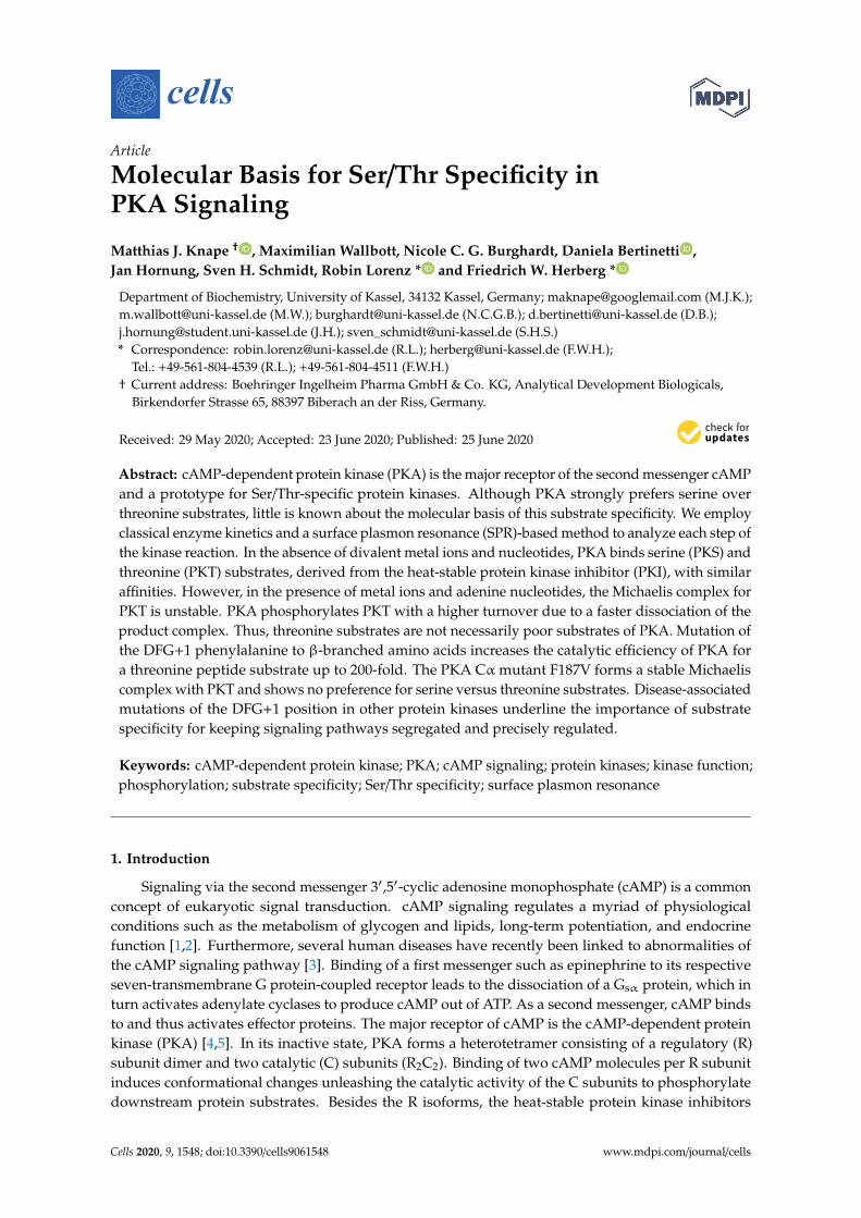
Figure 1. PKA prefers serine over threonine as a phosphoryl acceptor residue. (A) The AGC kinases
PKA, PKG, and PKC are serine-specific. Bar diagram showing the fraction of annotated phosphosites
for serine and threonine phosphoryl acceptors. Data were obtained from the PhosphoSitePlus®
database v6.5.9.1 [23]. (B) Alignment of the DFG motif (black box) and the DFG+1 residues (bold
letters) of human Ser/Thr protein kinases that prefer either serine (red) or threonine (black) as
phosphoryl acceptor. The alignment was generated with Clustal Omega [24]. UniProt IDs: P17612
(PKA Cα); Q13976 (PKG Iα); P17252 (PKCα); O96013 (PAK4); Q9P289 (MST4); P33981 (TTK); O15530
(PDK1); Q5S007 (LRRK2). (C) Crystal structure of the murine PKA Cα subunit with AMP-PNP, Mg2+,
Figure 1. PKA prefers serine over threonine as a phosphoryl acceptor residue. (A) The AGC kinasesPKA, PKG, and PKC are serine-specific. Bar diagram showing the fraction of annotated phosphosites forserine and threonine phosphoryl acceptors. Data were obtained from the PhosphoSitePlus®databasev6.5.9.1 [23]. (B) Alignment of the DFG motif (black box) and the DFG+1 residues (bold letters) ofhuman Ser/Thr protein kinases that prefer either serine (red) or threonine (black) as phosphoryl acceptor.The alignment was generated with Clustal Omega [24]. UniProt IDs: P17612 (PKA Cα); Q13976 (PKGIα); P17252 (PKCα); O96013 (PAK4); Q9P289 (MST4); P33981 (TTK); O15530 (PDK1); Q5S007 (LRRK2).
Cells 2020, 9, 1548 3 of 18
(C) Crystal structure of the murine PKA Cα subunit with AMP-PNP, Mg2+, and SP20 bound (PDB code:4DG0) [25]. A zoomed view (right panel) shows the DFG motif (residues 184-186) and the DFG+1residue (F187) interacting with Mg2AMP-PNP and the substrate SP20. S21 is the phosphoryl acceptorresidue of SP20. All structure images were generated using the PyMOL Molecular Graphics System(Version 2.2.2; Schrödinger, LLC, New York, NY, USA).
Originally, the lack of identified threonine substrates of PKA in vivo has been attributed to lowKM and kcat values for threonine peptides [20]. However, today, some important threonine substratesof PKA have been identified, amongst those the protein phosphatase 1 regulatory subunit 1B (alsoknown as dopamine- and cAMP-regulated neuronal phosphoprotein, DARPP32) [26].
How a Ser/Thr kinase differentiates between the two phosphoryl acceptors that only differ inone methyl group remained unclear for a long time. Chen and coworkers have recently shownthat the residue following the highly conserved DFG motif (DFG+1) is the main determinant forSer/Thr specificity (Figure 1B,C) [27,28]. Kinases that prefer serine as a phosphoryl acceptor carry alarge hydrophobic amino acid residue in this position, such as leucine, methionine, or phenylalanine.In contrast, kinases that prefer threonine substrates carry either β-branched or small amino acidresidues like isoleucine or valine in the DFG+1 position. Large, i.e., sterically more demanding, DFG+1residues of serine-specific kinases hinder the binding of threonine substrates, while β-branched aminoacids tolerate the additional methyl group of the threonine side chain [28].
Phosphoryl acceptor preference of protein kinases is generally described using basic enzymekinetics parameters. PKA strongly prefers serine over threonine residues as phosphoryl acceptor,which is indicated not only by 4-fold higher kcat values but also by a more than 30-fold increase in KM
for synthetic threonine substrate peptides compared to their serine-containing counterparts [20,29].Moreover, characterization of the individual steps of the phosphorylation reaction using viscosimetryas well as rapid quench kinetics led to the conclusion that serine- and threonine-containing peptideshave comparable affinities to the kinase [29].
Referring to KM values for serine and threonine peptides for PAK4 (p21-activated kinase 4) andMST4 (mammalian STE20-like protein kinase 4) kinases, Chen et al. reasoned that a threonine residuein the P0 position would not affect substrate affinity [28]. However, the Michaelis constant (KM) isthe substrate concentration at which the half-maximal enzyme activity is achieved, and therefore,KM values refer to the whole catalytic cycle including substrate association and release, phosphoryltransfer, and product release [29]. PKA is a highly dynamic enzyme, and thus, not every catalytic cycleresults in substrate phosphorylation leading to varying turnover rates [30]. For the PKA C subunit,the chemical reaction of peptide phosphorylation is fast, while the release of the product ADP isdiffusion-controlled, and thus the rate-determining step [31,32]. Accordingly, specific assumptionsmust be made to equate the KM with the equilibrium dissociation constant (KD) which describes(substrate) affinity. In the case of the PKA C subunit, the KD for Kemptide is at least one order ofmagnitude higher than the KM [33,34]. In this study, we, therefore, aimed to answer the fundamentalquestion, whether the phosphoryl acceptor residue itself influences substrate affinity. We used theCα subunit of PKA to analyze the phosphorylation and the binding kinetics of PKI-derived proteinsubstrates. In the presence of metal and nucleotide, the serine substrate PKS displays a higher affinityto PKA, when compared to the analogous threonine substrate, PKT. However, PKA phosphorylatesPKT faster. The tendency in affinities can be switched by mutation of the DFG+1 F187 to valine in thePKA C subunit (F187V). Thereby we demonstrate that binding affinity towards a substrate is indeedaffected by the phosphoryl acceptor residue. Our results suggest that the specificity of a Ser/Thr kinasedepends not only on the turnover but also on the substrate affinity.
Cells 2020, 9, 1548 4 of 18
2. Materials and Methods
2.1. Protein Preparation
GST fusion proteins were expressed and purified as described earlier [35]. Briefly, codingpGEX-KG plasmids were transformed in E. coli BL21 (DE3) cells and expression was induced with0.4 mM IPTG for 16 h at room temperature. Finally, the fusion proteins were purified using Protinoglutathione agarose 4B (MACHEREY-NAGEL, Düren, Germany) according to the manufacturer’sinstructions. The threonine substrate GST-PKT (=GST-PKI A21T) was generated by site-directedmutagenesis using the following primer pair: forward: 5′-CGACGTAACACCATCCACGATATCC-3′
and reverse: 5′-GGATATCGTGGATGGTGTTACGTCG-3′.Constructs of the PKA human Cα isoform (UniProt ID: P17612) were expressed and purified as
previously described [36,37]. Recombinant proteins were expressed in T7 Express Iq Competent E. colicells (New England Biolabs, Ipswich, MA, United States) for 16 h at room temperature after inductionwith 0.4 mM IPTG. The DFG+1 mutations F187V, F187I, and F187T were introduced by site-directedmutagenesis using the site-specific primers F187V_forward: 5′-GACTTCGGTGTCGCCAAGCGC-3′
and F187V_reverse: 5′-GCGCTTGGCGACACCGAAGTC-3′, F187I_forward:5′-GACTTCGGTATCGCCAAGCGC-3′, F187I_reverse: 5′-GCGCTTGGCGATACCGAAGTC-3′,F187T_forward: 5′-GACTTCGGTACCGCCAAGCGC-3′, and F187T_reverse:5′-GCGCTTGGCGGTACCGAAGTC-3′.
2.2. Western Blotting
The autophosphorylation status of recombinant PKA Cα wild type (wt) and F187V at positionT197 and S338 was investigated using Western blot analysis. Purified proteins were denaturedin SDS sample buffer and loaded onto SDS polyacrylamide gels. The transfer on a nitrocellulosemembrane was performed utilizing a semi-dry transfer system. For visualization, we used thepolyclonal rabbit IgG antibodies Phospho-PKA alpha/betaα-pT197 (44-988A; Cell Signaling Technology,Danvers, MA, USA) and Phospho-PKA beta α-pS338 (44-992G; Invitrogen, Thermo Fisher Scientific,Waltham, MA, USA). As a control, the PKA C subunits were detected using an α-PKA-Cα:scFv-Fc-Fusion (YumAb, human Fc region) protein (YumAb GmbH, Braunschweig, Germany).Secondary antibodies used were polyclonal α-rabbit IgG horseradish peroxidase antibodies (AmershamBioscience, Little Chalfont, UK) and polyclonal α-human IgG horseradish peroxidase antibodies fromgoat (Sigma-Aldrich, St. Louis, MO, USA).
2.3. Spectrophotometric Kinase Assay
To determine the Michaelis-Menten constant (KM) and the turnover number (kcat) of purified PKACα wt and the DFG+1 mutants for the peptide substrate Kemptide, a coupled spectrophotometric assaywas used [38]. As we were interested in the substrate specificity of the kinase, we tested two differentpeptide substrates: S-Kemptide (LRRASLG) as a serine substrate and T-Kemptide (LRRATLG) as athreonine substrate (GeneCust, Boynes, France). 50 nM PKA Cα wt were used when measured withT-Kemptide and 20 nM wt, F187I, or F187T when measured with S-Kemptide. In all other assays,the final kinase concentration was 10 nM of the respective kinase. All kinases were measured with aminimum of three independent replicates. The calculated turnover was plotted against the kinaseconcentration and analyzed with GraphPad Prism 8.0 (GraphPad Software, San Diego, CA, USA).
2.4. Phosphospecific Antibody-Based Kinase Assay
In vitro kinase assays were performed in 200µL reactions containing 20 mM MOPS, pH 7.0, 150 mMNaCl, 0.1 mM ATP or 0.2 mM AMP-PNP (adenylyl-imidodiphosphate), 1 mM MgCl2, and 1.5 µMsubstrate protein (GST-PKS or GST-PKT). The reaction was started by adding the kinase to a finalconcentration of 0.25–1.5 µM. The reaction was stopped after 5 min by adding 2× SDS sample buffer.The samples were loaded onto SDS polyacrylamide gels and transferred to a membrane for Western
Cells 2020, 9, 1548 5 of 18
blot analysis using either a phospho-PKA substrate antibody (α-RRXS*/T*; 100G7E, monoclonal rabbitIgG, Cell Signaling Technology, Danvers, MA, USA) or a polyclonal α-GST antibody (3998.1; CarlRoth, Karlsruhe, Germany). For visualization, an IRDye 800CW donkey α-rabbit IgG secondaryantibody (LI-COR, Lincoln, NE, USA) or a polyclonal α-rabbit IgG horseradish peroxidase (AmershamBioscience, Little Chalfont, UK) antibody were used.
2.5. Radioactive Kinase Assay
A radioisotopic kinase assay was performed as previously described following in principle themethod by Kish and Kleinsmith [35,39]. Briefly, the reaction mixture of 300 µl contained 30 µMGST-PKS or GST-PKT, and approximately 550 fmoles [γ-32P]-ATP (stock solution 110 TBq/mmol,HARTMANN ANALYTIC GmbH, Braunschweig, Germany) in 20 mM MOPS, pH 7.0, 150 mM NaCl,0.1 mM ATP, 1 mM MgCl2. The reaction was initiated by adding PKA Cα to a final concentrationof 5 nM. The mixture was incubated with shaking at 30 ◦C and 350 rpm. Samples of 50 µl weretaken after 20, 40, 60, and 80 min and mixed with 500 µl ice-cold ATP buffer solution (20 mM MOPS,pH 7.0, 150 mM NaCl, 1 mM ATP). Instantly, proteins were precipitated by adding 550 µl ice-cold10% trichloroacetic acid (TCA) plus 3% sodium pyrophosphate. The samples were sucked throughmixed cellulose ester membrane filters (MF-Millipore Membrane Filter, 0.45 µm, 25 mm diameter)presoaked in 1 mM ATP for 30 min. Each filter was washed twice with 5 mL of ice-cold 5% TCAcontaining 1.5% sodium pyrophosphate. Radioactivity of each filter was counted in 20 mL of distilledwater in a scintillation counter (Hidex 300 SL; Hidex, Turku, Finland), for 300 s or until a maximumof 10,000 counts was achieved) by detection of Cerenkov radiation. For each run, the total amountof radioactivity, and the blank (reaction mixture without kinase) was measured. For evaluation, allvalues (cpm) were blank value subtracted. Data evaluation was carried out using GraphPad Prism 8.0(GraphPad Software, San Diego, CA, USA).
2.6. Surface Plasmon Resonance (SPR)
SPR-based interaction analyses were performed using a Biacore T200 instrument (Cytiva,Marlborough, MA, USA) as previously described [35]. A polyclonal α-GST antibody (3998.1; CarlRoth, Karlsruhe, Germany) was immobilized to a CM5 sensor chip (S-series; Cytiva, Marlborough,MA, USA) surface by custom amine coupling. All measurements were performed at 25 ◦C with a flowrate of 30 µl/min using running buffer (20 mM MOPS, pH 7.0, 150 mM NaCl, 0.1 mM EDTA, 0.01% P20surfactant) supplemented with nucleotides and metal ions as indicated. In each measurement cycle,about 50 RU of GST fusion protein (GST-PKS, GST-PKT, or GST-PKI) was captured, before PKA Cα wasinjected for 150 s or 300 s (association). For the analysis of the product complex, GST fusion proteinsprephosphorylated with PKA Cα (GST-pPKS or GST-pPKT) were captured [36]. The dissociation phasewas induced by switching to running buffer for 150 s or 300 s. SPR signals from a blank flow cell withonly α-GST antibody immobilized as well as blank runs with buffer injections were subtracted (doublereferencing). The sensor chip surface was regenerated by injection of 10 mM glycine, pH 1.9 until thebaseline level was reached. Binding kinetics were analyzed with Biacore T200 Evaluation Software 3.0and BIAevaluation 4.1.1 (Cytiva, Marlborough, MA, USA) applying a 1:1 Langmuir binding model,global fit, or steady-state analysis.
On-chip phosphorylation was performed as previously described [36].
2.7. Docking Simulations
Structural models of PKA Cα wt and F187V in complex with AMP-PNP, Mg2+, and either SP20(serine-containing substrate peptide corresponding to the amino acid residues 5–24 of human PKIαisoform with the two mutations N20A and A21S; TTYADFIASGRTGRRASIHD [40]) or TP20 (SP20 withS21T mutation; TTYADFIASGRTGRRATIHD), were generated using the crystal structure of the murinemyristoylated PKA Cα, in complex with SP20 and AMP-PNP (PDB code: 4DG0) [25]. The respectiveamino acids were exchanged in YASARA (V.18.12.27) [41] and the side chain conformations were
Cells 2020, 9, 1548 6 of 18
optimized using the SCWALL approach [42] employing the SCWRL3 algorithm [43] before surfaceinteractions and solvation energies were optimized using the YASARA2 force field [42]. The structureswere subsequently disassembled into SP20 or TP20 and either PKA Cα wt or PKA Cα F187V. Energyminimization was performed with the (em_run) macro of YASARA [41], employing the Amber14forcefield [44] and the TIP3P water model [45]. Docking of SP20/TP20 to PKA Cα wt/F187V wasperformed with the dock_runlocal macro [41,46] which employs a YASARA-specific version [41] ofAutoDock VINA [47]. In this process, a simulation cell with a side length of x ≈ 44.185 Å, y ≈ 38.491 Å,and z ≈ 38.882 Å was placed around the SP20/TP20 and the binding site of PKA Cα wt/F187V. To furtheroptimize the conformation of all elements within the simulation cell, the NOVA force field [46] wasutilized by the dock_runlocal setup. The YASARA AutoSMILES algorithm was used for simulationswith a derivative of MOPAC [48] and the COSMO solvation model [49] before calculating and addingAM1BCCs [50] and applying the GAFF [51] to generate all values unknown to the NOVA forcefield [41,46,52]. Values for divalent metal ions were taken from [53]. After the docking was performed,each generated model was again submitted to the em_run macro [41].
3. Results
3.1. The DFG+1 Residue Determines the Serine Specificity of PKA
Early peptide studies based on radioactive kinase assays have already demonstrated that PKAprefers serine over threonine substrates [20]. Using a spectrophotometric kinase assay, we recapitulatedthat PKA Cαphosphorylates the peptide substrate S-Kemptide with lower KM and higher kcat comparedto T-Kemptide (Table 1, Figure S1) [20,29]. This leads to a more than 100-fold higher catalytic efficiency(kcat/KM) for the serine substrate peptide. To test the role of the DFG+1 phenylalanine (F187) forSer/Thr specificity, we mutated F187 to valine, isoleucine, and threonine, since β-branched amino acidsat this position were postulated to mediate threonine specificity [28]. Indeed, changing the DFG+1position resulted in higher catalytic efficiency for T-Kemptide compared to S-Kemptide (Table 1).This is partly because the kcat values for the threonine substrate were at least 5-fold increased andexceeded the kcat of PKA Cα wt for S-Kemptide. Interestingly, the mutation F187V also increasedthe kcat for phosphorylation of S-Kemptide compared to the wild type. Additionally, mutating theDFG+1 phenylalanine residue reduced the KM for T-Kemptide about 30-fold (Table 1). While KM
values for S-Kemptide were increased 4-fold for both PKA Cα F187V and F187I, the KM for PKA Cα
F187T remained unaffected.
Table 1. The catalytic efficiency of PKA Cα mutants for serine and threonine substrates. kcat andKM values were determined using a spectrophotometric kinase assay (Figure S1). The values arerepresented as means of at least three independent measurements with standard deviation (SD).
PKA Cα Substrate kcat (s−1) KM (µM) kcat/KM (×105 M−1s−1)
WtS-Kemptide 19.8 ± 1.0 19.8 ± 3.9 10.3 ± 1.7T-Kemptide 7.5 ± 0.8 861 ± 184 0.09 ± 0.02
F187VS-Kemptide 35.7 ± 0.7 81.3 ± 27.1 4.8 ± 1.6T-Kemptide 39.2 ± 3.7 31.5 ± 16.0 11.3 ± 4.0
F187IS-Kemptide 25.4 ± 5.8 92.2 ± 3.8 2.8 ± 0.8T-Kemptide 43.0 ± 4.7 19.8 ± 3.3 22.1 ± 4.5
F187TS-Kemptide 15.7 ± 0.3 23.0 ± 3.1 6.9 ± 1.2T-Kemptide 37.1 ± 4.5 25.0 ± 3.4 14.9 ± 0.6
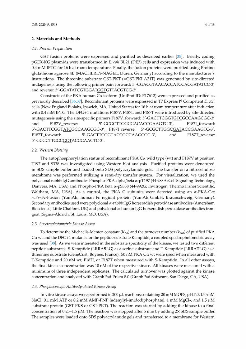
3.2. PKA Cα Phosphorylates PKT Faster than PKS
To study the substrate specificity of the PKA Cα subunit on protein substrates, we used PKI-derivedserine (PKI A21S, PKS) and threonine (PKI A21T, PKT) substrates. Previously, we have shown thatPKS is a high-affinity substrate of PKA [36].
Cells 2020, 9, 1548 7 of 18
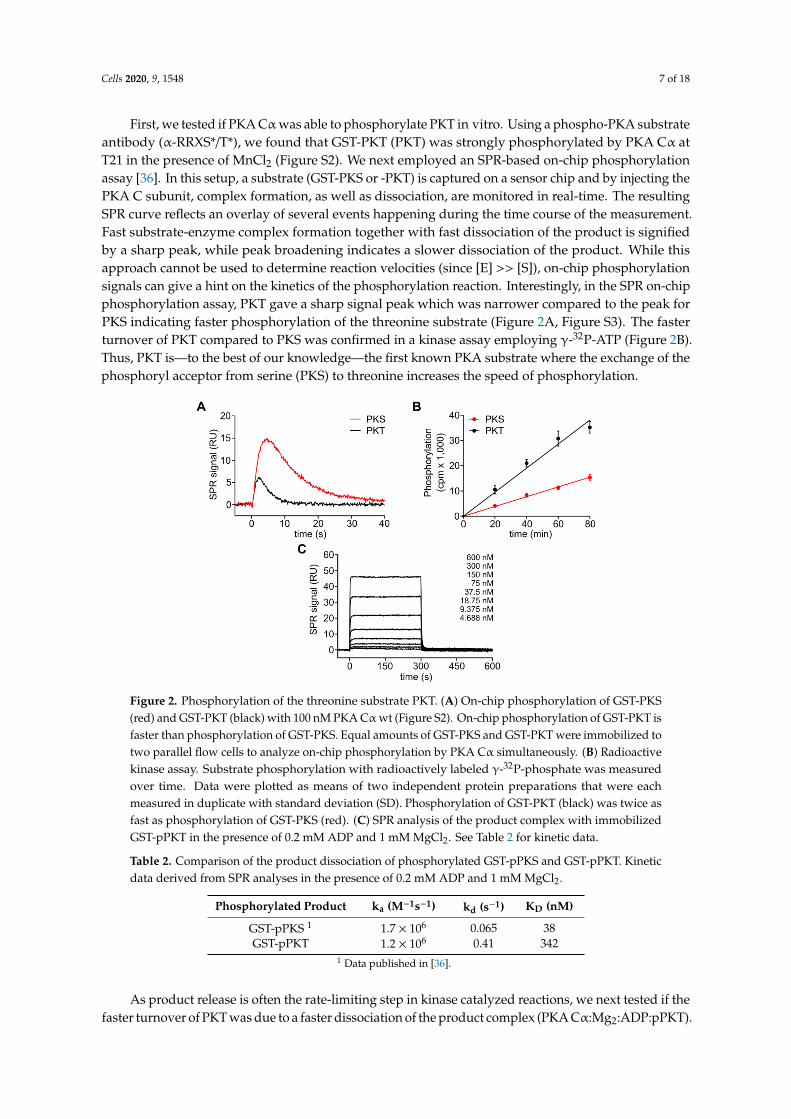
First, we tested if PKA Cαwas able to phosphorylate PKT in vitro. Using a phospho-PKA substrateantibody (α-RRXS*/T*), we found that GST-PKT (PKT) was strongly phosphorylated by PKA Cα atT21 in the presence of MnCl2 (Figure S2). We next employed an SPR-based on-chip phosphorylationassay [36]. In this setup, a substrate (GST-PKS or -PKT) is captured on a sensor chip and by injecting thePKA C subunit, complex formation, as well as dissociation, are monitored in real-time. The resultingSPR curve reflects an overlay of several events happening during the time course of the measurement.Fast substrate-enzyme complex formation together with fast dissociation of the product is signifiedby a sharp peak, while peak broadening indicates a slower dissociation of the product. While thisapproach cannot be used to determine reaction velocities (since [E] >> [S]), on-chip phosphorylationsignals can give a hint on the kinetics of the phosphorylation reaction. Interestingly, in the SPR on-chipphosphorylation assay, PKT gave a sharp signal peak which was narrower compared to the peak forPKS indicating faster phosphorylation of the threonine substrate (Figure 2A, Figure S3). The fasterturnover of PKT compared to PKS was confirmed in a kinase assay employing γ-32P-ATP (Figure 2B).Thus, PKT is—to the best of our knowledge—the first known PKA substrate where the exchange of thephosphoryl acceptor from serine (PKS) to threonine increases the speed of phosphorylation.
Cells 2020, 9, 1548 7 of 17
the product. While this approach cannot be used to determine reaction velocities (since [E] >> [S]),
on-chip phosphorylation signals can give a hint on the kinetics of the phosphorylation reaction.
Interestingly, in the SPR on-chip phosphorylation assay, PKT gave a sharp signal peak which was
narrower compared to the peak for PKS indicating faster phosphorylation of the threonine substrate
(Figure 2A, Figure S3). The faster turnover of PKT compared to PKS was confirmed in a kinase assay
employing γ-32P-ATP (Figure 2B). Thus, PKT is—to the best of our knowledge—the first known PKA
substrate where the exchange of the phosphoryl acceptor from serine (PKS) to threonine increases
the speed of phosphorylation.
Figure 2. Phosphorylation of the threonine substrate PKT. (A) On-chip phosphorylation of GST-PKS
(red) and GST-PKT (black) with 100 nM PKA Cα wt (Figure S2). On-chip phosphorylation of GST-
PKT is faster than phosphorylation of GST-PKS. Equal amounts of GST-PKS and GST-PKT were
immobilized to two parallel flow cells to analyze on-chip phosphorylation by PKA Cα
simultaneously. (B) Radioactive kinase assay. Substrate phosphorylation with radioactively labeled
γ-32P-phosphate was measured over time. Data were plotted as means of two independent protein
preparations that were each measured in duplicate with standard deviation (SD). Phosphorylation of
GST-PKT (black) was twice as fast as phosphorylation of GST-PKS (red). (C) SPR analysis of the
product complex with immobilized GST-pPKT in the presence of 0.2 mM ADP and 1 mM MgCl2. See
Table 2 for kinetic data.
As product release is often the rate-limiting step in kinase catalyzed reactions, we next tested if
the faster turnover of PKT was due to a faster dissociation of the product complex (PKA
Cα:Mg2:ADP:pPKT). We, therefore, compared the interaction of the C subunit with phosphorylated
GST-PKT (pPKT) in the presence of ADP and Mg2+ with our previously published data for pPKS [36].
The dissociation of the C subunit was at least 6-fold faster for pPKT than for pPKS, which resulted in
a 9-fold higher KD for the phosphorylated threonine product (Figure 2C, Table 2). This indicates that
indeed product release is the rate-limiting step and thus faster dissociation of pPKT leads to increased
turnover of PKT compared to PKS. Yet, since the phosphorylation of PKT was only twice as fast as
for PKS, other steps of the catalytic cycle may be slowed down with a threonine phosphoryl acceptor
(Figure 2B).
Figure 2. Phosphorylation of the threonine substrate PKT. (A) On-chip phosphorylation of GST-PKS(red) and GST-PKT (black) with 100 nM PKA Cα wt (Figure S2). On-chip phosphorylation of GST-PKT isfaster than phosphorylation of GST-PKS. Equal amounts of GST-PKS and GST-PKT were immobilized totwo parallel flow cells to analyze on-chip phosphorylation by PKA Cα simultaneously. (B) Radioactivekinase assay. Substrate phosphorylation with radioactively labeled γ-32P-phosphate was measuredover time. Data were plotted as means of two independent protein preparations that were eachmeasured in duplicate with standard deviation (SD). Phosphorylation of GST-PKT (black) was twice asfast as phosphorylation of GST-PKS (red). (C) SPR analysis of the product complex with immobilizedGST-pPKT in the presence of 0.2 mM ADP and 1 mM MgCl2. See Table 2 for kinetic data.
Table 2. Comparison of the product dissociation of phosphorylated GST-pPKS and GST-pPKT. Kineticdata derived from SPR analyses in the presence of 0.2 mM ADP and 1 mM MgCl2.
Phosphorylated Product ka (M−1s−1) kd (s−1) KD (nM)
GST-pPKS 1 1.7 × 10⁶ 0.065 38GST-pPKT 1.2 × 10⁶ 0.41 342
1 Data published in [36].
As product release is often the rate-limiting step in kinase catalyzed reactions, we next tested if thefaster turnover of PKT was due to a faster dissociation of the product complex (PKA Cα:Mg2:ADP:pPKT).
Cells 2020, 9, 1548 8 of 18
We, therefore, compared the interaction of the C subunit with phosphorylated GST-PKT (pPKT) inthe presence of ADP and Mg2+ with our previously published data for pPKS [36]. The dissociationof the C subunit was at least 6-fold faster for pPKT than for pPKS, which resulted in a 9-fold higherKD for the phosphorylated threonine product (Figure 2C, Table 2). This indicates that indeed productrelease is the rate-limiting step and thus faster dissociation of pPKT leads to increased turnover of PKTcompared to PKS. Yet, since the phosphorylation of PKT was only twice as fast as for PKS, other stepsof the catalytic cycle may be slowed down with a threonine phosphoryl acceptor (Figure 2B).
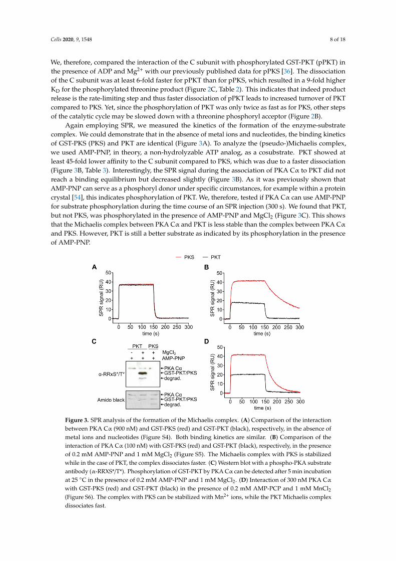
Again employing SPR, we measured the kinetics of the formation of the enzyme-substratecomplex. We could demonstrate that in the absence of metal ions and nucleotides, the binding kineticsof GST-PKS (PKS) and PKT are identical (Figure 3A). To analyze the (pseudo-)Michaelis complex,we used AMP-PNP, in theory, a non-hydrolyzable ATP analog, as a cosubstrate. PKT showed atleast 45-fold lower affinity to the C subunit compared to PKS, which was due to a faster dissociation(Figure 3B, Table 3). Interestingly, the SPR signal during the association of PKA Cα to PKT did notreach a binding equilibrium but decreased slightly (Figure 3B). As it was previously shown thatAMP-PNP can serve as a phosphoryl donor under specific circumstances, for example within a proteincrystal [54], this indicates phosphorylation of PKT. We, therefore, tested if PKA Cα can use AMP-PNPfor substrate phosphorylation during the time course of an SPR injection (300 s). We found that PKT,but not PKS, was phosphorylated in the presence of AMP-PNP and MgCl2 (Figure 3C). This showsthat the Michaelis complex between PKA Cα and PKT is less stable than the complex between PKA Cα
and PKS. However, PKT is still a better substrate as indicated by its phosphorylation in the presenceof AMP-PNP.
Cells 2020, 9, 1548 8 of 17
Table 2. Comparison of the product dissociation of phosphorylated GST-pPKS and GST-pPKT.
Kinetic data derived from SPR analyses in the presence of 0.2 mM ADP and 1 mM MgCl2.
Phosphorylated Product ka (M−1s−1) kd (s−1) KD (nM)
GST-pPKS 1 1.7 × 10⁶ 0.065 38
GST-pPKT 1.2 × 10⁶ 0.41 342
1 Data published in [36].
Again employing SPR, we measured the kinetics of the formation of the enzyme-substrate
complex. We could demonstrate that in the absence of metal ions and nucleotides, the binding
kinetics of GST-PKS (PKS) and PKT are identical (Figure 3A). To analyze the (pseudo-)Michaelis
complex, we used AMP-PNP, in theory, a non-hydrolyzable ATP analog, as a cosubstrate. PKT
showed at least 45-fold lower affinity to the C subunit compared to PKS, which was due to a faster
dissociation (Figure 3B, Table 3). Interestingly, the SPR signal during the association of PKA Cα to
PKT did not reach a binding equilibrium but decreased slightly (Figure 3B). As it was previously
shown that AMP-PNP can serve as a phosphoryl donor under specific circumstances, for example
within a protein crystal [54], this indicates phosphorylation of PKT. We, therefore, tested if PKA Cα
can use AMP-PNP for substrate phosphorylation during the time course of an SPR injection (300 s).
We found that PKT, but not PKS, was phosphorylated in the presence of AMP-PNP and MgCl2
(Figure 3C). This shows that the Michaelis complex between PKA Cα and PKT is less stable than the
complex between PKA Cα and PKS. However, PKT is still a better substrate as indicated by its
phosphorylation in the presence of AMP-PNP.
Figure 3. SPR analysis of the formation of the Michaelis complex. (A) Comparison of the interaction
between PKA Cα (900 nM) and GST-PKS (red) and GST-PKT (black), respectively, in the absence of
metal ions and nucleotides (Figure S4). Both binding kinetics are similar. (B) Comparison of the
interaction of PKA Cα (100 nM) with GST-PKS (red) and GST-PKT (black), respectively, in the
presence of 0.2 mM AMP-PNP and 1 mM MgCl2 (Figure S5). The Michaelis complex with PKS is
stabilized while in the case of PKT, the complex dissociates faster. (C) Western blot with a phospho-
PKA substrate antibody (α-RRXS*/T*). Phosphorylation of GST-PKT by PKA Cα can be detected after
5 min incubation at 25 °C in the presence of 0.2 mM AMP-PNP and 1 mM MgCl2. (D) Interaction of
300 nM PKA Cα with GST-PKS (red) and GST-PKT (black) in the presence of 0.2 mM AMP-PCP and
1 mM MnCl2 (Figure S6). The complex with PKS can be stabilized with Mn2+ ions, while the PKT
Michaelis complex dissociates fast.
Figure 3. SPR analysis of the formation of the Michaelis complex. (A) Comparison of the interactionbetween PKA Cα (900 nM) and GST-PKS (red) and GST-PKT (black), respectively, in the absence ofmetal ions and nucleotides (Figure S4). Both binding kinetics are similar. (B) Comparison of theinteraction of PKA Cα (100 nM) with GST-PKS (red) and GST-PKT (black), respectively, in the presenceof 0.2 mM AMP-PNP and 1 mM MgCl2 (Figure S5). The Michaelis complex with PKS is stabilizedwhile in the case of PKT, the complex dissociates faster. (C) Western blot with a phospho-PKA substrateantibody (α-RRXS*/T*). Phosphorylation of GST-PKT by PKA Cα can be detected after 5 min incubationat 25 ◦C in the presence of 0.2 mM AMP-PNP and 1 mM MgCl2. (D) Interaction of 300 nM PKA Cα
with GST-PKS (red) and GST-PKT (black) in the presence of 0.2 mM AMP-PCP and 1 mM MnCl2(Figure S6). The complex with PKS can be stabilized with Mn2+ ions, while the PKT Michaelis complexdissociates fast.
Cells 2020, 9, 1548 9 of 18
Table 3. Kinetic data for the formation of the Michaelis complex with PKA Cα wt in the presence of0.2 mM AMP-PNP and 1 mM MgCl2. Data derived from SPR analyses (Figure S5) of the interactionbetween PKA Cα and GST-PKS or GST-PKT, respectively.
Phosphoryl Acceptor ka (M−1s−1) kd (s−1) KD (nM)
GST-PKS 2.7 × 10⁶ 0.89 × 10⁻² 3.3GST-PKT 2.2 × 10⁶ 29 × 10−2 136
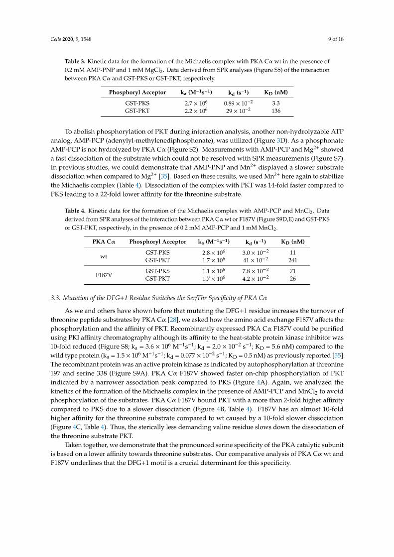
To abolish phosphorylation of PKT during interaction analysis, another non-hydrolyzable ATPanalog, AMP-PCP (adenylyl-methylenediphosphonate), was utilized (Figure 3D). As a phosphonateAMP-PCP is not hydrolyzed by PKA Cα (Figure S2). Measurements with AMP-PCP and Mg2+ showeda fast dissociation of the substrate which could not be resolved with SPR measurements (Figure S7).In previous studies, we could demonstrate that AMP-PNP and Mn2+ displayed a slower substratedissociation when compared to Mg2+ [35]. Based on these results, we used Mn2+ here again to stabilizethe Michaelis complex (Table 4). Dissociation of the complex with PKT was 14-fold faster compared toPKS leading to a 22-fold lower affinity for the threonine substrate.
Table 4. Kinetic data for the formation of the Michaelis complex with AMP-PCP and MnCl2. Dataderived from SPR analyses of the interaction between PKA Cα wt or F187V (Figure S9D,E) and GST-PKSor GST-PKT, respectively, in the presence of 0.2 mM AMP-PCP and 1 mM MnCl2.
PKA Cα Phosphoryl Acceptor ka (M−1s−1) kd (s−1) KD (nM)
wtGST-PKS 2.8 × 10⁶ 3.0 × 10⁻² 11GST-PKT 1.7 × 10⁶ 41 × 10−2 241
F187VGST-PKS 1.1 × 10⁶ 7.8 × 10⁻² 71GST-PKT 1.7 × 10⁶ 4.2 × 10⁻² 26
3.3. Mutation of the DFG+1 Residue Switches the Ser/Thr Specificity of PKA Cα
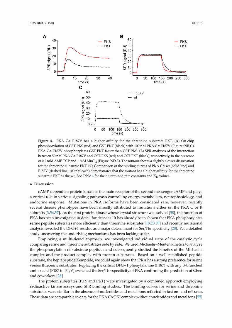
As we and others have shown before that mutating the DFG+1 residue increases the turnover ofthreonine peptide substrates by PKA Cα [28], we asked how the amino acid exchange F187V affects thephosphorylation and the affinity of PKT. Recombinantly expressed PKA Cα F187V could be purifiedusing PKI affinity chromatography although its affinity to the heat-stable protein kinase inhibitor was10-fold reduced (Figure S8; ka = 3.6 × 10⁶ M−1s−1; kd = 2.0 × 10⁻² s−1; KD = 5.6 nM) compared to thewild type protein (ka = 1.5 × 10⁶ M−1s−1; kd = 0.077 × 10⁻² s−1; KD = 0.5 nM) as previously reported [55].The recombinant protein was an active protein kinase as indicated by autophosphorylation at threonine197 and serine 338 (Figure S9A). PKA Cα F187V showed faster on-chip phosphorylation of PKTindicated by a narrower association peak compared to PKS (Figure 4A). Again, we analyzed thekinetics of the formation of the Michaelis complex in the presence of AMP-PCP and MnCl2 to avoidphosphorylation of the substrates. PKA Cα F187V bound PKT with a more than 2-fold higher affinitycompared to PKS due to a slower dissociation (Figure 4B, Table 4). F187V has an almost 10-foldhigher affinity for the threonine substrate compared to wt caused by a 10-fold slower dissociation(Figure 4C, Table 4). Thus, the sterically less demanding valine residue slows down the dissociation ofthe threonine substrate PKT.
Taken together, we demonstrate that the pronounced serine specificity of the PKA catalytic subunitis based on a lower affinity towards threonine substrates. Our comparative analysis of PKA Cα wt andF187V underlines that the DFG+1 motif is a crucial determinant for this specificity.
Cells 2020, 9, 1548 10 of 18Cells 2020, 9, 1548 10 of 17
Figure 4. PKA Cα F187V has a higher affinity for the threonine substrate PKT. (A) On-chip
phosphorylation of GST-PKS (red) and GST-PKT (black) with 100 nM PKA Cα F187V (Figure S9B,C).
PKA Cα F187V phosphorylates GST-PKT faster than GST-PKS. (B) SPR analyses of the interaction
between 50 nM PKA Cα F187V and GST-PKS (red) and GST-PKT (black), respectively, in the presence
of 0.2 mM AMP-PCP and 1 mM MnCl2 (Figure S9D,E). The mutant shows a slightly slower
dissociation for the threonine substrate PKT. (C) Comparison of the binding curves of PKA Cα wt
(solid line) and F187V (dashed line; 100 nM each) demonstrates that the mutant has a higher affinity
for the threonine substrate PKT as the wt. See Table 4 for the determined rate constants and KD values.
4. Discussion
cAMP-dependent protein kinase is the main receptor of the second messenger cAMP and plays
a critical role in various signaling pathways controlling energy metabolism, neurophysiology, and
endocrine response. Mutations in PKA isoforms have been considered rare, however, recently several
disease phenotypes have been directly attributed to mutations either on the PKA C or R subunits
[3,56,57]. As the first protein kinase whose crystal structure was solved [58], the function of PKA has
been investigated in detail for decades. It has already been shown that PKA phosphorylates serine
peptide substrates more efficiently than threonine substrates [18,20,59] and recently mutational
analysis revealed the DFG+1 residue as a major determinant for Ser/Thr specificity [28]. Yet a detailed
study uncovering the underlying mechanisms has been lacking so far.
Employing a multi-tiered approach, we investigated individual steps of the catalytic cycle
comparing serine and threonine substrates side by side. We used Michaelis–Menten kinetics to
analyze the phosphorylation of substrate peptides and subsequently studied the kinetics of the
Michaelis complex and the product complex with protein substrates. Based on a well-established
peptide substrate, the heptapeptide Kemptide, we could again show that PKA has a strong preference
for serine versus threonine substrates. Replacing the critical DFG+1 phenylalanine (F187) with any β-
branched amino acid (F187 to I/T/V) switched the Ser/Thr-specificity of PKA confirming the
prediction of Chen and coworkers [28].
The protein substrates (PKS and PKT) were investigated by a combined approach employing
radioactive kinase assays and SPR binding studies. The binding curves for serine and threonine
substrates were similar in the absence of nucleotides and metal ions reflected in fast on- and off-rates.
Those data are comparable to data for the PKA Cα:PKI complex without nucleotides and metal ions
[55] displaying an affinity in the higher nM range (GST-PKI:PKA Cα: KD = 450 nM [55], GST-PKS:PKA
Cα: KD = 124 nM; GST-PKT:PKA Cα: KD = 167 nM; Figure S4). It has been demonstrated that ATP and
Figure 4. PKA Cα F187V has a higher affinity for the threonine substrate PKT. (A) On-chipphosphorylation of GST-PKS (red) and GST-PKT (black) with 100 nM PKA Cα F187V (Figure S9B,C).PKA Cα F187V phosphorylates GST-PKT faster than GST-PKS. (B) SPR analyses of the interactionbetween 50 nM PKA Cα F187V and GST-PKS (red) and GST-PKT (black), respectively, in the presenceof 0.2 mM AMP-PCP and 1 mM MnCl2 (Figure S9D,E). The mutant shows a slightly slower dissociationfor the threonine substrate PKT. (C) Comparison of the binding curves of PKA Cα wt (solid line) andF187V (dashed line; 100 nM each) demonstrates that the mutant has a higher affinity for the threoninesubstrate PKT as the wt. See Table 4 for the determined rate constants and KD values.
4. Discussion
cAMP-dependent protein kinase is the main receptor of the second messenger cAMP and playsa critical role in various signaling pathways controlling energy metabolism, neurophysiology, andendocrine response. Mutations in PKA isoforms have been considered rare, however, recentlyseveral disease phenotypes have been directly attributed to mutations either on the PKA C or Rsubunits [3,56,57]. As the first protein kinase whose crystal structure was solved [58], the function ofPKA has been investigated in detail for decades. It has already been shown that PKA phosphorylatesserine peptide substrates more efficiently than threonine substrates [18,20,59] and recently mutationalanalysis revealed the DFG+1 residue as a major determinant for Ser/Thr specificity [28]. Yet a detailedstudy uncovering the underlying mechanisms has been lacking so far.
Employing a multi-tiered approach, we investigated individual steps of the catalytic cyclecomparing serine and threonine substrates side by side. We used Michaelis–Menten kinetics to analyzethe phosphorylation of substrate peptides and subsequently studied the kinetics of the Michaeliscomplex and the product complex with protein substrates. Based on a well-established peptidesubstrate, the heptapeptide Kemptide, we could again show that PKA has a strong preference for serineversus threonine substrates. Replacing the critical DFG+1 phenylalanine (F187) with any β-branchedamino acid (F187 to I/T/V) switched the Ser/Thr-specificity of PKA confirming the prediction of Chenand coworkers [28].
The protein substrates (PKS and PKT) were investigated by a combined approach employingradioactive kinase assays and SPR binding studies. The binding curves for serine and threoninesubstrates were similar in the absence of nucleotides and metal ions reflected in fast on- and off-rates.Those data are comparable to data for the PKA Cα:PKI complex without nucleotides and metal ions [55]
Cells 2020, 9, 1548 11 of 18
displaying an affinity in the higher nM range (GST-PKI:PKA Cα: KD = 450 nM [55], GST-PKS:PKACα: KD = 124 nM; GST-PKT:PKA Cα: KD = 167 nM; Figure S4). It has been demonstrated that ATPand two divalent metal ions are required to achieve a high-affinity complex between the catalyticsubunit and the pseudosubstrate inhibitor PKI [55,60]. In this conformation, the catalytic subunit islocked in the fully closed state. During the catalytic cycle, the kinase needs to toggle between openand closed conformations, which involves, in particular, the glycine-rich loop [36,61]. The role ofmetals and nucleotides for the closing of the glycine-rich loop has been demonstrated in differentcrystal structures and is reviewed in [61]. Hence, we speculate that PKA C subunits in the openconformation (i.e., without metal and nucleotide) do not discriminate between serine or threoninephosphoryl acceptors. SPR revealed that in the presence of metal ions and adenine nucleotides, PKACα binds threonine substrates (here PKT) with lower affinity compared to serine substrates (PKS).Exchanging the large phenylalanine residue in the DFG+1 position (F187) for a smaller valine residue(PKA Cα F187V) increased the affinity for the threonine substrate by reducing the off-rate, underliningthe importance of this position for substrate specificity. Based on KM values and crystal structures ofPAK4 wt and PAK4 F461V in complex with PAKtide-S and PAKtide-T it was assumed that threonineand serine peptides bind with similar affinities [28]. Decreased phosphorylation of threonine substrateswas attributed to conformational restrains in the catalytic center such as the DFG+1 phenylalanineresidue. This was also extrapolated to the catalytic subunit of PKA. While the PAK4 crystal structuresdepict the importance of the orientation of the phosphoryl acceptor, no nucleotide or metal was visiblein most structures with PAKtide, indicating that the kinase was not in the fully closed conformation [28].At least for PKA, we unambiguously demonstrate that the preference for serine versus threoninesubstrates is also reflected in their affinity.
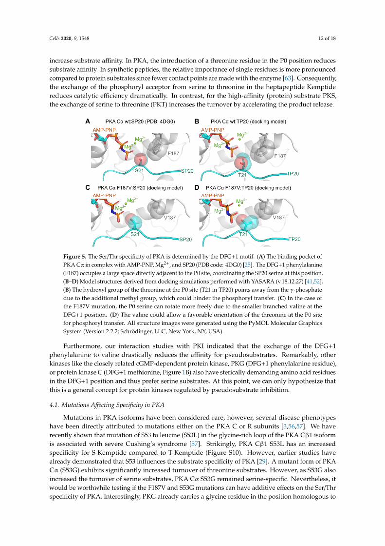
To gain insights into the protein kinase-substrate complex with metal and nucleotide bound to thekinase, we performed docking simulations to generate structural models of the Michaelis complexes ofboth PKA Cα wt and F187V with PKS and PKT, respectively (Figure 5). As a template, we used thePKA Cα wt:SP20 structure co-crystallized with AMP-PNP and Mg2+ (PDB code: 4DG0) [25]. In thecrystal structure of PKA Cα wt bound with SP20, F187 seems to limit the available space for the serineside chain, benefiting the orientation towards the AMP-PNP γ-phosphate (Figure 5A). In the caseof threonine as phosphoryl acceptor (T21 in TP20), the DFG+1 phenylalanine restricts the rotationalfreedom of the threonine side chain due to the increased steric demand between the additional methylgroup and the benzene ring of F187 (Figure 5B). Exchanging the DFG+1 phenylalanine for a valineresidue (PKA Cα F187V) increases the space and thus the freedom of rotation of the phosphorylacceptor (Figure 5C,D). In the case of the PKA Cα F187V:SP20 complex, the interaction of the S21side chain with V187 (Figure 5C) is weaker than the interaction with F187 (Figure 5A). This topologycould allow the entrance of additional water molecules leading to a less stable Michaelis complexcompared to PKA Cα wt [61]. In contrast, in the complex of PKA Cα F187V with TP20, V187 seemsto accommodate the P0 threonine and thus restricts free rotation (Figure 5D). In the model for PKACα F187V with TP20, the hydroxyl group is perfectly positioned for phosphoryl transfer contrarilyto the PKA Cα wt:TP20 complex (Figure 5B,D). As the F187V mutation reduced the affinity for PKSonly slightly, the specificity was not switched completely. We, therefore, hypothesize that the DFG+1position is a rather weak determinant for threonine specificity but strongly promotes serine specificity.Small or β-branched DFG+1 residues facilitate threonine phosphorylation rather than switching theselectivity towards threonine substrates.
One important finding of our study was that PKA Cα phosphorylated PKT faster than PKS,even in the presence of AMP-PNP. While the chemical step of phosphoryl transfer is considered to befast, the release of the phosphorylated product and ADP-Mg is rate-limiting [32]. Our data clearly showa faster release of pPKT compared to pPKS, which also explains the faster turnover. This demonstratesthat threonine substrates are not per se poor substrates of a serine-specific kinase. In line with this,DARPP32 is a good PKA substrate, although DARPP32-derived peptides are not [62]. Like thePKA RI subunits, DARPP32 carries additional arginine residues in positions P-3 and P-4 that may
Cells 2020, 9, 1548 12 of 18
increase substrate affinity. In PKA, the introduction of a threonine residue in the P0 position reducessubstrate affinity. In synthetic peptides, the relative importance of single residues is more pronouncedcompared to protein substrates since fewer contact points are made with the enzyme [63]. Consequently,the exchange of the phosphoryl acceptor from serine to threonine in the heptapeptide Kemptidereduces catalytic efficiency dramatically. In contrast, for the high-affinity (protein) substrate PKS,the exchange of serine to threonine (PKT) increases the turnover by accelerating the product release.Cells 2020, 9, 1548 12 of 17
Figure 5. The Ser/Thr specificity of PKA is determined by the DFG+1 motif. (A) The binding pocket
of PKA Cα in complex with AMP-PNP, Mg2+, and SP20 (PDB code: 4DG0) [25]. The DFG+1
phenylalanine (F187) occupies a large space directly adjacent to the P0 site, coordinating the SP20
serine at this position. (B–D) Model structures derived from docking simulations performed with
YASARA (v.18.12.27) [41,52]. (B) The hydroxyl group of the threonine at the P0 site (T21 in TP20)
points away from the γ-phosphate due to the additional methyl group, which could hinder the
phosphoryl transfer. (C) In the case of the F187V mutation, the P0 serine can rotate more freely due
to the smaller branched valine at the DFG+1 position. (D) The valine could allow a favorable
orientation of the threonine at the P0 site for phosphoryl transfer. All structure images were generated
using the PyMOL Molecular Graphics System (Version 2.2.2; Schrödinger, LLC, New York, NY, USA).
One important finding of our study was that PKA Cα phosphorylated PKT faster than PKS, even
in the presence of AMP-PNP. While the chemical step of phosphoryl transfer is considered to be fast,
the release of the phosphorylated product and ADP-Mg is rate-limiting [32]. Our data clearly show a
faster release of pPKT compared to pPKS, which also explains the faster turnover. This demonstrates
that threonine substrates are not per se poor substrates of a serine-specific kinase. In line with this,
DARPP32 is a good PKA substrate, although DARPP32-derived peptides are not [62]. Like the PKA
RI subunits, DARPP32 carries additional arginine residues in positions P-3 and P-4 that may increase
substrate affinity. In PKA, the introduction of a threonine residue in the P0 position reduces substrate
affinity. In synthetic peptides, the relative importance of single residues is more pronounced
compared to protein substrates since fewer contact points are made with the enzyme [63].
Consequently, the exchange of the phosphoryl acceptor from serine to threonine in the heptapeptide
Kemptide reduces catalytic efficiency dramatically. In contrast, for the high-affinity (protein)
substrate PKS, the exchange of serine to threonine (PKT) increases the turnover by accelerating the
product release.
Furthermore, our interaction studies with PKI indicated that the exchange of the DFG+1
phenylalanine to valine drastically reduces the affinity for pseudosubstrates. Remarkably, other
kinases like the closely related cGMP-dependent protein kinase, PKG (DFG+1 phenylalanine
residue), or protein kinase C (DFG+1 methionine, Figure 1B) also have sterically demanding amino
acid residues in the DFG+1 position and thus prefer serine substrates. At this point, we can only
hypothesize that this is a general concept for protein kinases regulated by pseudosubstrate inhibition.
4.1. Mutations Affecting Specificity in PKA
Mutations in PKA isoforms have been considered rare, however, several disease phenotypes
have been directly attributed to mutations either on the PKA C or R subunits [3,56,57]. We have
recently shown that mutation of S53 to leucine (S53L) in the glycine-rich loop of the PKA Cβ1 isoform
is associated with severe Cushing’s syndrome [57]. Strikingly, PKA Cβ1 S53L has an increased
Figure 5. The Ser/Thr specificity of PKA is determined by the DFG+1 motif. (A) The binding pocket ofPKA Cα in complex with AMP-PNP, Mg2+, and SP20 (PDB code: 4DG0) [25]. The DFG+1 phenylalanine(F187) occupies a large space directly adjacent to the P0 site, coordinating the SP20 serine at this position.(B–D) Model structures derived from docking simulations performed with YASARA (v.18.12.27) [41,52].(B) The hydroxyl group of the threonine at the P0 site (T21 in TP20) points away from the γ-phosphatedue to the additional methyl group, which could hinder the phosphoryl transfer. (C) In the case ofthe F187V mutation, the P0 serine can rotate more freely due to the smaller branched valine at theDFG+1 position. (D) The valine could allow a favorable orientation of the threonine at the P0 sitefor phosphoryl transfer. All structure images were generated using the PyMOL Molecular GraphicsSystem (Version 2.2.2; Schrödinger, LLC, New York, NY, USA).
Furthermore, our interaction studies with PKI indicated that the exchange of the DFG+1phenylalanine to valine drastically reduces the affinity for pseudosubstrates. Remarkably, otherkinases like the closely related cGMP-dependent protein kinase, PKG (DFG+1 phenylalanine residue),or protein kinase C (DFG+1 methionine, Figure 1B) also have sterically demanding amino acid residuesin the DFG+1 position and thus prefer serine substrates. At this point, we can only hypothesize thatthis is a general concept for protein kinases regulated by pseudosubstrate inhibition.
4.1. Mutations Affecting Specificity in PKA
Mutations in PKA isoforms have been considered rare, however, several disease phenotypeshave been directly attributed to mutations either on the PKA C or R subunits [3,56,57]. We haverecently shown that mutation of S53 to leucine (S53L) in the glycine-rich loop of the PKA Cβ1 isoformis associated with severe Cushing’s syndrome [57]. Strikingly, PKA Cβ1 S53L has an increasedspecificity for S-Kemptide compared to T-Kemptide (Figure S10). However, earlier studies havealready demonstrated that S53 influences the substrate specificity of PKA [29]. A mutant form of PKACα (S53G) exhibits significantly increased turnover of threonine substrates. However, as S53G alsoincreased the turnover of serine substrates, PKA Cα S53G remained serine-specific. Nevertheless, itwould be worthwhile testing if the F187V and S53G mutations can have additive effects on the Ser/Thrspecificity of PKA. Interestingly, PKG already carries a glycine residue in the position homologous to
Cells 2020, 9, 1548 13 of 18
S53 (G370 in PKG Iα), and a G370S mutation has been recently associated with thoracic aortic aneurysmand dissection (TAAD) [64].
Another mutation leading to Cushing’s syndrome in the Cα subunit of PKA is L205R [56],which disrupts the intramolecular allosteric network [65]. Consequently, PKA C L205R has a decreasedcatalytic efficiency for Kemptide and phosphorylates non-canonical substrates leading to a dysregulatedsignaling network in tumor cells [65–67].
4.2. Mutations Affecting Specificity in Other Kinases
Pathogenic mutations in protein kinases have been associated with a change in Ser/Thr specificity.The exchange between serine and threonine phosphoryl acceptor residues leads to altered cellularsignal transduction affecting, for example, mTOR (mammalian/mechanistic target of rapamycin)signaling [68] or glycogen metabolism [69,70].
The leucine-rich repeat kinase 2 (LRRK2), a Ser/Thr kinase, is linked to both familial and sporadicParkinson’s disease (PD) [71]. In contrast to PKA, LRRK2 prefers threonine over serine substrates,probably due to the β-branched isoleucine at the DFG+1 position (I2020; Figure 1B) [72]. Interestingly,the substitution of this isoleucine to threonine (I2020T) is one of the most common PD-associatedmutations in LRRK2 [73]. LRRK2 I2020T phosphorylates serine substrates more efficiently [74].Since both isoleucine and threonine are β-branched amino acids, it is likely, that the I2020T mutationdoes not affect Ser/Thr specificity. We revisited this situation in PKA Cα with the mutations F187I andF187T, and, in support of this idea, both mutations resulted in a threonine specificity with similar kcat
values for both substrates (Table 1). However, the F187I mutation increased the KM for S-Kemptide4-fold compared to wt, while the KM remained unaffected by the F187T mutation. The resulting highercatalytic efficiency (kcat/KM) of F187T could have a strong impact in a cellular context.
Mice heterozygous for the DFG+1 mutation L597V in the proto-oncogene BRAF have reducedbody weight and enlarged hearts [75]. In contrast to the well-known mutation V600E, L597V showsonly a slightly increased kinase activity depending on the investigated tissue. Mutation of the DFG+1residue could generally increase the affinity for non-canonical substrates leading to disease conditionslike cancer [76].
5. Conclusions
To maintain the integrity of intracellular signaling, pathways need to be segregated. In this line,protein kinase networks must be strictly separated based on their substrate specificity. In cellularsignaling pathways, protein kinases can not only distinguish between Tyr and Ser/Thr but alsobetween Ser and Thr substrates. This allows for further fine-tuning by phospho-specific bindingproteins such as FHA domains, 14-3-3 proteins, and SH2 domains, which specifically bind pThr,pSer, or pTyr, respectively [11–13]. Phosphatases have also been shown to have these kinds ofpreferences [77,78]. The specificity of the cAMP/PKA signaling pathway is ensured on different(regulatory) levels: (1) signaling can be tissue- or cell-specific due to the expression pattern of theinvolved proteins, (2) scaffold proteins like A-kinase anchoring proteins (AKAPs) contribute to thegeneration of microdomains and can even sequester substrates to specific intracellular sites, (3) theinvolvement/crosstalk of different second messengers, like cAMP and cGMP, and (4) the selectivityof the respective effector proteins for either ligand and finally, (5) specificity for downstream effectorsubstrates with the DFG+1 residue as a key determinant for the substrate specificity of Ser/Thrprotein kinases.
Supplementary Materials: The following are available online at http://www.mdpi.com/2073-4409/9/6/1548/s1,Figure S1: Michaelis-Menten kinetics of PKA Cα constructs, Figure S2: PKA Cα wt phosphorylates GST-PKTefficiently in vitro, Figure S3: On-chip phosphorylation with PKA Cα wt, Figure S4: Binding of PKA Cα toPKI-derived substrates in the absence of metal ions and nucleotides, Figure S5: Formation of the Michaelis complexin the presence of 0.2 mM AMP-PNP and 1 mM MgCl2, Figure S6: Formation of the Michaelis complex in thepresence of 0.2 mM AMP-PCP and 1 mM MnCl2, Figure S7: Steady-state analysis for the substrate binding ofPKA Cα wt in the presence of 0.2 mM AMP-PCP and 1 mM MgCl2, Figure S8: Interaction of PKA Cα F187V with
Cells 2020, 9, 1548 14 of 18
GST-PKI, Figure S9: Biochemical analyses of PKA Cα F187V, Figure S10: Cushing’s syndrome mutant PKA Cβ1S53L has increased serine specificity.
Author Contributions: Conceptualization, M.J.K. and F.W.H.; funding acquisition, F.W.H.; investigation, M.J.K.,M.W., N.C.G.B., D.B., and J.H.; methodology, M.J.K., M.W., N.C.G.B., and D.B.; project administration, F.W.H.;supervision, F.W.H.; visualization, M.J.K., M.W., J.H., and R.L.; Writing–original draft, M.J.K., M.W. and R.L.;Writing–review & editing, M.J.K., M.W., D.B., S.H.S., R.L., and F.W.H. All authors have read and agreed to thepublished version of the manuscript.
Funding: This research was supported by the collaborative research center PhosMOrg, funded by the Universityof Kassel, Germany. We acknowledge the Center for Interdisciplinary Nanostructure Science and Technology(CINSaT) of the University of Kassel for support of this work.
Acknowledgments: We acknowledge the excellent technical assistance of Michaela Hansch, Erik M. F. Machal,Daniel Abid, Oliver Bertinetti, Anna-Luisa Jedelhauser, and Jenny Tausch. Also, we would like to thank Ioannis V.Pavlidis and Jonas Peterle for scientific discussions and critical reading of the manuscript.
Conflicts of Interest: The authors declare no conflict of interest.
References
1. Beavo, J.A.; Brunton, L.L. Cyclic nucleotide research–still expanding after half a century. Nat. Rev. Mol.Cell Biol. 2002, 3, 710–718. [CrossRef] [PubMed]
2. Sassone-Corsi, P. The cyclic AMP pathway. Cold Spring Harb. Perspect. Biol. 2012, 4, 2012–2015. [CrossRef][PubMed]
3. Schernthaner-Reiter, M.H.; Trivellin, G.; Stratakis, C.A. Chaperones, somatotroph tumors and the cyclicAMP (cAMP)-dependent protein kinase (PKA) pathway. Mol. Cell. Endocrinol. 2020, 499, 110607. [CrossRef][PubMed]
4. Taylor, S.S.; Kim, C.; Cheng, C.Y.; Brown, S.H.J.; Wu, J.; Kannan, N. Signaling through cAMP andcAMP-dependent protein kinase: Diverse strategies for drug design. Biochim. Biophys. Acta 2008, 1784, 16–26.[CrossRef]
5. Kim, C.; Cheng, C.Y.; Saldanha, S.A.; Taylor, S.S. PKA-I holoenzyme structure reveals a mechanism forcAMP-dependent activation. Cell 2007, 130, 1032–1043. [CrossRef]
6. Walsh, D.A.; Ashby, C.D.; Gonzalez, C.; Calkins, D. Purification and characterization of a protein inhibitor ofadenosine 3’, 5’-monophosphate-dependent protein kinases. J. Biol. Chem. 1971, 246, 1977–1985.
7. Fantozzi, D.A.; Taylor, S.S.; Howard, P.W.; Maurer, R.A.; Feramisco, J.R.; Meinkoth, J.L. Effect of thethermostable protein kinase inhibitor on intracellular localization of the catalytic subunit of cAMP-dependentprotein kinase. J. Biol. Chem. 1992, 267, 16824–16828.
8. Kanev, G.K.; de Graaf, C.; de Esch, I.J.P.; Leurs, R.; Würdinger, T.; Westerman, B.A.; Kooistra, A.J.The Landscape of Atypical and Eukaryotic Protein Kinases. Trends Pharmacol. Sci. 2019, 40, 818–832.[CrossRef]
9. Kannan, N.; Taylor, S.S.; Zhai, Y.; Venter, J.C.; Manning, G. Structural and functional diversity of the microbialkinome. PLoS Biol. 2007, 5, e17. [CrossRef]
10. Lindberg, R.A.; Quinn, A.M.; Hunter, T. Dual-specificity protein kinases: Will any hydroxyl do?Trends Biochem. Sci. 1992, 17, 114–119. [CrossRef]
11. Durocher, D.; Taylor, I.A.; Sarbassova, D.; Haire, L.F.; Westcott, S.L.; Jackson, S.P.; Smerdon, S.J.;Yaffe, M.B. The molecular basis of FHA domain: Phosphopeptide binding specificity and implications forphospho-dependent signaling mechanisms. Mol. Cell 2000, 6, 1169–1182. [CrossRef]
12. Filippakopoulos, P.; Müller, S.; Knapp, S. SH2 domains: Modulators of nonreceptor tyrosine kinase activity.Curr. Opin. Struct. Biol. 2009, 19, 643–649. [CrossRef] [PubMed]
13. Yaffe, M.B.; Rittinger, K.; Volinia, S.; Caron, P.R.; Aitken, A.; Leffers, H.; Gamblin, S.J.; Smerdon, S.J.;Cantley, L.C.; Street, W. The Structural Basis for 14-3-3: Phosphopeptide Binding Specificity. Cell1997, 91, 961–971. [CrossRef]
14. Pinna, L.A.; Ruzzene, M. How do protein kinases recognize their substrates? Biochim. Biophys. Acta–Mol.Cell Res. 1996, 1314, 191–225. [CrossRef]
15. Shabb, J.B. Physiological substrates of cAMP-dependent protein kinase. Chem. Rev. 2001, 101, 2381–2411.[CrossRef]
Cells 2020, 9, 1548 15 of 18
16. Hennrich, M.L.; Marino, F.; Groenewold, V.; Kops, G.J.P.L.; Mohammed, S.; Heck, A.J.R. Universal quantitativekinase assay based on diagonal SCX chromatography and stable isotope dimethyl labeling provideshigh-definition kinase consensus motifs for PKA and human Mps1. J. Proteome Res. 2013, 12, 2214–2224.[CrossRef]
17. Kennelly, P.J.; Krebs, E.G. Consensus sequences as substrate specificity determinants for protein kinases andprotein phosphatases. J. Biol. Chem. 1991, 266, 15555–15558.
18. Zetterqvist, Ö.; Ragnarsson, U.; Engström, L. Substrate Specificity of Cyclic AMP-Dependent Protein Kinase.In Peptides and Protein Phosphorylation; Kemp, B.E., Ed.; CRC Press: Boca Raton, FL, USA, 1990; pp. 171–187.
19. Loog, M.; Oskolkov, N.; O’Farrell, F.; Ek, P.; Järv, J. Comparison of cAMP-dependent protein kinase substratespecificity in reaction with proteins and synthetic peptides. Biochim. Biophys. Acta—Proteins Proteomics2005, 1747, 261–266. [CrossRef]
20. Kemp, B.E.; Graves, D.J.; Benjamini, E.; Krebs, E.G. Role of multiple basic residues in determining thesubstrate specificity of cyclic AMP-dependent protein kinase. J. Biol. Chem. 1977, 252, 4888–4894.
21. Hjelmquist, G.; Andersson, J.; Edlund, B.; Engström, L. Amino acid sequence of a (32P)phosphopeptide frompig liver pyruvate kinase phosphorylated by cyclic 3′,5′-AMP-stimulated protein kinase and γ-(32P)ATP.Biochem. Biophys. Res. Commun. 1974, 61, 559–563. [CrossRef]
22. Mitchell, R.D.; Glass, D.B.; Wong, C.W.; Angelos, K.L.; Walsh, D.A. Heat-stable inhibitor protein derivedpeptide substrate analogs: Phosphorylation by cAMP-dependent and cGMP-dependent protein kinases.Biochemistry 1995, 34, 528–534. [CrossRef] [PubMed]
23. Hornbeck, P.V.; Zhang, B.; Murray, B.; Kornhauser, J.M.; Latham, V.; Skrzypek, E. PhosphoSitePlus, 2014:Mutations, PTMs and recalibrations. Nucleic Acids Res. 2015, 43, D512–D520. [CrossRef] [PubMed]
24. Sievers, F.; Wilm, A.; Dineen, D.; Gibson, T.J.; Karplus, K.; Li, W.; Lopez, R.; McWilliam, H.; Remmert, M.;Söding, J.; et al. Fast, scalable generation of high-quality protein multiple sequence alignments using ClustalOmega. Mol. Syst. Biol. 2011, 7, 539. [CrossRef]
25. Bastidas, A.C.; Deal, M.S.; Steichen, J.M.; Keshwani, M.M.; Guo, Y.; Taylor, S.S. Role of N-Terminalmyristylation in the structure and regulation of cAMP-dependent protein kinase. J. Mol. Biol.2012, 422, 215–229. [CrossRef] [PubMed]
26. Cohen, P.; Rylatt, D.B.; Nimmo, G.A. The hormonal control of glycogen metabolism: The amino acid sequenceat the phosphorylation site of protein phosphatase inhibitor-1. FEBS Lett. 1977, 76, 182–186. [CrossRef]
27. Modi, V.; Dunbrack, R.L. Defining a new nomenclature for the structures of active and inactive kinases.Proc. Natl. Acad. Sci. USA 2019, 116, 6818–6827. [CrossRef]
28. Chen, C.; Ha, B.H.; Thévenin, A.F.; Lou, H.J.; Zhang, R.; Yip, K.Y.; Peterson, J.R.; Gerstein, M.; Kim, P.M.;Filippakopoulos, P.; et al. Identification of a Major Determinant for Serine-Threonine Kinase PhosphoacceptorSpecificity. Mol. Cell 2014, 53, 140–147. [CrossRef]
29. Aimes, R.T.; Hemmer, W.; Taylor, S.S. Serine-53 at the tip of the glycine-rich loop of cAMP-dependent proteinkinase: Role in catalysis, P-site specificity, and interaction with inhibitors. Biochemistry 2000, 39, 8325–8332.[CrossRef]
30. Sims, P.C.; Moody, I.S.; Choi, Y.; Dong, C.; Iftikhar, M.; Corso, B.L.; Gul, O.T.; Collins, P.G.; Weiss, G.A.Electronic measurements of single-molecule catalysis by cAMP-dependent protein kinase A. J. Am. Chem. Soc.2013, 135, 7861–7868. [CrossRef]
31. Grant, B.D.; Adams, J.A. Pre-steady-state kinetic analysis of cAMP-dependent protein kinase using rapidquench flow techniques. Biochemistry 1996, 35, 2022–2029. [CrossRef]
32. Zhou, J.; Adams, J.A. Participation of ADP dissociation in the rate-determining step in cAMP- dependentprotein kinase. Biochemistry 1997, 36, 15733–15738. [CrossRef] [PubMed]
33. Whitehouse, S.; Feramisco, J.R.; Casnellie, J.E.; Krebs, E.G.; Walsh, D.A. Studies on the kinetic mechanism ofthe catalytic subunit of the cAMP-dependent protein kinase. J. Biol. Chem. 1983, 258, 3693–3701. [PubMed]
34. Masterson, L.R.; Cembran, A.; Shi, L.; Veglia, G. Allostery and binding cooperativity of the catalytic subunit ofprotein kinase a by NMR spectroscopy and molecular dynamics simulations. Adv. Protein Chem. Struct. Biol.2012, 87, 363–389. [PubMed]
35. Knape, M.J.; Ballez, M.; Burghardt, N.C.; Zimmermann, B.; Bertinetti, D.; Kornev, A.P.; Herberg, F.W. Divalentmetal ions control activity and inhibition of protein kinases. Metallomics 2017, 9, 1576–1584. [CrossRef][PubMed]
Cells 2020, 9, 1548 16 of 18
36. Knape, M.J.; Ahuja, L.G.; Bertinetti, D.; Burghardt, N.C.G.; Zimmermann, B.; Taylor, S.S.; Herberg, F.W.Divalent Metal Ions Mg2+ and Ca2+ Have Distinct Effects on Protein Kinase A Activity and Regulation.ACS Chem. Biol. 2015, 10, 2303–2315. [CrossRef] [PubMed]
37. Olsen, S.R.; Uhler, M.D. Affinity purification of the C alpha and C beta isoforms of the catalytic subunit ofcAMP-dependent protein kinase. J. Biol. Chem. 1989, 264, 18662–18666.
38. Cook, P.F.; Neville, M.E.; Vrana, K.E.; Hartl, F.T.; Roskoski, R. Adenosine cyclic 3’,5’-monophosphatedependent protein kinase: Kinetic mechanism for the bovine skeletal muscle catalytic subunit. Biochemistry1982, 21, 5794–5799. [CrossRef]
39. Kish, V.M.; Kleinsmith, L.J. Purification and Assay of Nuclear Protein Kinases. Methods Cell Biol.1978, 19, 101–107.
40. Madhusudan; Trafny, E.A.; Xuong, N.-H.; Adams, J.A.; Eyck, L.F.T.; Taylor, S.S.; Sowadski, J.M.cAMP-dependent protein kinase: Crystallographic insights into substrate recognition and phosphotransfer.Protein Sci. 1994, 3, 176–187.
41. Krieger, E.; Vriend, G. YASARA View—molecular graphics for all devices—from smartphones to workstations.Bioinformatics 2014, 30, 2981–2982. [CrossRef]
42. Krieger, E.; Joo, K.; Lee, J.; Lee, J.; Raman, S.; Thompson, J.; Tyka, M.; Baker, D.; Karplus, K. Improvingphysical realism, stereochemistry, and side-chain accuracy in homology modeling: Four approaches thatperformed well in CASP8. Proteins Struct. Funct. Bioinform. 2009, 77, 114–122. [CrossRef] [PubMed]
43. Canutescu, A.A.; Shelenkov, A.A.; Dunbrack, R.L. A graph-theory algorithm for rapid protein side-chainprediction. Protein Sci. 2003, 12, 2001–2014. [CrossRef] [PubMed]
44. Maier, J.A.; Martinez, C.; Kasavajhala, K.; Wickstrom, L.; Hauser, K.E.; Simmerling, C. ff14SB: Improvingthe Accuracy of Protein Side Chain and Backbone Parameters from ff99SB. J. Chem. Theory Comput.2015, 11, 3696–3713. [CrossRef]
45. Jorgensen, W.L.; Chandrasekhar, J.; Madura, J.D.; Impey, R.W.; Klein, M.L. Comparison of simple potentialfunctions for simulating liquid water. J. Chem. Phys. 1983, 79, 926–935. [CrossRef]
46. Krieger, E.; Koraimann, G.; Vriend, G. Increasing the precision of comparative models with YASARANOVA—A self-parameterizing force field. Proteins Struct. Funct. Genet. 2002, 47, 393–402. [CrossRef][PubMed]
47. Trott, O.; Olson, A.J. AutoDock Vina: Improving the speed and accuracy of docking with a new scoringfunction, efficient optimization, and multithreading. J. Comput. Chem. 2010, 31, 455–461. [CrossRef][PubMed]
48. James, J.P. Stewart MOPAC: A semiempirical molecular orbital program. J. Comput. Aided. Mol. Des.1990, 4, 1–103.
49. Klamt, A. Conductor-like screening model for real solvents: A new approach to the quantitative calculationof solvation phenomena. J. Phys. Chem. 1995, 99, 2224–2235. [CrossRef]
50. Jakalian, A.; Jack, D.B.; Bayly, C.I. Fast, efficient generation of high-quality atomic charges. AM1-BCC model:II. Parameterization and validation. J. Comput. Chem. 2002, 23, 1623–1641. [CrossRef]
51. Wang, J.; Wolf, R.M.; Caldwell, J.W.; Kollman, P.A.; Case, D.A. Development and Testing of a General AmberForce Field. J. Comput. Chem. 2004, 56531, 1157–1174. [CrossRef]
52. Krieger, E.; Vriend, G. New ways to boost molecular dynamics simulations. J. Comput. Chem.2015, 36, 996–1007. [CrossRef] [PubMed]
53. Li, P.; Roberts, B.P.; Chakravorty, D.K.; Merz, K.M. Rational design of particle mesh ewald compatiblelennard-jones parameters for +2 metal cations in explicit solvent. J. Chem. Theory Comput. 2013, 9, 2733–2748.[CrossRef] [PubMed]
54. Bastidas, A.C.; Deal, M.S.; Steichen, J.M.; Guo, Y.; Wu, J.; Taylor, S.S. Phosphoryl transfer by protein kinase Ais captured in a crystal lattice. J. Am. Chem. Soc. 2013, 135, 4788–4798. [CrossRef] [PubMed]
55. Zimmermann, B.; Schweinsberg, S.; Drewianka, S.; Herberg, F.W. Effect of metal ions on high-affinity bindingof pseudosubstrate inhibitors to PKA. Biochem. J. 2008, 413, 93–101. [CrossRef] [PubMed]
56. Beuschlein, F.; Fassnacht, M.; Assié, G.; Calebiro, D.; Stratakis, C.A.; Osswald, A.; Ronchi, C.L.; Wieland, T.;Sbiera, S.; Faucz, F.R.; et al. Constitutive activation of PKA catalytic subunit in adrenal cushing’s syndrome.N. Engl. J. Med. 2014, 370, 1019–1028. [CrossRef] [PubMed]
Cells 2020, 9, 1548 17 of 18
57. Espiard, S.; Knape, M.J.; Bathon, K.; Assié, G.; Rizk-Rabin, M.; Faillot, S.; Luscap-Rondof, W.; Abid, D.;Guignat, L.; Calebiro, D.; et al. Activating PRKACB somatic mutation in cortisol-producing adenomas.JCI Insight 2018, 3, e98296. [CrossRef]
58. Knighton, D.R.; Zheng, J.; Ten Eyck, L.F.; Ashford, V.A.; Xuong, N.H.; Taylor, S.S.; Sowadski, J.M. Crystalstructure of the catalytic subunit of cyclic adenosine monophosphate-dependent protein kinase. Science1991, 253, 407–414. [CrossRef]
59. Zetterqvist, Ö.; Ragnarsson, U.; Humble, E.; Berglund, L.; Engström, L. The minimum substrate of cyclicAMP-stimulated protein kinase, as studied by synthetic peptides representing the phosphorylatable site ofpyruvate kinase (type L) of rat liver. Biochem. Biophys. Res. Commun. 1976, 70, 696–703. [CrossRef]
60. Herberg, F.W.; Taylor, S.S. Physiological inhibitors of the catalytic subunit of cAMP-dependent protein kinase:Effect of MgATP on protein-protein interactions. Biochemistry 1993, 32, 14015–14022. [CrossRef]
61. Johnson, D.A.; Akamine, P.; Radzio-Andzelm, E.; Madhusudan, M.; Taylor, S.S. Dynamics of cAMP-dependentprotein kinase. Chem. Rev. 2001, 101, 2243–2270. [CrossRef]
62. Chessa, G.; Borin, G.; Marchiori, F.; Meggio, F.; Brunati, A.M.; Pinna, L.A. Synthetic peptides reproducingthe site phosphorylated by cAMP-dependent protein kinase in protein phosphatase inhibitor-1: Effect ofstructural modifications on the phosphorylation efficiency. Eur. J. Biochem. 1983, 135, 609–614. [CrossRef][PubMed]
63. Kemp, B.E.; Parker, M.W.; Hu, S.; Tiganis, T.; House, C. Substrate and pseudosubstrate interactions withprotein kinases: Determinants of specificity. Trends Biochem. Sci. 1994, 19, 440–444. [CrossRef]
64. Zhang, W.; Han, Q.; Liu, Z.; Zheou, W.; Cao, Q.; Zhou, W. Exome sequencing reveals a de novo PRKG1mutation in a sporadic patient with aortic dissection. BMC Med. Genet. 2018, 19, 218. [CrossRef] [PubMed]
65. Walker, C.; Wang, Y.; Olivieri, C.; Karamafrooz, A.; Casby, J.; Bathon, K.; Calebiro, D.; Gao, J.; Bernlohr, D.A.;Taylor, S.S.; et al. Cushing’s syndrome driver mutation disrupts protein kinase A allosteric network, alteringboth regulation and substrate specificity. Sci. Adv. 2019, 5, 1–12. [CrossRef]
66. Lubner, J.M.; Dodge-Kafka, K.L.; Carlson, C.R.; Church, G.M.; Chou, M.F.; Schwartz, D. Cushing’s syndromemutant PKA L205R exhibits altered substrate specificity. FEBS Lett. 2017, 591, 459–467. [CrossRef]
67. Bathon, K.; Weigand, I.; Vanselow, J.T.; Ronchi, C.L.; Sbiera, S.; Schlosser, A.; Fassnacht, M.; Calebiro, D.Alterations in protein kinase a substrate specificity as a potential cause of cushing syndrome. Endocrinology2019, 160, 447–459. [CrossRef]
68. Kang, S.A.; Pacold, M.E.; Cervantes, C.L.; Lim, D.; Lou, H.J.; Ottina, K.; Gray, N.S.; Turk, B.E.; Yaffe, M.B.;Sabatini, D.M. mTORC1 phosphorylation sites encode their sensitivity to starvation and rapamycin. Science2013, 341. [CrossRef]
69. Liu, J.; Wu, J.; Oliver, C.; Shenolikar, S.; Brautigan, D.L. Mutations of the serine phosphorylated in the proteinphosphatase-1-binding motif in the skeletal muscle glycogen-targeting subunit. Biochem. J. 2000, 346, 77–82.[CrossRef]
70. Dent, P.; Campbell, D.G.; Hubbard, M.J.; Cohen, P. Multisite phosphorylation of the glycogen-bindingsubunit of protein phosphatase-1G by cyclic AMP-dependent protein kinase and glycogen synthase kinase-3.FEBS Lett. 1989, 248, 67–72. [CrossRef]
71. Martin, I.; Kim, J.W.; Dawson, V.L.; Dawson, T.M. LRRK2 pathobiology in Parkinson’s disease. J. Neurochem.2014, 131, 554–565. [CrossRef]
72. Nichols, R.J.; Dzamko, N.; Hutt, J.E.; Cantley, L.C.; Deak, M.; Moran, J.; Bamborough, P.; Reith, A.D.;Alessi, D.R. Substrate specificity and inhibitors of LRRK2, a protein kinase mutated in Parkinson’s disease.Biochem. J. 2009, 424, 47–60. [CrossRef] [PubMed]
73. Zimprich, A.; Biskup, S.; Leitner, P.; Lichtner, P.; Farrer, M.; Lincoln, S.; Kachergus, J.; Hulihan, M.; Uitti, R.J.;Calne, D.B.; et al. Mutations in LRRK2 cause autosomal-dominant parkinsonism with pleomorphic pathology.Neuron 2004, 44, 601–607. [CrossRef] [PubMed]
74. Ray, S.; Bender, S.; Kang, S.; Lin, R.; Glicksman, M.A.; Liu, M. The Parkinson disease-linked LRRK2 proteinmutation I2020T stabilizes an active state conformation leading to increased kinase activity. J. Biol. Chem.2014, 289, 13042–13053. [CrossRef] [PubMed]
75. Andreadi, C.; Cheung, L.K.; Giblett, S.; Patel, B.; Jin, H.; Mercer, K.; Kamata, T.; Lee, P.; Williams, A.;McMahon, M.; et al. The intermediate-activity L597V BRAF mutant acts as an epistatic modifier of oncogenicRAS by enhancing signaling through the RAF/MEK/ERK pathway. Genes Dev. 2012, 26, 1945–1958. [CrossRef]

Cells 2020, 9, 1548 18 of 18
76. Howard, C.J.; Hanson-Smith, V.; Kennedy, K.J.; Miller, C.J.; Lou, H.J.; Johnson, A.D.; Turk, B.E.; Holt, L.J.Ancestral resurrection reveals evolutionary mechanisms of kinase plasticity. Elife 2014, 3, 1–22. [CrossRef]
77. Bremmer, S.C.; Hall, H.; Martinez, J.S.; Eissler, C.L.; Hinrichsen, T.H.; Rossie, S.; Parker, L.L.; Hall, M.C.;Charbonneau, H. Cdc14 phosphatases preferentially dephosphorylate a subset of cyclin-dependent kinase(Cdk) sites containing phosphoserine. J. Biol. Chem. 2012, 287, 1662–1669. [CrossRef]
78. Caunt, C.J.; Keyse, S.M. Dual-specificity MAP kinase phosphatases (MKPs): Shaping the outcome of MAPkinase signalling. FEBS J. 2013, 280, 489–504. [CrossRef]
© 2020 by the authors. Licensee MDPI, Basel, Switzerland. This article is an open accessarticle distributed under the terms and conditions of the Creative Commons Attribution(CC BY) license (http://creativecommons.org/licenses/by/4.0/).