Embed Size (px)
Citation preview
Molecular basis of differential target regulation bymiR-96 and miR-182: the Glypican-3 as a modelSandra Jalvy-Delvaille1,2, Marion Maurel1,2, Vanessa Majo1,2, Nathalie Pierre1,3,
Sandrine Chabas1,3, Chantal Combe1,2, Jean Rosenbaum1,2, Francis Sagliocco1,2 and
Christophe F. Grosset1,2,*
1Universite Bordeaux Segalen, 2INSERM, U1053 and 3INSERM, U869, Bordeaux, F-33076 Bordeaux, France
Received June 10, 2011; Revised September 20, 2011; Accepted September 21, 2011
ABSTRACT
Besides the fact that miR-96 and miR-182 belong tothe miR-182/183 cluster, their seed region (UUGGCA,nucleotides 2–7) is identical suggesting potentialcommon properties in mRNA target recognitionand cellular functions. Here, we used the mRNAencoding Glypican-3, a heparan-sulfate proteogly-can, as a model target as its short 30 untranslatedregion is predicted to contain one miR-96/182 site,and assessed whether it is post-transcriptionallyregulated by these two microRNAs. We found thatmiR-96 downregulated GPC3 expression by target-ing its mRNA 30-untranslated region and interactingwith the predicted site. This downregulatory effectwas due to an increased mRNA degradation anddepended on Argonaute-2. Despite its seed similar-ity with miR-96, miR-182 was unable to regulateGPC3. This differential regulation was confirmed ontwo other targets, FOXO1 and FN1. By site-directedmutagenesis, we demonstrated that the miRNA nu-cleotide 8, immediately downstream the UUGGCAseed, plays a critical role in target recognition bymiR-96 and miR-182. Our data suggest that becauseof a base difference at miRNA position 8, these twomicroRNAs control a completely different set ofgenes and therefore are functionally independent.
INTRODUCTION
Post-transcriptional regulations are complex cellular mech-anisms involving cis-acting RNA sequences locatedthroughout the messenger RNA, and their associatedtrans-regulatory factors (1,2). Typical cis-acting RNA se-quences are microRNA sites, the AU-rich element (ARE)or the major protein-coding region determinant of instability(1,3–5). AREs are mainly located in 30-untranslated regions
(UTR) and predicted to control 5–8% of cellular mRNAs(6). By comparison microRNA sites are particular in thesense that they are found in both untranslated and codingregions, and in many organisms, microRNAs (miRNAs)are predicted to exert their regulatory effects on at least50% of cellular mRNAs (5,7).
MiRNAs are �22-nt-long non-coding RNAs, originallydiscovered in worms and plants, which control expressionof a plethora of genes involved in transient and adaptablecellular processes, such as induced proliferation, metabol-ism or stress. Mainly described as translation regulatorsin earlier studies (8), more recent reports show that somemiRNAs downregulate protein output at the mRNA levelby inducing mRNA decay (7,9–12). Many mammalianmiRNAs are organized in miRNA families (13) or ingene clusters transcribed as a long polycistronic primarymiRNA (e.g. miR182-183 cluster or miR-17-92 cluster)(14). Clustered miRNAs are under intense post-transcriptional controls which fine tune their cellular abun-dance and biological functions (15). Finally, althoughdouble-stranded once matured, miRNAs are loaded intoan Argonaute (AGO) family protein as single-guidestrand, this association forming the active miRNA-induced silencing complex (miRISC) (7,12).
At a molecular level, miRNAs control gene expressionby annealing to their mRNA targets through perfect or im-perfect matching following base-pairing rules (5,12).However this interaction depends on many other molecu-lar features amongst which the miRNA-site neighbor-hood, proximity of poly(A) tail or of termination codon,proximal AU-richness, the number of miRNA sites, aswell as protein trans-regulatory factors [Argonautes,GW182/Trinucleotide repeat-containing gene 6A protein(TNRC6) and their accessory proteins] (5,7,12,13). Targetrecognition is mainly based on conserved and continuousWatson–Crick pairing centered on miRNA positions 2–7,the so-called seed sequence (5,7). Sometimes seed pairingcan tolerate wobble (i.e. Let-7a:MYC recognition) or re-quires an extra match at position 8 (5,9). In other cases,
*To whom correspondence should be addressed. Tel: +33 557 57 46 30; Fax: +33 556 51 40 77; Email: [email protected]
1356–1365 Nucleic Acids Research, 2012, Vol. 40, No. 3 Published online 18 October 2011doi:10.1093/nar/gkr843
� The Author(s) 2011. Published by Oxford University Press.This is an Open Access article distributed under the terms of the Creative Commons Attribution Non-Commercial License (http://creativecommons.org/licenses/by-nc/3.0), which permits unrestricted non-commercial use, distribution, and reproduction in any medium, provided the original work is properly cited.
Downloaded from https://academic.oup.com/nar/article-abstract/40/3/1356/1144990by gueston 29 March 2018
miRNAs can recognize their targets through contiguousbase pairing to their central region (16). A match atmiRNA position 1 seems not necessary for miRNA:target recognition, nor miRNA function. In fact themiRNA 50-end nucleotide is hidden in AGO and not ac-cessible for target recognition (5,7). Involvement of30-compensatory pairing in miRNA:target recognition isstill a matter of debate as functional data failed to dem-onstrate its general requirement for miRNA function (5).Intriguingly some mammalian miRNAs have multipleisoforms (paralogues) with the same seed region but avariable remaining sequence. Based on this feature, thosewere classified in families and are predicted to target thesame genes (5,13). However questions about the role ofsuch a functional redundancy at the level of a cell or awhole organism mostly remain unanswered.
Here, we evaluated the regulatory potential of specificmiRNAs selected by in silico approaches on theGlypican-3 (GPC3), a gene encoding a cell-membrane-embedded glycosyl-phosphatidylinositol-anchored extra-cellular glycoprotein which belongs to the heparan-sulfateproteoglycan family. GPC3 was chosen as model becauseof its involvement in various human pathologies (17,18).We found that miR-96 post-transcriptionally controlsGPC3 whereas miR-182, a miRNA bearing a 2–7 seed re-gion identical to that of miR-96, had no effect on GPC3expression. Using the lentiviral- and fluorescent reporter-based method named FunREG method (9,10,19) andmolecular approaches, we deciphered the differentialmechanism governing the regulation of GPC3 bymiR-96 and miR-182.
MATERIAL AND METHODS
Plasmid constructs
The pTRIP-eGFP plasmid has been described previously(9). pTRIP-eGFP-GPC3 was constructed as follows. TheGPC3 30-UTR was amplified with the primers 50-CAGACTCGAGCTGCCTGGTGCCCAGC-30 and 50-GAGAGGTACCCAAAGAAATCCATGCAAAGAG-30 using nor-mal liver cDNA. The PCR product was cloned into thepGEM-T plasmid (Promega) creating the pGEM-GPC3plasmid and integrity of the insert was controlled byDNA sequencing. The pGEM-GPC3 plasmid was digestedby XhoI and KpnI, and the resulting insert was gel purifiedand cloned into the pTRIP-eGFP. The plasmids pGEM-GPC3�A was constructed by PCR-site directed deletionusing the pGEM-GPC3 plasmid as template, two primersspanning the sequence to delete (50-CATATAGATTGTCCCCATCAAGTTGTGCC-30 and 50-GGCACAACTTGATGGGGACAATCTATATGC-30) as well as the primersspanning the GPC3 30-UTR (see above). The final PCRproduct was cloned into the pTRIP-eGFP plasmid asdescribed above. Similarly the pTRIP-eGFP-mutGPC3and pTRIP-eGFP-G>U GPC3 plasmids were constructedby PCR-site directed mutagenesis using the pTRIP-eGFP-GPC3 plasmid as template. The miRNA sitewas mutated using either the primers 50-CCATCAAGTTGTCCGATATTATTCTCCTATG-30 and 50-CATAGGAGAATAATATCGGACAACTTGATGG-30 or the
primers 50-CCATCAAGTTTTGCCAAATTAT-30 and50–ATAATTTGGCAAAACTTGATGG-30. Each PCRproduct was digested by XhoI and KpnI, cloned intopTRIP-eGFP and DNA sequenced as described above.
Cultures of cell lines and primary hepatocytes, small RNAsynthesis and transfection
The hepatocellular carcinoma (HCC)-derived HuH7 andSNU398 cell lines were grown in D-MEM medium(Invitrogen) containing 10% FCS and penicillin/strepto-mycin antibiotics. The Luciferase small interfering RNA(siLuc, sense 50-CGUACGCGGAAUACUUCGA-30) wasfrom Eurofin MWG Operon. The miRNA mimics andhairpin inhibitors, as well as Argonaute protein siRNAswere from Thermo Scientific Dharmacon Products. Whenmentioned, artificial double-strand wild type or mutatedmiRNAs (bearing a RNA backbone) were chemically syn-thesized and purified (Supplementary Table S2). Then thecorresponding strands were annealed. Small RNAs weretransferred into the target cells by reverse-transfectionusing Lipofectamine RNAi Max (Invitrogen) followingmanufacturer’s instructions at a final concentration of12 nM.
Lentiviral production, titration and cell transduction
Production and titration of infectious lentiviral particles,as well as biosafety considerations, procedures andpolicies have been described previously (9). Lentiviralparticles were added to the target cells and incubatedfor 24 h. Then the cells were washed twice in PBS andgrown in the presence of medium for 6 days before experi-mental use.
FunREG analysis
Flow cytometry. One week after transduction, cells werewashed in PBS, detached with trypsin/EDTA, collectedand analyzed by FACS using a BD FACSCanto II (BDBiosciences, San Jose, CA, USA) and the BD FACSDivasoftware as described previously (9).
Real-time quantitative PCR and RT–PCR. GenomicDNA and total RNA were respectively isolated with theNucleospin Tissue kit (Macherey-Nagel) and the TRIReagent (Sigma) following manufacturer’s instructions.Complementary DNA was synthesized with the AMVReverse Transcriptase (Promega). Real-time quantitativePCR (QPCR) amplifications were performed in 12-mlmultiplex PCR reactions containing 1� SYBR� GreenSupermix (Quanta Biosciences). The primers used wereas described previously (9). TaqMan microRNA assays(Applied Biosystems) were used to quantify the expressionlevels of mature miRNAs. The Albumin gene and 18sribosomal RNA served as internal controls for normaliza-tion when using, respectively, genomic DNA or cDNA astemplate (9). Subsequent data analyses were performedusing the Step One Plus Quantitative PCR System(Applied Biosystems).
Nucleic Acids Research, 2012, Vol. 40, No. 3 1357
Downloaded from https://academic.oup.com/nar/article-abstract/40/3/1356/1144990by gueston 29 March 2018
Antibodies and western blot analyses
Whole cell extracts were prepared by treating cells withRIPA buffer (Sigma). Proteins were separated by 10%SDS–PAGE and blotted onto nitrocellulose membrane(Protran, Whatman). After blotting, total loaded proteinswere quantified with SYPRO Ruby following manufactur-er’s instructions (Invitrogen). Stained membranes wereimaged with the Molecular Imager PharosFX PlusSystem (Biorad) and proteins were analyzed with theQuantity One (Biorad) basic software. Then membraneswere saturated in Odyssey Blocker and successivelyincubated with the indicated primary antibodies andadequate InfraRed-labeled secondary antibody (eitherIRDye-680 or -800 conjugated secondary antibodies) fol-lowing manufacturer’s instructions. Fluorescence signalswere detected and quantified using the Odyssey infraredimaging system. Blocker and Odyssey infrared imagingsystem were from LI-COR Biosciences (ScienceTec, LesUlis, France). Specific protein staining was normalizedto the quantity of total proteins. Anti-GPC3 was fromBiomosaics. Anti-FOXO1 (C-20) was from Santa CruzBiotechnology, anti-FN1 was from BD Biosciences andanti-ADCY6 (SAB2100054) was from Sigma.
Statistical analyses
All analyses were done using GraphPad Prism 5.0software. Data are represented as mean with standard de-viation (SD) from the indicated number of independentexperiments. When experiment contained three groups ofvalues or more, regular one-way analysis of variance(ANOVA) was used for the comparison of multiple means.Means were considered significantly different if theP< 0.05. NS means ‘not significant’. The ANOVA testwas followed by a Bonferroni’s multiple-comparisonpost-test and selected pairs of data were compared.Significant variations were represented by asterisks abovethe corresponding bar when comparing the test with thecontrol condition or above the line when comparing thetwo indicated conditions.
RESULTS
miR-96, but not miR-182, controls Glypican-3 expression
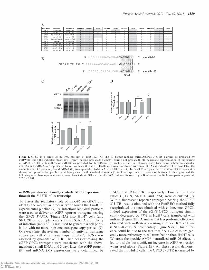
To find miRNAs involved in GPC3 post-transcriptionalregulation, we submitted the GPC3 mRNA to 10 establishedprediction programs gathered on miRWalk [miRWalk—ADatabase on Predicted and Validated microRNA Targets,(http://mirwalk.uni-hd.de)] and selected those targetingthe 30-UTR. As shown in Figure 1A, miR-96 appearedin top position with 6 positive predictions over 10.MiR-182, which displays the same 50-UUGGCA-30 seedsequence to positions 2–7 (Figure 1B) than miR-96,appeared just below miR-96 among the four top-rankedmiRNAs with 5 positive predictions over 10 (Figure 1A).By individually testing different programs, miR-96 andmiR-182 were predicted to target GPC3 using Targetscan(Figure 1B and Supplementary Figure S1A) (13), DianamicroT v.4 (Supplementary Figure S1B) (20), miRanda/mirSVR (Supplementary Figure S1C) (21) and Pictar
(Supplementary Figure S1E) (22), whereas only miR-96was found amongst the miRNAs predicted using PITA(23) and miRDB (24,25) (Supplementary Figure S1Dand F, respectively). The unique 50-UGCCAA-30
miRNA site located in the GPC3 30-UTR and predictedto pair with the seed of miR-96 or miR-182 is veryconserved amongst species (Supplementary Figure S2A)suggesting an apparent evolutionary-conserved partner-ship between GPC3 and these two miRNAs. In silico,miR-96:GPC3 30-UTR pairing is defined as an 8-mer siteby Targetscan, whereas miR-182:GPC3 30-UTR inter-action is defined as 7-mer-1A (13). The only differencebetween these two types of sites resides in the fact thatin miR-96, the nucleotide at position 8 immediately down-stream the seed can pair the GPC3 30-UTR (G–C pair),whereas that of miR-182 cannot (Figure 1B). FinallymiR-96 and miR-182, together with miR-183, belong tothe same intergenic miRNA cluster (miR-182-183,miRBase (26)).
Based on these observations, experimental analyses wereundertaken. Expression of miR-96 and miR-182 in HuH7and SNU398 cells, two human HCC-derived GPC3-expressing cell lines, was first confirmed by RT-qPCR(Supplementary Figure S2B). Results showed thatmiR-182 was apparently more abundant in both HCCcell lines than miR-96 (compare �Ct values inSupplementary Figure S2B) suggesting the existence ofpost-transcriptional regulations in the course of miR-182-183 cluster biogenesis. Then we tested the ability ofthe two miRNAs to control GPC3 expression in HCC-derived HuH7 cells by over-expressing each miRNAusing cell transfection. As shown in Figure 1C, miR-96significantly decreased GPC3-protein amount by morethan half. As expected, a specific hairpin inhibitor,AM96, counterbalanced the negative effect induced bymiR-96. However when used alone, AM96 slightly, butsignificantly, increased GPC3-protein expression suggest-ing that it efficiently interacted with the endogenousmiR-96 and inhibited its function. Comparable resultswere obtained when monitoring GPC3-transcriptamounts (Figure 1D) suggesting that miR-96 downregu-lated GPC3 expression at the mRNA level. Surprisingly,although miR-96 and miR-182 carry the same seed(Figure 1B), miR-182 had no effect on GPC3 protein ex-pression, nor mRNA level (Figure 1C and D). Thisabsence of effect could not originate from a poor cell-transfection efficiency as miR-182 overexpression wasobserved in the corresponding transfected HuH7 cells(Supplementary Figure S2C). To assess miR-182 function-ing, we studied its regulatory effect on Adenylate cyclasetype 6 (ADCY6), one of its validated target (27).Noticeably ADCY6 30-UTR is predicted to contain one8-mer site for miR-182 and three 8-mer sites for miR-96(Figure 4A). As shown in Supplementary Figure S2D,both miR-96 and miR-182 downregulated expression ofADCY6 mRNA and protein, despite the fact positionsof their predicted sites into the ADCY6 30-UTR remainto be confirmed. Together these results showed that(i) miR-96 specifically controls GPC3 expression at themRNA level, and (ii) miR-182 is functionally inefficienton GPC3.
1358 Nucleic Acids Research, 2012, Vol. 40, No. 3
Downloaded from https://academic.oup.com/nar/article-abstract/40/3/1356/1144990by gueston 29 March 2018
miR-96 post-transcriptionally controls GPC3 expressionthrough the 30-UTR of its transcript
To assess the regulatory role of miR-96 on GPC3 andidentify the molecular process, we followed the FunREGexperimental pipeline (9,19). Infectious lentiviral particleswere used to deliver an eGFP-reporter transgene bearingthe GPC3 30-UTR (Figure 2A) into HuH7 cells (andSNU398 cells, Supplementary Figure S3A). A multiplicityof infection (moi) of 0.5 was used to generate a cell popu-lation with no more than one transgene copy per cell (9).One week later the average number of lentiviral transgenecopies per cell (‘transgene copy number’, TCN) wasassessed by quantitative PCR. Then cells expressing theeGFP-GPC3 transgene were transfected with the above-mentioned small RNAs and 3 days later, the eGFP protein(P) and mRNA (M) expressions were determined by
FACS and RT-qPCR, respectively. Finally the threeratios (P/TCN, M/TCN and P/M) were calculated (9).With a fluorescent reporter transgene bearing the GPC330-UTR, results obtained with the FunREG method fullyrecapitulated the ones obtained with endogenous GPC3.Indeed expression of the eGFP-GPC3 transgene signifi-cantly decreased by 47% in HuH7 cells transfected withmiR-96 (Figure 2B). A similar but less profound effect wasobserved with miR-96 when using another HCC cell line(SNU398 cells, Supplementary Figure S3A). This differ-ence could be due to the fact that SNU398 cells are gen-erally more refractory to cell transfection than HuH7 cells.Whereas the specific AM96 neutralized miR-96 effect, itled to a slight but significant increase in eGFP expressionwhen used alone (Figure 2B). All these results demonst-rated that in HuH7 cells, the GPC3 30-UTR is targeted by
A
B
C D
Figure 1. GPC3 is a target of miR-96, but not of miR-182. (A) The 10 highest-ranking miRNA:GPC3-30-UTR pairings as predicted bymiRWalk using the indicated algorithms (1/grey: pairing predicted; 0/empty: pairing not predicted). (B) Schematic representation of the pairingof GPC3 30-UTR with miR-96 or miR-182 as predicted by TargetScan. In this figure and the following ones, base pairings between indicatedmRNAs and miRNAs are represented by vertical lines. (C and D): HuH7 cells were transfected with small RNAs as indicated. Three days later, theamounts of GPC3 protein (C) and mRNA (D) were quantified (ANOVA: P< 0.0001; n=6). In Panel C, a representative western blot experiment isshown on top and a bar graph recapitulating means with standard deviation (SD) of six experiments is shown on bottom. In this figure and thefollowing ones, bars represent means, error bars indicate SD and the ANOVA test was followed by a Bonferroni’s multiple comparison post-test.***P< 0.001.
Nucleic Acids Research, 2012, Vol. 40, No. 3 1359
Downloaded from https://academic.oup.com/nar/article-abstract/40/3/1356/1144990by gueston 29 March 2018
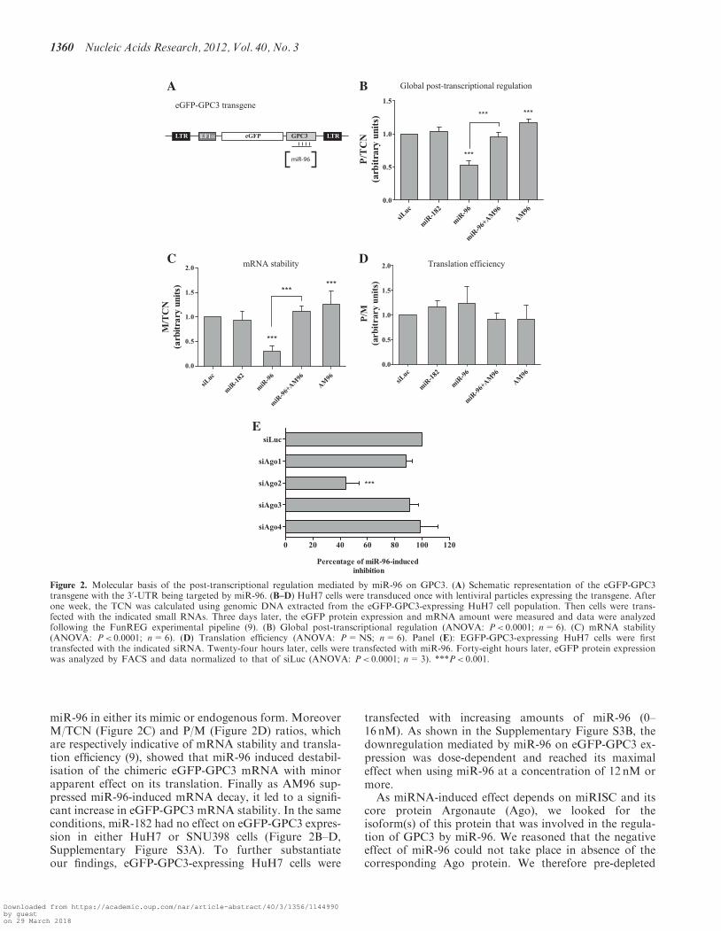
miR-96 in either its mimic or endogenous form. MoreoverM/TCN (Figure 2C) and P/M (Figure 2D) ratios, whichare respectively indicative of mRNA stability and transla-tion efficiency (9), showed that miR-96 induced destabil-isation of the chimeric eGFP-GPC3 mRNA with minorapparent effect on its translation. Finally as AM96 sup-pressed miR-96-induced mRNA decay, it led to a signifi-cant increase in eGFP-GPC3 mRNA stability. In the sameconditions, miR-182 had no effect on eGFP-GPC3 expres-sion in either HuH7 or SNU398 cells (Figure 2B–D,Supplementary Figure S3A). To further substantiateour findings, eGFP-GPC3-expressing HuH7 cells were
transfected with increasing amounts of miR-96 (0–16 nM). As shown in the Supplementary Figure S3B, thedownregulation mediated by miR-96 on eGFP-GPC3 ex-pression was dose-dependent and reached its maximaleffect when using miR-96 at a concentration of 12 nM ormore.
As miRNA-induced effect depends on miRISC and itscore protein Argonaute (Ago), we looked for theisoform(s) of this protein that was involved in the regula-tion of GPC3 by miR-96. We reasoned that the negativeeffect of miR-96 could not take place in absence of thecorresponding Ago protein. We therefore pre-depleted
A B
C
E
D
Figure 2. Molecular basis of the post-transcriptional regulation mediated by miR-96 on GPC3. (A) Schematic representation of the eGFP-GPC3transgene with the 30-UTR being targeted by miR-96. (B–D) HuH7 cells were transduced once with lentiviral particles expressing the transgene. Afterone week, the TCN was calculated using genomic DNA extracted from the eGFP-GPC3-expressing HuH7 cell population. Then cells were trans-fected with the indicated small RNAs. Three days later, the eGFP protein expression and mRNA amount were measured and data were analyzedfollowing the FunREG experimental pipeline (9). (B) Global post-transcriptional regulation (ANOVA: P< 0.0001; n=6). (C) mRNA stability(ANOVA: P< 0.0001; n=6). (D) Translation efficiency (ANOVA: P=NS; n=6). Panel (E): EGFP-GPC3-expressing HuH7 cells were firsttransfected with the indicated siRNA. Twenty-four hours later, cells were transfected with miR-96. Forty-eight hours later, eGFP protein expressionwas analyzed by FACS and data normalized to that of siLuc (ANOVA: P< 0.0001; n=3). ***P< 0.001.
1360 Nucleic Acids Research, 2012, Vol. 40, No. 3
Downloaded from https://academic.oup.com/nar/article-abstract/40/3/1356/1144990by gueston 29 March 2018
HuH7 cells of each Argonaute isoform with specificsiRNAs (Supplementary Figure S3C–F). Then depletedcells were transfected with miR-96. Results in Figure 2Eclearly showed that miR-96 mainly required Argonaute 2,as its inhibitory effect (shown at its maximum in the siLuccontrol condition) was decreased by 60% in absence ofAgo2. It should be noted that depletion of Ago2 inHuH7 cells was compensated by an increase of Ago1and Ago3 mRNAs (Supplementary Figure S3D).Altogether these results showed that miR-96 negativelycontrols GPC3 expression at a post-transcriptional levelby targeting the 30-UTR of its transcript and by inducingits degradation by an Ago-2-dependent mechanism.
Molecular basis of miR-96-target recognition
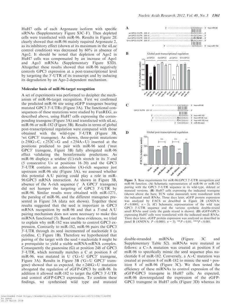
A set of experiments was performed to decipher the mech-anism of miR-96-target recognition. First we confirmedthe predicted miR-96 site using eGFP transgenes bearingmutated GPC3 30-UTRs (Figure 3A). The functional con-sequences of these mutations were studied by FunREG, asdescribed above, using HuH7 cells expressing the corres-ponding transgene (Figure 3A) and transfected with siLuc,miR-96 or miR-182 (Figure 3B). Results in terms of globalpost-transcriptional regulation were compared with thoseobtained with the wild-type 30-UTR (Figure 3B,‘wt GPC3’ transgene). As shown, three-point mutations(r.250G>C, r.252C>G and r.254A>U) inserted at thepositions predicted to pair with miR-96 seed (‘mutGPC3’ transgene, Figure 3B) fully abrogated miR-96effect validating the bioinformatic predictions. AsmiR-96 displays a uridine (U)-rich stretch in its 30-end(5 consecutive Us at positions 16–20) and the GPC330-UTR contains an adenosine (A)-rich sequence justupstream miR-96 site (Figure 3A), we assessed whetherthis potential A/U pairing could play a role in miR-96:GPC3 mRNA interaction. As shown in Figure 3B,absence of the A-rich sequence (‘�A GPC3’ transgene)did not hamper the targeting of GPC3 30-UTR bymiR-96. Similar results were obtained with miR-1271,the paralog of miR-96, and the different transgenes pre-sented in Figure 3A (data not shown). Together theseresults suggested that the seed is important in GPC3mRNA recognition by miR-96 and that a 30-end A/Upairing mechanism does not seem necessary to make thismiRNA functional (5). Based on these evidences, we triedto explain why miR-182 was unable to control GPC3 ex-pression. Contrarily to miR-182, miR-96 pairs the GPC330-UTR through its seed incremented of nucleotide 8 (acytidine, C; Figure 1B). Therefore we hypothesized thatpairing of the target with the seed+nucleotide 8 might bea prerequisite to yield a stable miRNA:mRNA complex.Consequently the guanosine (G) at position 248 of GPC330-UTR, which normally matches a C at position 8 ofmiR-96, was mutated in U (‘G>U GPC3’ transgene,Figure 3A). Results in Figure 3B (‘G>U GPC3’ trans-gene) showed that as expected, the r.248G>U mutationabrogated the regulation of eGFP-GPC3 by miR-96. Inaddition it allowed miR-182 to target the GPC3 30-UTRand control eGFP-GPC3 expression. To confirm thesefindings, we synthesised wild type and mutated
double-stranded miRNAs (Figure 3C andSupplementary Table S2). miRNAs were mutated asfollows: a C>A mutation was created at position 8 ofmiR-96 to specifically mimic the seed sequence plus nu-cleotide 8 of miR-182. Conversely, a A>C mutation wascreated at position 8 of miR-182 to mimic the seed+pos-ition 8 of miR-96 (Figure 3C). We then tested theefficiency of these miRNAs to control expression of theeGFP-GPC3 transgene in HuH7 cells. As expected,miR-96 downregulated the expression of the eGFP-GPC3 transgene in HuH7 cells (Figure 3D) whereas its
A
B
C
D
Figure 3. Base requirements for miR-96:GPC3 30-UTR recognition andmiR-96 function. (A) Schematic representation of miR-96 or miR-182pairing with the GPC3 30-UTR sequence in its wild-type, deleted ormutated versions. (B) HuH7 cells expressing the indicated transgene(shown above the bars; TCN value measured) were transfected withthe indicated small RNAs. Three days later, eGFP protein expressionwas analyzed by FACS as described in Figure 2B (ANOVA:P< 0.0001; n=3). (C) Schematic representation of the wild typeGPC3 30-UTR sequence and the various synthetic double-strandsmall RNAs used (only the guide strand is shown). (D) eGFP-GPC3-expressing HuH7 cells were transfected with the indicated small RNAs.Three days later, eGFP protein expression was analyzed as described inpanel B (ANOVA: P< 0.0001; n=5). **P< 0.01; ***P< 0.001.
Nucleic Acids Research, 2012, Vol. 40, No. 3 1361
Downloaded from https://academic.oup.com/nar/article-abstract/40/3/1356/1144990by gueston 29 March 2018
mutated counterpart (miR-96mut) was inefficient. Itshould be noted however that the synthetic miR-96 thatwe produced was less efficient than its commercial mimicform. Although miR-182 had still no effect, its mutatedcounterpart (miR-182mut) was fully functional anddecreased eGFP expression by 47%. Because of thecentral role of the first 8 nt of miR-96 in its functioning,we also tested the impact of transfecting cells with the8-mer oligonucleotide (‘miR-96 8-mer’, Figure 3C) oneGFP-GPC3 expression. Unexpectedly miR-96 8-mer didnot reduce, but rather slightly increased eGFP-GPC3 ex-pression (Figure 3D). This increase was similar to thatobtained with AM96 in Figure 2B suggesting that the8-mer nucleotide cannot recapitulate miR-96 activity,but rather acts as a specific competitive inhibitor.Finally we evaluated whether the differential regulation
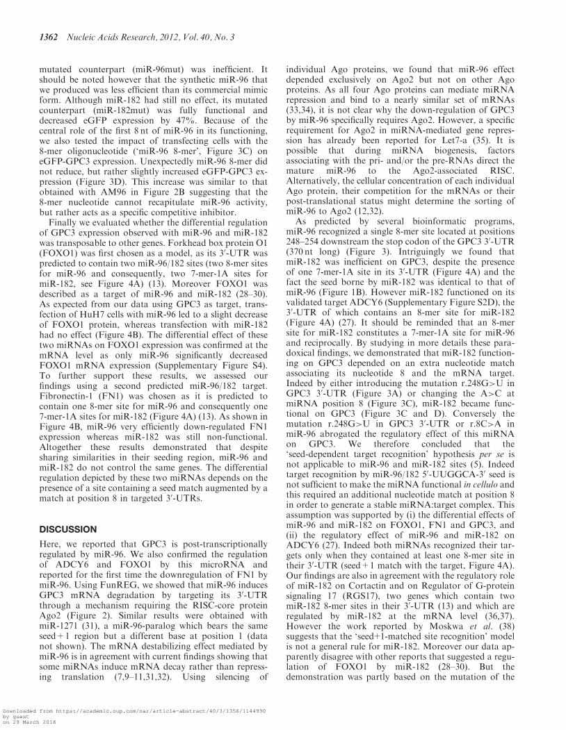
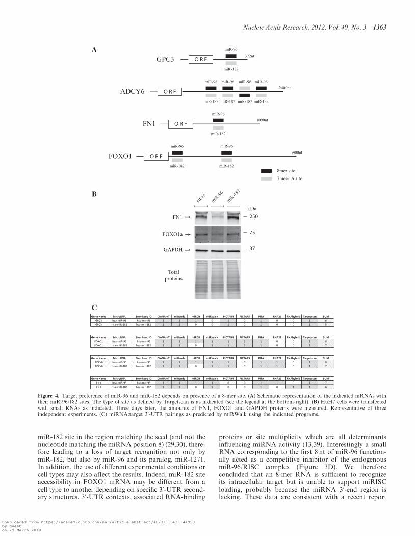
of GPC3 expression observed with miR-96 and miR-182was transposable to other genes. Forkhead box protein O1(FOXO1) was first chosen as a model, as its 30-UTR waspredicted to contain two miR-96/182 sites (two 8-mer sitesfor miR-96 and consequently, two 7-mer-1A sites formiR-182, see Figure 4A) (13). Moreover FOXO1 wasdescribed as a target of miR-96 and miR-182 (28–30).As expected from our data using GPC3 as target, trans-fection of HuH7 cells with miR-96 led to a slight decreaseof FOXO1 protein, whereas transfection with miR-182had no effect (Figure 4B). The differential effect of thesetwo miRNAs on FOXO1 expression was confirmed at themRNA level as only miR-96 significantly decreasedFOXO1 mRNA expression (Supplementary Figure S4).To further support these results, we assessed ourfindings using a second predicted miR-96/182 target.Fibronectin-1 (FN1) was chosen as it is predicted tocontain one 8-mer site for miR-96 and consequently one7-mer-1A sites for miR-182 (Figure 4A) (13). As shown inFigure 4B, miR-96 very efficiently down-regulated FN1expression whereas miR-182 was still non-functional.Altogether these results demonstrated that despitesharing similarities in their seeding region, miR-96 andmiR-182 do not control the same genes. The differentialregulation depicted by these two miRNAs depends on thepresence of a site containing a seed match augmented by amatch at position 8 in targeted 30-UTRs.
DISCUSSION
Here, we reported that GPC3 is post-transcriptionallyregulated by miR-96. We also confirmed the regulationof ADCY6 and FOXO1 by this microRNA andreported for the first time the downregulation of FN1 bymiR-96. Using FunREG, we showed that miR-96 inducesGPC3 mRNA degradation by targeting its 30-UTRthrough a mechanism requiring the RISC-core proteinAgo2 (Figure 2). Similar results were obtained withmiR-1271 (31), a miR-96-paralog which bears the sameseed+1 region but a different base at position 1 (datanot shown). The mRNA destabilizing effect mediated bymiR-96 is in agreement with current findings showing thatsome miRNAs induce mRNA decay rather than repress-ing translation (7,9–11,31,32). Using silencing of
individual Ago proteins, we found that miR-96 effectdepended exclusively on Ago2 but not on other Agoproteins. As all four Ago proteins can mediate miRNArepression and bind to a nearly similar set of mRNAs(33,34), it is not clear why the down-regulation of GPC3by miR-96 specifically requires Ago2. However, a specificrequirement for Ago2 in miRNA-mediated gene repres-sion has already been reported for Let7-a (35). It ispossible that during miRNA biogenesis, factorsassociating with the pri- and/or the pre-RNAs direct themature miR-96 to the Ago2-associated RISC.Alternatively, the cellular concentration of each individualAgo protein, their competition for the mRNAs or theirpost-translational status might determine the sorting ofmiR-96 to Ago2 (12,32).
As predicted by several bioinformatic programs,miR-96 recognized a single 8-mer site located at positions248–254 downstream the stop codon of the GPC3 30-UTR(370 nt long) (Figure 3). Intriguingly we found thatmiR-182 was inefficient on GPC3, despite the presenceof one 7-mer-1A site in its 30-UTR (Figure 4A) and thefact the seed borne by miR-182 was identical to that ofmiR-96 (Figure 1B). However miR-182 functioned on itsvalidated target ADCY6 (Supplementary Figure S2D), the30-UTR of which contains an 8-mer site for miR-182(Figure 4A) (27). It should be reminded that an 8-mersite for miR-182 constitutes a 7-mer-1A site for miR-96and reciprocally. By studying in more details these para-doxical findings, we demonstrated that miR-182 function-ing on GPC3 depended on an extra nucleotide matchassociating its nucleotide 8 and the mRNA target.Indeed by either introducing the mutation r.248G>U inGPC3 30-UTR (Figure 3A) or changing the A>C atmiRNA position 8 (Figure 3C), miR-182 became func-tional on GPC3 (Figure 3C and D). Conversely themutation r.248G>U in GPC3 30-UTR or r.8C>A inmiR-96 abrogated the regulatory effect of this miRNAon GPC3. We therefore concluded that the‘seed-dependent target recognition’ hypothesis per se isnot applicable to miR-96 and miR-182 sites (5). Indeedtarget recognition by miR-96/182 50-UUGGCA-30 seed isnot sufficient to make the miRNA functional in cellulo andthis required an additional nucleotide match at position 8in order to generate a stable miRNA:target complex. Thisassumption was supported by (i) the differential effects ofmiR-96 and miR-182 on FOXO1, FN1 and GPC3, and(ii) the regulatory effect of miR-96 and miR-182 onADCY6 (27). Indeed both miRNAs recognized their tar-gets only when they contained at least one 8-mer site intheir 30-UTR (seed+1 match with the target, Figure 4A).Our findings are also in agreement with the regulatory roleof miR-182 on Cortactin and on Regulator of G-proteinsignaling 17 (RGS17), two genes which contain twomiR-182 8-mer sites in their 30-UTR (13) and which areregulated by miR-182 at the mRNA level (36,37).However the work reported by Moskwa et al. (38)suggests that the ‘seed+1-matched site recognition’ modelis not a general rule for miR-182. Moreover our data ap-parently disagree with other reports that suggested a regu-lation of FOXO1 by miR-182 (28–30). But thedemonstration was partly based on the mutation of the
1362 Nucleic Acids Research, 2012, Vol. 40, No. 3
Downloaded from https://academic.oup.com/nar/article-abstract/40/3/1356/1144990by gueston 29 March 2018
miR-182 site in the region matching the seed (and not thenucleotide matching the miRNA position 8) (29,30), there-fore leading to a loss of target recognition not only bymiR-182, but also by miR-96 and its paralog, miR-1271.In addition, the use of different experimental conditions orcell types may also affect the results. Indeed, miR-182 siteaccessibility in FOXO1 mRNA may be different from acell type to another depending on specific 30-UTR second-ary structures, 30-UTR contexts, associated RNA-binding
proteins or site multiplicity which are all determinantsinfluencing miRNA activity (13,39). Interestingly a smallRNA corresponding to the first 8 nt of miR-96 function-ally acted as a competitive inhibitor of the endogenousmiR-96/RISC complex (Figure 3D). We thereforeconcluded that an 8-mer RNA is sufficient to recognizeits intracellular target but is unable to support miRISCloading, probably because the miRNA 30-end region islacking. These data are consistent with a recent report
A
B
C
Figure 4. Target preference of miR-96 and miR-182 depends on presence of a 8-mer site. (A) Schematic representation of the indicated mRNAs withtheir miR-96/182 sites. The type of site as defined by Targetscan is as indicated (see the legend at the bottom-right). (B) HuH7 cells were transfectedwith small RNAs as indicated. Three days later, the amounts of FN1, FOXO1 and GAPDH proteins were measured. Representative of threeindependent experiments. (C) miRNA:target 30-UTR pairings as predicted by miRWalk using the indicated programs.
Nucleic Acids Research, 2012, Vol. 40, No. 3 1363
Downloaded from https://academic.oup.com/nar/article-abstract/40/3/1356/1144990by gueston 29 March 2018
showing that small locked nucleic acids (LNA) targetingmiRNA seed can abrogate miRNA function (40).Therefore any interference in miRNA:target recognitionby using either seed complementary or 8-mer mimic(comprising the seed flanked by a base at positions 1and 8) LNAs should specifically block miRNA action, aproperty which could lead to therapeutic effects (40). Atlast we showed that miR-96:target pairing does notrequire a 30-compensatory mechanism. Indeed bothmiR-96 and miR-182mut (as well as miR-1271, data notshown) controlled GPC3 expression, although the remain-ing sequences downstream nucleotide 8 are completely dif-ferent (Figure 3). Together these observations are inaccordance with the proposed ‘seed nucleation’ modelwhere the miRNA 30-end is necessary for the biologicalfunction, but does not participate in target recognition (5).Therefore such a 30-compensatory mechanism seems notto be required in target recognition by miR-96 nor miR-182.Because of the central role played by the miRNA seed+1region in our model, we used the RNAhybrid softwareto estimate the strength of these miRNA-seed:target hybrid-isations (41). The analyses showed that the free energyhybridisations of miR-96:GPC3 (nucleotides 2–8) andmiR-182:GPC3 (nucleotides 2–7) complexes were �14.8and �12.8 kCal/mol, respectively. In the same condition,the free energy hybridisation of the miR-182mut:GPC3(nucleotides 2–8) complex was �13.7 kCal/mol.Therefore in our experimental conditions, a minimumfree energy of at least �13.7 kCal/mol is apparentlyrequired to make these miRNAs functional. Togetherour data demonstrated that the nucleotide 8 of miR-96and miR-182 is as critical as nucleotides 2–7 for targetrecognition and miRNA:mRNA complex stabilization.Our results are in accordance with other reports showingthat introducing single nucleotide changes in the target sitecomplementary to nucleotides 2–8 of Human miR-96/1271or Drosophila miR-7 abrogates gene regulation (31,42).Therefore although dissimilar (Figure 1B), the seeds ofmiR-96 and miR-182 comprise nucleotides 2–8(31,32,43) rather than 2–7 (5) and their matching sitecan be classified as 50 dominant seed site (42) or 8-mersite (5). However we cannot exclude the possibility thatthese miRNA could act differently under specific circum-stances as previously reported for miR-182 (38).By taking into account all these findings, we can specu-
late that the sets of cellular genes regulated by miR-96 andby miR-182 are profoundly different with the exception ofthose carrying at least one 8-mer site for both miRNAs(e.g. ADCY6, Figure 4A). Moreover it can be proposedthat any C>A variations at position 8 of miR-96 (or con-versely A>C change in miR-182) or any G>U changes inthe target at the corresponding matching position mightlead to a gene reprogramming with deep cellular conse-quences. Indeed such miRNA modifications might takeplace in physiological conditions (i.e. edition (12)) andplay a role in cell plasticity especially during organismdevelopment or cell differentiation. They may also belinked to evolutionary mechanisms such as single nucleo-tide polymorphisms or to post-transcriptional RNA modi-fications. Finally they may also be linked to human illnesssince several single nucleotide mutations involving
miRNA:target recognition have been reported lately,some involving miR-96 or miR-433 (10,44,45). With theadvent of genomic deep sequencing programs and therecent discovery of gene deregulations associated with30-UTR mutations, maybe some of these questions willfind answers.
Our data also point to the lack of accuracy of the bio-informatic prediction tools gathered on miRWalk (http://mirwalk.uni-hd.de). Indeed none of the algorithms inFigure 4C were in complete accordance with our function-al data: the regulatory effect of miR-96 on GPC3,ADCY6, FOXO1 and FN1, and that of miR-182 onADCY6. It should be specified however that miRDB(24,25) gave the best prognostication as it efficiently pre-dicted a regulation of GPC3, FN1 and FOXO1 bymiR-96, and not by miR-182. However it missed the regu-lation of ADCY6 by miR-182. Because the number of celltypes used in our work was limited, we cannot pretendthat our results reflect a general mechanism. However itis clear that room remains for bioinformatic tool improve-ment and that functional studies, associated with newvaluable experimental and screening tools, shouldgreatly contribute to their enhancement.
SUPPLEMENTARY DATA
Supplementary Data are available at NAR Online:Supplementary Tables 1 and 2, Supplementary Figures1–4, Supplementary References (13,20–25).
ACKNOWLEDGEMENTS
Helene Jacquemin-Sablon, Violaine Moreau, FredericSaltel, Patrick Lestienne and Eric Chevet are warmlyacknowledged for their critical comments of themanuscript.
FUNDING
This work was supported by the Agence Nationale pour laRecherche (ANR)—Programme Jeunes Chercheurs(Paris, France) (Grant no: JC07_184264 to C.G.) and byLa Ligue Nationale Contre le Cancer. M.M. and V.M.were recipients of fellowships from the Ministere del’Enseignement Superieur et de la Recherche (MESR).Funding for open access charge: INSERM.
Conflict of interest statement. None declared.
REFERENCES
1. Barreau,C., Paillard,L. and Osborne,H.B. (2005) AU-richelements and associated factors: are there unifying principles?Nucleic Acids Res., 33, 7138–7150.
2. Garneau,N.L., Wilusz,J. and Wilusz,C.J. (2007) The highwaysand byways of mRNA decay. Nat. Rev. Mol. Cell Biol., 8,113–126.
3. Grosset,C., Chen,C.Y., Xu,N., Sonenberg,N., Jacquemin-Sablon,H. and Shyu,A.B. (2000) A mechanism for translationallycoupled mRNA turnover: interaction between the poly(A) tailand a c-fos RNA coding determinant via a protein complex. Cell,103, 29–40.
1364 Nucleic Acids Research, 2012, Vol. 40, No. 3
Downloaded from https://academic.oup.com/nar/article-abstract/40/3/1356/1144990by gueston 29 March 2018
4. Grosset,C., Boniface,R., Duchez,P., Solanilla,A., Cosson,B. andRipoche,J. (2004) In vivo studies of translational repressionmediated by the granulocyte-macrophage colony-stimulatingfactor AU-rich element. J. Biol. Chem., 279, 13354–13362.
5. Bartel,D.P. (2009) MicroRNAs: target recognition and regulatoryfunctions. Cell, 136, 215–233.
6. Bakheet,T., Williams,B.R. and Khabar,K.S. (2006) ARED 3.0:the large and diverse AU-rich transcriptome. Nucleic Acids Res.,34, D111–D114.
7. Huntzinger,E. and Izaurralde,E. (2011) Gene silencing bymicroRNAs: contributions of translational repression and mRNAdecay. Nat. Rev. Genet., 12, 99–110.
8. Bartel,D.P. (2004) MicroRNAs: genomics, biogenesis, mechanism,and function. Cell, 116, 281–297.
9. Laloo,B., Simon,D., Veillat,V., Lauzel,D., Guyonnet-Duperat,V.,Moreau-Gaudry,F., Sagliocco,F. and Grosset,C. (2009) Analysisof post-transcriptional regulations by a functional, integrated, andquantitative method. Mol. Cell Proteomics, 8, 1777–1788.
10. Simon,D., Laloo,B., Barillot,M., Barnetche,T., Blanchard,C.,Rooryck,C., Marche,M., Burgelin,I., Coupry,I., Chassaing,N.et al. (2010) A mutation in the 30-UTR of the HDAC6 geneabolishing the post-transcriptional regulation mediated byhsa-miR-433 is linked to a new form of dominant X-linkedchondrodysplasia. Hum Mol Genet., 19, 2015–2027.
11. Guo,H., Ingolia,N.T., Weissman,J.S. and Bartel,D.P. (2010)Mammalian microRNAs predominantly act to decrease targetmRNA levels. Nature, 466, 835–840.
12. Krol,J., Loedige,I. and Filipowicz,W. (2010) The widespreadregulation of microRNA biogenesis, function and decay.Nat. Rev. Genet., 11, 597–610.
13. Grimson,A., Farh,K.K., Johnston,W.K., Garrett-Engele,P.,Lim,L.P. and Bartel,D.P. (2007) MicroRNA targeting specificity inmammals: determinants beyond seed pairing. Mol. Cell, 27, 91–105.
14. Kim,V.N., Han,J. and Siomi,M.C. (2009) Biogenesis of smallRNAs in animals. Nat. Rev. Mol. Cell Biol., 10, 126–139.
15. Slezak-Prochazka,I., Durmus,S., Kroesen,B.J. and van denBerg,A. (2010) MicroRNAs, macrocontrol: regulation of miRNAprocessing. RNA, 16, 1087–1095.
16. Shin,C., Nam,J.W., Farh,K.K., Chiang,H.R., Shkumatava,A. andBartel,D.P. (2010) Expanding the microRNA targeting code:functional sites with centered pairing. Mol. Cell, 38, 789–802.
17. Filmus,J. and Capurro,M. (2008) The role of glypican-3 in theregulation of body size and cancer. Cell Cycle, 7, 2787–2790.
18. Jakubovic,B.D. and Jothy,S. (2007) Glypican-3: From themutations of Simpson-Golabi-Behmel genetic syndrome to atumor marker for hepatocellular carcinoma. Exp. Mol. Pathol.,82, 184–189.
19. Laloo,B., Maurel,M., Jalvy-Delvaille,S., Sagliocco,F. andGrosset,C.F. (2010) Analysis of post-transcriptional regulationusing the FunREG method. Biochem. Soc. Trans., 38, 1608–1614.
20. Maragkakis,M., Reczko,M., Simossis,V.A., Alexiou,P.,Papadopoulos,G.L., Dalamagas,T., Giannopoulos,G., Goumas,G.,Koukis,E., Kourtis,K. et al. (2009) DIANA-microT web server:elucidating microRNA functions through target prediction.Nucleic Acids Res., 37, W273–W276.
21. Betel,D., Koppal,A., Agius,P., Sander,C. and Leslie,C. (2010)Comprehensive modeling of microRNA targets predicts functionalnon-conserved and non-canonical sites. Genome Biol., 11, R90.
22. Krek,A., Grun,D., Poy,M.N., Wolf,R., Rosenberg,L.,Epstein,E.J., MacMenamin,P., da Piedade,I., Gunsalus,K.C.,Stoffel,M. et al. (2005) Combinatorial microRNA targetpredictions. Nat. Genet., 37, 495–500.
23. Kertesz,M., Iovino,N., Unnerstall,U., Gaul,U. and Segal,E. (2007)The role of site accessibility in microRNA target recognition.Nat. Genet., 39, 1278–1284.
24. Wang,X. (2008) miRDB: a microRNA target prediction andfunctional annotation database with a wiki interface. RNA, 14,1012–1017.
25. Wang,X. and El Naqa,I.M. (2008) Prediction of both conservedand nonconserved microRNA targets in animals. Bioinformatics,24, 325–332.
26. Griffiths-Jones,S., Saini,H.K., van Dongen,S. and Enright,A.J.(2008) miRBase: tools for microRNA genomics. Nucleic AcidsRes., 36, D154–D158.
27. Xu,S., Witmer,P.D., Lumayag,S., Kovacs,B. and Valle,D. (2007)MicroRNA (miRNA) transcriptome of mouse retina andidentification of a sensory organ-specific miRNA cluster.J. Biol. Chem., 282, 25053–25066.
28. Myatt,S.S., Wang,J., Monteiro,L.J., Christian,M., Ho,K.K.,Fusi,L., Dina,R.E., Brosens,J.J., Ghaem-Maghami,S. andLam,E.W. (2010) Definition of microRNAs that repressexpression of the tumor suppressor gene FOXO1 in endometrialcancer. Cancer Res., 70, 367–377.
29. Guttilla,I.K. and White,B.A. (2009) Coordinate regulation ofFOXO1 by miR-27a, miR-96, and miR-182 in breast cancer cells.J. Biol. Chem., 284, 23204–23216.
30. Stittrich,A.B., Haftmann,C., Sgouroudis,E., Kuhl,A.A.,Hegazy,A.N., Panse,I., Riedel,R., Flossdorf,M., Dong,J.,Fuhrmann,F. et al. (2010) The microRNA miR-182 is induced byIL-2 and promotes clonal expansion of activated helper Tlymphocytes. Nat. Immunol., 11, 1057–1062.
31. Jensen,K.P. and Covault,J. (2011) Human miR-1271 is a miR-96paralog with distinct non-conserved brain expression pattern.Nucleic Acids Res., 39, 701–711.
32. Fabian,M.R., Sonenberg,N. and Filipowicz,W. (2010) Regulationof mRNA translation and stability by microRNAs. Annu. Rev.Biochem., 79, 351–379.
33. Su,H., Trombly,M.I., Chen,J. and Wang,X. (2009) Essential andoverlapping functions for mammalian Argonautes in microRNAsilencing. Genes Dev., 23, 304–317.
34. Landthaler,M., Gaidatzis,D., Rothballer,A., Chen,P.Y., Soll,S.J.,Dinic,L., Ojo,T., Hafner,M., Zavolan,M. and Tuschl,T. (2008)Molecular characterization of human Argonaute-containingribonucleoprotein complexes and their bound target mRNAs.RNA, 14, 2580–2596.
35. Schmitter,D., Filkowski,J., Sewer,A., Pillai,R.S., Oakeley,E.J.,Zavolan,M., Svoboda,P. and Filipowicz,W. (2006) Effects ofDicer and Argonaute down-regulation on mRNA levels in humanHEK293 cells. Nucleic Acids Res., 34, 4801–4815.
36. Zhang,L., Liu,T., Huang,Y. and Liu,J. (2011) microRNA-182inhibits the proliferation and invasion of human lungadenocarcinoma cells through its effect on human corticalactin-associated protein. Int. J. Mol. Med., 28, 381–388.
37. Sun,Y., Fang,R., Li,C., Li,L., Li,F., Ye,X. and Chen,H. (2010)Hsa-mir-182 suppresses lung tumorigenesis throughdown regulation of RGS17 expression in vitro.Biochem. Biophys. Res. Commun., 396, 501–507.
38. Moskwa,P., Buffa,F.M., Pan,Y., Panchakshari,R., Gottipati,P.,Muschel,R.J., Beech,J., Kulshrestha,R., Abdelmohsen,K.,Weinstock,D.M. et al. (2011) miR-182-mediated downregulationof BRCA1 impacts DNA repair and sensitivity to PARPinhibitors. Mol. Cell, 41, 210–220.
39. Brodersen,P. and Voinnet,O. (2009) Revisiting the principlesof microRNA target recognition and mode of action.Nat. Rev. Mol. Cell Biol., 10, 141–148.
40. Obad,S., dos Santos,C.O., Petri,A., Heidenblad,M., Broom,O.,Ruse,C., Fu,C., Lindow,M., Stenvang,J., Straarup,E.M. et al.(2011) Silencing of microRNA families by seed-targeting tinyLNAs. Nat. Genet., 43, 371–378.
41. Rehmsmeier,M., Steffen,P., Hochsmann,M. and Giegerich,R.(2004) Fast and effective prediction of microRNA/target duplexes.RNA, 10, 1507–1517.
42. Brennecke,J., Stark,A., Russell,R.B. and Cohen,S.M. (2005)Principles of microRNA-target recognition. PLoS Biol., 3, e85.
43. Filipowicz,W., Bhattacharyya,S.N. and Sonenberg,N. (2008)Mechanisms of post-transcriptional regulation by microRNAs: arethe answers in sight? Nat. Rev. Genet., 9, 102–114.
44. Mencia,A., Modamio-Hoybjor,S., Redshaw,N., Morin,M., Mayo-Merino,F., Olavarrieta,L., Aguirre,L.A., del Castillo,I., Steel,K.P.,Dalmay,T. et al. (2009) Mutations in the seed region of humanmiR-96 are responsible for nonsyndromic progressive hearing loss.Nat. Genet., 41, 609–613.
45. Lewis,M.A., Quint,E., Glazier,A.M., Fuchs,H., De Angelis,M.H.,Langford,C., van Dongen,S., Abreu-Goodger,C., Piipari,M.,Redshaw,N. et al. (2009) An ENU-induced mutation of miR-96associated with progressive hearing loss in mice. Nat. Genet., 41,614–618.
Nucleic Acids Research, 2012, Vol. 40, No. 3 1365
Downloaded from https://academic.oup.com/nar/article-abstract/40/3/1356/1144990by gueston 29 March 2018