Embed Size (px)
Citation preview
Published: May 27, 2011
r 2011 American Chemical Society 7940 dx.doi.org/10.1021/jp200330z | J. Phys. Chem. B 2011, 115, 7940–7949
ARTICLE
pubs.acs.org/JPCB
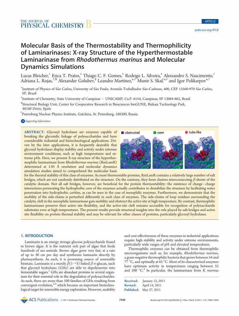
Molecular Basis of the Thermostability and Thermophilicityof Laminarinases: X-ray Structure of the HyperthermostableLaminarinase from Rhodothermus marinus and MolecularDynamics SimulationsLucas Bleicher,† Erica T. Prates,‡ Thiago C. F. Gomes,‡ Rodrigo L. Silveira,‡ Alessandro S. Nascimento,†
Adriana L. Rojas,†,§ Alexander Golubev,|| Leandro Martínez,*,† Munir S. Skaf,*,‡ and Igor Polikarpov*,†
†Institute of Physics of S~ao Carlos, University of S~ao Paulo, Avenida Trabalhador S~ao-Carlense, 400, CEP 13560-970 S~ao Carlos,SP, Brazil‡Institute of Chemistry, State University of Campinas � UNICAMP, Cx.P. 6154, Campinas, SP 13084-862, Brazil§Structural Biology Unit, Center for Cooperative Research in Biosciences bioGUNE, Bizkaia Technology Park,48160 Derio, Spain
)Petersburg Nuclear Physics Institute, Gatchina, St. Petersburg, 188300, Russia
bS Supporting Information
1. INTRODUCTION
Laminarin is an energy storage glucose polysaccharide foundin brown algae. It is the nutrient rich part of algae that feedshundreds of sea animals. Brown algae grow very fast, at a rateof up to 30 cm per day and synthesize laminarin directly byphotosynthesis. As such, it is promising source of renewablebiomass. Laminarin is a mostly β(1f3) linked β-D-glucan, suchthat glycosyl hydrolases (GHs) are able to depolymerize intofermentable sugars.1 GHs are abundant proteins in several organ-isms for their essential role in the degradation of polysaccharides.As such, there are more than 100 families of GHs resulting fromconvergent evolution,2,3 which became an important biotechno-logical target for renewable energy exploration.However, usability
and cost-effectiveness of these enzymes in industrial applicationsrequire high stability and activity under extreme environments,particularly wide ranges of pH and elevated temperatures.
Thermophilic enzymes can be obtained from thermophilicmicroorganisms such as, for example, Rhodothermus marinus,a gram-negative thermophilic bacteria that grows between 54 and77 �C, and optimally at 65 �C. Most of its characterized enzymeshave optimum activity in temperatures ranging between 55and 100 �C.4 In particular, the laminarinase from R. marinus
Received: January 12, 2011Revised: April 14, 2011
ABSTRACT: Glycosyl hydrolases are enzymes capable ofbreaking the glycosidic linkage of polysaccharides and haveconsiderable industrial and biotechnological applications. Dri-ven by the later applications, it is frequently desirable thatglycosyl hydrolases display stability and activity under extremeenvironment conditions, such as high temperatures and ex-treme pHs. Here, we present X-ray structure of the hyperther-mophilic laminarinase from Rhodothermus marinus (RmLamR)determined at 1.95 Å resolution and molecular dynamicssimulation studies aimed to comprehend the molecular basisfor the thermal stability of this class of enzymes. As most thermostable proteins, RmLamR contains a relatively large number of saltbridges, which are not randomly distributed on the structure. On the contrary, they form clusters interconnecting β-sheets of thecatalytic domain. Not all salt bridges, however, are beneficial for the protein thermostability: the existence of charge�chargeinteractions permeating the hydrophobic core of the enzymes actually contributes to destabilize the structure by facilitating waterpenetration into hydrophobic cavities, as can be seen in the case of mesophilic enzymes. Furthermore, we demonstrate that themobility of the side-chains is perturbed differently in each class of enzymes. The side-chains of loop residues surrounding thecatalytic cleft in the mesophilic laminarinase gain mobility and obstruct the active site at high temperature. By contrast, thermophiliclaminarinases preserve their active site flexibility, and the active-site cleft remains accessible for recognition of polysaccharidesubstrates even at high temperatures. The present results provide structural insights into the role played by salt-bridges and activesite flexibility on protein thermal stability and may be relevant for other classes of proteins, particularly glycosyl hydrolases.
7941 dx.doi.org/10.1021/jp200330z |J. Phys. Chem. B 2011, 115, 7940–7949
The Journal of Physical Chemistry B ARTICLE
(RmLamR) is a member of GH family 16, which was cloned,expressed, and characterized in 1998 by Krah et al.,5 displayingoptimal activity in pH 5.5 and 88 �C. Therefore, RmLamR is ahyperthermophilic enzyme, and as such bears a considerablepromise for industrial and biotechnological applications.
The optimal temperature for the stability and activity of eachenzyme results from an intricate balance between charged, polar,nonpolar, and solvent interactions, which are affected differentlyby temperature. In general terms, it has been observed thatthermostable proteins display enhanced polar interactions withthe solvent and increased hydrophobic cores.1,2 However, theenergetic balance is subtle, and some interactions may destabilizefolded structures at different temperatures.6 For instance, surfacesalt bridges (SBs) do not contribute to protein stability at roomtemperature, but they do at high temperatures due to decreaseddesolvation penalties of charged groups.7�11 Therefore, under-standing the interplay between different components of theprotein�solvent and residue�residue interactions is a key factorfor unveiling the molecular and structural reasons of a givenenzyme’s thermostability and thermophilicity.
Here, we discuss the molecular basis of the thermal stabilityand thermophilicity of family 16 laminarinases by solving theX-ray structure of the hyperthermostable RmLamR and by per-forming comparative molecular dynamics (MD) simulations ofthree structural homologues: the hyperthermophilic RmLamRfrom R. marinus, a thermostable laminarinase from alkaliphilicNocardiopsis, and a mesophilic laminarinase from Phanerochaetechrysosporium. From the combination of structural and MDanalysis we show that both the number and spatial distributionof SBs are determinant of protein thermal stability, and that distinctexposure of the active site occurs for laminarinases at differenttemperatures. The structural and dynamical features promotingthermal stability observed here can be useful for the rationaliza-tion of protein design, particularly for GHs.
2. MATERIALS AND METHODS
2.1. Experimental Section.The laminarinase from R. marinuswas cloned, expressed, purified, and crystallized as described.12
The X-ray diffraction images from the crystal were collected atthe MX-1 beamline at the Brazilian National Synchrotron LightLaboratory (LNLS), in Campinas, Brazil.13 The data set was indexedand integrated usingMOSFLM,14 scaled in SCALA,15 and analyzedusing phenix.xtriage.16,17 During this process it was detected thatthe crystal presented a rare case of pseudomerohedral twinning.12
The structure of endo-β-1,3-glucanase from Nocardiopsis sp.(PDB code: 2HYK) was used as the search model for a molecularreplacement in MOLREP.18 The structure was rebuilt with thecorrect sequence for RmLamR (GenBank: AAC69707.1; GI:2896144) using RESOLVE19,20 as implemented in phenix.autobuild.21 After real-space model manipulation using COOT22
alternated with least-squares twin refinement as implemented inphenix.refine,23 a final model consisting of two chains with 251residues each, a glycerol molecule and two calcium ions convergedat Rfactor = 16.7% and Rfree = 19.8%. The full set of crystallographicparameters is listed in Table 1. The final crystallographic modeland structure factors were deposited in the Protein Data Bankunder PDB code 3ILN.Secondary structural elements were assessed using Stride.24
Analysis of GHF16 amino acid distributions was evaluated usingversion 23.0 of the PFAM database,25 family PF00722. SSMSUPERPOSE26 was used for three-dimensional superposition ofprotein structures.2.2. Molecular Dynamics. The simulation boxes of the three
homologous laminarinases were built using their crystallographicstructures. The structure of laminarinase from R. marinus wasobtained as described above. Structural models 2HYK and 2CL2were obtained from the protein data bank for the alcaliphilicNocardiopsis sp. strain F96 (aNLam)27 and for laminarinase Lam16A from Phanerochaete chrysosporium (PcLam),28 respectively.Initially, the tridimensional aNLam structure had to be completedfor missing residues at the extremities (TESDMR sequencepeptide at the N-terminus and dipeptide LG at the C-terminus),according to its primary sequence. The coordinates of theseresidues were modeled by alignment of 2HYK to the RmLamRR-carbons. The positions of the other missing atoms were modeledaccording to the CHARMM27 topology file.29 The enzymes werehydrated by 15 000 water molecules in a cubic box with sides ofapproximately 80 Å using Packmol,30,31 such that the solventlayer around the protein surface is at least 12 Å thick.To set the ionization states of the ionizable residues (K, R, H,
D, and E), the correspondent pKa values were determinedaccording to the specified pH 7 and the molecular environment(high dielectric constant at the protein surface and low dielectricconstant in its interior) using the Hþþ server.32�34 Specialattention was paid to the choice of the ionization states of theresidues in the catalytic site. The residues D131 and E134 ofRmLamR were considered protonated, so that they may interactwith each other and with the substrate by means of hydrogenbonding. The nucleophile residue E129 was kept in its chargedform. For all the simulated systems, there were 50 chloride and50 sodium basal ions, in addition to the crystallographic calciumion. To keep the system’s electroneutrality, an excess of 1, 13, and3 sodium ions were added in the RmLamR, 2HYK (aNLam), and2CL2 (PcLam) systems, respectively. The resulting salt concen-tration is approximately 0.16 M.
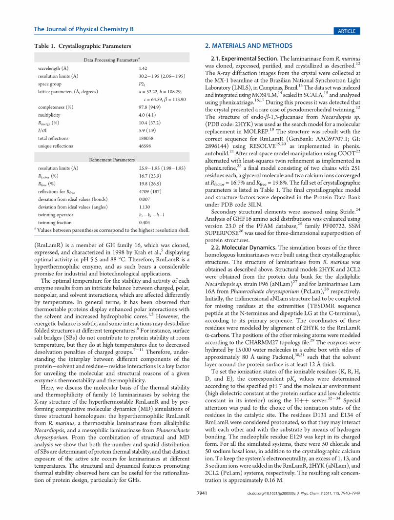
Table 1. Crystallographic Parameters
Data Processing Parametersa
wavelength (Å) 1.42
resolution limits (Å) 30.2�1.95 (2.06�1.95)
space group P21lattice parameters (Å, degrees) a = 52.22, b = 108.29,
c = 64.59, β = 113.90
completeness (%) 97.8 (94.9)
multiplicity 4.0 (4.1)
Rmerge (%) 10.4 (37.2)
I/σI 5.9 (1.9)
total reflections 188058
unique reflections 46598
Refinement Parameters
resolution limits (Å) 25.9�1.95 (1.98�1.95)
Rfactor (%) 16.7 (23.9)
Rfree (%) 19.8 (26.5)
reflections for Rfree 4709 (187)
deviation from ideal values (bonds) 0.007
deviation from ideal values (angles) 1.130
twinning operator h, �k, �h�l
twinning fraction 0.404aValues between parentheses correspond to the highest resolution shell.
7942 dx.doi.org/10.1021/jp200330z |J. Phys. Chem. B 2011, 115, 7940–7949
The Journal of Physical Chemistry B ARTICLE
The energy of the system was initially minimized by 500 stepsof the conjugate gradient (CG) method35,36 as implementedin NAMD37 to eliminate bad contacts. After minimization, weperformed equilibration runs consisting of three phases: In thefirst 10 ps, the positions of all residues, except the modeled ones,were kept fixed; in the next 100 ps, the lateral chains were released;and in the last 890 ps, the entire system was allowed to move. Torun simulations at higher temperature (363 K), the equilibrationperiod were extended in two other steps: 500 ps of equilibrationat an intermediate temperature (330.5 K) and 1 ns of equilibra-tion at 363 K. From the equilibrated systems, we carried out 10independent 12 ns simulations for RmLam, eight 12 ns simula-tions for aNLam, and eight 9 ns runs for PcLam at 25 �C. At90 �C, we performed five independent 9 ns simulations for eachprotein.All simulations were performed with NAMD37 applying
periodic boundary conditions and CHARMM parameters. TheTIP3Pmodel was used for water.38 Langevin dynamics and Nos�e-Hoover Langevin piston methods39,40 were used to keep thetemperature and pressure constant. The RESPA multiple-timestep algorithm41 was used with the shortest time step of 2 fs. Allhydrogen-to-heavy-atom bonds were kept rigid using SHAKE.42
A 12 Å cutoffwith smooth switching function starting at 10 Å wasused for the van der Waals interactions, whereas electrostaticforces were treated via the particle mesh Ewald method.43
3. RESULTS AND DISCUSSION
3.1. The Structure of R. marinus Laminarinase: A Memberof GH Family 16. The laminarinase from R. marinus (RmLamR)was crystallized in space group P21, with a dimer in the asymmetric
unit.12 This dimer and all other possible quaternary assembliesformed by contacts in the crystal are unlikely to be found insolution, as indicated by theoretical estimates of the solvation freeenergy gain upon interface formation for each assembly obtainedusing the PISA server.44 As previously observed by Krah et al.,5
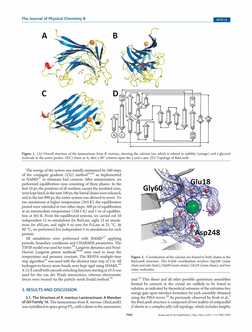
the RmLamR structure is composed of two leaflets of antiparallelβ-sheets in a complex jelly roll topology, which includes lengthy
Figure 1. (A) Overall structure of the laminarinase from R. marinus, showing the calcium ion, which is related to stability (orange) and a glycerolmolecule in the active pocket. (B,C) Same as A, after a 90� rotation upon the y and x axis. (D) Topology of RmLamR.
Figure 2. Coordination of the calcium ion bound to both chains in theRmLamR structure. The 6-fold coordination involves Asp240 (mainchain and side chain), Gly60 (main chain), Glu18 (main chain), and twowater molecules.
7943 dx.doi.org/10.1021/jp200330z |J. Phys. Chem. B 2011, 115, 7940–7949
The Journal of Physical Chemistry B ARTICLE
loop connections, segmented β-strands, and three small helixsegments, as shown in Figure 1. Its secondary structure content ismuch closer to that of B. macerans endo-1,3-1,4-β-glucanase thanto that predicted by circular dichroism spectra:5 instead of 16%R-helices and 37% β-strands, our reported structure contains 1%R-helices (resulting solely from the small 310 helix segment) and48% β-strands, similar to the 3% and 47%, respectively, in theB. macerans homologue.Calcium ions bound to each monomer were clearly identified
(Figure 2), in equivalent sites of each polypeptide chain in theasymmetric unit cell (ASU), and displaying the same coordina-tion patterns. The calcium ions are known to be important forprotein stability of the GH 16 family members.45,46 Coordinationof the calcium ion is 6-fold, and involves Gly60 (main chain),Asp248 (main chain and side chain), Glu18 (main chain), andtwo water molecules.A search using the DALI server47 shows that the endo-β-1,
3-glucanase from alkaliphilic Nocardiopsis sp. strain F96 (PDBcode 2HYK), used in molecular replacement as a search model, isindeed the most similar structure in the PDB, but in addition tothe expected similarity to other GH 16 structures, RmLamR alsoshares three-dimensional similarity to lectins and agglutinins(e.g., PDB codes 1FAY and 2E53) and also the Botulinumneurotoxin type B (PDB code 2NP0).3.2. The RmLamR Active Site. Sequence analysis of members
of the GH family 16 and site-directed mutagenesis have shownthat catalytic activity of RmLamR is dependent on two glutamateand one aspartate residues located in a highly conserved motif(WX1�4E[LIV]D[LIVF]X0�1EX1�3 [GQ]).
5 Moreover, the GHfamily 16 can be divided in two groups, which differ by thepresence of an additional catalytic residue, usually a methioninepreceding the second glutamate. The Met residue is responsiblefor a β-strand distortion, resulting in a β-bulge.48,49 The laminar-inase fromR. marinus has this additional methionine. Its presenceresults in one of six β-bulges in RmLamR, as deduced from thehydrogen bonding pattern between β-sheet containing Met133and the neighboring β-sheet. As for other members of GH family16,49,50 this β-bulge does not imply conformational changes inactive site residues. The RmLamR structure also reveals a glycerolmolecule fortuitously bound to the active site of the enzymemolecule. Anchoring of glycerol molecules by several hydrogenbonds to active site residues has been previously observed in theX-ray structure of the endo-β-1,3-glucanase fromNocardiopsis sp.strain F96.27
3.3. Structure and Thermostability.One common feature ofproteins bearing high thermal stability is the low content of loopregions.51 RmLamR, on the contrary, displays a large number ofloops of considerable lengths (Figure 1). Remarkably, all loopsand turns can be readily modeled from the electron density maps,even in regions where side-chains are barely seen. Thus, numberand lengths of loops do not imply an abundance of disorderedregions, and loops do not adopt multiple conformations in thecrystal.The sequence of RmLamR contains an increased number of
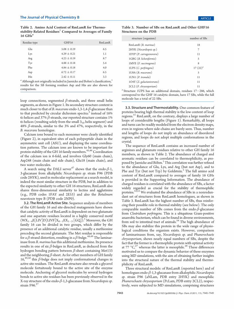
arginines and glutamate residues relative to other GH family 16members, as shown in Table 2. The abundance of charged andaromatic residues can be correlated to thermophilicity, as pro-posed by Jaenicke and B€ohm.1 This correlation was further refinedto the abundance of Glu, Lys, and Arg (but not Asp), and toPhe and Tyr (but not Trp) by Goldstein.2 The full amino acidcontent of RmLamR compared to averages of family 16 GHsis provided in the Supporting Information. The abundance ofcharged residues is correlated with the abundance of SBs, a factorwidely regarded as crucial for the stability of thermophilicproteins.52,53 We evaluated the abundance of SBs in a nonredun-dant set of structures from RmLamR homologues, as shown inTable 3. RmLamR has the highest number of SBs, thus reinfor-cing their possible role in thermal stability (see below). The onlycomparable number of SBs comes from the endo-β-glucanasefrom Clostridium perfringens. This is a ubiquitous Gram-positiveanaerobic bacterium, which can be found in diverse environments,from soil to intestinal tract of animals.54 The high abundance ofSBs may also stabilize this protein in the wide range of physio-logical conditions the organism exists. However, comparisonof laminarinases from, say, Nocardiopsis sp. and Phanerochaetechrysosporium, shows nearly equal numbers of SBs, despite thefact that the former is a thermophilic protein with optimal activityat 77 �C,27 whereas the latter is mesophile.28 These differencesmotivated us to compare the dynamic behavior of these enzymesusing MD simulations, with the aim of obtaining further insightsinto the structural nature of the thermal stability and thermo-philicity of RmLamR.Three structural models: of RmLamR (reported here) and of
homologues endo-β-1,3-glucanase from alkalophilicNocardiopsissp. strain F96 (aNLam, PDB entry 2HYK) and mesophilicPhanerochaete chrysosporium (PcLam, PDB entry 2CL2), respec-tively, were subjected to MD simulations, comprising structures
Table 2. Amino Acid Content of RmLamR for Thermo-stability-Related Residues1 Compared to Averages of Family16 GHsa
Residue type GHF16 RmLamR
Glu 5.08 ( 0.19 6.5
Lys 4.29 ( 0.21 1.1
Arg 4.23 ( 0.19 8.7
Tyr 4.88 ( 0.18 5.4
Phe 4.84 ( 0.18 4.0
Asp 6.72 ( 0.17 6.5
His 2.42 ( 0.11 3.3aAlthough not originally included in Jaenicke and Bohm’s classification,1
results for the SB forming residues Asp and His are also shown forcomparison.
Table 3. Number of SBs on RmLamR and Other GHF16Structures on the PDB
structure (organism) number of SBs
RmLamR (R. marinus) 18
2HYK (Nocardiopsis sp.) 7
1DYP (P. carrageenovora) 11
1GBG (B. licheniformis) 5
1MVE (F. succinogenes) 8
1UPS (C. perfringens) 17 (22)a
1U0A (B. macerans) 5
1UN1 (P. tremula) 11
1O4Y (Z. galactanivorans) 11
2CL2 (P. chrysosporium) 11a Structure 1UPS has an additional domain, residues 17�286, whichcorrespond to the GHF 16 catalytic domain, have 17 SBs, while the fullmolecule has a total of 22 SBs.
7944 dx.doi.org/10.1021/jp200330z |J. Phys. Chem. B 2011, 115, 7940–7949
The Journal of Physical Chemistry B ARTICLE
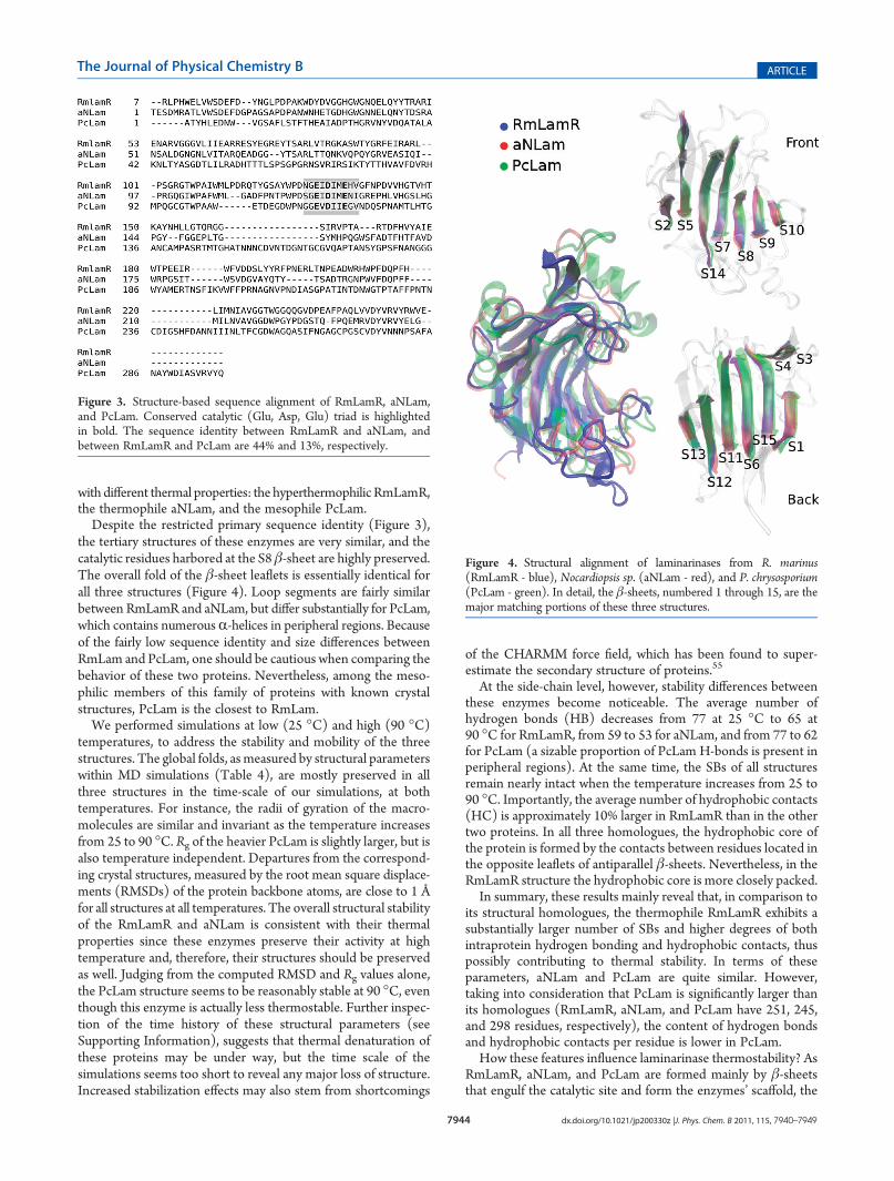
with different thermal properties: the hyperthermophilic RmLamR,the thermophile aNLam, and the mesophile PcLam.Despite the restricted primary sequence identity (Figure 3),
the tertiary structures of these enzymes are very similar, and thecatalytic residues harbored at the S8 β-sheet are highly preserved.The overall fold of the β-sheet leaflets is essentially identical forall three structures (Figure 4). Loop segments are fairly similarbetween RmLamR and aNLam, but differ substantially for PcLam,which contains numerousR-helices in peripheral regions. Becauseof the fairly low sequence identity and size differences betweenRmLam and PcLam, one should be cautious when comparing thebehavior of these two proteins. Nevertheless, among the meso-philic members of this family of proteins with known crystalstructures, PcLam is the closest to RmLam.We performed simulations at low (25 �C) and high (90 �C)
temperatures, to address the stability and mobility of the threestructures. The global folds, as measured by structural parameterswithin MD simulations (Table 4), are mostly preserved in allthree structures in the time-scale of our simulations, at bothtemperatures. For instance, the radii of gyration of the macro-molecules are similar and invariant as the temperature increasesfrom 25 to 90 �C. Rg of the heavier PcLam is slightly larger, but isalso temperature independent. Departures from the correspond-ing crystal structures, measured by the root mean square displace-ments (RMSDs) of the protein backbone atoms, are close to 1 Åfor all structures at all temperatures. The overall structural stabilityof the RmLamR and aNLam is consistent with their thermalproperties since these enzymes preserve their activity at hightemperature and, therefore, their structures should be preservedas well. Judging from the computed RMSD and Rg values alone,the PcLam structure seems to be reasonably stable at 90 �C, eventhough this enzyme is actually less thermostable. Further inspec-tion of the time history of these structural parameters (seeSupporting Information), suggests that thermal denaturation ofthese proteins may be under way, but the time scale of thesimulations seems too short to reveal any major loss of structure.Increased stabilization effects may also stem from shortcomings
of the CHARMM force field, which has been found to super-estimate the secondary structure of proteins.55
At the side-chain level, however, stability differences betweenthese enzymes become noticeable. The average number ofhydrogen bonds (HB) decreases from 77 at 25 �C to 65 at90 �C for RmLamR, from 59 to 53 for aNLam, and from 77 to 62for PcLam (a sizable proportion of PcLam H-bonds is present inperipheral regions). At the same time, the SBs of all structuresremain nearly intact when the temperature increases from 25 to90 �C. Importantly, the average number of hydrophobic contacts(HC) is approximately 10% larger in RmLamR than in the othertwo proteins. In all three homologues, the hydrophobic core ofthe protein is formed by the contacts between residues located inthe opposite leaflets of antiparallel β-sheets. Nevertheless, in theRmLamR structure the hydrophobic core is more closely packed.In summary, these results mainly reveal that, in comparison to
its structural homologues, the thermophile RmLamR exhibits asubstantially larger number of SBs and higher degrees of bothintraprotein hydrogen bonding and hydrophobic contacts, thuspossibly contributing to thermal stability. In terms of theseparameters, aNLam and PcLam are quite similar. However,taking into consideration that PcLam is significantly larger thanits homologues (RmLamR, aNLam, and PcLam have 251, 245,and 298 residues, respectively), the content of hydrogen bondsand hydrophobic contacts per residue is lower in PcLam.How these features influence laminarinase thermostability? As
RmLamR, aNLam, and PcLam are formed mainly by β-sheetsthat engulf the catalytic site and form the enzymes’ scaffold, the
Figure 3. Structure-based sequence alignment of RmLamR, aNLam,and PcLam. Conserved catalytic (Glu, Asp, Glu) triad is highlightedin bold. The sequence identity between RmLamR and aNLam, andbetween RmLamR and PcLam are 44% and 13%, respectively.
Figure 4. Structural alignment of laminarinases from R. marinus(RmLamR - blue), Nocardiopsis sp. (aNLam - red), and P. chrysosporium(PcLam - green). In detail, the β-sheets, numbered 1 through 15, are themajor matching portions of these three structures.
7945 dx.doi.org/10.1021/jp200330z |J. Phys. Chem. B 2011, 115, 7940–7949
The Journal of Physical Chemistry B ARTICLE
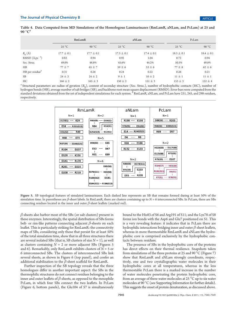
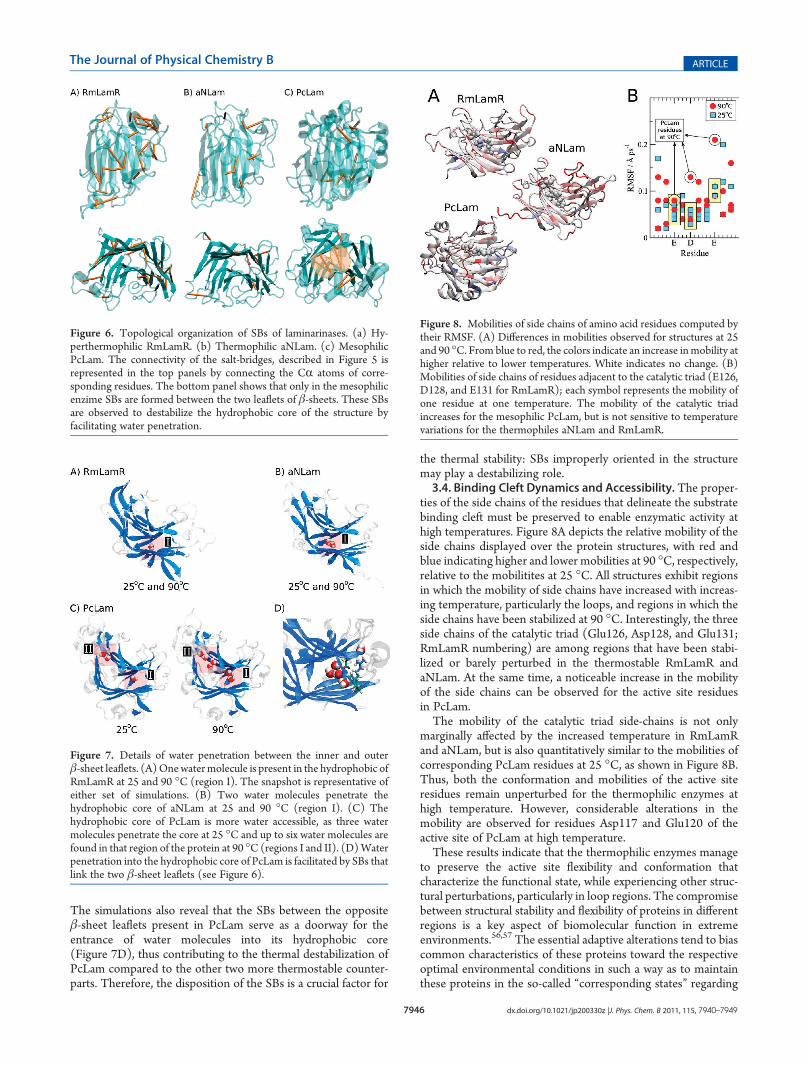
β-sheets also harbor most of the SBs (or salt clusters) present inthese enzymes. Interestingly, the spatial distribution of SBs formsbelt- or rim-like patterns connecting adjacent β-sheets on eachleaflet. This is particularly striking for RmLamR: the connectivitymaps of SBs, considering only those that persist for at least 50%of the total simulation time, show that in all three structures thereare several isolated SBs (that is, SB clusters of sizeN = 1), as wellas clusters containing N = 2 or more adjacent SBs (Figures 5and 6). Remarkably, only RmLamR exhibits clusters of N = 5 or6 interconnected SBs. The clusters of interconnected SBs linkseveral sheets, as shown in Figure 6 (top panel), and confer anadditional stabilization to the β-sheet scaffold for RmLamR.Further inspection of the SB topology reveals that the three
homologues differ in another important aspect: the SBs in thethermophilic structures do not connect residues belonging to theinner and outer leaflets of β-sheets, as opposed to the mesophilePcLam, in which four SBs connect the two leaflets. In PcLam(Figure 6, bottom panels), the Glu104 of S7 is simultaneously
bound to the His83 of S6 and Arg191 of S11; and the Lys78 of S8forms ion bonds with the Asp6 and Glu7 positioned on S1. Thisis a very revealing feature: it indicates that in PcLam there arehydrophilic interactions bridging inner and outer β-sheet leaflets,whereas in more thermostable RmLamR and aNLam the hydro-phobic core is comprised exclusively by the hydrophobic con-tacts between residues.The presence of SBs in the hydrophobic core of the proteins
has direct effects on their thermal resilience. Snapshots takenfrom simulations of the three proteins at 25 and 90 �C (Figure 7)show that RmLamR and aNLam strongly coordinate, respec-tively, one and two crystallographic water molecules in theirhydrophobic cores at all temperatures, whereas in the lessthermostable PcLam there is a marked increase in the numberof water molecules penetrating the protein hydrophobic core,from an average of three water molecules at 25 �C up to six watermolecules at 90 �C(see Supporting Information for further details).This suggests the onset of protein denaturation, as discussed above.
Table 4. Data Computed from MD Simulations of the Homologous Laminarinases (RmLamR, aNLam, and PcLam) at 25 and90 �Ca
RmLamR aNLam PcLam
25 �C 90 �C 25 �C 90 �C 25 �C 90 �C
Rg (Å) 17.7 ( 0.1 17.7 ( 0.1 17.3 ( 0.1 17.4 ( 0.1 18.3 ( 0.1 18.4 ( 0.1
RMSD (Å.ps�1) 0.82 0.94 0.92 1.04 0.72 0.94
Sec. Struc. 49.0% 48.8% 45.6% 44.2% 50.5% 49.8%
HB 77 ( 7 65 ( 7 59 ( 6 53 ( 6 77 ( 8 62 ( 6
HB per residueb 0.31 0.26 0.24 0.22 0.26 0.21
SB 24 ( 3 24 ( 2 9 ( 1 10 ( 2 11 ( 1 11 ( 1
HC 166 ( 2 165 ( 3 150 ( 2 151 ( 3 153 ( 3 152 ( 4a Structural parameters are radius of gyration (Rg), content of seconday structure (Sec. Struc.), number of hydrophobic contacts (HC), number ofhydrogen bonds (HB), average number of salt bridges (SB), and backbone root mean square displacement (RMSD). Error bars were computed from thestandard deviations obtained from the set of independent simulations for each system. bRmLamR, aNLam, and PcLam have 251, 245, and 298 residues,respectively.
Figure 5. SB topological features of simulated laminarinases. Each dashed line represents an SB that remains formed during at least 50% of thesimulation time. In parentheses are β-sheet labels. In RmLamR, there are clusters containing up to N = 6 interconnected SBs. In PcLam, there are SBsconnecting residues located in the inner and outer β-sheet leaflets (marked red).
7946 dx.doi.org/10.1021/jp200330z |J. Phys. Chem. B 2011, 115, 7940–7949
The Journal of Physical Chemistry B ARTICLE
The simulations also reveal that the SBs between the oppositeβ-sheet leaflets present in PcLam serve as a doorway for theentrance of water molecules into its hydrophobic core(Figure 7D), thus contributing to the thermal destabilization ofPcLam compared to the other two more thermostable counter-parts. Therefore, the disposition of the SBs is a crucial factor for
the thermal stability: SBs improperly oriented in the structuremay play a destabilizing role.3.4. Binding Cleft Dynamics and Accessibility. The proper-
ties of the side chains of the residues that delineate the substratebinding cleft must be preserved to enable enzymatic activity athigh temperatures. Figure 8A depicts the relative mobility of theside chains displayed over the protein structures, with red andblue indicating higher and lower mobilities at 90 �C, respectively,relative to the mobilitites at 25 �C. All structures exhibit regionsin which the mobility of side chains have increased with increas-ing temperature, particularly the loops, and regions in which theside chains have been stabilized at 90 �C. Interestingly, the threeside chains of the catalytic triad (Glu126, Asp128, and Glu131;RmLamR numbering) are among regions that have been stabi-lized or barely perturbed in the thermostable RmLamR andaNLam. At the same time, a noticeable increase in the mobilityof the side chains can be observed for the active site residuesin PcLam.The mobility of the catalytic triad side-chains is not only
marginally affected by the increased temperature in RmLamRand aNLam, but is also quantitatively similar to the mobilities ofcorresponding PcLam residues at 25 �C, as shown in Figure 8B.Thus, both the conformation and mobilities of the active siteresidues remain unperturbed for the thermophilic enzymes athigh temperature. However, considerable alterations in themobility are observed for residues Asp117 and Glu120 of theactive site of PcLam at high temperature.These results indicate that the thermophilic enzymes manage
to preserve the active site flexibility and conformation thatcharacterize the functional state, while experiencing other struc-tural perturbations, particularly in loop regions. The compromisebetween structural stability and flexibility of proteins in differentregions is a key aspect of biomolecular function in extremeenvironments.56,57 The essential adaptive alterations tend to biascommon characteristics of these proteins toward the respectiveoptimal environmental conditions in such a way as to maintainthese proteins in the so-called “corresponding states” regarding
Figure 6. Topological organization of SBs of laminarinases. (a) Hy-perthermophilic RmLamR. (b) Thermophilic aNLam. (c) MesophilicPcLam. The connectivity of the salt-bridges, described in Figure 5 isrepresented in the top panels by connecting the CR atoms of corre-sponding residues. The bottom panel shows that only in the mesophilicenzime SBs are formed between the two leaflets of β-sheets. These SBsare observed to destabilize the hydrophobic core of the structure byfacilitating water penetration.
Figure 7. Details of water penetration between the inner and outerβ-sheet leaflets. (A)Onewatermolecule is present in the hydrophobic ofRmLamR at 25 and 90 �C (region I). The snapshot is representative ofeither set of simulations. (B) Two water molecules penetrate thehydrophobic core of aNLam at 25 and 90 �C (region I). (C) Thehydrophobic core of PcLam is more water accessible, as three watermolecules penetrate the core at 25 �C and up to six water molecules arefound in that region of the protein at 90 �C (regions I and II). (D)Waterpenetration into the hydrophobic core of PcLam is facilitated by SBs thatlink the two β-sheet leaflets (see Figure 6).
Figure 8. Mobilities of side chains of amino acid residues computed bytheir RMSF. (A) Differences in mobilities observed for structures at 25and 90 �C. From blue to red, the colors indicate an increase inmobility athigher relative to lower temperatures. White indicates no change. (B)Mobilities of side chains of residues adjacent to the catalytic triad (E126,D128, and E131 for RmLamR); each symbol represents the mobility ofone residue at one temperature. The mobility of the catalytic triadincreases for the mesophilic PcLam, but is not sensitive to temperaturevariations for the thermophiles aNLam and RmLamR.
7947 dx.doi.org/10.1021/jp200330z |J. Phys. Chem. B 2011, 115, 7940–7949
The Journal of Physical Chemistry B ARTICLE
folding topology, structural flexibility, and solvent exposure.58 Asdiscussed above, the RmLamR, aNLam, and PcLamdisplay active-site flexibilities that are similar at temperatures of correspondingoptimal activity.The side-chain mobility of residues in the vicinity of the active
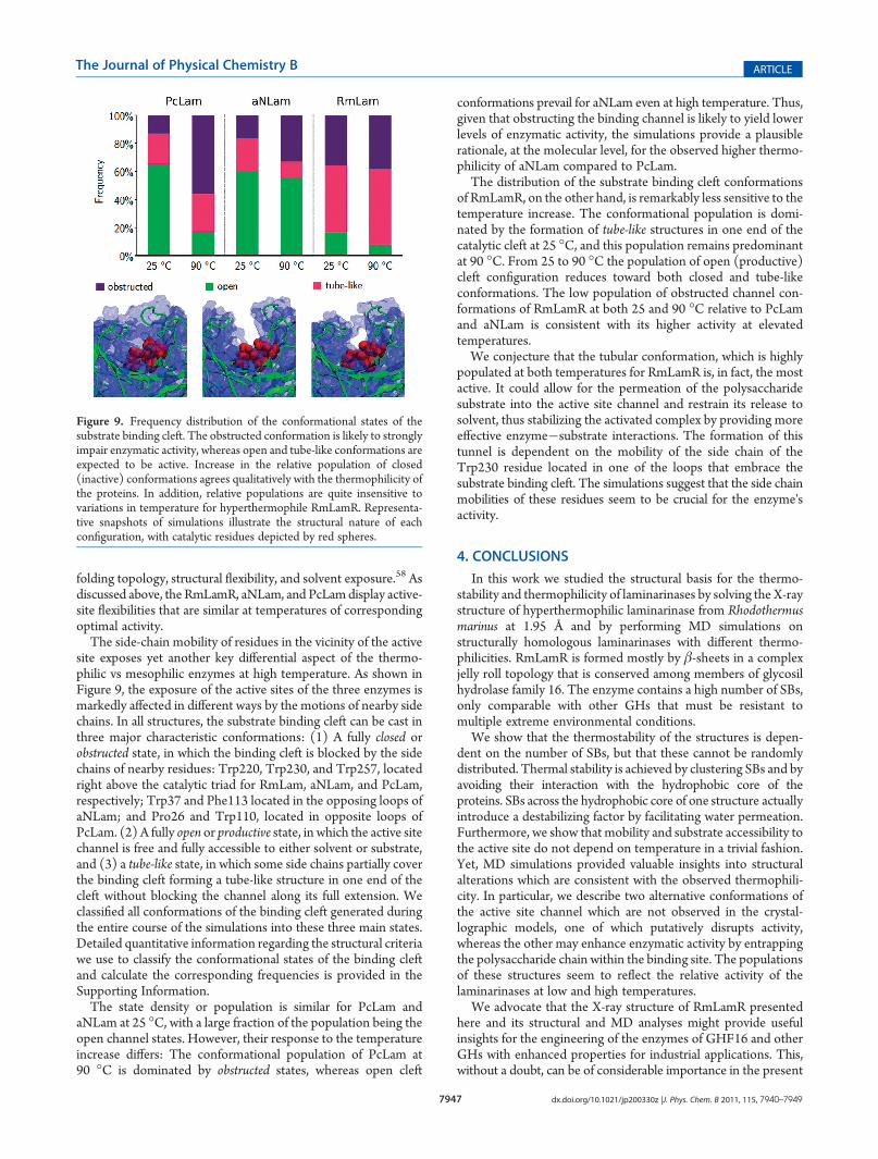
site exposes yet another key differential aspect of the thermo-philic vs mesophilic enzymes at high temperature. As shown inFigure 9, the exposure of the active sites of the three enzymes ismarkedly affected in different ways by the motions of nearby sidechains. In all structures, the substrate binding cleft can be cast inthree major characteristic conformations: (1) A fully closed orobstructed state, in which the binding cleft is blocked by the sidechains of nearby residues: Trp220, Trp230, and Trp257, locatedright above the catalytic triad for RmLam, aNLam, and PcLam,respectively; Trp37 and Phe113 located in the opposing loops ofaNLam; and Pro26 and Trp110, located in opposite loops ofPcLam. (2) A fully open or productive state, in which the active sitechannel is free and fully accessible to either solvent or substrate,and (3) a tube-like state, in which some side chains partially coverthe binding cleft forming a tube-like structure in one end of thecleft without blocking the channel along its full extension. Weclassified all conformations of the binding cleft generated duringthe entire course of the simulations into these three main states.Detailed quantitative information regarding the structural criteriawe use to classify the conformational states of the binding cleftand calculate the corresponding frequencies is provided in theSupporting Information.The state density or population is similar for PcLam and
aNLam at 25 �C, with a large fraction of the population being theopen channel states. However, their response to the temperatureincrease differs: The conformational population of PcLam at90 �C is dominated by obstructed states, whereas open cleft
conformations prevail for aNLam even at high temperature. Thus,given that obstructing the binding channel is likely to yield lowerlevels of enzymatic activity, the simulations provide a plausiblerationale, at the molecular level, for the observed higher thermo-philicity of aNLam compared to PcLam.The distribution of the substrate binding cleft conformations
of RmLamR, on the other hand, is remarkably less sensitive to thetemperature increase. The conformational population is domi-nated by the formation of tube-like structures in one end of thecatalytic cleft at 25 �C, and this population remains predominantat 90 �C. From 25 to 90 �C the population of open (productive)cleft configuration reduces toward both closed and tube-likeconformations. The low population of obstructed channel con-formations of RmLamR at both 25 and 90 �C relative to PcLamand aNLam is consistent with its higher activity at elevatedtemperatures.We conjecture that the tubular conformation, which is highly
populated at both temperatures for RmLamR is, in fact, the mostactive. It could allow for the permeation of the polysaccharidesubstrate into the active site channel and restrain its release tosolvent, thus stabilizing the activated complex by providing moreeffective enzyme�substrate interactions. The formation of thistunnel is dependent on the mobility of the side chain of theTrp230 residue located in one of the loops that embrace thesubstrate binding cleft. The simulations suggest that the side chainmobilities of these residues seem to be crucial for the enzyme'sactivity.
4. CONCLUSIONS
In this work we studied the structural basis for the thermo-stability and thermophilicity of laminarinases by solving the X-raystructure of hyperthermophilic laminarinase from Rhodothermusmarinus at 1.95 Å and by performing MD simulations onstructurally homologous laminarinases with different thermo-philicities. RmLamR is formed mostly by β-sheets in a complexjelly roll topology that is conserved among members of glycosilhydrolase family 16. The enzyme contains a high number of SBs,only comparable with other GHs that must be resistant tomultiple extreme environmental conditions.
We show that the thermostability of the structures is depen-dent on the number of SBs, but that these cannot be randomlydistributed. Thermal stability is achieved by clustering SBs and byavoiding their interaction with the hydrophobic core of theproteins. SBs across the hydrophobic core of one structure actuallyintroduce a destabilizing factor by facilitating water permeation.Furthermore, we show that mobility and substrate accessibility tothe active site do not depend on temperature in a trivial fashion.Yet, MD simulations provided valuable insights into structuralalterations which are consistent with the observed thermophili-city. In particular, we describe two alternative conformations ofthe active site channel which are not observed in the crystal-lographic models, one of which putatively disrupts activity,whereas the other may enhance enzymatic activity by entrappingthe polysaccharide chain within the binding site. The populationsof these structures seem to reflect the relative activity of thelaminarinases at low and high temperatures.
We advocate that the X-ray structure of RmLamR presentedhere and its structural and MD analyses might provide usefulinsights for the engineering of the enzymes of GHF16 and otherGHs with enhanced properties for industrial applications. This,without a doubt, can be of considerable importance in the present
Figure 9. Frequency distribution of the conformational states of thesubstrate binding cleft. The obstructed conformation is likely to stronglyimpair enzymatic activity, whereas open and tube-like conformations areexpected to be active. Increase in the relative population of closed(inactive) conformations agrees qualitatively with the thermophilicity ofthe proteins. In addition, relative populations are quite insensitive tovariations in temperature for hyperthermophile RmLamR. Representa-tive snapshots of simulations illustrate the structural nature of eachconfiguration, with catalytic residues depicted by red spheres.
7948 dx.doi.org/10.1021/jp200330z |J. Phys. Chem. B 2011, 115, 7940–7949
The Journal of Physical Chemistry B ARTICLE
era of protein engineering research applied to biotechnology andto second-generation biofuel technologies.
’ASSOCIATED CONTENT
bS Supporting Information. Further simulation details areprovided in Table S1. The full amino acid content of RmLamR isprovided in Table S2. Structural parameters reported in Table 4as functions of time for each independent simulation are shownin Figures S1�S7. Details on the calculation of the hydrationlevels of the hydrophobic core are provided, and the results asfunctions of time are shown in Figure S8. A geometric criterionfor the conformational states of the binding cleft is introducedand illustrated by Figures S9�S11. This information is availablefree of charge via the Internet at http://pubs.acs.org.
’AUTHOR INFORMATION
Corresponding Author*E-mail addresses: [email protected] (I.P.); [email protected] (M.S.S.); [email protected] (L.M.).
Author ContributionsL. Bleicher, E. T. Prates, T. C. F. Gomes, and R. L. Silveiracontributed equally to this work.
’ACKNOWLEDGMENT
We thank the Brazilian funding agencies CNPq and FAPESP(Grant Nos. 08/56225-9, 10/16947-9, 10/18849-4, 09/14107-6,10/08680-2, 10/16947-9) and the MCT/CNPq/FAPESP EU-Brazil Collaboration program in Second Generation Biofuels(Grant No. 490022/2009-0) for financial support. We also thankthe Brazilian National Synchrotron Laboratory for the utilizationof theMX1 beamline. L.B. also thanks Ralf Grosse-Kunstleve andPeter Swart.
’REFERENCES
(1) Jaenicke, R.; Bohm, G. Curr. Opin. Struct. Biol. 1998, 8, 738–748.(2) Goldstein, R. A. Protein Sci. 2007, 16, 1887–1895.(3) Davies, G.; Henrissat, B. Structure 1995, 3, 853–859.(4) Bjornsdottir, S. H.; Blondal, T.; Hreggvidsson, G. O.; Eggertsson,
G.; Petursdottir, S.; Hjorleifsdottir, S.; Thorbjarnardottir, S. H.; Kristjansson,J. K. Extremophiles 2006, 10, 1–16.(5) Krah, M.; Misselwitz, R.; Politz, O.; Thomsen, K. K.; Welfle, H.;
Borriss, R. Eur. J. Biochem. 1998, 257, 101–111.(6) Argos, P.; Rossmann, G. M.; Grau, U. M.; Zuber, H.; Frank, G.;
Tratschin, J. D. Biochemistry 1979, 18, 5698–5703.(7) Vogt, G.; Woell, S.; Argos, P. J. Mol. Biol. 1997, 269, 631–643.(8) Strop, P.; Mayo, S. L. Biochemistry 2000, 39, 1251–1255.(9) Elcock, A. H. J. Mol. Biol. 1998, 284, 489–502.(10) Priyakumar, U. D.; Ramakrishna, S.; Nagarjuna, K. R.; Reddy,
K. S. J. Phys. Chem. B 2010, 114, 1707–1718.(11) Xiao, L.; Honig, B. J. Mol. Biol. 1999, 289, 1435–1444.(12) Golubev, A. M.; Rojas, A. L.; Nascimento, A. S.; Bleicher, L.;
Kulminskaya, A. A.; Eneyskaya, E. V.; Polikarpov, I. Protein Pept. Lett.2008, 15, 1142–1144.(13) Polikarpov, I.; Perles, L. A.; Oliveira, R. T.; Oliva, G.;
Castellano, E. E.; Garratt, R. C.; Craievich, A. J. Synchrotron Radiat.1998, 5, 72–76.(14) Leslie, A. G. Acta Crystallogr. D 1999, 55, 1696–1702.(15) Evans, P. Acta Crystallogr. D 2006, 62, 72–82.(16) Zwart, P. H.; Grosse-Kunstleve, R. W.; Adams, P. D. CCP4
Newsletter 2005, 42, 58–67.
(17) Zwart, P. H.; Grosse-Kunstleve, R. W.; Adams, P. D. CCP4Newsletter 2005, 43, 27–35.
(18) Vagin, A.; Teplyakov, A. J. Appl. Crystallogr. 1997, 30,1022–1025.
(19) Terwilliger, T. C. Acta Crystallogr. D 2003, 59, 38–44.(20) Terwilliger, T. C. Acta Crystallogr. D 2003, 59, 45–49.(21) Terwilliger, T. C.; Grosse-Kunstleve, R. W.; Afonine, P. V.;
Moriarty, N. W.; Zwart, P. H.; Hung, L. W.; Read, R. J.; Adams, P. D.Acta Crystallogr. D 2008, 64, 61–69.
(22) Emsley, P.; Cowtan, K. Acta Crystallogr. D 2004, 60,2126–2132.
(23) Afonine, P. V.; Grosse-Kunstleve, R. W.; Adams, P. D. CCP4Newsletter 2005, 42, 43–49.
(24) Frishman, D.; Argos, P. Proteins 1995, 23, 566–579.(25) Finn, R. D.; Tate, J.; Mistry, J.; Coggill, P. C.; Sammut, S. J.;
Hotz, H. R.; Ceric, G.; Forslund, K.; Eddy, S. R.; Sonnhammer, E. L. L.;Bateman, A. Nucleic Acids Res. 2008, 36, D281–D288.
(26) Krissinel, E.; Henrick, K. Acta Crystallogr. D 2004, 60,2256–2268.
(27) Fibriansah, G.;Masuda, S.; Koizumi, N.; Nakamura, S.; Kumasaka,T. Proteins 2007, 69, 683–690.
(28) Vasur, J.; Kawai, R.; Larsson, A. M.; Igarashi, K.; Sandgren, M.;Samejima, M.; Stahlberg, J. Acta Crystallogr. D 2006, 62, 1422–1429.
(29) MacKerell, A. D., Jr.; Bashford, D.; Bellott, M.; Dunbrack, R. L.,Jr.; Evanseck, J. D.; Field, M. J.; Fischer, S.; Gao, J.; Guo, H.; Ha, S.;Joseph-McCarthy, D.; Kuchnir, L.; Kuczera, K.; Lau, F. T. K.; Mattos, C.;Michnick, S.; Ngo, T.; Nguyen, D. T.; Prodham, B.; Reiher, W. E., III;Roux, B.; Schlenkrich, M.; Smith, J. C.; Stote, R.; Straub, J.; Wantanabe,M.; Wi�orkiewicz-Kuczera, J.; Yin, D.; Karplus, M. J. Phys. Chem. B 1998,102, 3586–3616.
(30) Martínez, J. M.; Martínez, L. J. Comput. Chem. 2003, 24,819–825.
(31) Martínez, L.; Andrade, R.; Birgin, E. G.; Martínez, J. M.J. Comput. Chem. 2009, 30, 2157–2164.
(32) Hþþ Server. http://biophysics.cs.vt.edu/Hþþ. AccessedNovember 26, 2010.
(33) Gordon, J. C.; Myers, J. B.; Folta, T.; Shoja, V.; Heath, L. S.;Onufriev, A. Nucleic Acids Res. 2005, 33, W368–W371.
(34) Anandakrishnan, R.; Onufriev, A. J. Comput. Biol. 2008, 15,165–184.
(35) Fletcher, R. Practical Methods of Optimization, 2nd ed.; Wiley:Padstow, 2000.
(36) Fletcher, R.; Reeves, C. M. Comput. J. 1964, 7, 149–154.(37) Phillips, J. C.; Braun, R.; Wang, W.; Gumbart, J.; Tajkhorshid,
E.; Villa, E.; Chipot, C.; Skeel, R. D.; Kale, L.; Schulten, K. J. Comput.Chem. 2005, 26, 1781–1802.
(38) Jorgensen, W. L.; Chandrasekhar, J.; Madura, J. D.; Impey,R. W.; Klein, M. L. J. Chem. Phys. 1983, 79, 926–935.
(39) Schneider, T.; Stoll, E. Phys. Rev. B 1978, 17, 1302–1322.(40) Martyna, G. J.; Tobias, D. J.; Klein, M. L. J. Chem. Phys. 1994,
101, 4177–4189.(41) Tuckerman, M. E.; Berne, B. J.; Martyna, G. J. J. Chem. Phys.
1992, 97, 1990–2001.(42) Ryckaert, J. P.; Ciccotti, G.; Berendsen, H. J. C. J. Comput. Phys.
1977, 23, 327–341.(43) Darden, T.; York, D.; Pedersen, L. J. Chem. Phys. 1993, 98,
10089–10092.(44) Krissinel, E.; Henrick, K. J. Mol. Biol. 2007, 372, 774–797.(45) Keitel, T.;Meldgaard,M.; Heinemann, U.Eur. J. Biochem. 1994,
222, 203–214.(46) Tsai, L. C.; Shyur, L. F.; Lee, S. H.; Lin, S. S.; Yuan, H. S. J. Mol.
Biol. 2003, 330, 607–620.(47) Holm, L.; Kaariainen, S.; Rosenstrom, P.; Schenkel, A. Bioinfor-
matics 2008, 24, 2780–2781.(48) Neustroev, K. N.; Golubev, A. M.; Sinnott, M. L.; Borriss, R.;
Krah, M.; Brumer, H., III; Eneyskaya, E. V.; Shishlyannikov, S.; Shabalin,K. A.; Peshechonov, V. T.; Korolev, V. G.; Kulminskaya, A. A. Glyco-conjugate J. 2006, 23, 501–511.
7949 dx.doi.org/10.1021/jp200330z |J. Phys. Chem. B 2011, 115, 7940–7949
The Journal of Physical Chemistry B ARTICLE
(49) Allouch, J.; Jam, M.; Helbert, W.; Barbeyron, T.; Kloareg, B.;Henrissat, B.; Czjzek, M. J. Biol. Chem. 2003, 278, 47171–47180.(50) Vasur, J.; Kawai, R.; Hanna, K.; Jonsson, M.; Widmalm, G.;
Engstr€om, A.; Frank, M.; Andersson, E.; Hansson, H.; Forsberg, Z.;Igarashi, K.; Samejima, M.; Sandgren, M.; St�ahlberg, J. J. Am. Chem. Soc.2010, 132, 1724–1730.(51) Thompson, M. J.; Eisenberg, D. J. Mol. Biol. 1999, 290,
595–604.(52) Kumar, S.; Nussinov, R. ChemBioChem 2002, 3, 604–617.(53) Dominy, B. N.; Minoux, H.; Brooks, C. L., III. Proteins 2004,
57, 128–141.(54) Petit, L.; Gibert, M.; Popoff, M. R. Trends Microbiol. 1999, 7,
104–110.(55) MacKerell, A. D., Jr.; Feig, M.; Brooks, C. L., III. J. Comput.
Chem. 2004, 25, 1400–1415.(56) Tsou, C.-L. Biochemistry 1988, 27, 1809–1812.(57) Tsou, C.-L. Biochemistry 1998, 63, 253–258.(58) Jaenicke, R. Eur. J. Biochem. 1991, 202, 715–728.