Embed Size (px)
Citation preview
Molecular Biological and Physiological
Investigations of Heterotrophic Bacteria
Associated with Marine Filamentous
Cyanobacteria
Dissertation
zur Erlangung des Grades eines
Doktors der Naturwissenschaften
(Dr. rer. nat.)
dem Fachbereich Biologie / Chemie
der Universität Bremen
vorgelegt von
Annina E. Hube Stade
November 2009

2
1. Gutachter: Prof. Dr. Ulrich Fischer 2. Gutachter: PD Dr. Jens Harder Tag des Promotionskolloquiums: 16. Dezember 2009
Table of contents
3
Table of contents
Table of contents ........................................................................................................................ 3 List of abbreviations................................................................................................................... 4 Abstract ...................................................................................................................................... 5 Zusammenfassung...................................................................................................................... 7 1 Introduction ........................................................................................................................ 9
1.1 The Baltic Sea ............................................................................................................. 9 1.2 Bloom forming and benthic Baltic Sea cyanobacteria .............................................. 10 1.3 Cyanobacteria and heterotrophic bacteria ................................................................. 12 1.4 Aerobic anoxygenic phototrophic bacteria ............................................................... 14
1.4.1 Characterisation and physiology of aerobic anoxygenic phototrophic bacteria . 14 1.4.2 Evolution of AAnP.............................................................................................. 15 1.4.3 Taxonomy, phylogeny, and distribution of AAnP .............................................. 16
1.5 Objective of the thesis ............................................................................................... 17 2 Material and Methods....................................................................................................... 18 3 Manuscripts ...................................................................................................................... 20 4 General Discussion........................................................................................................... 72
4.1 Characterisation of heterotrophic bacteria with respect to their association with cyanobacteria............................................................................................................. 72
4.1.1 Characterisation of Porphyrobacter.................................................................... 72 4.1.2 Characterisation of Roseobacter ......................................................................... 74 4.1.3 Characterisation of strain Bo10-19 ..................................................................... 75 4.1.4 Characterisation of Muricauda............................................................................ 76
4.2 Characterisation of cyanobact. with respect to their association with heterotrophs . 78 4.3 Conclusions ............................................................................................................... 80 4.4 Future prospects ........................................................................................................ 81
5 References ........................................................................................................................ 83 6 Annex ............................................................................................................................... 92 7 Acknowledgements .......................................................................................................... 96

List of abbreviations
4
List of abbreviations
AAnP aerobic anoxygenic phototrophic bacteria
AB BBLTM Actinomyces Broth
AnAnP anaerobic anoxygenic phototrophic bacteria
ASNIII artificial seawater nutrient culture medium of half salinity
EPS exopolysaccharides
FISH fluorescence in situ hybridisation
MB DifcoTM Marine Broth culture medium
ME meat extract
n.d. not determined
PEP phosphoenolpyruvate
PSU practical salinity units
RuBisCO ribulose-1,5-bisphosphate carboxylase/oxygenase
YE yeast extract
Abstract
5
Abstract
Cyanobacteria have long been known to live in coexistence with heterotrophic bacteria.
However, to date little is known about the functionality of these associations.
Oscillatoria brevis strain Bo10 and Nodularia harveyana strain Bo53, two benthic
filamentous cyanobacteria from the Baltic Sea, were chosen for investigation. First, the
composition of the heterotrophic community within the cultures of both cyanobacteria was
investigated. On this account, 51 heterotrophic strains were isolated and phylogenetically
characterised, 30 from the Nodularia and 21 from the Oscillatoria culture. Both communities
were dominated by Alphaproteobacteria (10 out of the 24 isolates tested from Nodularia and
8 out of 20 from Oscillatoria), followed by Bacteroidetes bacteria (7/24 and 7/20
respectively) and Gammaproteobacteria (3/24 and 3/20 respectively). Four heterotrophic
strains were chosen for further investigations: a red (strain Bo53-33), pink (Bo10-20), and
colourless one (Bo10-19), grouping with Porphyrobacter, Roseobacter, and Rhodobacter
respectively (all Alphaproteobacteria), and a yellow-pigmented one (Bo10-09) that grouped
with Muricauda (Bacteroidetes). The Porphyrobacter and Roseobacter isolates were shown
to belong to the group of the so called “aerobic anoxygenic phototrophic bacteria” (AAnP).
For further investigations specific fluorescence in situ hybridisation probes were designed for
the genus Muricauda and for the family Erythrobacteraceae, comprising the genus
Porphyrobacter.
The mutual influence of the cyanobacteria and the heterotrophs was investigated with all four
heterotrophic bacterial strains. An improved method was developed to prepare axenic cultures
of Nodularia Bo53 and Oscillatoria Bo10, which then were mixed with single pure cultures
of the four different heterotrophs to examine the growth behaviour of both partners. A
detrimental effect was determined only for Oscillatoria with increasing amounts of
heterotrophs added, but not for Nodularia. The abundances of heterotrophs within the
cyanobacterial cultures were found to be self-regulated. It could be shown that in all cases
distinct new heterotrophic communities developed in the cyanobacterial cultures in the course
of the experiment. The occurrence of Porphyrobacter- and Roseobacter-related bacteria as
well as Muricauda was studies with ten further cyanobacterial cultures from the culture
collection of the department “Marine Mikrobiolgie”. A relation between heterotrophic
occurrence and cyanobacterial origin, morphology, or diazotrophy could not be observed for
Abstract
6
any of the groups tested, with the exception that Muricauda could not be found on unicellular
cyanobacteria.
Additionally, the heterotrophs were characterised concerning their morphological,
biochemical, and physiological properties with respect to their possible function for such
connections. Three of the heterotrophs were shown to be able to live on cyanobacterial
exopolysaccharides. This behaviour could not be demonstrated for Rhodobacter. Based on
pigment analysis, 14 different carotenoids were determined in the Porphyrobacter isolate,
five in the Roseobacter isolate, and one in the Muricauda isolate. Porphyrobacter and
Roseobacter possessed bacteriochlorophyll a as well. The results obtained for Rhodobacter
suggest that this strain might comprise a new species.
Zusammenfassung
7
Zusammenfassung
Koexistenzen zwischen Cyanobakterien und heterotrophen Bakterien sind seit langem
bekannt. Trotzdem weiß man bis heute relativ wenig über die Zusammenhänge innerhalb
dieser Gemeinschaften.
Für die Untersuchungen in der vorliegende Arbeit wurden die zwei aus der Ostsee
stammenden benthischen filamentösen Cyanobakterien Oscillatoria brevis Stamm Bo10 und
Nodularia harveyana Stamm Bo53 ausgewählt. Zuerst wurde die Zusammensetzung der
heterotrophen Gemeinschaft in beiden Kulturen untersucht. Dabei wurden 51 heterotrophe
Stämme isoliert und phylogenetisch charakterisiert, 30 davon aus der Nodularia-Kultur und
21 von Oscillatoria. Beide Gemeinschaften wurden von Alphaproteobakterien dominiert (10
von den 24 untersuchten Isolaten von Nodularia und 8 von 20 im Fall von Oscillatoria),
gefolgt von Bacteroidetes-Bakterien (jeweils 7/24 und 7/20) und Gammaproteobakterien
(jeweils 3/24 und 3/20). Vier Isolate wurden für weitere Untersuchungen ausgewählt: jeweils
ein rotes (Bo53-33), ein rosa-farbiges (Bo10-20) und ein farbloses (Bo10-19), welche in die
Genera Porphyrobacter, Roseobacter und Rhodobacter (alle drei Alphaproteobacteria)
eingeordnet wurden, sowie ein gelbes (Bo10-09), welches dem Genus Muricauda
(Bacteroidetes) zugeordnet wurde. Für das Porphyrobacter- und Roseobacter-Isolat konnte
jeweils die Zugehörigkeit zur Gruppe der “aerob anoxygenen phototrophen Bakterien“
(AAnP) nachgewiesen werden. Für die weiteren Untersuchungen wurden spezifische
Fluoreszenz-in situ-Hybridisierungs-Sonden für den Genus Muricauda und für die Familie
Erythrobacteraceae, welche auch den Genus Porphyrobacter einschließt, entwickelt.
Mit allen vier heterotrophen Isolaten wurden die gegenseitigen Beeinflussungen von
Cyanobakterien und Heterotrophen untersucht. Es wurde eine verbesserte Methode
entwickelt, um axenische Cyanobakterienkulturen herzustellen. Diese wurden dann einzeln
mit den Reinkulturen der Heterotrophen versetzt. In den Ansätzen wurde das
Wachstumsverhalten beider Partner analysiert. Es konnte gezeigt werden, dass die
Heterotrophen einen schädlichen Einfluss auf das Wachstum von Oscillatoria hatten, nicht
aber auf Nodularia. Ferner wurde festgestellt, dass sich die Abundanzen der Heterotrophen in
den Cyanobakterienkulturen selbst regulierten. Im Laufe des Experiments entwickelten sich
neue heterotrophe Gemeinschaften in den cyanobakteriellen Kulturen. Zehn weitere
Cyanobakterienkulturen aus der Stammsammlung der Abteilung Marine Mikrobiologie
wurden auf das Vorkommen von Porphyrobacter, Roseobacter und Muricauda hin
Zusammenfassung
8
untersucht. Allerdings konnten keine Zusammenhänge zwischen dem Vorkommen von
Heterotrophen und der Herkunft der Cyanobakterien, sowie deren Morphologie oder ihrer
Diazotrophie beobachtet werden. Eine Ausnahme bildete Muricauda. Dieser Organismus kam
in keiner der untersuchten unizellulären Cyanobakterienkulturen vor.
Zusätzlich wurden die Heterotrophen auf verschiedene morphologische, biochemische und
physiologische Eigenschaften hin untersucht, um weitere Rückschlüsse zu erhalten, die auf
eine Interaktion zwischen Cyanobakterien und Heterotrophen hindeuten könnten. Für drei der
Heterotrophen konnte gezeigt werden, dass sie in der Lage sind, von den cyanobakteriellen
Exopolysacchariden zu leben. Lediglich für Rhodobacter konnte ein derartiges Verhalten
nicht nachgewiesen werden. Mittels Pigmentanalyse wurden in Porphyrobacter Bo53-33 14
verschiedene Carotinoide gefunden, in dem Roseobacter Bo10-20 fünf verschiedene und
eines in dem Muricauda Bo10-09. In den Porphyrobacter- und dem Roseobacter-Isolaten
wurde zusätzlich noch Bacteriochlorophyll a nachgewiesen. Die Untersuchungsergebnisse
von Rhodobacter, deuten darauf hin, dass es sich bei diesem Stamm um eine neue Spezies
handelt.

Introduction
9
1 Introduction
1.1 The Baltic Sea
The Baltic Sea, located in Northern Europe, is the world’s largest brackish water
environment. It developed as a huge fresh-water lake about 14,000 years ago when the
glaciers of the last glacial period melted. During its history several salt and fresh water phases
alternated, before it became brackish (Bursa 1968; Schiewer 2008). The water temperature
increased from early to modern times as well (von Storch and Omstedt 2008).
Nowadays, the Baltic Sea is a shallow semi-enclosed intra-continental shelf area made up of a
series of large basins comprising a “microtidal” system. The average daily tidal range is
15 cm, but seiches, caused by air pressure variations or influence of the wind, can lead to
changes in sea level of up to 4 m (Schiewer 2008). The Baltic surface salinity ranges between
6 - 8 practical salinity units (PSU; corresponding to 6-8 ‰) in the central Baltic Sea Proper
and 2 - 3 PSU in the northernmost parts (Larsson et al. 2001; Wasmund and Uhlig 2003). The
deep-water (below 60 m) salinity is higher and can increase to 10 – 13 PSU in the Baltic
Proper (Schiewer 2008; Stal et al. 2003). Due to this permanent isohaline, the lower-salt
surface water and the saltier bottom water are constantly separated. This cuts the deeper
basins off from the supply of atmospheric oxygen and can lead to anoxic regions below a
depth of 130 m (Schiewer 2008). In summer, the surface layer heats up, leading to a thermally
stratified water body (Stal et al. 2003). New fresh water coming from rivers and less salty
lakes is then kept at the surface by the low salinity and the thermal stratification (Schiewer
2008). Due to its huge north-south extension of more than 1,200 km, the Baltic Sea exhibits a
strong temperature gradient from the north to the south (Schiewer 2008). The average
temperature in the upmost layer in summer typically ranges from 15 to 18 °C. At
exceptionally calm and warm weather conditions, an additional thin warm surface layer with
temperatures of up to 22 °C can develop (Sto� et al. 2002; Wasmund and Uhlig 2003). By the
end of fall, the water temperature decreases to mean temperatures of 3 to 4 °C (Mašín et al.
2006) and during winter, the eastern and northern part is regularly covered with ice (Schiewer
2008). Near Warnemünde at the German Baltic coast, the mean annual temperature is
approximately + 8.4 °C.
Due to the fact that all countries in the catchment area are developed industrial states and
some of them also possess highly developed agricultural systems, the Baltic Sea is strongly
influenced by anthropogenic pollution. In this context, it is mainly nitrogen and phosphorus
Introduction
10
that is of interest, but also other industrially produced allochthonous substances occur
(Schiewer 2008). Since the exchange of water masses with the North Sea is limited, most of
the introduced substances remain trapped in the Baltic Sea and accumulate in the sediments,
which has recently led to the endangerment of the coastal biotopes in particular (Schiewer
2008). The nitrogen and phosphorus eutrophication has also resulted in an increased
occurrence of cyanobacteria (Kahru et al. 1994; Larsson et al. 1985).
Shallow shore areas are characterised by extensive mixing of the water-sediment interface by
tides or wind stress. Resuspension of sediment particles into the water column is followed by
periods of sedimentation. Sharp changes in salt content occur, caused by flooding incidents or
through river water inputs. But also variations in light intensity or temperature (Schiewer
2008), nutrient as well as oxygen concentrations can emerge from the shallowness of the
water at the shores. Microorganisms, found in these regions, are faced with these pronounced
fluctuations and need to be able to adapt to them.
The Salzhaff, where the cyanobacteria investigated in this study derived from, is a relatively
enclosed water body with an area of approximately 21 km² and a depth of 2.3 to 10 m. Its
average temperature in summer is around 20 °C. The opening to the Baltic Sea is about
1.5 km wide and 4 m deep and accounts for an intensive exchange of water. The fresh water
inflow and thereby the anthropogenic pollution is low in this area. It is typically well mixed
and therefore well supplied with oxygen. Nevertheless, oxygen deficiency periods have been
observed as well. Recently, massive local occurrences of cyanobacteria, such as Spirulina and
Oscillatoria, caused by increasing eutrophication have been observed in this area (Schiewer
2008).
All these changing conditions are circumstances that the Baltic organisms had and still have
to adapt to. Especially the prokaryotes are sufficiently adaptive organisms showing different
strategies to cope with these relatively hostile conditions.
1.2 Bloom-forming and benthic Baltic Sea cyanobacteria
Blooms are defined to be mass occurrences of microalgae (Stal et al. 2003). Cyanobacterial
blooms are aggregations of cyanobacteria that mainly occur in eutrophic lakes and seas. The
cyanobacteria involved develop in large numbers to form loose, visible aggregates that may
cover large areas. Some blooms release substances toxic to fish and other organisms. But even
blooms of non-toxic cyanobacteria can cause fish kills by excluding light, necessary for

Introduction
11
photosynthesis in the lower water layers and thereby preventing release of oxygen, or by
depletion of the oxygen in the cause of their decay (Stal et al. 2003).
Cyanobacteria are ancient organisms (Xiong 2007) which might have occurred in the Baltic
Sea from early times (Dippner and Vuorinen 2008) and which survived and adapted to the
changing conditions that emerged in former as well as in modern times. But even though, they
have occurred from primordial times on, there is evidence that, due to recently increased
nutrient contamination and global warming, cyanobacterial blooms have augmented and
gained in importance for the whole ecosystem (Dippner and Vuorinen 2008; Wasmund and
Uhlig 2003).
Two blooms typically occur in the Baltic Sea annually. The spring bloom (March till May) is
generally dominated by dinophytes and/or diatoms under participation of chlorophytes,
cryptophytes, euglenophytes, and cyanobacteria. The autumn bloom (September till October),
which is much more diverse, is dominated by cyanobacteria accompanied by dinophytes,
cryptophytes, and chlorophytes as other main contributors. The cyanobacteria mainly found in
these blooms are Aphanizomenon, Merismopedia, Gomphosphaeria, Microcystis (Stal et al.
2003; Sto� et al 2002), Anabaena (Halinen et al. 2008), and Synechococcus (Stal et al. 2003),
as well as Nodularia spumigena (Kahru et al. 1994; Stal et al. 2003). However, the
compositions are known to vary considerably from year to year (Dippner and Vuorinen 2008).
The cyanobacterial blooms are not triggered primarily by a surplus of nitrogen, since most of
the bloom-forming cyanobacteria are diazotrophs. They are generally limited by phosphorus
and iron (Sivonen et al. 2007; Stal et al. 1999). The blooms are typically set off by the
picoplanktonic cyanobacteria, which are followed by the filamentous ones (Schiewer 2008),
and they generally end in the depletion of inorganic nutrients, especially nitrogen (Sto� et al.
2002; Wasmund and Uhlig 2003).
Since these bloom-forming planktonic cyanobacteria are mainly of public interest, the benthic
mat-forming ones remained largely unexplored so far. These benthic mats are defined to be
multilayer vertically stratified microbial communities, which are usually dominated by
phototrophic bacteria. In most cases, cyanobacteria are the main mat-forming organisms (Stal
et al. 1985). These mats represent complex ecosystems enclosing photoautotrophic,
photoheterotrophic, chemoautotrophic, and heterotrophic microorganisms (algae and
bacteria). However, in comparison to other ecosystems, within mats, the microbial and
chemical zonations and thus also the nutrient cycles occur on much smaller scales (Canfield
and Des Marais 1993). Halinen and co-workers (2008) and Sivonen and co-workers (2007)
presumed the benthic cyanobacteria to be more diverse than planktonic ones, and even though
Introduction
12
they do not produce microcystins or nodularins, they seem to contain other potentially
harmful cytotoxins (Surakka et al. 2005). But apart from that, their ecological characteristics
are not well-known yet.
Nodularia is a diazotrophic heterocystous cyanobacterium. During bloom events, N.
spumigena forms aggregates as large as 10 cm in diameter (Stal et al. 2003). Due to the
possession of gas vesicles and its formation of aggregates, N. spumigena together with
Aphanizomenon floats to the surface during periods of calm weather and can form thick
surface accumulations (Walsby et al. 1997), which then can be transported by wind over long
distances and thus become widely distributed (Stal et al. 2003). But in the Baltic Sea, the two
benthic species N. sphaerocarpa and N. harveyana occur as well (Lyra et al. 2005; Stal et al.
2003). However, N. harveyana, which was used in the present study, does not possess gas
vesicles and is normally found in shallow coastal waters, where it forms the microbial mats
and only occurs occasionally but in much lower numbers than N. spumigena (Stal et al. 2003).
Oscillatoria, the other genus used in the present study, can be found generally to a much
lesser extent in the Baltic Sea. Major occurrences of Oscillatoria species seem to be more
local events as described by Schiewer for the Salzhaff (2008). Oscillatoria is a mainly
planktonic cyanobacterium that is known to fix nitrogen but does not form heterocysts
(Carpenter and Price 1976).
Cyanobacterial growth in general is known to be influenced by nutrient availability, salinity,
turbulence, and temperature (Dippner and Vuorinen 2008; Stal et al. 2003). But it is as well
strongly influenced by the abundance of various heterotrophic bacteria (Paerl and Fulton
2006). On the other hand, all cyanobacteria produce and excrete also labile organic substances
and thus represent nutrient-rich hotspots in the Baltic water which attract heterotrophic
bacteria.
1.3 Cyanobacteria and heterotrophic bacteria
Heterotrophic bacteria can be found attached to cyanobacterial trichomes as well as imbedded
in the mucopolysaccharide layer surrounding the trichomes (Nausch 1996; Paerl et al. 1989)
or unicellular cyanobacteria (Brunberg 1999). It has been shown that the bacterioplankton
during blooms is controlled primarily by the availability of labile dissolved organic carbon
produced by the phytoplankton (Heinänen et al. 1995). But these partnerships seem to provide
advantages for both partners: While the cyanobacteria supply the heterotrophs with organic

Introduction
13
substances, the latter ones provide remineralised nutrients (Tuomainen et al. 2006). It is
known that the associations can range from the general presence of heterotrophs in the
surroundings and in the mucilaginous sheaths of the cyanobacteria to highly specific
associations (Paerl and Gallucci 1985). But even though cyanobacterial blooms have
intensively been studied within the last decades, the exact role of the cyanobacteria-associated
heterotrophs has remained virtually unexplored so far (Tuomainen et al. 2006).
Most cyanobacteria are able to fix atmospheric nitrogen and provide oxygen and organic
matter, which might support heterotrophic growth, especially in nutrient deficient
environments (Hietanen et al. 2002). Marine waters are generally considered to be nitrogen
limited (Capone 2000). Therefore, living on nitrogen-fixing organisms seems to be an
advantage for non-nitrogen-fixing heterotrophic bacteria. Larsson and Hagström (1982)
showed that in the Baltic Sea a substantial part of the energy necessary of heterotrophic
growth derived from phytoplankton exudates, and they suggest the existence of very effective
bacterial mechanisms for the use of exudates as substrates for growth. Even a bacterial
chemotactic response to the concentration gradient surrounding phytoplankton has been
proposed (Larsson and Hagström 1982; Paerl and Gallucci 1985). Various investigations
indicate that bacteria living attached to particles show higher activity (measured by thymidine
incorporation rates) and higher exoenzyme concentrations than free-living ones (Bidle and
Fletcher 1995; Griffith et al. 1994). However, there are also studies contradicting these
findings (Hietanen et al. 2002; Nausch 1996).
Bidle and Fletcher (1995) described significant differences between the composition of free-
living and particle-associated bacterial communities. Variations among different particle-
associated communities were low compared with those among the free-living ones. This
indicates that attached associations may comprise very characteristic assemblages of
microorganisms which are specifically adapted to growth on particles, for example by their
substrate utilisation capabilities or by attachment characteristics (Bidle and Fletcher 1995;
Cole 1982). Other studies however lead to the assumption that bacteria living attached to
phytoplankton might not necessarily be functionally distinct to free-living ones (Worm et al.
2001).
The heterotrophic community on Nodularia, comprising mainly Alpha-, Beta-, and
Gammaproteobacteria, as well as Actinobacteria (Salomon et al. 2003), has only roughly
been investigated so far. Tuomainen and co-workers (2006) additionally found Bacteroidetes
bacteria and Gram-positives. It has been demonstrated in particular for N. harveyana that this
cyanobacterium does not produce the toxin nodularin. However, the production of compounds
Introduction
14
with antimicrobial features has been detected (Lyra et al. 2005; Pushparaj et al. 1999). This
also indicates a very specifically adapted community on this cyanobacterium, since the
heterotrophs, particularly beneficial for the cyanobacterium, need to be resistant to these
compounds. The heterotrophic community on Oscillatoria has not been investigated so far.
1.4 Aerobic anoxygenic phototrophic bacteria
1.4.1 Characterisation and physiology of aerobic anoxygenic phototrophic bacteria
One group of bacteria that is known to live often in association with cyanobacteria are the so
called aerobic anoxygenic phototrophic bacteria (AAnP) (Jiao et al. 2007; Waidner and
Kirchman 2007).
AAnP were discovered 1979 in the sediments of Tokyo Bay, Japan (Shiba et al. 1979). Even
though the oxygenic photosynthesis of plants and cyanobacteria and the bacterial anaerobic
anoxygenic photosynthesis had been known for a long time, this was the first time that the
third pathway of photosynthesis had been described for AAnP (Karl 2002; Shiba et al. 1979).
AAnP seem to be ubiquitous in the euphotic zone of the ocean (Kolber et al. 2000; Kolber et
al. 2001) and seem to represent a large fraction of the prokaryotic community, irrespective of
the trophic status of the water masses (Lami et al. 2007).
Members of these phototrophic bacteria are obligate aerobes with unusually high
concentrations and a great variety of carotenoids, low cellular contents of bacteriochlorophyll
a, and while containing photosynthetic reaction centres and light harvesting complex I, they
often lack light harvesting complex II (Yurkov and Beatty 1998). Like anaerobic anoxygenic
phototrophic bacteria (AnAnP), they do not use water as photosynthetic electron donor so that
no oxygen is produced (Karl 2002). However, they are not able to use their
bacteriochlorophyll for anaerobic photosynthetic growth (Nishimura et al. 1996). AAnP
synthesise their bacteriochlorophyll a in the presence of oxygen and carry out photosynthesis
under oxygenic conditions (Yurkov and Beatty 1998). Bacteriochlorophyll synthesis is
inhibited by light (Beatty 2002; Nishimura et al. 1996; Yurkov and Gemerden 1993), but
growth rates increase in the light due to its utilisation as additional energy source (Yurkov and
Gemerden 1993), indicating that AAnP are well adapted to live in environments with
alternating light/dark conditions. Kolber and co-workers (2001) found out that AAnP are able
to control the expression of their photosynthetic apparatus. The authors described the AAnP

Introduction
15
to be facultative phototrophs, switching to a mostly heterotrophic metabolism under organic-
rich conditions, where photosynthesis presumably offers fewer advantages. However, Yurkov
and Beatty (1998) characterised these organisms to mainly grow heterotrophically and to use
light as an additional source of energy when organic carbon is scarce. In any case, the
possibility to live phototrophically and heterotrophically (photoheterotrophy) allows AAnP to
operate with significantly lower carbon requirements than obligate heterotrophs (Goericke
2002). When living heterotrophically, most AAnP exhibit chemoorganoheterotrophy (Yurkov
and Beatty 1998) or mixotrophy (chemolithoheterotrophy) (Swingley et al. 2007).
AAnP do not possess a Calvin cycle and are unable to feed on inorganic carbon. Nevertheless,
under deficiency conditions they are able to fix small amounts of atmospheric carbon in a
light-depending manner (Kolber et al. 2001; Yurkov 2006), with the help of pyruvate-
orthophosphate dikinase, phosphoenolpyruvate (PEP) carboxylase, or other functionally
diverged carboxylases (Swingley et al. 2007; Yurkov and Beatty 1998). A light-stimulated
reverse citric acid cycle may also account for some carbon fixation (Yurkov and Beatty 1998).
Data from Kolber and coworkers (2001) indicate that AAnP contribute significantly to the
global carbon cycle, but those from Goericke (2002) and Schwalbach and Fuhrman (2005)
suggest only a minor role.
1.4.2 Evolution of AAnP
It has been proposed that all proteobacteria descended from a common purple photosynthetic
bacterial ancestor (Xiong et al. 2000), and it is assumed that the first AAnP evolved from
AnAnP after the accumulation of oxygen in the earth’s atmosphere (Beatty 2002) or after the
oceans became enriched of dissolved organic carbon (Jiao et al. 2007). Beatty (2002) and
Woese (1987) suggested that phototrophy is ancestral in all Proteobacteria and that non-
phototrophs arose from the loss of photosynthesis. The loss of the ribulose-1,5-bisphosphate
carboxylase/oxygenase (RuBisCO) genes was maybe caused by a combination of RuBisCO
inhibition by oxygen and the decreasing need for carbon assimilation (Swingley et al. 2007).
However, it is questionable whether it is possible to draw unambiguous conclusions from
modern existing bacteria onto potential ancestors, since the possibility of lateral gene transfer
needs to be taken into consideration as well (Nagashima et al. 1997; Raymond et al. 2002).
This aspect is also supported by the fact that the puf genes, coding for the photosynthetic
apparatus, were found to be located on plasmids (Pradella et al. 2004).
Introduction
16
1.4.3 Taxonomy, phylogeny, and distribution of AAnP
After only few alphaproteobacterial AAnP had been discovered initially (Shiba et al. 1979),
some years ago Béjà and co-workers (2002) described the group of AAnP to be much more
diverse than expected. Nowadays, it is known that the group mainly consists of
Alphaproteobacteria including the six marine genera Erythrobacter, Roseobacter,
Citromicrobium, Rubrimonas, Roseovarius, Roseivivax and the six freshwater genera
Erythromicrobium, Roseococcus, Porphyrobacter, Acidiphilum, Erythromonas, Roseateles,
and Sandaracinobacter, as well as the two soil genera Craurococcus and Paracraurococcus
(Yurkov 2006). Additionally, some Gammaproteobacteria (Csotonyi et al. 2008; Hu et al.
2006) and one betaproteobacterium (Mašín et al 2006; Suyama et al. 1999; Suyama et al.
2002) have been found as well. In most of these genera, the AAnP are intermixed with both
phototrophic and non-phototrophic bacteria.
AAnP are presumed to constitute as much as 10% of all microbial cells in the oceans (Cottrell
et al. 2006; Kolber et al. 2000; Kolber et al. 2001) and seem to be widely distributed (Lami et
al. 2007). This wide distribution is probably due to the fact that phototrophy in AAnP is
combined with a wide range of other metabolic capacities (Lami et al. 2007). However, in
oligotrophic situations, their photosynthesis provides an advantage in competition with
strictly heterotrophic organisms (Beatty 2002). Additionally, AAnP have been found often to
live in association with phytoplankton. Schwalbach and Fuhrman (2005) as well as Sieracki
and co-workers (2006) described a coupling between algal blooms and the numbers of AAnP
occurring. Dinoflagellates, for example, represent an important ecological niche for AAnP.
Since dinoflagellates are phototrophic organisms themselves and can swim actively, they
probably provide suitable light conditions for the associated bacteria (Allgaier et al. 2003).
AAnP can also use the DMSP produced by dinoflagellates as readily available carbon source
(Yurkov and Csotonyi 2008). But AAnP also have been found often to live attached to
particles (Waidner and Kirchman 2008), other phytoplankton, algae, and sea grasses (Shiba et
al. 1979; Shiba et al. 1991). However, the exact function of these interactions has not been
investigated so far.
Introduction
17
1.5 Objective of the thesis
Associations between cyano- and heterotrophic bacteria have been known for a long time, but
the interactions between both organisms have not been investigated in detail yet.
Therefore, the approaches of the present study were
i) to identify the heterotrophic bacteria living on two different filamentous
cyanobacteria,
ii) to find out which influences single phylogenetically and physiologically
different bacteria have upon the cyanobacterial strains examined and vice
versa, and
iii) to analyse what factors exactly determine the mutual influences.
Therefore, i) heterotrophs from marine filamentous heterocystous and non-heterocystous
cyanobacterial cultures were supposed to be isolated, phylogenetically characterised by means
of various molecular biological methods, and a selection of distinct isolates was assumed to
be sequenced. The sequence data was also to be used to design specific rRNA probes for the
different heterotrophs for further investigations. It was planned ii) to develop a method to
obtain axenic cyanobaceria. Cyanobacteria, treated with this method, were supposed to be
used to check the influence that heterotrophic bacteria and cyanobacteria have upon each
other. In this regard, also the occurrences and abundances of heterotrophs in these and as well
in other cyanobacterial cultures of the department’s culture collection were to be examined.
The heterotrophs iii) were supposed to be tested regarding various biochemical and
physiological properties that might have an influence upon their association with
cyanobacteria.
Material and Methods
2 Material and Methods
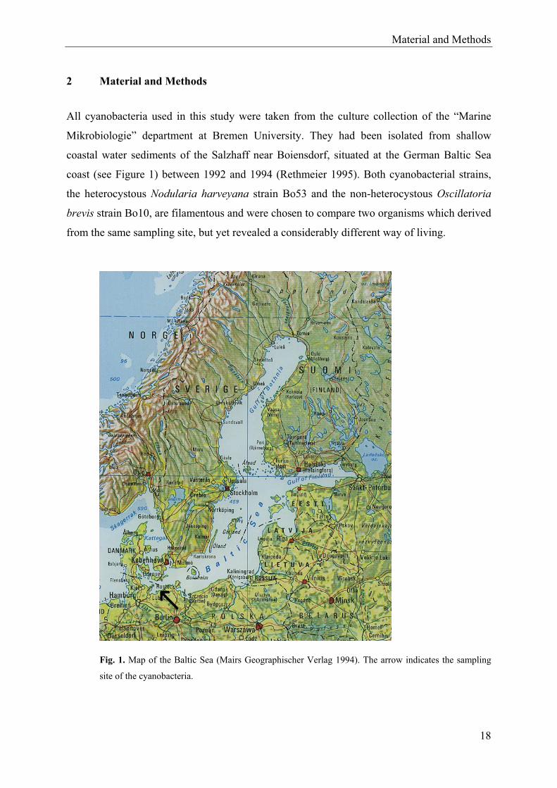
All cyanobacteria used in this study were taken from the culture collection of the “Marine
Mikrobiologie” department at Bremen University. They had been isolated from shallow
coastal water sediments of the Salzhaff near Boiensdorf, situated at the German Baltic Sea
coast (see Figure 1) between 1992 and 1994 (Rethmeier 1995). Both cyanobacterial strains,
the heterocystous Nodularia harveyana strain Bo53 and the non-heterocystous Oscillatoria
brevis strain Bo10, are filamentous and were chosen to compare two organisms which derived
from the same sampling site, but yet revealed a considerably different way of living.
Fig. 1. Map of the Baltic Sea (Mairs Geographischer Verlag 1994). The arrow indicates the sampling
site of the cyanobacteria.
18
Material and Methods
19
It was the aim of the study to isolate and analyse the heterotrophic community in two different
filamentous cyanobacterial cultures and to elucidate their role inside the community.
To determine the heterotrophic community within the cyanobacterial cultures, heterotrophic
cultures were obtained by streaking the supernatant of the cyanobacterial culture (before and
after ultrasonication) onto solid media. This cultivation dependent approach was chosen to
allow further investigations on the influence of the isolated heterotrophs upon their host
cyanobacterial strains.
In the course of the identification of the associated bacteria living in culture with the
cyanobacteria mentioned above, 51 different heterotrophic strains were obtained which were
phylogenetically characterised by fluorescence in situ hybridisation (FISH), fingerprinting
methods, and sequencing of the 16S rRNA genes. Four phylogenetically and physiologically
different strains, one non-coloured (strain Bo10-19) and two differently coloured (strains
Bo10-20 and Bo53-33) Alphaproteobacteria together with one coloured Bacteroidetes
bacterium (strain Bo10-09), were chosen for further comparable investigations to study their
possibly different influences on cyanobacterial growth behaviour. Afterwards, an improved
method was developed to obtain axenic cyanobacterial cultures. The axenic cultures were
mixed with single pure cultures of the heterotrophic strains mentioned above to study the
influence of the heterotrophic bacteria upon the cyanobacterial growth behaviour and vice
versa.
Pigment analyses, proofs for production of catalase and extracellular enzymes, and utilisation
of carbon sources were accomplished as well to characterise the heterotrophic bacteria and to
get information on their possible role in their coexistence with cyanobacteria. More detailed
information on material and methods used can be found in the manuscripts (see chapter 3).
Manuscripts
20
3 Manuscripts
Explanation to my own contribution to each manuscript.
Manuscript I
Hube AE, Heyduck-Söller B, Fischer U (2009) Phylogenetic classification of heterotrophic
bacteria associated with filamentous marine cyanobacteria in culture. Syst Appl Microbiol
32:256-265
This manuscript describes and compares the composition of heterotrophic bacteria living
attached to and in culture with the two different cyanobacterial strains examined.
I developed the experimental set-up for this investigation and carried out the practical work.
Laboratory work was supported by the second author. The manuscript was written in
discussion with both other authors.
Manuscript II
Hube AE, Fischer U (2009) Interactions between heterotrophic bacterial and cyanobacteria.
Submitted to “Aquatic Microbial Ecology” in November 2009.
In the second manuscript results concerning the influence of four different heterotrophic
bacteria upon cyanobacteria and vice versa are presented.
The idea for the experimental set-up was designed by me. I also conducted all the laboratory
work. The data were analysed and the manuscript written in discussion together with the
second author.
Manuscripts
21
Manuscript III
Hube AE, Heyduck-Söller B, Fischer U (2009) Characterisation of phylogenetically different
heterotrophic bacteria isolated from marine filamentous cyanobacteria. Ready to be submitted
to “Aquatic Microbial Ecology”.
This manuscript describes a variety of biochemical and physiological properties of the
heterotrophic bacteria in order to get information on the possible nature of influences,
heterotrophic bacteria carry out upon cyanobacteria.
All experiments were developed and conducted by me. The pigment analysis was supported
by the second author. The manuscript was prepared in discussion with both other authors.
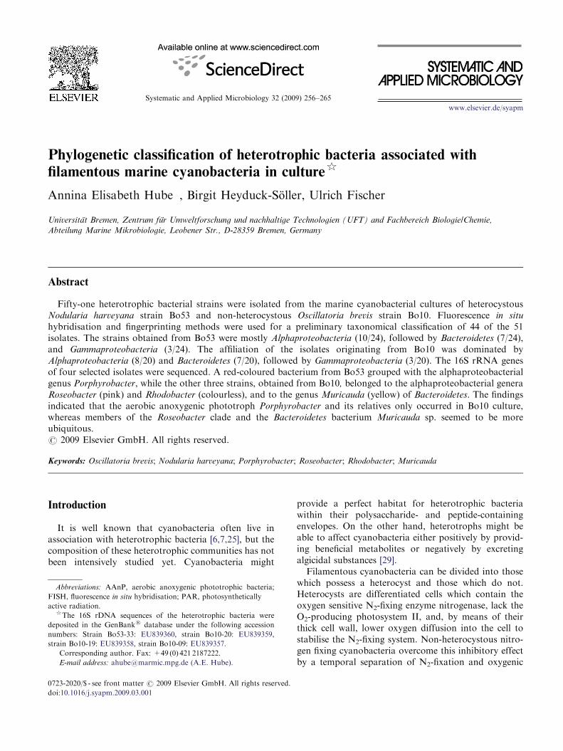
Systematic and Applied Microbiology 32 (2009) 256–265
Phylogenetic classification of heterotrophic bacteria associated with
filamentous marine cyanobacteria in culture$
Annina Elisabeth Hube�, Birgit Heyduck-Soller, Ulrich Fischer
Universitat Bremen, Zentrum fur Umweltforschung und nachhaltige Technologien (UFT) and Fachbereich Biologie/Chemie,
Abteilung Marine Mikrobiologie, Leobener Str., D-28359 Bremen, Germany
Abstract
Fifty-one heterotrophic bacterial strains were isolated from the marine cyanobacterial cultures of heterocystousNodularia harveyana strain Bo53 and non-heterocystous Oscillatoria brevis strain Bo10. Fluorescence in situ
hybridisation and fingerprinting methods were used for a preliminary taxonomical classification of 44 of the 51isolates. The strains obtained from Bo53 were mostly Alphaproteobacteria (10/24), followed by Bacteroidetes (7/24),and Gammaproteobacteria (3/24). The affiliation of the isolates originating from Bo10 was dominated byAlphaproteobacteria (8/20) and Bacteroidetes (7/20), followed by Gammaproteobacteria (3/20). The 16S rRNA genesof four selected isolates were sequenced. A red-coloured bacterium from Bo53 grouped with the alphaproteobacterialgenus Porphyrobacter, while the other three strains, obtained from Bo10, belonged to the alphaproteobacterial generaRoseobacter (pink) and Rhodobacter (colourless), and to the genus Muricauda (yellow) of Bacteroidetes. The findingsindicated that the aerobic anoxygenic phototroph Porphyrobacter and its relatives only occurred in Bo10 culture,whereas members of the Roseobacter clade and the Bacteroidetes bacterium Muricauda sp. seemed to be moreubiquitous.r 2009 Elsevier GmbH. All rights reserved.
Keywords: Oscillatoria brevis; Nodularia harveyana; Porphyrobacter; Roseobacter; Rhodobacter; Muricauda
Introduction
It is well known that cyanobacteria often live inassociation with heterotrophic bacteria [6,7,25], but thecomposition of these heterotrophic communities has notbeen intensively studied yet. Cyanobacteria might
provide a perfect habitat for heterotrophic bacteriawithin their polysaccharide- and peptide-containingenvelopes. On the other hand, heterotrophs might beable to affect cyanobacteria either positively by provid-ing beneficial metabolites or negatively by excretingalgicidal substances [29].
Filamentous cyanobacteria can be divided into thosewhich possess a heterocyst and those which do not.Heterocysts are differentiated cells which contain theoxygen sensitive N2-fixing enzyme nitrogenase, lack theO2-producing photosystem II, and, by means of theirthick cell wall, lower oxygen diffusion into the cell tostabilise the N2-fixing system. Non-heterocystous nitro-gen fixing cyanobacteria overcome this inhibitory effectby a temporal separation of N2-fixation and oxygenic
ARTICLE IN PRESS
www.elsevier.de/syapm
0723-2020/$ - see front matter r 2009 Elsevier GmbH. All rights reserved.
doi:10.1016/j.syapm.2009.03.001
Abbreviations: AAnP, aerobic anoxygenic phototrophic bacteria;
FISH, fluorescence in situ hybridisation; PAR, photosynthetically
active radiation.$The 16S rDNA sequences of the heterotrophic bacteria were
deposited in the GenBanks database under the following accession
numbers: Strain Bo53-33: EU839360, strain Bo10-20: EU839359,
strain Bo10-19: EU839358, strain Bo10-09: EU839357.�Corresponding author. Fax: +49 (0) 421 2187222.
E-mail address: [email protected] (A.E. Hube).
photosynthesis together with other oxygen protectionmechanisms [33].
It may be an additional advantage, especially forheterocystous cyanobacteria, that heterotrophic bacteriamight lower the oxygen partial pressure so that thenitrogenase is better protected against the harmful effectof oxygen. It has been shown that some heterotrophs arespecialised to live in contact with heterocysts, usingcyanobacterial excretion products on the one hand andenhancing cyanobacterial nitrogenase activity by con-suming oxygen on the other hand [24].
Salomon and co-workers [29] and Delucca andMcCracken [7] could demonstrate that the associatedheterotrophic community of the main bloom formingheterocystous Nodularia spumigena influenced thegrowth behaviour of the phototroph partner. Theyreported either stimulation or inhibition or no effect atall [7,29]. Concerning the heterotrophic community onnon-heterocystous cyanobacteria, such as Oscillatoria, itcould be demonstrated that the growth rates of thephototrophs were all positively affected by the hetero-trophs [7,14].
To our knowledge, the community structure ofheterotrophs and heterocystous or non-heterocystouscyanobacteria has never been compared before. There-fore, it was the aim of the present work to examinecomparatively the heterotrophic communities of cul-tures of nitrogen-fixing (Nodularia harveyana strainBo53) and non-nitrogen-fixing (Oscillatoria brevis strainBo10) filamentous cyanobacteria by applying molecularbiological methods for their taxonomical grouping.
Materials and methods
Cultivation of cyanobacteria
The filamentous cyanobacteria Oscillatoria brevis
strain Bo10 and Nodularia harveyana strain Bo53 weretaken from the culture collection of the ‘‘MarineMikrobiologie’’ department at Bremen University. Bothorganisms originated from sediments of shallow coastalwaters of the Baltic Sea (Boiensdorf, Germany),sampled between 1992 and 1994 [27]. Cyanobacteriacontaining sediment samples were streaked onto ASNIII
(see below) containing agar plates [28]. Repeatedtransfers were performed until single filaments orcolonies could be picked under a stereomicroscopewith a sterile Pasteur pipette to pass unicyanobacterialisolates into liquid ASNIII medium (stock culture).Approximately 3ml of these stock cultures wereregularly transferred into 50ml Erlenmeyer flaskscontaining 17ml of medium every four weeks.The cyanobacteria were cultivated at 21 1C and aphoton flow density of either 15 to 20 mEm�2 s�1
photosynthetically active radiation (PAR) (Oscillatoria)or �10mEm�2 s�1 PAR (Nodularia) in ASNIII medium[28] of half salinity containing 12.5 g/l NaCl, 1 g/lMgCl2� 6H2O, 0.25 g/l KCl, 1.75 g/l MgSO4� 7H2O,0.25 g/l CaCl2� 2H2O, 0.75 g/l NaNO3 (only forO. brevis), 0.12g/l Na2CO3, 0.01g/l K2HPO4�H2O,1.5mg/l Fe-NH4-citrate, 5mg/l vitamin B12, and 0.5ml ofa trace metal mix solution according to Rippka et al. [28].
Isolation of heterotrophic bacteria
To obtain free-living heterotrophs, 50 ml aliquots ofeither undiluted cyanobacterial culture or 1:10, 1:100,and 1:1000 dilutions were directly streaked onto solidmedia. The remaining culture was washed three timeswith medium (centrifugation: 5min at 2,250� g and21 1C) and then subjected twice to ultrasonication atroom temperature for 1min (Elmas Transsonic Digital,highest intensity) to detach heterotrophic bacteria fromaggregates. After this treatment, undiluted and diluted50 ml supernatants (see above) were streaked onto agarplates. The following media were used: ASNIII enrichedwith either 1% or 0.1% (w/v) meat or yeast extract,DifcoTM Marine Broth undiluted or 1:10 and 1:100diluted, and BBLTM Actinomyces Broth. The agarplates were incubated at 21 1C for 4 to 10 days. Colonieswhich macroscopically appeared to be distinguishablefrom each other were used to obtain pure culturesby repeated plating on agar plates. Pure cultures weretested for their Gram staining behaviour and thenfrozen in stocks containing 17.4% glycerol.
Fluorescence in situ hybridisation
The respective cultures were fixed in 4% formalde-hyde for one hour on ice and filtrated on polycarbonatefilters (GTBP black, 0.2 mm pore size, Millipore).Thereafter, the cells were washed with PBS buffer(22.8 g/l NaCl, 1.334 g/l NaH2PO4�H2O, 3.8 g/l Na2H-PO4). Fluorescence in situ hybridisation (FISH) wascarried out as described by Glockner and co-workers[10]. The probes and the formamide concentrations inthe hybridisation buffer used are given in Table 1.Hybridised bacterial samples were analysed with a ZeissAxiolab or a Zeiss Axioskop epifluorescence micro-scope.
DNA extraction and PCR amplification of 16S
rRNA genes
DNA extraction was carried out either with thePrestoSpin D Bug kit (Molzym), according to themanufacturer’s instructions, or by phenol chloroformextraction, modified after Mikolajczak and co-workers[20] and Neilan [23]. To remove potential RNAs, the
ARTICLE IN PRESSA.E. Hube et al. / Systematic and Applied Microbiology 32 (2009) 256–265 257
samples were afterwards incubated with 1ml RNase A(30 mg/ml, Molzym) at 37 1C in a water bath for 1 h.RNase activity was inactivated at 65 1C for 10min.The reaction mixture (50 ml total volume) for thePCR contained: 5 ml 10�PCR buffer without MgCl2(Invitrogen), 4mM MgCl2, 0.2mM deoxynucleotidetriphosphates, 0.4mM of each primer, 0.001mg/mlbovine serum albumin, 0.04U/ml AmpliTaq GoldTM
polymerase (Applied Biosystems), 10–20 ng DNA(except for strains Bo10-20 and Bo53-33, where theDNA was diluted to 0.1–0.2 ng, respectively, and0.01–0.02 ng for strain Bo10-19), and it was filled withsterile distilled water. The following primer pairs wereused: 8F (50-AGAGTTTGATCMTGGC-30) and 1507R(50-TACCTTGTTACGACTT-30) [21] for strain Bo10-09, R1n (50-GCTCAGATTGAACGCTGGCG-30) [34]and DG74 (50-AGGAGGTGATCCAACCGCA-30) [11]for strain Bo10-19, and 8F and 1542R (50-AGAAAG-GAGGTGATCCARCC-30) [15] for strains Bo10-20 andBo53-33. The PCR reaction was started at 95 1C for15min to activate the polymerase, followed by 35 cyclesfor 30 s at 95 1C, 30 s at 50 1C, and 30 s at 72 1C. Thereaction was completed by a 5min extension at 72 1C.The PCR products were electrophoresed at 100V ona 1% agarose gel prepared with 1�TBE buffer [30](pH 8.0), stained with ethidium bromide (0.5 mg/ml), andanalysed with a Biometra Fluo_Link Transilluminator.
Fingerprinting methods
Amplified ribosomal DNA restriction analysis (AR-DRA) was carried out by using a 25–50 ng 16S rRNAgene amplicon and 3U of the restriction enzymes AluI,Hin6I or HpaII (all Fermentas) in a total reactionvolume of 10 ml. The incubation was conducted for 3 hat 37 1C. Random amplified polymorphic DNAPCR (RAPD-PCR) was carried out with 10–20 ngof undiluted genomic DNA and the primers CRA22
(50-CCGCAGCCAA-30) and CRA23 (50-GCGATCCC-CA-30) [23] (the 50 ml reaction volume contained: 5 ml10�PCR buffer without MgCl2 (Invitrogen), 3mMMgCl2, 0.2mM deoxynucleotide triphosphates, 0.4 mMof each primer, 1mg/ml bovine serum albumine, 0.02U/mlPlatinums Taq DNA polymerase (Invitrogen), and itwas filled with sterile distilled water). PCR amplificationinvolved a 2min denaturation at 95 1C followed by 30cycles at 94 1C for 20 s, 45 1C for 30 s, and 72 1C for 60 s.Thereafter, a 5min extension at 72 1C was performed.The products of both fingerprinting methods wereelectrophoresed at 60–100V on a 2% agarose gelprepared with 1�TBE buffer [30] (pH 8.0). Stainingand analysis were performed as described above. Thegels were evaluated with the computer programmeTotalLab TL120 (Nonlinear Dynamics Ltd.).
Purification and sequencing
Purification of 16S rRNA gene PCR products wascarried out with the QIAquick PCR Purification Kit(Qiagen), following the manufacturer’s protocol. Forsequencing, 30 ml PCR product aliquots with a concen-tration of at least 10 ng/ml were prepared. The DNAconcentration of the PCR products was estimated in a1% agarose gel using the MassRulerTM DNA LadderMix (Fermentas). Sequencing was conducted by GATCBiotec AG, Konstanz, Germany. The following primerswere used: 517F (CCAGCAGCCGCGGTAATAC),1099F (GCAACGAGCGCAACCC), 534R (ATTAC-CGCGGCTGCTGGC), and 803R (CTACAAGGG-TATCTAATCC) [39]. The four overlapping partialsequences obtained were assembled with the computerprogramme ChromasPro Version 1.34 (TechnelysiumPty Ltd) and the consensus sequence was checked withthe BLAST database (National Centre for Biotechnol-ogy Information, US National Library of Medicine), aswell as with the database of the Ribosomal Database
ARTICLE IN PRESS
Table 1. Description of oligonucleotide probes.
Probe name Probe sequence (50-30) Specificity Formamide concentration (%) Reference
EUB338 (GCTGCCTCCCGTAGGAGT) Bacteria 0 [2]
ARCH915 (GTGCTCCCCCGCCAATTCCT) Archaea 0 [32]
ALF968 (GGTAAGGTTCTGCGCGTT) Alphaproteo bacteria 20 [22]
GAM42aa (GCCTTCCCACATCGTTT) Gammaproteo bacteria 35 [18]
BET42ab (GCCTTCCCACTTCGTTT) Betaproteo bacteria 35 [19]
CF319a (TGGTCCGTGTCTCAGTAC) Bacteroidetes group 35 [19]
ROS537 (CAACGCTAACCCCCTCC) Marine alpha cluster 35 [8]
ERY150 (CCGAAGACATTATCCGGT) Erythro bacteraceae 20 This study
MUR88 (GTTCCATACGCGTTCCGC) Muricauda sp. 70 This study
aUsed together with competitor BET42a (GCCTTCCCACTTCGTTT).bUsed together with competitor GAM42a (GCCTTCCCACATCGTTT).
A.E. Hube et al. / Systematic and Applied Microbiology 32 (2009) 256–265258

Project II, Release 9.50 (Centre for Microbial Ecology,Michigan State University). Phylogenetic trees wereconstructed with the programme provided by theRibosomal Database Project II and calculated with theneighbour-joining algorithm.
Probe design
Probes were designed by using the 16S sequence dataof the heterotrophic strains and sequences from theHugenholtz database (http://greengenes.lbl.gov/cgi-bin/nph-index.cgi). Primrose version 2.17 served as acomputer programme. The specificity of the probeswas reverified by comparison with the database of theNational Centre for Biotechnology Information(NCBI), US National Library of Medicine, Bethesda,USA (http://www.ncbi.nlm.nih.gov), the Hugenholtzdatabase (http://greengenes.lbl.gov/cgi-bin/nph-index.cgi), the database of the Ribosomal Database ProjectII (http://rdp.cme.msu.edu), and the SILVA database(www.arb-silva.de). The accessibility of the newlydesigned probes was checked with data published byBehrens and co-workers [3] and Fuchs and co-workers[9]. Other properties of the probes were checked with theprogramme Oligonucleotide Properties Calculator(http://www.basic.northwestern.edu/biotools/OligoCalc.html).
The optimal formamide concentration for the hybri-disation buffer was determined for both probes, asdescribed by Hugenholtz and co-workers [16].
Results
A total of 30 heterotrophic bacterial strains weresuccessfully isolated from the Nodularia harveyana
Bo53 culture, of which two strains were red andsix were light or dark yellow coloured. Red pigmentedisolates grew in DifcoTM Marine Broth from theundiluted and the 1:1000 diluted supernatant of thecyanobacterial culture. The yellow appearing oneswere obtained from undiluted as well as from the1:100 diluted supernatant in DifcoTM Marine Brothand ASNIII with 0.1 and 1% meat extract. Thecolourless isolates grew from all dilutions performedin DifcoTM Marine Broth, ASNIII with 1% meat extractand 0.1 or 1% yeast extract, and BBLTM ActinomycesBroth. Twenty-one heterotrophic strains were obtainedfrom the Oscillatoria brevis Bo10 culture. Two ofthem were pink and grew only from the 1:100 dilutionof the supernatant on ASNIII with 1% meat extract,while five yellow pigmented ones grew from alldilution rates on DifcoTM Marine Broth. The remainingcolourless isolates were obtained from all supernatantdilutions and could be cultivated on DifcoTM Marine
Broth, ASNIII with 1% meat or yeast extract, or BBLTM
Actinomyces Broth.All red and pink isolates originated only from
aggregates, while colourless and yellow colonies origi-nated both from the supernatant and from the attachedcells. The attachment of heterotrophic bacteria to thefilamentous cyanobacteria N. harveyana strain Bo53(A) and O. brevis strain Bo10 (B) is illustrated in Fig. 1.No Gram-positive bacteria were found from among theheterotrophic isolates.
FISH was applied first with 24 isolates from the Bo53and 20 from the Bo10 culture by using the probes forBacteria (EUB338) and Archaea (ARCH915). Allstrains hybridised well with the first mentioned probe,but not with the other one. Application of a set of groupprobes (see Table 1) indicated that most of the isolatesbelonged to Alphaproteobacteria (41.7% of isolates fromBo53 and 40% of the isolates from Bo10), theBacteroidetes (29.2% or 35%, respectively), andGammaproteobacteria (12.5% or 15%, respectively).Betaproteobacteria could not be found. As can be seenfrom Tables 2 and 3, the Alphaproteobacteria showedmuch more diversity in pigmentation than the Bacter-
oidetes group in which only yellow colonies occurred.Nine yellow colonies were separated into two groups
by ARDRA, whereas two did not group with any othercolonies. Ten of the red, pink, and colourless isolatescould be clustered into three groups. The resultsobtained with the three restriction enzymes appliedwere consistent (data not shown). Due to the fact thatthe primers used did not bind to the DNA of any red,pink or colourless isolate, RAPD-PCR was evaluatedonly for the yellow ones. The results were not inaccordance with those of ARDRA. The fingerprints ofthe RAPD-PCR revealed that six yellow isolatesstill clustered into two groups, whereas the remainingisolates did not comprise any cluster (see Figs. S1–S3).From this analysis, it can be deduced that the following
ARTICLE IN PRESS
Fig. 1. Photomicrographs of (A) filamentous heterocystous
Nodularia harveyana (strain Bo53) and (B) filamentous non-
heterocystous Oscillatoria brevis (strain Bo10) with attached
heterotrophic bacteria, respectively.
A.E. Hube et al. / Systematic and Applied Microbiology 32 (2009) 256–265 259
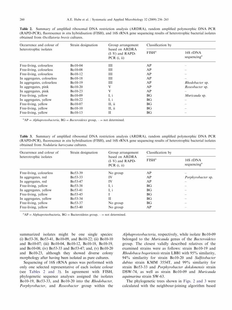
summarized isolates might be one single species:(i) Bo53-38, Bo53-41, Bo10-09, and Bo10-22; (ii) Bo10-10and Bo10-07; (iii) Bo10-04, Bo10-12, Bo10-18, Bo10-19,and Bo10-08; (iv) Bo53-33 and Bo53-47; and, (v) Bo10-20and Bo10-23, although they showed diverse colonymorphology after having been isolated as pure cultures.
Sequencing of 16S rRNA genes was performed withonly one selected representative of each isolate colour(see Tables 2 and 3). In agreement with FISH,phylogenetic sequence analyses assigned the isolatesBo10-19, Bo53-33, and Bo10-20 into the Rhodobacter,
Porphyrobacter, and Roseobacter group within the
Alphaproteobacteria, respectively, while isolate Bo10-09belonged to the Muricauda genus of the Bacteroidetes
group. The closest validly described relatives of theexamined strains were as follows: strain Bo10-19 andRhodobaca bogoriensis strain LBB1 with 95% similarity,94% similarity for strain Bo10-20 and Sulfitobacter
dubius strain KMM 3554T, and 99% similarity forstrain Bo53-33 and Porphyrobacter dokdonensis strainDSW-74, as well as strain Bo10-09 and Muricauda
aquimarina strain SW-63.The phylogenetic trees shown in Figs. 2 and 3 were
calculated with the neighbour-joining algorithm based
ARTICLE IN PRESS
Table 2. Summary of amplified ribosomal DNA restriction analysis (ARDRA), random amplified polymorphic DNA PCR
(RAPD-PCR), fluorescence in situ hybridisation (FISH), and 16S rRNA gene sequencing results of heterotrophic bacterial isolates
obtained from Oscillatoria brevis cultures.
Occurrence and colour of
heterotrophic isolates
Strain designation Group arrangement
based on ARDRA
(I–V) and RAPD-
PCR (i, ii)
Classification by
FISHa 16S rDNA
sequencinga
Free-living, colourless Bo10-04 III AP –
Free-living, colourless Bo10-08 III AP –
Free-living, colourless Bo10-12 III AP –
In aggregates, colourless Bo10-18 III AP –
In aggregates, colourless Bo10-19 III AP Rhodobacter sp.
In aggregates, pink Bo10-20 V AP Roseobacter sp.
In aggregates, pink Bo10-23 V AP –
Free-living, yellow Bo10-09 I, i BG Muricauda sp.
In aggregates, yellow Bo10-22 I, i BG –
Free-living, yellow Bo10-07 II, ii BG –
Free-living, yellow Bo10-10 II, ii BG –
Free-living, yellow Bo10-13 II BG –
aAP ¼ Alphaproteobacteria, BG ¼ Bacteroidetes group, – ¼ not determined.
Table 3. Summary of amplified ribosomal DNA restriction analysis (ARDRA), random amplified polymorphic DNA PCR
(RAPD-PCR), fluorescence in situ hybridisation (FISH), and 16S rRNA gene sequencing results of heterotrophic bacterial isolates
obtained from Nodularia harveyana cultures.
Occurrence and colour of
heterotrophic isolates
Strain designation Group arrangement
based on ARDRA
(I–V) and RAPD-
PCR (i, ii)
Classification by
FISHa 16S rDNA
sequencinga
Free-living, colourless Bo53-39 No group AP –
In aggregates, red Bo53-33 IV AP Porphyrobacter sp.
In aggregates, red Bo53-47 IV AP –
Free-living, yellow Bo53-38 I, i BG –
In aggregates, yellow Bo53-41 I, i BG –
Free-living, yellow Bo53-45 I BG –
In aggregates, yellow Bo53-34 II BG –
Free-living, yellow Bo53-37 No group BG –
Free-living, yellow Bo53-40 No group AP –
aAP ¼ Alphaproteobacteria, BG ¼ Bacteroidetes group, – ¼ not determined.
A.E. Hube et al. / Systematic and Applied Microbiology 32 (2009) 256–265260
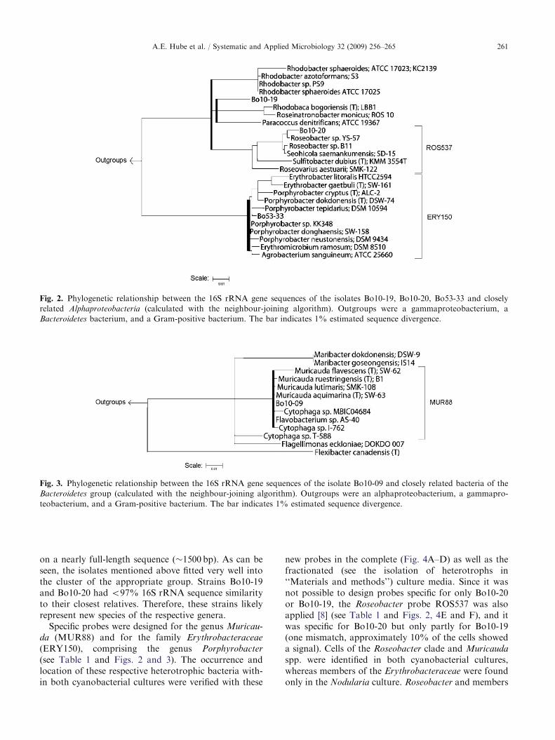
on a nearly full-length sequence (�1500 bp). As can beseen, the isolates mentioned above fitted very well intothe cluster of the appropriate group. Strains Bo10-19and Bo10-20 had o97% 16S rRNA sequence similarityto their closest relatives. Therefore, these strains likelyrepresent new species of the respective genera.
Specific probes were designed for the genus Muricau-
da (MUR88) and for the family Erythrobacteraceae
(ERY150), comprising the genus Porphyrobacter
(see Table 1 and Figs. 2 and 3). The occurrence andlocation of these respective heterotrophic bacteria with-in both cyanobacterial cultures were verified with these
new probes in the complete (Fig. 4A–D) as well as thefractionated (see the isolation of heterotrophs in‘‘Materials and methods’’) culture media. Since it wasnot possible to design probes specific for only Bo10-20or Bo10-19, the Roseobacter probe ROS537 was alsoapplied [8] (see Table 1 and Figs. 2, 4E and F), and itwas specific for Bo10-20 but only partly for Bo10-19(one mismatch, approximately 10% of the cells showeda signal). Cells of the Roseobacter clade and Muricauda
spp. were identified in both cyanobacterial cultures,whereas members of the Erythrobacteraceae were foundonly in the Nodularia culture. Roseobacter and members
ARTICLE IN PRESS
Fig. 2. Phylogenetic relationship between the 16S rRNA gene sequences of the isolates Bo10-19, Bo10-20, Bo53-33 and closely
related Alphaproteobacteria (calculated with the neighbour-joining algorithm). Outgroups were a gammaproteobacterium, a
Bacteroidetes bacterium, and a Gram-positive bacterium. The bar indicates 1% estimated sequence divergence.
Fig. 3. Phylogenetic relationship between the 16S rRNA gene sequences of the isolate Bo10-09 and closely related bacteria of the
Bacteroidetes group (calculated with the neighbour-joining algorithm). Outgroups were an alphaproteobacterium, a gammapro-
teobacterium, and a Gram-positive bacterium. The bar indicates 1% estimated sequence divergence.
A.E. Hube et al. / Systematic and Applied Microbiology 32 (2009) 256–265 261
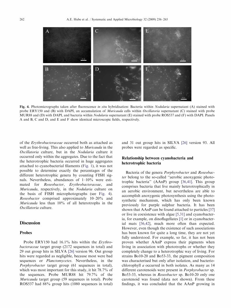
of the Erythrobacteraceae occurred both as attached aswell as free-living. This also applied to Muricauda in theOscillatoria culture, but in the Nodularia culture itoccurred only within the aggregates. Due to the fact thatthe heterotrophic bacteria occurred in huge aggregatesattached to cyanobacterial filaments (Fig. 1), it was notpossible to determine exactly the percentages of thedifferent heterotrophic genera by counting FISH sig-nals. Nevertheless, abundances of 1–10% were esti-mated for Roseobacter, Erythrobacteraceae, andMuricauda, respectively, in the Nodularia culture onthe basis of FISH microphotographs (see Fig. 4).Roseobacter comprised approximately 10–20% andMuricauda less than 10% of all heterotrophs in theOscillatoria culture.
Discussion
Probes
Probe ERY150 had 16.1% hits within the Erythro-
bacteraceae target group (2172 sequences in total) and29 out group hits in SILVA [26] version 96. Out grouphits were regarded as negligible, because most were badsequences or Planctomycetes. Nevertheless, in thePorphyrobacter target group (61 sequences in total),which was most important for this study, it hit 78.7% ofthe sequences. Probe MUR88 hit 79.7% of theMuricauda target group (59 sequences in total). ProbeROS537 had 88% group hits (1080 sequences in total)
and 31 out group hits in SILVA [26] version 93. Allprobes were regarded as specific.
Relationship between cyanobacteria and
heterotrophic bacteria
Bacteria of the genera Porphyrobacter and Roseobac-
ter belong to the so-called ‘‘aerobic anoxygenic photo-trophic bacteria’’ (AAnP) group [36,41]. This groupcomprises bacteria that live mainly heterotrophically inan aerobic environment, but nevertheless are able toaccomplish anoxygenic photosynthesis using the photo-synthetic mechanism, which has only been knownpreviously for purple sulphur bacteria. It has beenshown that AAnP can be found attached to particles [37]or live in coexistence with algae [5,31] and cyanobacter-ia, for example, on dinoflagellates [1] or in cyanobacter-ial mats [36,42], much more often than expected.However, even though the existence of such associationshas been known for quite a long time, they are not yetfully understood. For example, so far, it has not beenproven whether AAnP express their pigments whenliving in association with phototrophs or whether theycompletely change to a heterotrophic way of living. Forstrains Bo10-20 and Bo53-33, the pigment compositionwas characterised but only after isolation, and bacterio-chlorophyll a occurred in both isolates. As many as 14different carotenoids were present in Porphyrobacter sp.Bo53-33, whereas in Roseobacter sp. Bo10-20 only onecarotenoid was found (data not shown). From thesefindings, it was concluded that the AAnP growing in
ARTICLE IN PRESS
Fig. 4. Photomicrographs taken after fluorescence in situ hybridisation: Bacteria within Nodularia supernatant (A) stained with
probe ERY150 and (B) with DAPI, an accumulation of Muricauda cells within Oscillatoria supernatant (C) stained with probe
MUR88 and (D) with DAPI, and bacteria within Nodularia supernatant (E) stained with probe ROS537 and (F) with DAPI. Panels
A and B, C and D, and E and F show identical microscopic fields, respectively.
A.E. Hube et al. / Systematic and Applied Microbiology 32 (2009) 256–265262
association with cyanobacteria might indeed expresstheir pigments. It is not possible to extract andsubsequently analyse the AAnP’s pigments from insidethe cyanobacterial cultures because they will always beoverlaid by the much more abundant cyanobacterialpigments.
For members of the Roseobacter lineage, it has longbeen assumed that they establish relationships withproducers of organic carbon [1] or sulphur compounds(organic or inorganic) [5,36]. Another reason for suchcoexistences might be that the partners are phototrophsthemselves and that AAnP are exposed to a suitablelight intensity on their surface. However, this cannotexplain why the Roseobacter isolates were found in bothcyanobacterial cultures, while Porphyrobacter occurredonly on the nitrogen-fixing Nodularia, even though bothcyanobacterial strains were derived from the sameshallow coastal water site in the Baltic Sea. The samebehaviour was also observed with other Nodularia andOscillatoria cultures from the department’s culturecollection (data not shown), which might imply aspecific dependency. The detection of Roseobacter inthe marine habitat contradicts the report of Hagstromand co-workers [13] who did not find any Roseobacter orclosely related isolates in the Baltic Sea, even thoughthey also used a cultivation dependent [17] approach, asperformed in the present study.
The genus Muricauda has been described onlyrecently. Coexistence with filamentous cyanobacteriahas not been mentioned yet, although it has beenreported for some of their relatives [4]. To date,Muricauda has only been isolated from the GermanWadden Sea [4] and from a salt lake near HwajinpoBeach of the East Sea in Korea [40]. Bruns and co-workers [4] presume that the special appendages of theMuricauda cells might be used for attachment to a givensubstratum. Our results affirm this assumption, sinceMuricauda were often found within the aggregates.Other members of the Bacteroidetes group also live inassociation with cyanobacteria [7] or algae [12], andit is assumed that they are involved in particledegradation [8,38].
In contrast to the results of Salomon and co-workers[29] and Tuomainen and co-workers [35], we could notdetect heterotrophs belonging to the Betaproteobacteria
or to the phylum Firmicutes associated with nitrogen-fixing Nodularia species. On the other hand, althoughcyanobacteria-associated AAnP representatives werefound in our study, the authors cited above did notfind them [24,35]. These differences in the communitystructure might be due to different sampling sites. Whileour cyanobacterial strain originated from the benthos ofcoastal waters, the other strains derived from the openwater column [29,35].
Future prospects
Cyanobacteria are not easily cultivable withoutaccompanying heterotrophs. They often die when theabundance of heterotrophs decreases to a certainamount [25]. Since the present work provided hetero-trophs from the phototrophic cyanobacteria commu-nity, it will now be possible to elucidate the potentialinfluences of these bacteria on the cyanobacterial host.
Acknowledgments
We thank Birgit Lubben for technical assistance andMartina Stickan for editorial help. Furthermore, wethank Rudolf Amann and Bernhard Fuchs (Max PlanckInstitute for Marine Microbiology, Bremen, Germany)for additional editorial and technical help.
Appendix A. Supplementary materials
The online version of this article contains additionalsupplementary data. Please visit doi:10.1016/j.syapm.2009.03.001.
References
[1] M. Allgaier, H. Uphoff, A. Felske, I. Wagner-Dobler,Aerobic anoxygenic photosynthesis in Roseobacter clade
bacteria from diverse marine habitats, Appl. Environ.
Microbiol. 69 (2003) 5051–5059.
[2] R.I. Amann, B.J. Binder, R.J. Olson, S.W. Chrisholm,
R. Devereux, D.A. Stahl, Combination of 16S rRNA-
targeted oligonucleotide probes with flow cytometry for
analyzing mixed microbial populations, Appl. Environ.
Microbiol. 56 (1990) 1919–1925.
[3] S. Behrens, C. Ruhland, J. Inacio, H. Huber, A. Fonseca,I. Spencer-Martins, B.M. Fuchs, R. Amann, In situ
accessibility of small-subunit rRNA of members of the
domains Bacteria, Archaea, and Eucarya to Cy3-labeled
oligonucleotide probes, Appl. Environ. Microbiol. 69
(2003) 1748–1758.
[4] A. Bruns, M. Rohde, L. Berthe-Corti, Muricauda
ruestringensis gen. nov., sp. nov., a facultatively anaero-
bic, appendaged bacterium from German North Sea
intertidal sediment, Int. J. Syst. Evol. Microbiol. 51 (2001)
1997–2006.
[5] A. Buchan, J.M. Gonzalez, M.A. Moran, Overview of the
marine Roseobacter lineage, Appl. Environ. Microbiol. 71
(2005) 5665–5677.
[6] J.J. Cole, Interactions between bacteria and algae in aquatic
ecosystems, Ann. Rev. Ecol. Syst. 13 (1982) 291–314.
[7] R. Delucca, M.D. McCracken, Observations on interac-
tions between naturally-collected bacteria and several
species of algae, Hydrobiologia. 55 (1977) 71–75.
ARTICLE IN PRESSA.E. Hube et al. / Systematic and Applied Microbiology 32 (2009) 256–265 263
[8] H. Eilers, J. Pernthaler, J. Peplies, F.O. Glockner, G.
Gerdts, R. Amann, Isolation of novel pelagic bacteria
from the German bight and their seasonal contributions
to surface picoplankton, Appl. Environ. Microbiol. 67
(2001) 5134–5142.
[9] B.M. Fuchs, F.O. Glockner, J. Wulf, R. Amann,
Unlabeled helper oligonucleotides increase the in situ
accessibility to 16S rRNA of fluorescently labeled
oligonucleotide probes, Appl. Environ. Microbiol. 66
(2000) 3603–3607.
[10] F.O. Glockner, R. Amann, A. Alfreider, J. Pernthaler,
R. Psenner, K. Trebesius, K.-H. Schleifer, An in situ
hybridization protocol for detection and identification of
planktonic bacteria, Syst. Appl. Microbiol. 19 (1996)
403–406.
[11] K. Greisen, M. Loeffelholz, A. Purohit, D. Leong,
PCR primers and probes for the 16S rRNA gene of most
species of pathogenic bacteria, including bacteria found in
cerebrospinal fluid, J. Clinic. Microbiol. 32 (1994)
335–351.
[12] H.-P. Grossart, F. Levold, M. Allgaier, M. Simon,
T. Brinkhoff, Marine diatom species harbour distinct
bacterial communities, Environ. Microbiol. 7 (2005)
860–873.
[13] (A. Hagstrom, J. Pinhassi, U.L. Zweifel, Biogeographical
diversity among marine bacterioplankton, Aquat. Mi-
crob. Ecol. 21 (2000) 231–244.
[14] V. Herbst, J. Overbeck, Metabolic coupling between the
alga Oscillatoria redekei and accompanying bacteria,
Naturwissenschaften 65 (1978) 598–599.
[15] A. Heuchert, Physiologische, chemotaxonomische und
molekularbiologische Eigenschaften mariner Partikel-as-
soziierter Bakterien aus dem Atlantik, Ph.D. Thesis,
Department of Marine Microbiology, University of
Bremen, Bremen, Germany, 2004.
[16] P. Hugenholtz, G.W. Tyson, L.L. Blackall, Design and
evaluation of 16S rRNA-targeted oligonucleotide probes
for fluorescence in situ hybridisation, Meth. Molec. Biol.
176 (2001) 29–42.
[17] H.W. Jannasch, G.E. Jones, Bacterial populations in sea
water as determined by different methods of enumeration,
Limn. Oceanogr. 4 (1959) 128–139.
[18] W. Manz, R. Amann, W. Ludwig, M. Wagner,
K.-H. Schleifer, Phylogenetic oligodeoxynucleotide probes
for the major subclasses of Proteobacteria: problems and
solutions, Syst. Appl. Microbiol. 15 (1992) 593–600.
[19] W. Manz, R. Amann, W. Ludwig, M. Vancanneyt,
K.-H. Schleifer, Application of a suite of 16S rRNA-specific
oligonucleotide probes designed to investigate bacteria of
the phylum Cytophaga-Flavobacter-Bacteroides in the nat-
ural environment, Microbiol. 142 (1996) 1097–1106.
[20] A. Mikolajczak, R. Soller, D. Blohm, U. Fischer,
Molecular biological characterization of unicellular mar-
ine cyanobacteria from the Baltic Sea, Bull. Inst.
Oceanogr. Monaco 19 (1999) 95–100.
[21] G. Muyzer, A. Teske, C.O. Wirsen, H.W. Jannasch,
Phylogenetic relationships of Thiomicrospira species and
their identification in deep-sea hydrothermal vent samples
by denaturing gradient gel electrophoresis of 16S rDNA
fragments, Arch. Microbiol. 164 (1995) 165–172.
[22] A. Neef, Anwendung der in situ Einzelzell-Identifizierung
von Bakterien zur Populationsanalyse in komplexen
mikrobiellen Biozonosen, Ph.D. Thesis, Technische Uni-
versitat Munchen, Munich, Germany, 1997.
[23] B.A. Neilan, Identification and phylogenetic analysis of
toxigenic cyanobacteria by Multiplex Randomly Ampli-
fied Polymorphic DNA PCR, Appl. Environ. Microbiol.
61 (1995) 2286–2291.
[24] H.W. Paerl, Role of heterotrophic bacteria in promoting
N2 fixation by Anabaena in aquatic habitats, Microb.
Ecol. 4 (1978) 215–231.
[25] K.A. Palinska, C. Becker, W.E. Krumbein, Isolation and
purification techniques for benthic marine cyanobacter-
ia—biotechnology potential, Bull. Inst. Oceanogr. Mon-
aco 19 (1999) 585–592.
[26] E. Pruesse, C. Quast, K. Knittel, B.M. Fuchs, W. Ludwig,
J. Peplies, F.O. Glockner, SILVA: a comprehensive online
resource for quality checked and aligned ribosomal RNA
sequence data compatible with ARB, Nucl. Acids Res. 35
(2007) 7188–7196.
[27] J. Rethmeier, Untersuchungen zur Okologie und zum
Mechanismus der Sulfidadaptation mariner Cyanobakterien
der Ostsee, Ph.D. Thesis, Department of Marine Micro-
biology, University of Bremen, Bremen, Germany, 1995.
[28] R. Rippka, J. Deruelles, J.B. Waterbury, R.Y. Stanier,
Generic assignments, strain histories and properties of pure
cultures of cyanobacteria, J. Gen. Microb. 111 (1979) 1–61.
[29] P.S. Salomon, S. Janson, E. Graneli, Molecular identifi-
cation of bacteria associated with filaments of Nodularia
spumigena and their effect on the cyanobacterial growth,
Harmful Algae 2 (2003) 261–272.
[30] J. Sambrook, E.F. Fritsch, T. Maniatis, Molecular cloning.
A laboratory manual, second ed, Cold Spring Harbor
Laboratory Press, Cold Spring Harbor, USA, 1989.
[31] T. Shiba, U. Simidu, N. Taga, Distribution of aerobic
bacteria which contain bacteriochlorophyll a, Appl.
Environ. Microbiol. 38 (1979) 43–45.
[32] D.A. Stahl, R. Amann, Development and application of
nucleic acid probes, in: E. Stackebrandt, M. Goodfellow
(Eds.), Nucleic acid techniques in bacterial systematics, John
Wiley & Sons Ltd, Chichester, UK, 1991, pp. 205–248.
[33] L.J. Stal, Cyanobacterial mats and stromatolites, in:
B.A. Whitton, M. Potts (Eds.), The Ecology of Cyano-
bacteria, Kluwer Academic Publishers, Dordrecht, The
Netherlands, 2000, pp. 61–120.
[34] H.V. Tichy, R. Simon, Effiziente Analyse von Mikroor-
ganismen mit PCR-Fingerprint-Verfahren, Bioforum 17
(1994) 499–505.
[35] J. Tuomainen, S. Hietanen, J. Kuparinen, P.J. Martikai-
nen, K. Servomaa, Community structure of the bacteria
associated with Nodularia sp. (cyanobacteria) aggregates
in the Baltic Sea, Microb. Ecol. 52 (2006) 513–522.
[36] I. Wagner-Dobler, H. Biel, Environmental biology of the
marine Roseobacter lineage, Annu. Rev. Microbiol. 60
(2006) 255–280.
[37] L.A. Waidner, D.L. Kirchman, Aerobic anoxygenic
phototrophic bacteria attached to particles in turbid
waters of the Delaware and Chesapeake estuaries, Appl.
Environ. Microbiol. 73 (2007) 3936–3944.
ARTICLE IN PRESSA.E. Hube et al. / Systematic and Applied Microbiology 32 (2009) 256–265264
[38] A. Wichels, C. Hummert, M. Elbrachter, B. Luckas, C.Schutt, G. Gerdts, Bacterial diversity in toxic Alexan-
drium tamarense blooms off the Orkney Isles and the
Firth of Forth, Helgol. Mar. Res. 58 (2004) 93–103.
[39] A. Wilmotte, G. Van der Auwera, R. De Wachter,
Structure of the 16S ribosomal RNA of the thermophilic
cyanobacterium Chlorogloeopsis HTF (‘Mastigocladus
laminosus HTF’) strain PCC7518, and phylogenetic
analysis, FEBS Lett. 317 (1993) 96–100.
[40] J.-H. Yoon, M.-H. Lee, T.-K. Oh, Y.-H. Park, Muricauda
flavescens sp. nov., and Muricauda aquimarina sp. nov.,
isolated from a salt lake near Hwajinpo Beach of the East Sea
in Korea, and emended description of the genus Muricauda,
Int. J. Syst. Evol. Microbiol. 55 (2005) 1015–1019.
[41] V.V. Yurkov, Aerobic phototrophic proteobacteria, in: M.
Dworkin, S. Falkow, E. Rosenberg, K.-H. Schleifer, E.
Stackebrandt (Eds.), The Prokaryotes, third ed, Springer,
Berlin Heidelberg, New York, 2006, pp. 562–584.
[42] V.V. Yurkov, J.T. Beatty, Aerobic anoxygenic photo-
trophic bacteria, Microbiol. Mol. Biol. Rev. 62 (1998)
695–724.
ARTICLE IN PRESSA.E. Hube et al. / Systematic and Applied Microbiology 32 (2009) 256–265 265
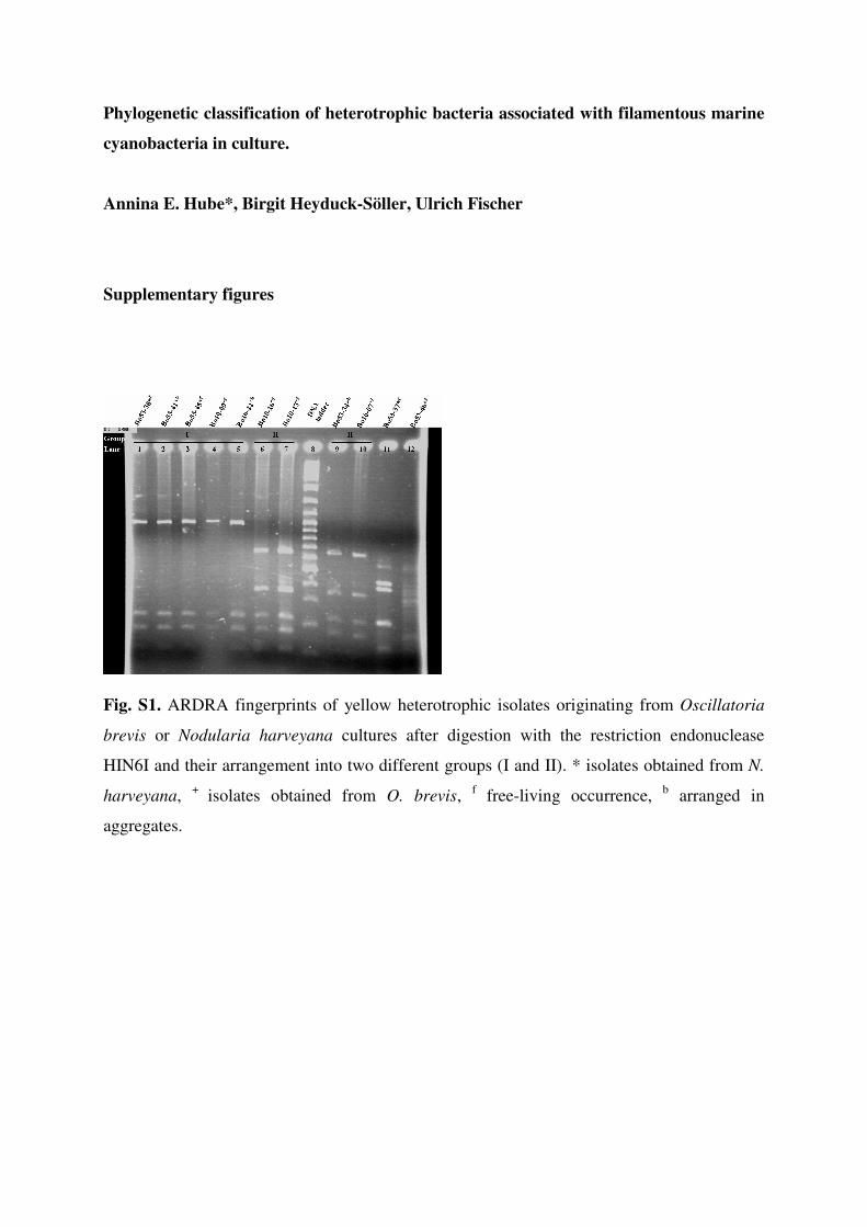
Phylogenetic classification of heterotrophic bacteria associated with filamentous marine
cyanobacteria in culture.
Annina E. Hube*, Birgit Heyduck-Söller, Ulrich Fischer
Supplementary figures
Fig. S1. ARDRA fingerprints of yellow heterotrophic isolates originating from Oscillatoria
brevis or Nodularia harveyana cultures after digestion with the restriction endonuclease
HIN6I and their arrangement into two different groups (I and II). * isolates obtained from N.
harveyana, + isolates obtained from O. brevis, f free-living occurrence, b arranged in
aggregates.
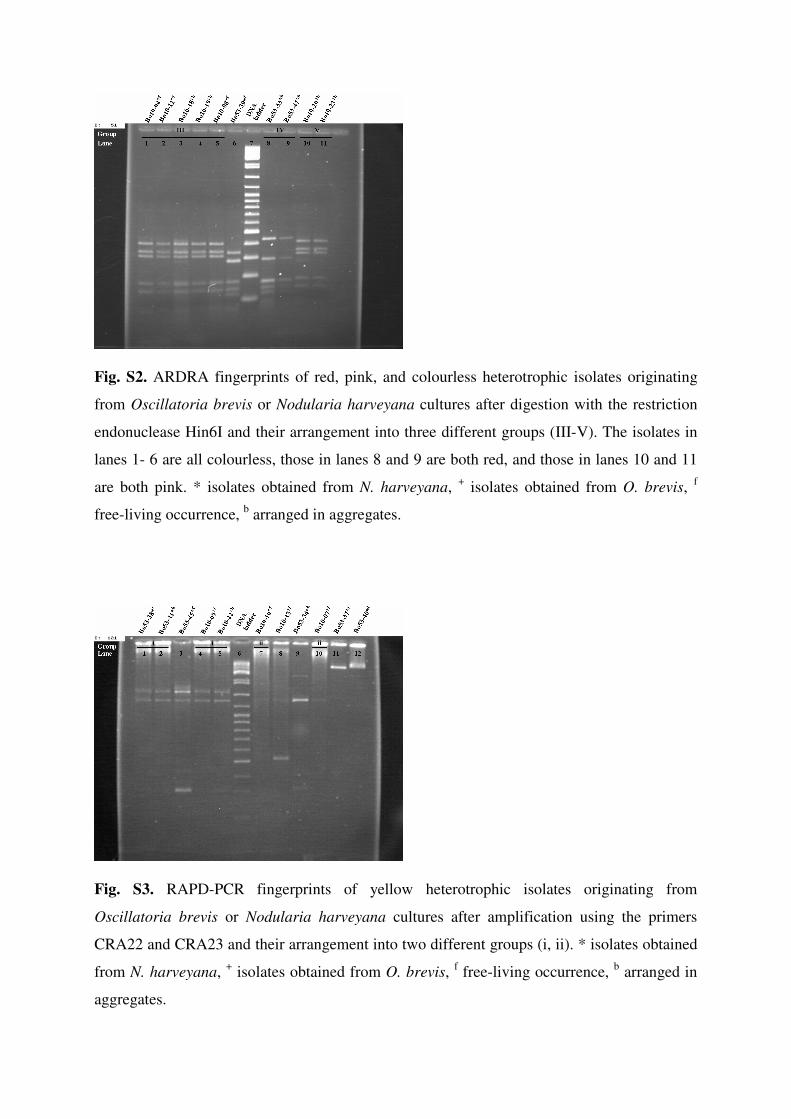
Fig. S2. ARDRA fingerprints of red, pink, and colourless heterotrophic isolates originating
from Oscillatoria brevis or Nodularia harveyana cultures after digestion with the restriction
endonuclease Hin6I and their arrangement into three different groups (III-V). The isolates in
lanes 1- 6 are all colourless, those in lanes 8 and 9 are both red, and those in lanes 10 and 11
are both pink. * isolates obtained from N. harveyana, + isolates obtained from O. brevis, f
free-living occurrence, b arranged in aggregates.
Fig. S3. RAPD-PCR fingerprints of yellow heterotrophic isolates originating from
Oscillatoria brevis or Nodularia harveyana cultures after amplification using the primers
CRA22 and CRA23 and their arrangement into two different groups (i, ii). * isolates obtained
from N. harveyana, + isolates obtained from O. brevis, f free-living occurrence, b arranged in
aggregates.
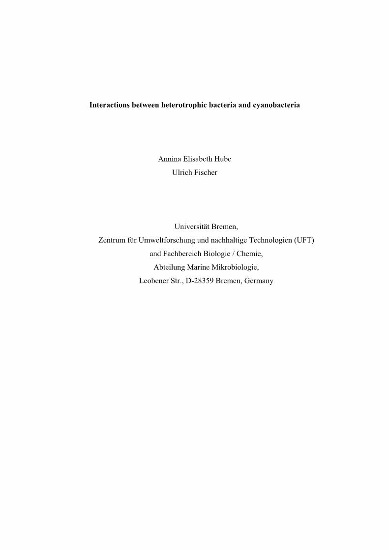
Interactions between heterotrophic bacteria and cyanobacteria
Annina Elisabeth Hube
Ulrich Fischer
Universität Bremen,
Zentrum für Umweltforschung und nachhaltige Technologien (UFT)
and Fachbereich Biologie / Chemie,
Abteilung Marine Mikrobiologie,
Leobener Str., D-28359 Bremen, Germany
Manuscripts
ABSTRACT
Associations of cyanobacteria and heterotrophic bacteria have been observed for a long time,
but such biocoenoses have not fully been understood so far. Here, we present some data, how
heterotrophic bacteria and cyanobacteria influence each other’s respective growth behaviour.
Specific FISH probes were used to detect members of the family Erythrobacteraceae and the
genera Muricauda and Roseobacter in various cyanobacterial cultures. For any of the
heterotrophic groups, a dependence on cyanobacterial origin, morphology, or diazotrophy
could be observed, except that Muricauda was not found together with unicellular
cyanobacteria. Furthermore, an improved method to obtain virtually axenic cyanobacterial
cultures was established and applied to cultures of the heterocystous Nodularia harveyana
strain Bo53 and the non-heterocystous Oscillatoria brevis strain Bo10. Both of these axenic
cyanobacterial species were mixed with single pure cultures of Muricauda sp., Roseobacter
sp., and Rhodobacter sp., which had been obtained from the Oscillatoria culture, and
Porphyrobacter sp., which had been obtained from the Nodularia one. All heterotrophic
bacteria exhibited with increasing abundance only a negative effect on the growth of
Oscillatoria but not on that of Nodularia.
KEY WORDS
Cyanobacteria-associated heterotrophs, aerobic anoxygenic phototrophic bacteria, Muricauda,
Roseobacter, Porphyrobacter
35
Manuscripts
INTRODUCTION
It has long been known that cyanobacteria often live in association with heterotrophic bacteria
(Delucca & McCracken 1977, Cole 1982, Palinska et al. 1999). For some of the heterotrophs
it has been shown that they are able to live without their phototrophic partner (Hube et al.
2009). However, there must be a reason for them to live in such associations. Palinska and co-
workers (1999) demonstrated for cyanobacteria that it is not easily possible to keep axenic
cultures alive. A lot has been speculated so far about such interferences (Paerl 1978, Larsson
& Hagström 1982, Lignell 1990). Hietanen and co-workers (2002) suggested a mutualistic
relationship with cyanobacteria providing substrates for the heterotrophs and the latter
providing a source of remineralised nutrients for the phototrophs. Larsson and Hagström
(1982) showed that in the Baltic Sea a substantial part of the energy, necessary for
heterotrophic growth, derived from phytoplankton exudates and they assumed the existence of
very effective bacterial mechanisms for the use of exudates as growth substrates. Sánchez and
co-workers (2005) reported that heterotrophs living in the sheath of Microcoleus
chthonoplastes are responsible for nitrogen fixation and hydrogen compound degradation,
while Microcoleus provides a habitat and source of oxygen and organic matter. Up to now,
the entire nature of these dependencies has not completely been understood. Recently, Hube
and co-workers (2009) could demonstrate the occurrence of heterotrophic species of the
genera Muricauda, Porphyrobacter, Rhodobacter, and Roseobacter living in association with
the marine filamentous cyanobacteria Nodularia harveyana and Oscillatoria brevis.
Roseobacter and relatives had been known for a long time to be aggressive colonisers, since
they are able to form biofilms and even inhibit growth of other colonisers by producing
antibacterial substances (Gram et al. 2002, Buchan et al. 2005, Bruhn et al. 2007). The ability
to attach to phytoplankton had previously only been assumed for Muricauda, based solely on
their morphology (Bruns et al. 2001). Porphyrobacter and Roseobacter belong to the aerobic
anoxygenic phototrophic bacteria (AAnP). This group of bacteria has been known to
comprise effective colonisers (Jiao et al. 2007, Waidner & Kirchman 2007), but to our
knowledge, this never had been shown for Porphyrobacter itself. In either case, nothing is
known so far about factors enabling heterotrophs to colonise cyanobacteria.
Therefore, the aim of this study was to investigate twelve cyanobacterial species, representing
members of all five cyanobacterial subspecies as described by Castenholz (2001), concerning
the occurrence of heterotrophic species and to find out what kind of influence the particular
heterotrophs may have upon the cyanobacteria and vice versa.
36
Manuscripts
MATERIALS AND METHODS
Isolation and cultivation of cyanobacteria and heterotrophs
All cyanobacterial strains (Table 2) were taken from the culture collection of the department
“Marine Mikrobiologie” at Bremen University. With the exception of Geitlerinema sp. and
Fischerella ambigua, all other strains originated from shallow coastal waters of the Baltic Sea
and were sampled between 1992 and 1994 (Rethmeier 1995). Unicyanobacterial cultures were
prepared as described by Hube and co-workers (Hube et al. 2009). Geitlerinema sp. derived
from a North American Atlantic coast mangrove wood and Fischerella ambigua was provided
by Prof. Gabriele M. König (University of Bonn) in 2000 and derived from a fresh water lake.
The cyanobacteria were cultivated in ASNIII medium (Rippka et al. 1979) of half salinity at
21 °C and 1 μE m-2 s-1 photosynthetically active radiation (PAR) photon flow density for
Chroococcus, Synechocystis, Myxosarcina, and Geitlerinema; 1 – 5 μE m-2 s-1 PAR for
Lyngbya, Anabaena, Oscillatoria laetevirens, and Nodularia sp.; ~10 μE m-2 s-1 PAR for
Nodularia harveyana; 10 – 15 μE m-2 s-1 PAR for Fischerella ambigua, and 15 - 20 μE m-2 s-1
PAR for Oscillatoria brevis. The medium for Chroococcus, Synechocystis, Myxosarcina,
Geitlerinema, Lyngbya, and Oscillatoria contained 0.75 g/l NaNO3, while the other strains
were kept without an inorganically bound nitrogen source. Fischerella ambigua was
cultivated in BG11 medium (Rippka et al. 1979) containing 1.5 g/l NaNO3 and was
continuously shaken at 70 rpm. Every four weeks, approximately 3 ml of the cyanobacterial
cultures were transferred into 50 ml Erlenmeyer flasks containing 17 ml medium.
Heterotrophs used in this study and the source of their origins are documented in Table 1.
Their isolation, identification, and cultivation have been described by Hube and co-workers
(2009).
Fluorescence in situ hybridisation
Sample preparation and fluorescence in situ hybridisation (FISH) were carried out as
described by Hube and co-workers (2009), using probes ERY150, MUR88 (Hube et al. 2009),
and ROS537 (Eilers et al. 2001). Heterotrophic cell numbers in the FISH samples were
determined by counting the signals in photomicrographs taken microscopically with a Zeiss
Axioskop 2 epifluorescence microscope equipped with a Zeiss AxioCam MRm and the
AxioVision 4.3 software.
37
Manuscripts
Preparation of axenic cyanobacterial cultures
Biomasses of Oscillatoria brevis strain Bo10 and Nodularia harveyana strain Bo53 cultures
of the stationary growth phase (approximately four weeks old) were divided into portions of
between 20 and 230 mg wet weight and transferred into a test tube containing 2 ml of fresh
medium each under sterile conditions. These samples were ultrasonicated at room temperature
for 10 min with highest intensity (Elma® Transsonic Digital) and afterwards washed four
times with culture medium by centrifugation (10 min at 9,000 x g and 20 °C). Only the
Nodularia culture was ultrasonicated (see above) after each washing step. These steps were
repeated until mainly single filaments could microscopically be observed. Then, the
cyanobacteria were resuspended in 200 μl fresh culture medium and again ultrasonicated (see
above) before they were centrifuged in a PercollTM (GE Healthcare) solution, which was
prepared as follows: 1 ml of 10 x concentrated culture medium was mixed with 9 ml of the
PercollTM solution. 1.8 ml of this mixture were pipetted into a 2 ml test tube, and the
cyanobacterial sample (0.2 ml) was added on top. The samples prepared in this way were
centrifuged for 3 h at 17,530 x g and 20 °C and with deceleration adjusted to the lowest
intensity (Beckman GS-15R). After centrifugation, the PercollTM solution on top was removed
and the cyanobacteria band transferred into a new test tube, containing 5 ml of fresh culture
medium. Subsequently, 0.61 μmol Rifampicin (stocksolution 10 mg/ml in distilled water)
were added to the culture which was cultivated for 24 h under standard conditions (see
above). Then, this culture was washed four times (see above) and the cell pellet suspended in
5 ml culture medium.
Preparation of cyanobacteria-heterotroph mixtures
This mixture was prepared directly after the above mentioned cyanobacterial cultures
Oscillatoria brevis strain Bo10 and Nodularia harveyana strain Bo53 had been made axenic.
The cell numbers of the heterotrophs were determined with a Neubauer improved
haemocytometer. Each axenic cyanobacterial culture was divided into two nearly equal parts.
These parts were mixed either with 1,000 or 10,000 heterotrophic cells per μg cyanobacterial
wet weight of single two days old heterotrophic cultures (Table 3). Cyanobacterial cultures
supplemented with mean volumes of heterotrophic culture medium served as control. Studies
performed in this way were evaluated after two weeks of incubation under standard
conditions (see above) by macroscopic comparison and fluorescence in situ hybridisation
analysis, including determination of heterotrophic cell numbers. Each experiment was
conducted three times (later referred to as three parallel assays).
38
Manuscripts
RESULTS
Twelve different cyanobacterial cultures were investigated concerning the occurrence of
Muricauda sp., Roseobacter sp., and members of the Erythrobacteraceae. As can be seen in
Table 2, Muricauda and the Erythrobacteraceae could each be detected in 50% of the
cultures tested (in 3 out of the 8 none-nitrogen-fixing cyanobacterial cultures and in 3 out of
the 4 nitrogen-fixing ones in both cases), but Roseobacter only in one third (3 out of the 8
none-nitrogen-fixing cultures and 1 out of the 4 nitrogen-fixing ones). Muricauda was not
found in any of the three unicellular cyanobacterial cultures tested, whereas
Erythrobacteraceae bacteria could be found in one and Roseobacter in two of them. Of the
filamentous cyanobacterial cultures, Muricauda sp. was the dominant heterotroph (66.7%),
followed by the Erythrobacteraceae bacteria (55.6%), and Roseobacter sp. (22.2%).
The macroscopic evaluation of the growth behaviour of axenic cyanobacteria mixed with
single heterotrophic cultures showed almost no influence of the heterotrophic bacteria upon
Nodularia. Even within the control assay (no addition of heterotrophs), good cyanobacterial
growth was observed. For the Oscillatoria culture, a toxic effect of the heterotrophs was
detectable, especially in the approaches with 10,000 heterotrophic cells per μg cyanobacteria.
Within the control assays, a cyanobacterial decay could be observed as well. None of the
heterotrophs revealed an outstanding influence within the cyanobacterial cultures. They
seemed to influence the cyanobacteria to similar degrees (Table 3).
Occurrence respectively reappearance of each heterotroph within the cyanobacterial cultures
was checked after 14 days of incubation within one of the three parallel assays. The presence
of Porphyrobacter, Muricauda, and Roseobacter was proved with the probes mentioned in
the legend to Table 4. The numbers of the respective heterotrophs were counted within the
cyanobacterial cultures they had been added to at the beginning of the experiment. It could be
shown that in all cases a mixed heterotrophic community developed in the cyanobacterial
cultures in the course of the experiment. In the Oscillatoria and Nodularia cultures total
numbers of between 8.2 x 10⁵ and 6.65 x 10⁶ respectively 7.6 x 10⁵ to 5.59 x 10⁶ cells were
found (obtained by counting the DAPI stained cells, data not shown). The cell numbers of the
heterotrophic bacteria which had been added at the beginning of the experiment decreased. As
can be seen in Table 4, the amount of Porphyrobacter and Roseobacter cells showed widely
diverse values (1.22 x 10⁴ – 3.24 x 10⁵ in the case of Porphyrobacter and 1.86 x 10⁴ - 1.08 x
106 that of Roseobacter) at the end of the experiment, independent of the amount of bacteria
added and the respective cyanobacterial culture, while Muricauda reached cell numbers
39
Manuscripts
between 4.01 x 10⁴ and 1.26 x 10⁶ in these cultures. Even though these total cell numbers did
not show a significant difference between the diverse heterotrophs, a calculation on percental
base shows that Muricauda cells stabilised to a level of 1.58 – 1.88% of the heterotrophic
community in the Nodularia culture and of 18.9 - 23.4% in the Oscillatoria culture. In the
case of Porphyrobacter and Roseobacter, no stable percentages could be observed.
DISCUSSION
Preparation of axenic cyanobacterial cultures
Although known to be difficult, it has been tried several times to detach filamentous
cyanobacteria from their contaminants (Vaara et al. 1979, Kim et al. 1996, Vázquez-Martínez
et al. 2004, Choi et al. 2008). Additional challenges occur for some of the filamentous ones
which form tight aggregates together with their accompanying heterotrophs and which are
difficult to access with common methods. Even though approaches of getting axenic
cyanobacteria succeeded (Vaara et al. 1979, Shirai et al. 1989, Bolch & Blackburn 1996, Kim
et al. 1999, Vázquez-Marínez et al. 2004, Choi et al. 2008), none of the authors reported to be
able to keep the axenic cultures over a longer period alive.
By combining parts of the methods already described, we developed special adjusted methods
suitable for our strains (see MATERIALS AND METHODS). Since in case of the
Oscillatoria cultures the heterotrophs and cyanobacterial filaments seem to be comparatively
loosely connected our developed procedure does not need to be repeated very often with this
culture. However, most heterotrophs live in large tightly organised aggregates surrounding the
cyanobacterial filaments in Nodularia cultures. Therefore, the procedure has to be repeated
several times with this culture to disperse the aggregates to make the heterotrophs accessible
for antibiotics. To circumvent the problem of keeping cultures axenic and alive, it is necessary
to prepare the axenic cultures directly before running the experiment.
Influence of heterotrophic bacteria upon cyanobacteria
It has been reported before that some heterotrophic bacteria inhibit cyanobacterial growth by
competition for limiting nutrients, lysis of cyanobacterial cells, and modification of the
environment (Rhee 1972, Cole 1982, Drakare 2002). On the other hand, some heterotrophic
bacteria can also stimulate cyanobacterial growth by nutrient regeneration, vitamin
production, and maintenance of low oxygen concentrations in the vicinity of the heterocyst
40
Manuscripts
(Paerl 1978, Cole 1982, Paerl et al. 1989). Delucca and McCracken (1977) demonstrated a
stimulating effect of various heterotrophs upon Oscillatoria. These authors were also able to
show a positive impact, as well as no reaction at all after addition of various heterotrophs to
Anabaena flos-aquae. Paerl (1978) also described a bacteria-promoted enhancement of
heterocystous Anabaena growth rates in most of his studies.
In the present study, we were able to show clear differences between the growth behaviour of
Nodularia and Oscillatoria after the addition of heterotrophs. The fact that the absence or
addition of heterotrophs only seem to have an effect on Oscillatoria but not on Nodularia
might be due to the latter one’s diazotrophy. While for Nodularia unlimited atmospheric
nitrogen is present, Oscillatoria needs to compete with the heterotrophs for available bound
nitrogen sources.
Moreover, we were able to show for Oscillatoria that the addition of higher amounts of
heterotrophs caused a deterioration of its growth which did not happen when lower amounts
were added. However, none of the heterotrophs seems to have a conspicuously stronger
influence on the cyanobacteria than the others. We therefore assume for Oscillatoria that
single heterotrophic colonisers are rather detrimental. When Oscillatoria cultures grow with
their natural composition of heterotrophic colonisers, the latter might prevent each other from
gaining an exceedingly strong influence.
Influence of cyanobacteria on heterotrophic bacteria
For some cyanobacteria it has been shown that they are able to inhibit bacterial growth, for
instance by alteration of the macroenvironment or production of antibiotics (Østensvik et al.
1998). The latter has been reported for Nodularia harveyana which exhibited antibacterial
activity upon Gram-positive bacteria (Pushparaj et al. 1999). But cyanobacteria can also
stimulate bacterial growth by supplying nutrients (Paerl 1978) in the form of exudates
(Larsson & Hagström 1982, Lignell 1990), by the disposal of soluble compounds during
growth (Cole 1982, Cole et al. 1982), or by the decomposition of cyanobacterial cells. In
oligotrophic environments, heterotrophs even depend on phytoplankton-produced organic
carbon to a certain extent (Drakare 2002).
Another advantage of living attached to cyanobacteria has been reported especially for the
AAnP. Cyanobacteria are phototrophs, so they might be able to transport the AAnP towards
zones with appropriate light conditions by means of buoyancy regulations (Walsby et al.
1995). Kolber and co-workers (2001) additionally supposed a nutrient cycle for AAnP, tightly
linked to that of oxygenic phototrophs. We detected Erythrobacteraceae in various
41
Manuscripts
cyanobacterial cultures, independently of the cyanobacterial origin, their morphology, or
utilisation of atmospheric or bound nitrogen sources. The factors regulating such colonisation
still remain unknown. Porphyrobacter originally did not occur in our Oscillatoria cultures.
But when we added our heterotrophic strain Bo53-33 to Oscillatoria strain Bo10,
Porphyrobacter survived well, even though the cell numbers varied considerably after two
weeks (see Table 4). Porphyrobacter may originally have occurred in Oscillatoria-associated
communities but was eliminated by other heterotrophs in the cultures for unknown reasons.
By means of their cell morphology it has been assumed for Muricauda sp. that they might
colonise particles or phytoplankton (Bruns et al. 2001), but to our knowledge this has never
been investigated. In the present study, Muricauda was detected only on some of the
filamentous cyanobacteria investigated but not on the unicellular ones. Furthermore, no
relation to cyanobacterial diazotrophy or to the origin could be observed. The fact that
Muricauda was the only strain which seemed to have become a stable part of respective
communities in the cyanobacterial cultures after two weeks of incubation, might indicate that
they are fast colonisers.
Roseobacter is known to be an effective and aggressive coloniser (Bruhn et al. 2007, Buchan
et al. 2005, Gram et al. 2002). However, our investigations revealed that it does not occur in
all cyanobacterial cultures examined. But a relation to cyanobacterial diazotrophy, origin, or
morphology could not be demonstrated either. Since Roseobacter’s cell number had not
settled to a stable level after the incubation either, this may indicate that it emerges as a stable
part of the community rather slowly.
These findings indicate that Erythrobacteraceae, Muricauda as well as Roseobacter aim at
certain unknown factors when colonising cyanobacteria which still needs to be elucidated.
Neither the cyanobacterial morphology nor the use of atmospheric or bound nitrogen sources
seems to play a role in the distribution of the heterotrophic bacteria. Since none of the
heterotrophic bacteria tested could be found to be ubiquitously present in all cyanobacterial
cultures tested, other dependencies seem to exist than the ones mentioned above.
In the culture of Geitlerinema sp., the only cyanobacterial strain from the Atlantic Ocean,
Erythrobacteriaceae as well as Muricauda occurred (Table 2). This might indicate an
ubiquity of these two groups in marine cyanobacteria-associated communities. In the
Fischerella ambigua culture, the only fresh water cyanobacterial strain, none of the
heterotrophs tested could be found. Therefore, they might not occur in fresh water
cyanobacteria-associated communities.
42
Manuscripts
We could demonstrate that heterotrophs reappeared in high amounts in the cyanobacterial
cultures they were not added to within two weeks after our axenic cyanobacterial cultures had
been mixed with single heterotrophic cultures. This finding indicates that i) it was not
possible to remove all heterotrophs from these filamentous cyanobacteria, even by using an
adjusted combination of different purification methods and ii) it is not possible to keep
filamentous cyanobacterial cultures axenic without further treatment. A distinct, even though
not yet fully developed, new heterotrophic community is capable to form itself within two
weeks.
In conclusion, the relationship between Nodularia and its heterotrophic partners might be of
commensal behaviour, being of benefit for the heterotrophs, while that between Oscillatoria
and heterotrophs seems to be more complex, having a kind of symbiotic character but only
within certain limits.
Future prospects
It has been shown before that in some cases cyanobacteria respond differently to single- than
to multiple-bacterial cultures which they were exposed to (Delucca & McCracken 1977, Cole
1982). On this account, results gained with the addition of only one single bacterial strain
need to be handled with caution. Continuative experiments should therefore include testings
concerning the effect of combinations of heterotrophic bacteria on cyanobacteria.
Furthermore, the effect of single bacterial components or dead bacteria on cyanobacteria
needs to be studied, to find out more about the nature of the mutual influence.
Additionally, the abundances of different heterotrophic bacteria living in association with
cyanobacteria still remain to be analysed in more detail. These investigations might give hint
towards the importance of the heterotroph for the cyanobacterium as well as towards the
relationship of the different heterotrophic partners among themselves.
ACKNOWLEDGEMENTS
The authors like to thank Dr. B. Heyduck-Söller for stimulating discussions and Mrs M.
Stickan for editorial help.
43
Manuscripts
LITERATURE CITED
� Berg, KA, Lyra C, Sivonen K, Paulin L, Suomalainen S, Tuomi P, Rapala J (1999) High
diversity of cultivable heterotrophic bacteria in association with cyanobacterial water
blooms. The ISME J 3:314-325
� Bolch CJS, Blackburn SI (1996) Isolation and purification of Australian isolates of the
toxic cyanobacterium Microcystis aeruginosa Kütz. J Appl Phycol 8:5-13
� Bruhn JB, Gram L, Belas R (2007) Production of antibacterial compounds and biofilm
formation by Roseobacter species are influenced by culture conditions. Appl Environ
Microbiol 73:442-450
� Bruns A, Rohde M, Berthe-Corti L (2001) Muricauda ruestringensis gen. nov., sp. nov., a
facultatively anaerobic, appendaged bacterium from German North Sea intertidal
sediment. Int J Syst Evol Microbiol 51:1997-2006
� Buchan A, González JM, Moran MA (2005) Overview of the marine Roseobacter lineage.
Appl Environ Microbiol 71:5665-5677
� Castenholz RW (2001) Phylum BX. Cyanobacteria. In: Garrity GM, Boone DR,
Castenholz RW (eds.) Bergey’s Manual of Systematic Bacteriology, 2nd edn. Springer,
Berlin Heidelberg New York, pp 473-599
� Choi GG, Bae MS, Ahn CY, Oh HM (2008) Induction of axenic culture of Arthrospira
(Spirulina) platensis based on antibiotic sensitivity of contaminating bacteria. Biotechnol
Lett 30:87-92
� Cole JJ (1982) Interactions between bacteria and algae in aquatic ecosystems. Ann Rev
Ecol Syst 13:291-314
� Cole JJ, Likens GE, Strayer DL (1982) Photosynthetically produced dissolved organic
carbon: an important carbon source for planktonic bacteria. Limnol Oceanogr 27:1080-
1090
� Delucca R, McCracken MD (1977) Observations on interactions between naturally-
collected bacteria and several species of algae. Hydrobiologia 55:71-75
� Drakare S (2002) Competition between picoplanktonic cyanobacteria and heterotrophic
bacteria along crossed gradients of glucose and phosphate. Microb Ecol 44:327-335
� Eilers H, Pernthaler J, Peplies J, Glöckner FO, Gerdts G, Amann R (2001) Isolation of
novel pelagic bacteria from the German bight and their seasonal contributions to surface
picoplankton. Appl Environ Microbiol 67:5134-5142
44
Manuscripts
� Gram L, Grossart HP, Schlingloff A, Kiørboe T (2002) Possible quorum sensing in
marine snow bacteria: production of acylated homoserine lactones by Roseobacter strains
isolated from marine snow. Appl Environ Microbiol 68:4111-4116
� Hietanen S, Moisander PH, Kuparinen J, Tuominen L (2002) No sign of denitrification in
a Baltic Sea cyanobacterial bloom. Mar Ecol Prog Ser 242:73-82
� Hube AE, Heyduck-Söller B, Fischer U (2009) Phylogenetic classification of
heterotrophic bacteria associated with filamentous marine cyanobacteria in culture. Syst
Appl Microbiol 32:256-265
� Jiao N, Zhang Y, Zeng Y, Hong N, Liu R, Chen F, Wang P (2007) Distinct distribution
pattern of abundance and diversity of aerobic anoxygenic phototrophic bacteria in the
global ocean. Environ Microbiol 9:3091-3099
� Kim JS, Park YH, Yoon BD, Oh HM (1999) Establishment of axenic cultures of
Anabaena flos-aque and Aphanothece nidulans (cyanobacteria) by lysozyme treatment. J
Phycol 35:865-869
� Kolber ZS, Plumley FG, Lang AS, Beatty JT, Blankenship RE, Van Dover CL, Vetriani
C, Koblížek M, Rathgeber C, Falkowski PG (2001) Contribution of aerobic
photoheterotrophic bacteria to the carbon cycle in the ocean. Science 292:2492-2495
� Larsson U, Hagström Å (1982) Fractionated phytoplankton primary production, exudate
release and bacterial production in a Baltic eutrophication gradient. Mar Biol 67:57-70
� Lignell R (1990) Excretion of organic carbon by phytoplankton: its relation to algal
biomass, primary productivity and bacterial secondary productivity in the Baltic Sea. Mar
Ecol Prog Ser 68:85-99
� Østensvik Ø, Skulberg OM, Underdal B, Hormazabal V (1998) Antibacterial properties of
extracts from selected planktonic freshwater cyanobacteria – a comparative study of
bacterial bioassays. J Appl Microbiol 84:1117-1124
� Paerl HW (1978) Role of heterotrophic bacteria in promoting N2 fixation by Anabaena in
aquatic habitats. Microb Ecol 4:215-231
� Paerl HW, Bebout BM, Prufert LE (1989) Bacterial associations with marine Oscillatoria
sp. (Trichodesmium sp.) populations: ecophysiological implications. J Physiol 25:773-784
� Palinska KA, Becker C, Krumbein WE (1999) Isolation and purification techniques for
benthic marine cyanobacteria – biotechnology potential. Bull Inst Océanogr Monaco
19:585-592
� Pushparaj B, Pelosi E, Jüttner F (1999) Toxicological analysis of the marine
cyanobacterium Nodularia harveyana. J Appl Phycol 10:527-530
45

Manuscripts
� Rethmeier J (1995) Untersuchungen zur Ökologie und zum Mechanismus der
Sulfidadaptation mariner Cyanobakterien der Ostsee. PhD thesis, Department of Marine
Microbiology, University of Bremen, Bremen
� Rhee GY (1972) Competition between an alga and an aquatic bacterium for phosphate.
Limnol Oceanogr 17:505-514
� Rippka R, Deruelles J, Waterbury JB, Stanier RY (1979) Generic assignments, strain
histories and properties of pure cultures of cyanobacteria. J Gen Microb 111:1-61
� Sánchez O, Diestra E, Esteve I, Mas J (2005) Molecular characterization of an oil-
degrading cyanobacterial consortium. Microb Ecol 50:580-588
� Shirai M, Matumaru K, Ohotake A, Takamura Y, Aida T, Nakano M (1989) Development
of a solid medium for growth and isolation of axenic Microcystis strains (cyanobacteria).
Appl Environ Microbiol 55:2569-2571
� Vaara T, Vaara M, Niemelä S (1979) Two improved methods for obtaining axenic
cultures of cyanobacteria. Appl Environ Microbiol 38:1011-1014
� Vázquez-Martínez G, Rodriguez MH, Hernández-Hernández F, Ibarra JE (2004) Strategy
to obtain axenic cultures from field-collected samples of the cyanobacterium Phormidium
animalis. J Microb Meth 57:115-121
� Waidner LA, Kirchman DL (2007) Aerobic anoxygenic phototrophic bacteria attached to
particles in turbid waters of the Delaware and Chesapeake estuaries. Appl Environ
Microbiol 73:3936-3944
� Walsby AE, Hayes PK, Boje R (1995) The gas vesicles, buoyancy and vertical
distribution of cyanobacteria in the Baltic Sea. Eur J Phycol 30:87-94
46

Manuscripts
TABLES
Table 1. Heterotrophic strains used in the mixed culture experiment and source of isolation
Heterotrophic strain Source
Muricauda strain Bo10-09 Oscillatoria brevis strain Bo10
Rhodobacter strain Bo10-19 Oscillatoria brevis strain Bo10
Roseobacter strain Bo10-20 Oscillatoria brevis strain Bo10
Porphyrobacter strain Bo53-33 Nodularia harveyana strain Bo53
Table 2. Occurrence of members of the family Erythrobacteraceae and the genera Muricaudaand Roseobacter in various cyanobacterial cultures of the culture collection of the Department of “Marine Mikrobiologie” at Bremen University (Germany) by using the probes ERY150, MUR88, and ROS537. + = present, - = not present.
Cyanobacterial strain and classification
subsections* (given in parentheses)
Erythro-
bacteraceae
Muricauda
sp.
Roseo-
bacter clade
Unicellular
Chroococcus turgidus Bo1 (I) - - +
Synechocystis sp. Bo79 (I) + - -
Myxosarcina chroococoides AH149 (II) - - +
Filamentous without heterocysts
Geitlerinema sp. Flo1 (III) + + -
Lyngbya semiplenta Fä8 (III) + - -
Oscillatoria brevis Bo10 (III) - + +
Oscillatoria laetevirens Bo17 (III) - + -
Filamentous with heterocysts
Anabaena flos aquae Hi35 (IV) + - +
Anabaena variabilis Hi4 (IV) - + -
Nodularia harveyana Bo53 (IV) + + -
Nodularia sp. Fä19 (IV) + + -
Fischerella ambigua (V) - - -
* Subsections I-V according to Castenholz (2001)
47
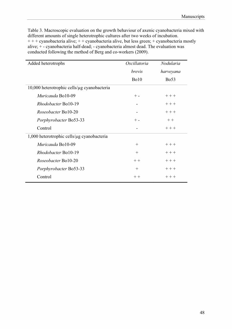
Manuscripts
48
Table 3. Macroscopic evaluation on the growth behaviour of axenic cyanobacteria mixed with different amounts of single heterotrophic cultures after two weeks of incubation. + + + cyanobacteria alive; + + cyanobacteria alive, but less green; + cyanobacteria mostly alive; + - cyanobacteria half-dead; - cyanobacteria almost dead. The evaluation was conducted following the method of Berg and co-workers (2009).
Added heterotrophs Oscillatoria
brevis
Bo10
Nodularia
harveyana
Bo53
10,000 heterotrophic cells/μg cyanobacteria
Muricauda Bo10-09 + - + + +
Rhodobacter Bo10-19 - + + +
Roseobacter Bo10-20 - + + +
Porphyrobacter Bo53-33 + - + +
Control - + + +
1,000 heterotrophic cells/μg cyanobacteria
Muricauda Bo10-09 + + + +
Rhodobacter Bo10-19 + + + +
Roseobacter Bo10-20 + + + + +
Porphyrobacter Bo53-33 + + + +
Control + + + + +
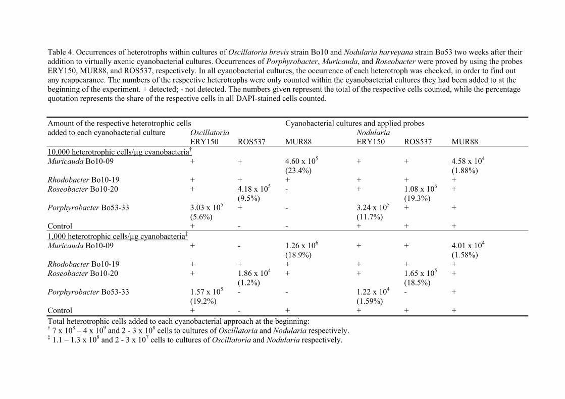
Table 4. Occurrences of heterotrophs within cultures of Oscillatoria brevis strain Bo10 and Nodularia harveyana strain Bo53 two weeks after their addition to virtually axenic cyanobacterial cultures. Occurrences of Porphyrobacter, Muricauda, and Roseobacter were proved by using the probes ERY150, MUR88, and ROS537, respectively. In all cyanobacterial cultures, the occurrence of each heterotroph was checked, in order to find out any reappearance. The numbers of the respective heterotrophs were only counted within the cyanobacterial cultures they had been added to at the beginning of the experiment. + detected; - not detected. The numbers given represent the total of the respective cells counted, while the percentage quotation represents the share of the respective cells in all DAPI-stained cells counted.
Amount of the respective heterotrophic cells Cyanobacterial cultures and applied probes added to each cyanobacterial culture Oscillatoria Nodularia
ERY150 ROS537 MUR88 ERY150 ROS537 MUR88 10,000 heterotrophic cells/μg cyanobacteria†
Muricauda Bo10-09 + + 4.60 x 10⁵ + + 4.58 x 10⁴
(23.4%) (1.88%) Rhodobacter Bo10-19 + + + + + + Roseobacter Bo10-20 + 4.18 x 10⁵ - + 1.08 x 10⁶ + (9.5%) (19.3%) Porphyrobacter Bo53-33 3.03 x 10⁵ + - 3.24 x 10⁵ + + (5.6%) (11.7%) Control + - - + + + 1,000 heterotrophic cells/μg cyanobacteria‡
Muricauda Bo10-09 + - 1.26 x 10⁶ + + 4.01 x 10⁴
(18.9%) (1.58%) Rhodobacter Bo10-19 + + + + + + Roseobacter Bo10-20 + 1.86 x 10⁴ + + 1.65 x 10⁵ + (1.2%) (18.5%) Porphyrobacter Bo53-33 1.57 x 10⁵ - - 1.22 x 10⁴ - + (19.2%) (1.59%) Control + - + + + + Total heterotrophic cells added to each cyanobacterial approach at the beginning: † 7 x 10⁸ – 4 x 10⁹ and 2 - 3 x 10⁸ cells to cultures of Oscillatoria and Nodularia respectively. ‡ 1.1 – 1.3 x 10⁸ and 2 - 3 x 10⁷ cells to cultures of Oscillatoria and Nodularia respectively.
Characterisation of phylogenetically different heterotrophic bacteria
isolated from marine filamentous cyanobacteria
Annina Elisabeth Hube
Birgit Heyduck-Söller
Ulrich Fischer
Universität Bremen,
Zentrum für Umweltforschung und nachhaltige Technologien (UFT)
and Fachbereich Biologie / Chemie,
Abteilung Marine Mikrobiologie,
Leobener Str., D-28359 Bremen, Germany
Manuscripts
ABSTRACT
Little is known about the connection of bacteria accompanying filamentous cyanobacteria and
their phototrophic partners. Morphological, biochemical, and physiological properties of four
bacteria (Porphyrobacter Bo53-33, Roseobacter Bo10-20, Muricauda Bo10-09, and strain
Bo10-19, closely related to Rhodobacter) deriving from two distinct cyanobacterial cultures
were investigated to highlight different aspects that might contribute to such a relationship.
Fourteen different carotenoids were detected in Porphyrobacter, five in Roseobacter, and
only one in Muricauda. Porphyrobacter and Roseobacter contained bacteriochlorophyll a as
well. Flexirubin could not be detected. The physiological investigations showed that
Porphyrobacter had only a very restricted potential with respect to its utilisation of carbon
substrates. On the other hand, Muricauda degraded all kinds of sugars, but none of the
alcohols offered. Extracellular amylases were detected in Porphyrobacter, Roseobacter, and
Muricauda. The latter one was also shown to excrete DNase. None of the isolates tested
produced peptidases or lipases. All four strains were shown to possess a catalase. Many of the
properties shown are beneficial when living in association with phytoplankton. Therefore, the
compiled data set indicates a complex and multifarious adaptation to this way of living.
KEY WORDS
Porphyrobacter, Roseobacter, Muricauda, cyanobacteria-associated heterotrophs, aerobic
anoxygenic phototrophic bacteria
51

Manuscripts
INTRODUCTION
Cyanobacteria have long been known to live in associations with heterotrophic bacteria
(Delucca & McCracken 1977, Cole 1982). However, the dynamics of these co-existences
have not intensively been studied yet. To at least partially fill this knowledge gap, we
characterised four different heterotrophic strains, isolated from associations with two unlike
cyanobacterial isolates from the Baltic Sea. Of these heterotrophic strains, the phylogenetical
identities and influence on the cyanobacterial growth behaviour have been investigated before
(Hube et al. 2009, Hube & Fischer 2009).
One important group known to be living attached to cyanobacteria are the aerobic anoxygenic
phototrophic bacteria (AAnP) (Yurkov & Beatty 1998, Wagner-Döbler & Biebl 2006, Hube
et al. 2009). AAnP are a unique group of microorganisms in the marine environment whose
ecological role is presently poorly understood. Little is known about their physiology and
metabolisms and their role in energy flow and carbon cycling in the ecosystem. AAnP are
known to live aerobically. Nevertheless, they are able to perform anoxygenic photosynthesis
to complement heterotrophic metabolism (Yurkov 2006). In most of them, phototrophy is
combined with a wide range of other metabolic capabilities (Yurkov 2006, Lami et al. 2007).
Even though the ribulose-1,5-bisphosphate carboxylase/oxygenase (RuBisCO) has never been
detected in any of them, some AAnP are able to fix small amounts of CO2 via other pathways
(Kolber et al. 2001, Swingley et al. 2007). Aerobic anoxygenic photosynthesis might
constitute an advantage for the organism in carbon-limited environments (Buchan et al. 2005).
Due to the presence of photopigments, less organic carbon sources are required for catabolic
processes, hence more is available for growth (Yurkov & van Gemerden 1993). Since AAnP
are known to be widely distributed (Kolber et al. 2000, Schwalbach & Fuhrman 2005) and
possess a unique function (Yurkov & Beatty 1998), a better understanding of these
microorganisms may change our current perspective on the energy and oxygen balance in the
ocean. Roseobacter in particular was described by Swingley and co-workers (2007) to be a
generalist, and the authors suggest a comparatively complex contribution to the global carbon
cycle. In addition, the character of the connection between the AAnP and the cyanobacteria,
which they are often found in association with, has also not been fully understood yet.
Muricauda sp., another heterotroph isolated from cyanobacterial cultures (Hube et al. 2009),
which belongs to the Bacteroidetes group, is not capable of performing photosynthesis. For
many of its relatives it is known that they are able to degrade natural polymers such as cell
52
Manuscripts
wall components and exudates from algae (Johansen et al. 1999, Bruns et al. 2001).
Nevertheless, concerning Muricauda’s relation to its cyanobacterial partners, nothing is
known so far.
Rhodobacter strain Bo10-19, to be characterised in the present study is an
alphaproteobacterial isolate (Hube et al. 2009). Despite the close relationship to phototrophic
purple bacteria and AAnP, strain Bo10-19 lives heterotrophically and does not possess
considerable amounts of pigments. However, it seems to be to some extent specialised in
living in association with cyanobacteria as well (Hube et al. 2009, Hube & Fischer 2009).
Based on these facts about the physiologically differing strains mentioned, we decided to
investigate some of the strains’ morphological, biochemical, and physiological properties to
be able to specify their relationship with cyanobacteria.
MATERIALS AND METHODS
Isolation and cultivation
Heterotrophic bacteria were isolated from the cultures of Oscillatoria brevis strain Bo10
(Muricauda sp. strain Bo10-09, Rhodobacter sp. strain Bo10-19, and Roseobacter sp. strain
Bo10-20) and Nodularia harveyana strain Bo53 (Porphyrobacter sp. strain Bo53-33) in 2005.
Their isolation and identification, as well as their cultivation has been described by Hube and
co-workers (2009).
Proof of slime capsules
Appearance of bacterial capsules was proved by i) mixing a bacterial culture scraped from an
agar plate with either one drop of royal ink or a 10% nigrosin solution on a slide and ii)
staining with crystal violet. For this procedure, bacterial cells were mixed with one drop of a
skimmed milk powder suspension [100 g/l], dried, and thermal fixed on a slide, covered with
one drop of 0.1% crystal violet and rinsed with a 20% copper sulphate solution. Then, the
sample was air dried, embedded with one drop of immersion oil and afterwards covered with
a cover slip. All preparations were evaluated by bright field microscopy (Zeiss Axiolab).
Slime capsules appeared as clear zones around the organism and the dark blue background.
53
Manuscripts
Pigment analysis
Cultures of 100 ml medium were grown at 21 °C under continuous light at a photon flow
density of 10 – 15 μE m-2 s-1 photosynthetically active radiation (PAR) and shaken at 70 rpm
for two days, centrifuged (6,500 x g at 10 °C for 15 min) and washed once in ASNIII culture
medium (Rippka et al. 1979, Hube et al. 2009) without addition of meat extract and NaNO3.
Finally, cells were centrifuged at 8,500 x g and 10 °C for 15 min. The pellets were
resuspended in 8 ml methanol and incubated at 4 °C in the dark for approximately 2.5 h.
Afterwards, the samples were centrifuged again (17,530 x g at 4 °C for 5 min) and the
supernatants stored in the dark on ice until further use. Pigments were separated by thin layer
chromatography at room temperature for 1 h in the dark using Silica gel plates (pre-coated
TLC plates SIL G-25 UV254, Macherey-Nagel) and a mixture of acetone and n-hexane (40/60
v/v) as running solvent. The separated pigments were scraped off the plates, solved in ice cold
methanol, and centrifuged (3,000 x g at 4 °C for 5 min). The supernatants were stored in the
dark on ice until further use. The absorption spectra of the extracts were recorded with a
Beckman DU®640 spectrophotometer from 200 – 900 nm.
In vivo spectra (200 – 900 nm) were obtained by resuspending the pellet in a 50% sucrose
solution from 1 ml of a 2 days old centrifuged culture (15,000 x g at room temperature for 30
sec).
Identification of pigments was accomplished by Reversed Phase HPLC as described by
Heuchert (2004). Pigments of each organism were extracted by adding 10 ml of an acetone
methanol mixture (7/2 v/v) to the centrifuged cell pellets (8.500 x g at 10 °C for 15 min)
obtained from 225 ml of four days old cultures. Immediately after the addition of the
extraction solvent, cells were disrupted by ultrasonication (Elma® Transsonic Digital, highest
intensity at 4 °C for 3 min). The samples were incubated and the pigments separated as
described above for the thin layer chromatography, before they were applied to the HPLC
treatment. After scraping them off the TLC plate, the pigments were resuspended in ice cold
acetone methanol mixture (see above).
Presence of flexirubin was tested by applying one drop of 10% KOH solution onto agar plates
overgrown with the used isolate (modified from Reichenbach et al. 1980). The colour of
bacteria containing the pigment was supposed to change to red.
Anaerobic and autotrophic growth
Anaerobic and autotrophic growth behaviour of the heterotrophs was tested in six different
approaches (i – vi) containing 10 ml of ASNIII culture medium modified as described by Hube
54
Manuscripts
and co-workers (2009) for two weeks either in the dark (i and ii) or (iii-vi) under continuous
light conditions (~ 1 μE m-2 s-1 PAR). The approaches were treated and supplemented as
follows:
i) with 1% meat extract and degassed for 10 min with nitrogen
ii) with 1% meat extract, aerobically
iii) with 1% meat extract, degassed for 10 min with nitrogen
iv) with 1% meat extract, aerobically
v) with nitrate (0.75 g/l), aerated for 10 min with CO2
vi) with nitrate (0.75 g/l), aerobically
Utilisation of carbon sources
Utilisation of various carbon sources was tested with api® 50 CH kits (Biomérieux). Bacterial
cultures (20 ml) were grown in ASNIII culture medium (Rippka et al. 1979, Hube et al. 2009)
enriched with 1% meat extract at 30 °C for two days. The bacterial cells were washed with a
0.9% NaCl solution and resuspended in a medium without additional carbon sources for the
api® test kits. The medium used contained 13.19 g/l NaCl, 2.84 g/l MgCl2 x 6 H2O, 3.4 g/l
MgSO4 x 7 H2O, 0.095 g/l NaHCO3, 0.74 g/l CaCl2 x 2H2O, 0.36 KCl, 0.5 g/l KH2PO4, 0.5
g/l NH4Cl, 0.05 mg/l vitamin B12, 0.05 mg/l riboflavin, 0.05 mg/l thiamine, 4.2 mg/l FeSO4,
10.4 mg/l Na2-EDTA, 0.06 mg/l H3BO3, 0.2 mg/l MnCl2 x 4 H2O, 0.38 mg/l CoCl2 x 6 H2O,
0.048 mg/l NiCl2 x 6 H2O, 0.02 mg/l CuCl2 x 2 H2O, 0.288 mg/l ZnSO4 x 7 H2O, 0.072 mg/l
Na2MoO4 x 2 H2O, 0.2 mg/l NaOH, 0.018 mg/l Na2SeO3 x 5 H2O, 0.018 mg/l Na2WO4 x 2
H2O, 0.414 mg/l NaH2PO4, 1.246 mg/l Na2HPO4, 0.04 mg/l 4-aminobenzoic acid, 0.02 mg/l
D-(+)-biotin, 0.1 mg/l nicotinic acid, 0.05 mg/l Ca-D-(+)-pantothenate, 0.15 mg/l pyridoxine
hydrochloride, 0.04 mg/l folic acid, and 0.02 mg/l lipoic acid. The resuspended bacteria were
transferred into the microtubes of the api kits according to the manufacturer’s instructions and
incubated at 30 °C for seven respectively fourteen days. Acidification of the medium was
evaluated after one, three, seven, and fourteen days of incubation.
Utilisation of fructose, galactose, and sucrose (0.5 mM final concentration) was additionally
proved in 50 ml Erlenmeyer flasks, containing 20 ml of the medium described above or in a
modified ASNIII culture medium as described by Hube and co-workers (2009). The ASNIII
medium was prepared with 0.75 g/l Na2NO3 but without the addition of Na2CO3 and ferric
ammonium citrate. Additionally, 0.18 g/l phenol red and varying carbon sources (see above)
were added to both media and the pH was adjusted with 1N HCl or 1N NaOH to 8.
55

Manuscripts
Catalase and extracellular enzyme activities
The presence of catalase was proved with the Bactident® Katalase Kit (Merck) according to
the manufacturer’s instructions.
Extracellular enzyme activities were detected by plating assays. ASNIII culture medium
(Rippka et al. 1979, Hube et al. 2009) with 15 g/l agar was used for all tests. Amylase was
tested as described by Smibert and Krieg (1981) with 2 g/l starch and 10 g/l peptone
supplemented to the agar medium. Plates were incubated at 21 °C for 2 - 9 days. Then, plates
were covered with Lugol’s iodine (I2:KI = 1:2) for 2 min, after the bacteria had been removed
from the plates. In a positive test, colourless zones were visible after the iodine solution had
been decanted. DNase activity was determined according to the method described by Schreier
(1969) by adding 100 mg/l toluidine, 10 g/l meat extract, and 0.5 g/l DNA to the medium.
Plates were incubated at 21 °C for up to 3 weeks. In a positive test, colonies were surrounded
by a pink zone. Peptidase activity was proved as described by DeShazer and co-workers
(1999) by adding 10 g/l yeast extract and 20 g/l skimmed milk powder to the agar medium.
Evaluation was carried out after the plates had been incubated for up to 3 weeks at 21 °C. In a
positive test, colonies were surrounded by a clear zone. Lipase activity was determined on
agar medium plates supplemented with 10 g/l meat extract, 25 g/l olive oil, and 0.01 g/l
rhodamine B according to the method of Kouker and Jaeger (1987). Plates were incubated for
up to 4 weeks at 21 °C. Lipase-positive strains were identified by means of their fluorescence
under UV irradiation (360 nm).
RESULTS
Morphological and biochemical characterisation
The AAnP strains Bo53-33 and Bo10-20 were both rod-shaped with 1.5 – 3.0 μm in length
and ~ 0.5 μm in width. Both strains formed low-convex, circular, smooth colonies on agar
plates. Colonies of Bo53-33 were brownish orange, whereas those of Bo10-20 were pink
coloured. Bo10-19 had short rod shaped cells with 1.3 – 1.7 μm length and 0.4 – 0.5 μm
width. Strain Bo10-19 cells formed thick milky colonies with a flattened, even rim on agar.
Bo10-09 cells were rod shaped, 0.4 – 0.5 μm wide and 1.5 - 10.0 μm long. Single cells were
able to form long chains and globular structures (Fig. 1). Its colonies were low-convex,
circular, smooth, and yellow coloured. Capsules were observed for all four strains tested. All
were motile and able to grow at 21 °C and at 30 °C.
56
Manuscripts
Absorption spectra of methanol- or acetone/methanol extracts of strains Bo53-33, Bo10-20,
and Bo10-09 revealed that they contain as pigments mainly the carotenoids zeaxanthin (or
�,�-carotene) or spheroidenone besides a variety of other ones. The correct classification of
the others was not possible with respect to available data from literature. Based on their
retention times and absorption maxima, 14 respectively 5 pigments could be detected in the
Porphyrobacter strain Bo53-33 and the Roseobacter strain Bo10-20 (Table 1, Fig. 2A). In
addition to the carotenoids, bacteriochlorophyll a was found in these two strains as well. The
Muricauda strain Bo10-09 (Fig. 2B) seems to contain only a yellow carotenoid (one
maximum at 465 nm), which could not be identified more precisely by comparison with
literature data. Flexirubin was not found in strain Bo10-09, the only organism tested for. The
pigment composition of Bo10-19 was not examined because of its pale colour.
Physiological characterisation
As can be seen from Table 2, the Muricauda strain Bo10-09 can use many of the offered
carbon sources for growth while the ability to do so is limited to only a few ones by the
Porphyrobacter strain Bo53-33. Strain Bo10-09 has a preference for all kinds of sugars but
not for alcoholic carbon sources. Strain Bo53-33 could use only a few of the carbon sources
offered but seems to have a preference for disaccharides. When strain Bo53-33 was grown in
liquid media in Erlenmeyer flasks, the organism could grow with fructose, galactose, and
sucrose (data not shown) which was not the case in the api test kit. This test kit was not
applied with strains Bo10-19 and Bo10-20, since preliminary tests had shown that the pH in
their culture media did not change either.
Although strain Bo10-19 has a close relationship to purple non-sulphur bacteria, our strain
could not grow anaerobically and autotrophically in the media applied (see Materials and
Methods), growth of this is dependent on oxygen and organic carbon sources. Concerning
enzymatic equipment, all four strains possessed catalase activity, whereas peptidases and
lipases did not occur in any of the strains tested. Amylase was only absent in strain Bo10-19,
while DNase was found only in Bo10-09.
57
Manuscripts
DISCUSSION
All four strains investigated in this study have been isolated and first described to live on
cyanobacteria by Hube and co-workers (2009). They had been characterised phylogenetically
by means of various molecular methods. The strains Bo53-33 and Bo10-20 had been shown
to belong to the AAnP affiliated alphaproteobacterial genera Porphyrobacter and
Roseobacter. Strain Bo10-20 belongs to the Bacteroidetes genus Muricauda, and for Bo10-
19 its close relationship to the alphaproteobacterial purple non-sulphur genus Rhodobacter
had been shown.
The morphological observations of our AAnP strains largely correspond to former
descriptions (Lafay et al. 1995, Hiraishi et al. 2002, Rainey et al. 2003, Yoon et al. 2004,
Buchan et al. 2005, Yoon et al. 2006). These properties also apply to our Muricauda strain
(Bruns et al. 2001, Yoon et al. 2008), including its ability to form chains and globular
structures (Bruns et al. 2001, Müller et al. 2001). In contrast, the morphological
characterisation of our strain Bo10-19 varied considerably compared to that of its next
relatives Rhodobaca (Milford et al. 2000) and Rodobacter (Hiraishi & Ueda 1994). The cells
of our isolate were noticeably larger and the colonies were lacking the typical yellow or
brownish colour.
The main function of carotenoids in all photosynthetic organisms is to provide photooxidative
protection. However, they also have a light-harvesting function (Britton 1995a, Armstrong
1997). Carotenoids harvest light of wavelengths between 460 and 550 nm, which can
penetrate to about 50 m depth in eutrophic coastal waters, but up to 200 m depth in clear
oligotrophic oceanic waters (Kolber et al. 2001, Ackleson 2003).
Although carotenoids are abundant in AAnP, they only play a minor role in harvesting light
energy in these organisms (Yurkov & Beatty 1998, Kolber et al. 2001).
A variety of carotenoids has been described in Porphyrobacter strains (Hanada et al. 1997,
Hiraishi et al. 2002, Rainey et al. 2003, Yoon et al. 2006, Rathgeber et al. 2007), but only few
of them have been identified so far. Hanada and co-workers (1997) described the occurrence
of carotenoid sulphates, derivates of hydroxyl-monoketo-ß-carotene, hydroxyl-ß-carotene as
well as nostoxanthin and bacteriorubixanthinal for Porphyrobacter tepidarius. The latter one
was also found in closely related Erythrobacter isolates, along with erythroxanthin sulphate,
zeaxanthin, and ß,ß-carotene (Koblížek et al. 2003). Comparison with carotenoid absorption
maxima from literature (Britton 1995b) showed that at least zeaxanthin and ß,ß-carotene are
58
Manuscripts
likely to be present in the light yellow or orange TLC bands obtained from our strains as well.
The occurrence of spheroidenone as major carotenoid has already been described for
Roseobacter (Shiba 1991). This might as well be in accordance with our results, because one
pigment (no. 19 in Table 1) shows a peak pattern similar to that of spheroidenone, compared
with data from Britton (1995b). Additionally, Bruhn and co-workers (2005, 2007) as well as
Gram and co-workers (2002) detected a brownish pigment in various Roseobacter strains,
assuming that it has antibacterial potential and is produced when living in biofilms on marine
snow and phytoplankton cells. This pigment was detected in the supernatant by measuring the
absorbance at 398 nm. However, this pigment could not be detected in significant amounts in
our Roseobacter strain. This might be due to inappropriate culture conditions or that our strain
might not be able to produce this substance.
To our knowledge, the carotenoids in Muricauda have never been investigated before, but we
were able to show that our strain Bo10-09 possesses at least one yellow carotenoid, even
though we could not identify it precisely.
Although we were not able to qualitatively determine all pigments in our isolates, we could
correctly determine the number of different carotenoids in our strains belonging to AAnP and
Muricauda for the first time.
Nishimura and co-workers (1996) as well as Yurkov and Beatty (1998) described that the
expression of photosynthetic pigments in AAnP can vary, due to the regulation by
environmental parameters. The puf genes, encoding for bacteriochlorophyll a, even have been
found to be coded on plasmids (Pradella et al. 2004). Additionally, Allgaier and co-workers
(2003) reported the occurrence of pufM genes (encoding for the reaction centre) only in
Roseobacter isolates deriving from dinoflagellate cultures and not in those deriving from
water samples or biofilms. We were able to show the expression of bacteriochlorophyll a for
our AAnP isolates when they were grown as pure cultures. Nevertheless, it was not possible
to investigate the pigment expression of these strains when living in association with
cyanobacteria. Since cyanobacteria are phototrophs themselves and are able to move to areas
of suitable light condition, it is likely that they show photosynthetic activity when living
associated. However, bright light restrains the production of bacteriochlorophyll in AAnP
(Beatty 2002), and the availability of large amounts of carbon sources excreted by
cyanobacteria might as well cause the AAnP to stop their photosynthetic activity. If this also
applied to our isolates, we at least could proof that they are able to change back to a partly
phototrophic way of living when separated from the cyanobacteria.
59
Manuscripts
The enzyme catalase prevents cells from damage inflicted by harmful oxygen species
(Halliwell & Gutteridge 1999). The ability to form this protective enzyme, which was found
in all our heterotrophs tested, can be a special adaptation of bacteria living in oxygen rich
environments, as in associations with phototrophs. Therefore, the heterotrophs might as well
support cyanobacterial antioxidative defence (Hünken et al. 2008) and therefore be useful for
the cyanobacteria.
Heterotrophic bacteria have long been known to live on carbon compounds released by
primary producers (Azam et al. 1983). Cellular uptake rates of sugars (glucose) and amino
acids (glutamate) of attached bacteria have been found to sometimes be higher than those of
surrounding free-living bacteria (Kirchman & Mitchell 1982). Moreover, bacteria living on
particles show higher activities and higher exoenzyme concentrations (Karner & Herndl 1992,
Smith et al. 1992, Bidle & Fletcher 1995). In the present study, we tried to more specifically
determine the carbon utilisation of the cyanobacteria associated heterotrophs and to draw
conclusions concerning the correlation with excreted products of cyanobacteria.
The api® 50 CH test kit based on the fermentation or aerobic oxidation of the available carbon
sources, resulting in the production of acids. Since a slight positive reaction (change of
colour) was observed in the control assay without any carbon source after fourteen days, we
decided to evaluate the test already after seven days of incubation. Additional studies with
Bo53-33 as liquid culture in Erlenmeyer flasks have shown that in this case the pH in the
culture medium does not necessarily change when the strain metabolises carbohydrates such
as fructose, galactose, or sucrose. This might be an indication for the strains capacity to
regulate the pH in its medium. Nevertheless, our Porphyrobacter strain Bo53-33 was shown
to metabolise only some hexoses, disaccharides, and trisaccharides. Growth on pentoses,
polysaccharides, amino acids, or alcohols could not be observed. These findings largely
correspond to other descriptions of Porphyrobacter isolates (Fuerst et al. 1993, Hanada et al.
1997, Hiraishi et al. 2002, Rainey et al. 2003, Yoon et al. 2004, Yoon et al. 2006, Rathgeber
et al. 2007). The Muricauda strain Bo10-09 was shown to be able to degrade almost all
sugars, but none of the alcohols offered. These findings are partly in accordance with
corresponding properties of other Muricauda isolates (Bruns et al. 2001, Yoon et al. 2005,
Yoon et al. 2008). Muricauda generally seems to be able to live on di- and trisaccharides but
not on alcohols. Our isolate was also able to degrade almost all monosaccharides it was tested
for, whereas Bruns and co-workers (2001) and Yoon and co-workers (2005 and 2008)
observed that their isolates only used a few of them.
60
Manuscripts
Cyanobacteria are known to excrete organic compounds, such as amino acids, peptides,
alkaloids, carbohydrates, and lipopolysaccharides (Paerl & Gallucci 1985). It is known that
attached bacteria can degrade these organic compounds released by their hosts (Cole 1982,
Cole et al. 1982, Kolber et al 2001). Sutherland and Tait (1992) described the occurrence of a
xyloglucan, consisting of glucose and xylose (ratio 8:1), and another more complex
polysaccharide containing uronic acid, glucose, xylose, and ribose (ratio 10:6:1:1) in
Anabaena flos-aquae’s exopolysaccharides (EPS) and a complex proteoglycan containing
uronic acids, glucose, xylose, mannose, galactose, and fucose as major components in Nostoc
calcicola’s EPS. If these substances occur in the EPS of our Oscillatoria and Nodularia
strains as well, Muricauda in the cyanobacterial cultures presumably lives on these carbon
moieties, while Porphyrobacter seems able to utilise only the glucose of the EPS and maybe
depends on other carbon sources as well. The findings that strains Bo10-09, Bo53-33, and
Bo10-20 produce extracellular amylases corroborate this assumption. Only Bo10-19 seems to
live on distinctly different carbon sources. Since neither peptidases nor lipases were found in
any of our isolates, these heterotrophs do not seem to live on amino acids or lipids excreted by
cyanobacteria.
All findings of the present study might shed some light on possible interactions between
cyanobacteria and their attached heterotrophic bacteria. Even though Worm and co-workers
(2001) stated that bacteria living attached to phytoplankton may not necessarily be
functionally distinct to free-living ones, our results indicate that most of the properties tested
might at least be of advantage when living in association with cyanobacteria. Additionally,
our results indicate that the interaction between heterotrophs and their phytoplanktonic
partners appears to be based not only on a single factor, but seems to be much more complex.
Therefore, adjacent to the pigment analysis, investigations on the pufM genes and their
expression need to be done to detect photosynthetic activity. Thus, it might as well be
possible to draw more precise conclusions regarding the photosynthetic activity of the AAnP
within the cyanobacterial cultures. It also would be useful to analyse the EPS of the Nodularia
and Oscillatoria strains to get precise information of their composition and ratios of
carbohydrates to each other.
61

Manuscripts
ACKNOWLEDGEMENTS
Thanks are due to Ms Tatjana Votteler, Ms Christina Zinnowitsch, and Ms Clara Hoppe for
their help with respect to extracellular enzyme activity determination, pigment analyses, and
the tests for carbon source utilisation.
Special thanks go to colleagues of the department for helpful discussions and to Ms Martina
Stickan for critical English reading of the manuscript
LITERATURE CITED
� Ackleson SG (2003) Light in shallow water: A brief research review. Limnol
Oceanogr 48:323-328
� Allgaier M, Uphoff H, Felske A, Wagner-Döbler I (2003) Aerobic anoxygenic
photosynthesis in Roseobacter clade bacteria from diverse marine habitats. Appl
Environ Microbiol 69:5051-5059
� Armstrong GA (1997) Genetics of eubacterial carotenoid biosynthesis: A colorful
Tale. Annu Rev Microbiol 51:629-659
� Azam F, Fenchel T, Field JG, Gray JS, Meyer-Reil LA, Thingstad F (1983) The
ecological role of water-column microbes in the sea. Mar Ecol 10:257-263
� Beatty JT (2002) On the natural selection and devolution of the aerobic phototrophic
bacteria. Photosynthesis Research 73:109-114
� Bidle KD, Fletcher M (1995) Comparison of free-living and particle-associated
bacterial communities in the Chesapeake Bay by stable low-molecular-weight RNA
analysis. Appl Environ Microbiol 61:944-952
� Britton G (1995a) Structure and properties of carotenoids in relation to function. The
FASEB Journal 9:1551-1558
� Britton G (1995b) UV/visible spectroscopy. In: Britton G, Liaaen-Jensen S, Pfander H
(eds) Carotenoids, Volume 1B: Spectroscopy. Birkhäuser, Basel Boston Berlin, p 13-
62
� Bruhn JB, Nielsen KF, Hjelm M, Hansen M, Bresciani J, Schulz S, Gram L (2005)
Ecology, inhibitory activity, and morphogenesis of a marine antagonistic bacterium
belonging to the Roseobacter clade. Appl Environ Microbiol 71:7263-7270
62
Manuscripts
� Bruhn JB, Gram L, Belas R (2007) Production of antibacterial compounds and biofilm
formation by Roseobacter species are influenced by culture conditions. Appl Environ
Microbiol 73:442-450
� Bruns A, Rohde M, Berthe-Corti L (2001) Muricauda ruestringensis gen. nov., sp.
nov., a facultatively anaerobic, appendaged bacterium from German North Sea
intertidal sediment. Int J Syst Evol Microbiol 51:1997-2006
� Buchan A, González JM, Moran MA (2005) Overview of the marine Roseobacter
lineage. Appl Environ Microbiol 71:5665-5677
� Cole JJ (1982) Interactions between bacteria and algae in aquatic ecosystems. Ann
Rev Ecol Syst 13:0291-314
� Cole JJ, Likens GE, Strayer DL (1982) Photosynthetically produced dissolved organic
carbon: an important carbon source for planktonic bacteria. Limnol Oceanogr
27:1080-1090
� Delucca R, McCracken MD (1977) Observations on interactions between naturally-
collected bacteria and several species of algae. Hydrobiologia 55:71-75
� DeShazer D, Brett PJ, Burtnick MN, Woods DE (1999) Molecular characterization of
genetic loci required for selection of exoproducts in Burkholderia pseudomallei. J
Bacteriol 181:4661-4664
� Fuerst JA, Hawkins JA, Holmes A, Sly LI, Moore CJ, Stackebrandt E (1993)
Porphyrobacter neustonensis gen. nov., sp. nov., an aerobic bacteriochlorophyll-
synthesizing budding bacterium from fresh water. Int J Syst Bacteriol 43:125-134
� Gram L, Grossart HP, Schlingloff A, Kiorboe T (2002) Possible quorum sensing in
marine snow bacteria: production of acylated homoserine lactones by Roseobacter
strains isolated from marine snow. Appl Environ Microbiol 68:4111-4116
� Halliwell B, Gutteridge JMC (1999) Free radicals in biology and medicine. Third
edition. University press, Oxford
� Hanada S, Kawase Y, Hiraishi A, Takichi S, Matsuura K, Shimada K, Nagashima
KVP (1997) Porphyrobacter tepidarius sp. nov., a moderately thermophilic aerobic
phototrophic bacterium isolated from a hot spring. Int J Syst Bacteriol 47:408-413
� Heuchert A (2004) Physiologische, chemotaktische und molekularbiologische
Eigenschaften mariner Partikel-assoziierter Bakterien aus dem Atlantik. PhD
dissertation, University of Bremen, Bremen
63
Manuscripts
� Hiraishi A, Ueda Y (1994) Intrageneric structure of the genus Rhodobacter: Transfer
of Rhodobacter sulfidophilus and related marine species to the genus Rhodovulum
gen. nov.. Int J Syst Bacteriol 44:15-23
� Hiraishi A, Yonemitsu Y, Matsushita M, Shin YK, Kuraishi H, Kawahara K (2002)
Characterization of Porphyrobacter sanguineus sp. nov., an aerobic
bacteriochlorophyll-containing bacterium capable of degrading biphenyl and
dibenzofuran. Arch Microbiol 178:45-52
� Hube AE, Fischer U (2009) Interactions between heterotrophic bacteria and
cyanobacteria. Submitted to Aquat Microb Ecol
� Hube AE, Heyduck-Söller B, Fischer U (2009) Phylogenetic classification of
heterotrophic bacteria associated with filamentous marine cyanobacteria in culture.
Syst Appl Microbiol 32:256-265
� Hünken M, Harder J, Kirst GO (2008) Epiphytic bacteria on the Antarctic ice diatom
Amphiprora kufferathii Manguin cleave hydrogen peroxide produced during algal
photosynthesis. Plant Biol 10:519-526
� Johansen JE, Nielsen P, Sjøholm C (1999) Description of Cellulophaga baltica gen.
nov., sp. nov. and Cellulophaga fucicola gen. nov., sp. nov. and reclassification of
[Cytophaga] lytica to Cellulophaga lytica gen. nov., comb nov.. Int J Syst Bacteriol
49:1231-1240
� Karner M, Herndl GJ (1992) Extracellular enzymatic activity and secondary
production in free-living and marine-snow-associated bacteria. Mar Biol 113:341-347
� Kirchman D, Mitchell R (1982) Contribution of particle-bound bacteria to total
microheterotrophic activity in five ponds and two marshes. Mar Biol 113:341-347
� Koblížek M, Béjà O, Bidigare RR, Christensen S, Benitez-Nelson B, Vetriani C,
Kolber MK, Falkowski PG, Kolber ZS (2003) Isolation and characterization of
Erythrobacter sp. strains from the upper ocean. Arch Microbiol 180:327-338
� Kolber ZS, Plumley FG, Lang AS, Beatty JT, Blankenship RE, Van Dover CL,
Vetriani C, Koblížek M, Rathgeber C, Falkowski PG (2001) Contribution of the
aerobic photoheterotrophic bacteria to the carbon cycle in the ocean. Science
292:2492-2495
� Kolber ZS, Van Dover CL, Niederman RA, Falkowski PG (2000) Bacterial
photosynthesis in surface waters of the open ocean. Nature 40:177-179
� Kouker G, Jaeger KE (1987) Specific and sensitive plate assay for bacterial lipases.
Appl Environ Microbiol 53:211-213
64
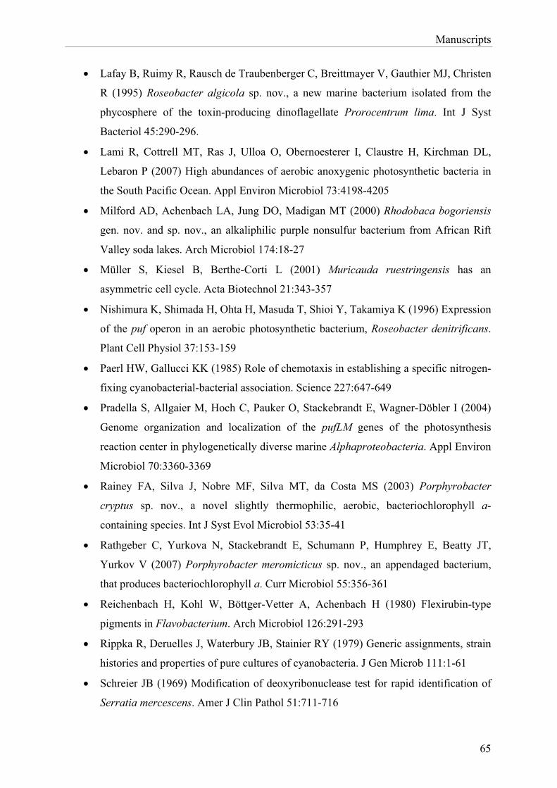
Manuscripts
� Lafay B, Ruimy R, Rausch de Traubenberger C, Breittmayer V, Gauthier MJ, Christen
R (1995) Roseobacter algicola sp. nov., a new marine bacterium isolated from the
phycosphere of the toxin-producing dinoflagellate Prorocentrum lima. Int J Syst
Bacteriol 45:290-296.
� Lami R, Cottrell MT, Ras J, Ulloa O, Obernoesterer I, Claustre H, Kirchman DL,
Lebaron P (2007) High abundances of aerobic anoxygenic photosynthetic bacteria in
the South Pacific Ocean. Appl Environ Microbiol 73:4198-4205
� Milford AD, Achenbach LA, Jung DO, Madigan MT (2000) Rhodobaca bogoriensis
gen. nov. and sp. nov., an alkaliphilic purple nonsulfur bacterium from African Rift
Valley soda lakes. Arch Microbiol 174:18-27
� Müller S, Kiesel B, Berthe-Corti L (2001) Muricauda ruestringensis has an
asymmetric cell cycle. Acta Biotechnol 21:343-357
� Nishimura K, Shimada H, Ohta H, Masuda T, Shioi Y, Takamiya K (1996) Expression
of the puf operon in an aerobic photosynthetic bacterium, Roseobacter denitrificans.
Plant Cell Physiol 37:153-159
� Paerl HW, Gallucci KK (1985) Role of chemotaxis in establishing a specific nitrogen-
fixing cyanobacterial-bacterial association. Science 227:647-649
� Pradella S, Allgaier M, Hoch C, Pauker O, Stackebrandt E, Wagner-Döbler I (2004)
Genome organization and localization of the pufLM genes of the photosynthesis
reaction center in phylogenetically diverse marine Alphaproteobacteria. Appl Environ
Microbiol 70:3360-3369
� Rainey FA, Silva J, Nobre MF, Silva MT, da Costa MS (2003) Porphyrobacter
cryptus sp. nov., a novel slightly thermophilic, aerobic, bacteriochlorophyll a-
containing species. Int J Syst Evol Microbiol 53:35-41
� Rathgeber C, Yurkova N, Stackebrandt E, Schumann P, Humphrey E, Beatty JT,
Yurkov V (2007) Porphyrobacter meromicticus sp. nov., an appendaged bacterium,
that produces bacteriochlorophyll a. Curr Microbiol 55:356-361
� Reichenbach H, Kohl W, Böttger-Vetter A, Achenbach H (1980) Flexirubin-type
pigments in Flavobacterium. Arch Microbiol 126:291-293
� Rippka R, Deruelles J, Waterbury JB, Stainier RY (1979) Generic assignments, strain
histories and properties of pure cultures of cyanobacteria. J Gen Microb 111:1-61
� Schreier JB (1969) Modification of deoxyribonuclease test for rapid identification of
Serratia mercescens. Amer J Clin Pathol 51:711-716
65
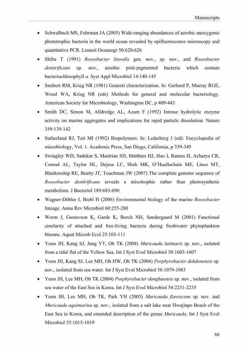
Manuscripts
� Schwalbach MS, Fuhrman JA (2005) Wide-ranging abundances of aerobic anoxygenic
phototrophic bacteria in the world ocean revealed by epifluorescence microscopy and
quantitative PCR. Limnol Oceanogr 50:620-626
� Shiba T (1991) Roseobacter litoralis gen. nov., sp. nov., and Roseobacter
denitrificans sp. nov., aerobic pink-pigmented bacteria which contain
bacteriochlorophyll a. Syst Appl Microbiol 14:140-145
� Smibert RM, Krieg NR (1981) General characterization. In: Gerhard P, Murray RGE,
Wood WA, Krieg NR (eds) Methods for general and molecular bacteriology.
American Society for Microbiology, Washington DC, p 409-443
� Smith DC, Simon M, Alldredge AL, Azam F (1992) Intense hydrolytic enzyme
activity on marine aggregates and implications for rapid particle dissolution. Nature
359:139-142
� Sutherland RJ, Tait MI (1992) Biopolymers. In: Lederberg J (ed): Encyclopedia of
microbiology, Vol. 1. Academic Press, San Diego, California, p 339-349
� Swingley WD, Sadekar S, Mastrian SD, Matthies HJ, Hao J, Ramos H, Acharya CR,
Conrad AL, Taylor HL, Dejesa LC, Shah MK, O’Huallachain ME, Lince MT,
Blankenship RE, Beatty JT, Touchman JW (2007) The complete genome sequence of
Roseobacter denitrificans reveals a mixotrophic rather than photosynthetic
metabolism. J Bacteriol 189:683-690
� Wagner-Döbler I, Biebl H (2006) Environmental biology of the marine Roseobacter
lineage. Annu Rev Microbiol 60:255-280
� Worm J, Gustavson K, Garde K, Borch NH, Søndergaard M (2001) Functional
similarity of attached and free-living bacteria during freshwater phytoplankton
blooms. Aquat Microb Ecol 25:103-111
� Yoon JH, Kang SJ, Jung YT, Oh TK (2008) Muricauda lutimaris sp. nov., isolated
from a tidal flat of the Yellow Sea. Int J Syst Evol Microbiol 58:1603-1607
� Yoon JH, Kang SJ, Lee MH, Oh HW, Oh TK (2006) Porphyrobacter dokdonensis sp.
nov., isolated from sea water. Int J Syst Evol Microbiol 56:1079-1083
� Yoon JH, Lee MH, Oh TK (2004) Porphyrobacter donghaensis sp. nov., isolated from
sea water of the East Sea in Korea. Int J Syst Evol Microbiol 54:2231-2235
� Yoon JH, Lee MH, Oh TK, Park YH (2005) Muricauda flavescens sp. nov. and
Muricauda aquimarina sp. nov., isolated from a salt lake near Hwajinpo Beach of the
East Sea in Korea, and emended description of the genus Muricauda. Int J Syst Evol
Microbiol 55:1015-1019
66

Manuscripts
� Yurkov VV (2006) Aerobic phototrophic proteobacteria. In: Dworkin M, Falkow S,
Rosenberg E, Schleifer KH, Stackebrandt E (eds) The Prokaryotes, 3rd ed. Springer,
Berlin Heidelberg New York, p 562-584
� Yurkov VV, Beatty JT (1998) Aerobic anoxygenic phototrophic bacteria. Microbiol
Mol Biol Rev 62:695-724
� Yurkov VV, van Gemerden H (1993) Impact of light/dark regimen on growth rate,
biomass formation and bacteriochlorophyll synthesis in Erythromicrobium
hydrolyticum. Arch Microbiol 159:84-89
67
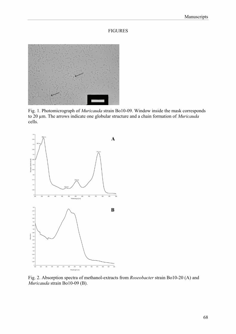
Manuscripts
FIGURES
Fig. 1. Photomicrograph of Muricauda strain Bo10-09. Window inside the mask corresponds to 20 μm. The arrows indicate one globular structure and a chain formation of Muricauda cells.
Fig. 2. Absorption spectra of methanol-extracts from Roseobacter strain Bo10-20 (A) and Muricauda strain Bo10-09 (B).
68
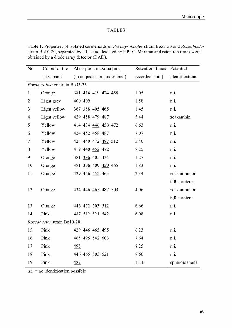
Manuscripts
TABLES
Table 1. Properties of isolated carotenoids of Porphyrobacter strain Bo53-33 and Roseobacter strain Bo10-20, separated by TLC and detected by HPLC. Maxima and retention times were obtained by a diode array detector (DAD). No. Colour of the
TLC band
Absorption maxima [nm]
(main peaks are underlined)
Retention times
recorded [min]
Potential
identifications
Porphyrobacter strain Bo53-33
1 Orange 381 414 419 424 458 1.05 n.i.
2 Light grey 400 409 1.58 n.i.
3 Light yellow 367 388 405 465 1.45 n.i.
4 Light yellow 429 458 479 487 5.44 zeaxanthin
5 Yellow 414 434 446 458 472 6.63 n.i.
6 Yellow 424 452 458 487 7.07 n.i.
7 Yellow 424 440 472 487 512 5.40 n.i.
8 Yellow 419 440 452 472 8.25 n.i.
9 Orange 381 396 405 434 1.27 n.i.
10 Orange 381 396 409 429 465 1.83 n.i.
11 Orange 429 446 452 465 2.34 zeaxanthin or
ß,ß-carotene
12 Orange 434 446 465 487 503 4.06 zeaxanthin or
ß,ß-carotene
13 Orange 446 472 503 512 6.66 n.i.
14 Pink 487 512 521 542 6.08 n.i.
Roseobacter strain Bo10-20
15 Pink 429 446 465 495 6.23 n.i.
16 Pink 465 495 542 603 7.64 n.i.
17 Pink 495 8.25 n.i.
18 Pink 446 465 503 521 8.60 n.i.
19 Pink 487 13.43 spheroidenone
n.i. = no identification possible
69
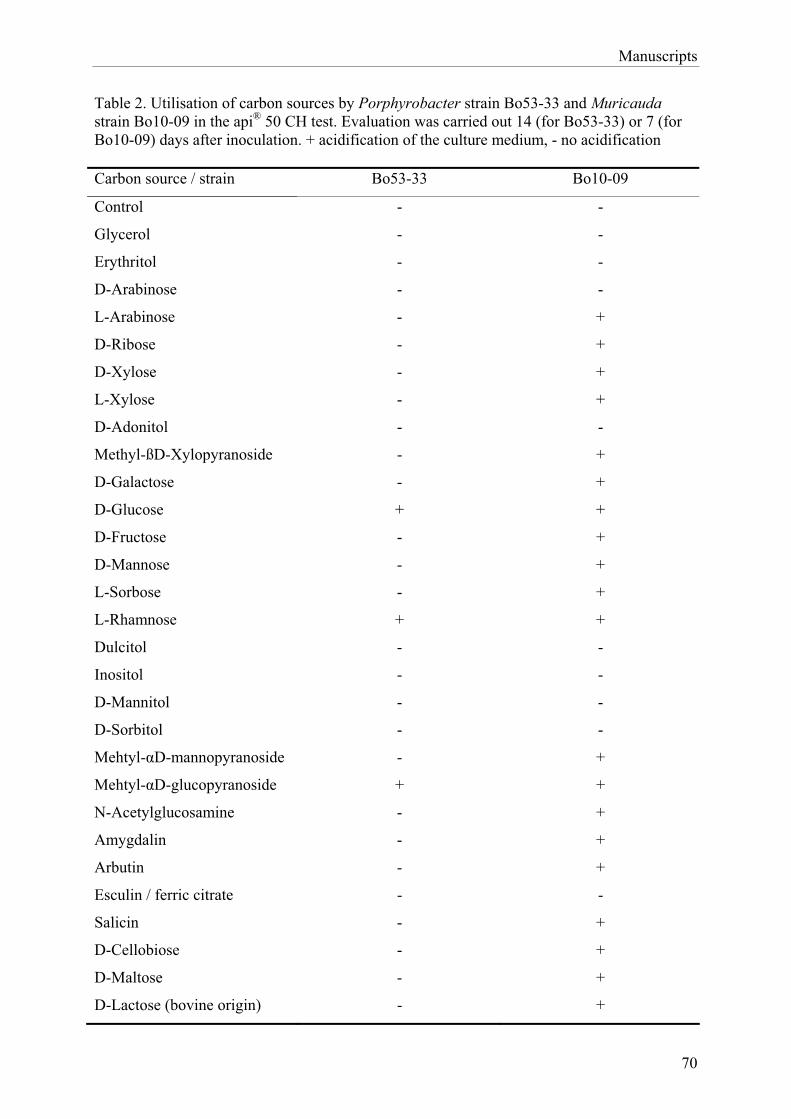
Manuscripts
Table 2. Utilisation of carbon sources by Porphyrobacter strain Bo53-33 and Muricauda strain Bo10-09 in the api® 50 CH test. Evaluation was carried out 14 (for Bo53-33) or 7 (for Bo10-09) days after inoculation. + acidification of the culture medium, - no acidification
Carbon source / strain Bo53-33 Bo10-09
Control - -
Glycerol - -
Erythritol - -
D-Arabinose - -
L-Arabinose - +
D-Ribose - +
D-Xylose - +
L-Xylose - +
D-Adonitol - -
Methyl-ßD-Xylopyranoside - +
D-Galactose - +
D-Glucose + +
D-Fructose - +
D-Mannose - +
L-Sorbose - +
L-Rhamnose + +
Dulcitol - -
Inositol - -
D-Mannitol - -
D-Sorbitol - -
Mehtyl-�D-mannopyranoside - +
Mehtyl-�D-glucopyranoside + +
N-Acetylglucosamine - +
Amygdalin - +
Arbutin - +
Esculin / ferric citrate - -
Salicin - +
D-Cellobiose - +
D-Maltose - +
D-Lactose (bovine origin) - +
70
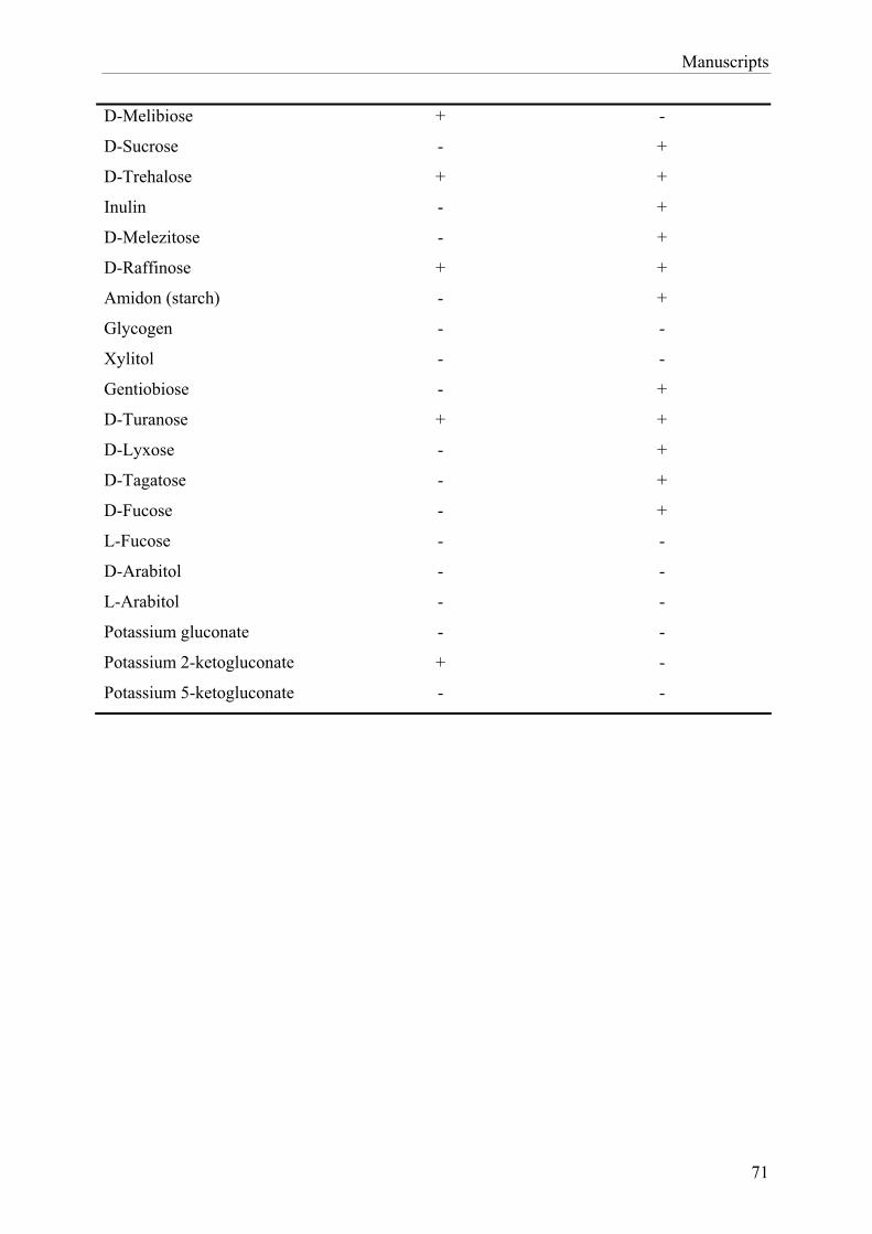
Manuscripts
71
D-Melibiose + -
D-Sucrose - +
D-Trehalose + +
Inulin - +
D-Melezitose - +
D-Raffinose + +
Amidon (starch) - +
Glycogen - -
Xylitol - -
Gentiobiose - +
D-Turanose + +
D-Lyxose - +
D-Tagatose - +
D-Fucose - +
L-Fucose - -
D-Arabitol - -
L-Arabitol - -
Potassium gluconate - -
Potassium 2-ketogluconate + -
Potassium 5-ketogluconate - -
General Discussion
4 General Discussion
4.1 Characterisation of heterotrophic bacteria with respect to their association with
cyanobacteria
To be able to compare various interactions that can occur between heterotrophs and
cyanobacteria, four heterotrophic strains were chosen after their isolation to be investigated in
more detail. Two of these strains, Porphyrobacter strain Bo53-33 and Roseobacter strain
Bo10-20, were differing AAnP strains. Strain Bo10-19 was a colourless strain closely related
to AAnP, while the coloured bacterium Muricauda strain Bo10-09 did not seem to be an
AAnP.
4.1.1 Characterisation of Porphyrobacter
The genus Porphyrobacter was designated in 1993 by Fuerst and co-workers. It includes
bacteriochlorophyll containing aerobes and branches with Erythrobacter longus within the
Erythrobacteraceae (Lee et al. 2005) in the Alphaproteobacteria. All Porphyrobacter species
described so far belong to the AAnP (Fuerst et al. 1993; Hanada et al. 1997; Hiraishi et al.
2002; Rainey et al. 2003; Rathgeber et al. 2007; Yoon et al. 2004; Yoon et al. 2006). But even
though it has long been known that AAnP often live in association with phytoplankton, to my
knowledge this has never been described before for Porphyrobacter. So far, only Navarro and
co-workers (2008) detected a bacterial community containing Porphyrobacter and the
unicellular cyanobacterium Synechococcus in the ephemeral hypereutrophic Mojave Dessert
Playa Lake.
Porphyrobacter strain Bo53-33, described in the present study, derived from the
cyanobacterial Nodularia harveyana strain Bo53 culture and showed highest 16S gene
sequence similarity to Porphyrobacter dokdonensis (99% similarity to strain DSW-74T (Yoon
et al. 2006), see manuscript I). Yoon and co-workers (2006) isolated their Porphyrobacter
strain from the Sea of Japan and did not observe any kind of association with phytoplankton.
However, for the strain used in the present study, characteristics were found that are at least
advantageous for this way of living. The extracellular amylase production and glucose
utilisation (see manuscript III) for example enable the organism to metabolise amylose and
hence live on readily available cyanobacterial exopolysaccharides (EPS). The high amount of
carotenoids detected in strain Bo53-33 is advantageous when living in environments with an
72
General Discussion
increased requirement for a protection against photooxidation (Armstrong 1997; Kolber et al.
2001; Yurkov and Beatty 1998), as it is usual for phototrophic organisms. However, the
cyanobacterial hosts, which generally move phototactically to light conditions that are
approximately 1 – 10% of the daylight (Stal et al. 1985), might provide a certain protection as
well.
The most significant properties were found when investigating the occurrence of
Porphyrobacter and closely related species in various cyanobacterial cultures (see manuscript
I and II). These organisms were only found in some cultures. In both Nodularia cultures
tested they were detected, but in none of the Oscillatoria ones, even though these derived
from the same sampling site as our Nodularia harveyana strain. But 14 days after the cultures
of Nodularia harveyana strain Bo53 and Oscillatoria brevis strain Bo10 were made axenic,
Porphyrobacter and relatives reoccurred in the Bo53 culture, but as well in the Bo10 culture
cells were found sporadically (see manuscript II). This indicates that Porphyrobacter is able
to live on both cyanobacteria’s excretory products and is able to develop fast. However, in the
naturally occurring community on Oscillatoria competitors might prevent the spread of
Porphyrobacter. It can be assumed that the competitors grow more slowly than
Porphyrobacter does and, therefore, would constrain the growth of the latter one in the
formally axenic cultures within a certain time as well.
The fact that Porphyrobacter was also found in the culture of Geitlerinema strain Flo1,
deriving from a mangrove area in Florida south of Miami, might be an evidence for an
ubiquitous distribution and is in accordance with reports that Porphyrobacter isolates were
obtained from various worldwide places, such as a brackish hot spring in Japan (Hanada et al.
1997), a hot spring in Portugal (Rainey et al. 2003), and the sea of Japan (Yoon et al. 2004;
Yoon et al. 2006). Porphyrobacter could not be found in the culture of Fischerella ambigua
from the department’s culture collection, the only fresh water cyanobacterial culture tested.
However, this does not necessarily mean that this heterotroph will not occur in any fresh
water communities, since Fuerst and co-workers (1993) and Rathgeber and co-workers (2007)
isolated their Porphyrobacter strains from freshwater.
In summary, there is evidence that strain Bo53-33 occurs in association with cyanobacteria
and possesses some capabilities useful for a beneficial coexistence in such a community, but
does not depend on such way of living. Porphyrobacter seems to be a fast growing,
ubiquitously distributed organism, and does not seem to be specialised to a certain living
condition. However, Porphyrobacter appears to be sensitive to interfere with other bacteria.
73
General Discussion
4.1.2 Characterisation of Roseobacter
The genus Roseobacter was established by Shiba in 1991 and belongs to the �-3 subclass of
the Proteobacteria. This genus is physiologically heterogeneous and it is known to comprise
some of the most abundant members of the AAnP, such as the first described species
Roseobacter litoralis and Roseobacter denitrificans (Allgaier et al. 2003; Brinkhoff et al.
2008; Buchan et al. 2005; Oz et al. 2005). However, nowadays it is known that not all
Roseobacter species are able to produce bacteriochlorophyll (Lafay et al. 1995). It is as well
known that members of the Roseobacter clade dominate among marine phytoplankton-
associated bacteria (Buchan et al. 2005). They are described to be effective colonisers that are
able to form biofilms and even inhibit other bacteria by producing antibacterial substances
(Bruhn et al. 2007; Gram et al. 2002).
Roseobacter strain Bo10-20, isolated during the present study (see manuscript I), derived
from the culture of Oscillatoria brevis strain Bo10 and showed high 16S gene sequence
similarities with some non-characterised Roseobacter clone sequences. However, the most
closely related validly described species to strain Bo10-20 was Sulfitobacter dubius strain
KMM 3554T (Ivanova et al. 2004) (94% similarity).
As previously described for Porphyrobacter (see chapter 4.1.1), the physiological and
biochemical investigations of Roseobacter strain Bo10-20 also revealed characteristics that
are useful when living in association with cyanobacteria, such as the possession of several
carotenoids and the ability to produce extracellular amylases (see above). Nevertheless, it
could be demonstrated that the occurrence of Roseobacter in the cyanobacterial cultures (see
manuscript I and II) showed some noticeable differences between this heterotroph and
Porphyrobacter.
Even though the Roseobacter clade has been described to be ubiquitously distributed in
marine environments (Buchan et al. 2005; Wagner-Döbler and Biebl 2006), in the present
study it was not possible to detect Roseobacter species in each of the cyanobacterial cultures
tested. Among the ten marine Baltic cyanobacterial strains sampled, Roseobacter spp. could
neither be detected in cultures of Synechocystis, Lyngbya semiplenta, Oscillatoria laetevirens,
Anabaena variabilis, nor in any Nodularia culture. (But it must be mentioned that after the
separation of attached and free-living heterotrophs from the Nodularia harveyana Bo53
culture, traces of Roseobacter cells were found in the supernatants (see manuscript I).) In the
culture of Geitlerinema strain Flo1, Roseobacter was not found either. Additionally, in
experiments with axenic cultures, this AAnP did not reappear in all of the Oscillatoria brevis
cultures after 14 days of incubation. One explanation for the absence in some of the
74
General Discussion
cyanobacterial cultures might be a lack of certain essential nutrients. However, more likely is
the explanation that certain competitors prevent the growth of Roseobacter as described for
Porphyrobacter before (see above). The observation that Roseobacter appeared in almost all
formally axenic Nodularia harveyana cultures, even though originally only scarcely found
with this culture, is also in accordance with this assumption that its growth is often inhibited
in natural heterotrophic communities, and as well with the observations for Porphyrobacter
(see chapter 4.1.1).
Nevertheless, the observation that Roseobacter was not detected in the culture of the
freshwater cyanobacterium Fischerella ambigua is in accordance with other reports for
freshwater environments (Buchan et al. 2005).
In summary, it can be said that, even though Roseobacter strain Bo10-20 exhibited some
benefiting features, it was not possible to maintain the behaviour of Roseobacter to be an
aggressive, ubiquitously occurring coloniser. It became apparent that this organism reacts
discriminative to various cyanobacterial cultures or the respective heterotrophic colonisers
and in several cases its growth was inhibited.
4.1.3 Characterisation of strain Bo10-19
Strain Bo10-19 was a colourless alphaproteobacterium clustering with the Rhodobacter clade
with highest 16S gene sequence similarity of only 95% to Rhodobaca bogoriensis (see
manuscript I). It also derived from a culture of Oscillaturia brevis strain Bo10.
Rhodobaca bogoriensis is known to be a yellow to pink coloured purple non-sulphur
bacterium, possessing bacteriochlorophyll a and carotenoids (Milford et al. 2000; Takaichi et
al. 2001). To my knowledge, it has never been described living in coexistence with
phytoplankton. However, Navarro and co-workers (2008) were at least able to detect
Rhodobaca and Rhodobacter bacteria in samples from Mojave Desert Playa Lake (California)
along with the unicellular cyanobacterium Synechococcus.
Strain Bo10-19 did not bear a strong resemblance with the photoheterotrophic Rhodobaca
bogoriensis as described by Milford and co-workers (2000) and its relatives (Hiraishi and
Ueda 1994). In contrast to Rhodobaca bogoriensis, strain Bo10-19 was colourless and grew
exclusively chemoorganotrophically under aerobic conditions. Light did not seem to influence
its growth behaviour (see manuscript III).
75
General Discussion
Neither extracellular amylases, nor DNases, peptidases, or lipases were found to be produced
by strain Bo10-19 by testing with standardised plating methods (see manuscript III). This
might indicate that within the cyanobacterial cultures this strain lives on very specific
substances produced by the cyanobacteria. However, it should be noted that it was possible to
cultivate strain Bo10-19 and related ones in pure culture on undefined media (see manuscript
I).
Even though it was not possible to design a probe specific for strain Bo10-19, other
experiments revealed that it is unlikely that Bo10-19-like bacteria occurred in Nodularia
harveyana Bo53 culture as well. In the course of the strain’s isolation from the cyanobacteria,
several isolates with similar morphological properties were found in the Oscillatoria brevis
Bo10 culture, being closely related to strain Bo10-19 when compared by fingerprinting
methods. However, from the Nodularia harveyana Bo53 culture no similarly looking isolates
were obtained (see manuscript I).
In conclusion, it can be said that strain Bo10-19 seems to be a heterotroph which is very
specifically adapted to some particular living conditions such as association with Oscillatoria
brevis. Since the 16S gene sequence similarity between Rhodobaca bogoriensis and strain
Bo10-19 is only 95% and they do not share many other features either, strain Bo10-19 might
comprise a new species. However, to pinpoint this assumption some more physiological
investigations need to be performed.
4.1.4 Characterisation of Muricauda
The genus Muricauda was described in 2001 by Bruns and co-workers and belongs to the
Bacteroidetes group. The genus comprises four species that are known to be marine, yellow
pigmented, and strictly aerobic or facultatively anaerobic bacteria. Strains described so far are
cosmpolitans and occurred e.g. in the German Wadden Sea (Bruns et al. 2001), the Sea of
Japan (Yoon et al. 2005), the Yellow Sea, Korea (Yoon et al. 2008), and the Mediterranean
Sea (Agogué et al. 2005). Even though it is known for some of their relatives (Delucca and
McCracken 1977; Grossart et al. 2005), this way of living has never been shown for any of
the Muricauda strains described. Nevertheless, Bruns and co-workers (2001) first observed
appendages which they assumed to be meant for colonisation of given substrates.
Muricauda strain Bo10-09 was isolated from a culture of Oscillatoria brevis strain Bo10 and
showed highest 16S gene sequence similarity with Muricauda aquimarina (Yoon et al. 2005)
76
General Discussion
(99%, see manuscript I). The biochemical and physiological investigations (see manuscript
III) revealed that Bo10-09 contains one yellow carotenoid. Furthermore, this strain was able
to metabolise almost all sugars offered, which seems to be advantageous when living on
cyanobacterial EPS, but none of the alcohols tested. Extracellular enzymes detected were
amylases and DNases. Especially the production of amylases is beneficial for the utilisation of
cyanobacterial excretory products. Additionally, structures were observed by light
microscopical observation studies that might be similar to those vesicle-like structures at the
end of the appendages of Muricauda ruestringensis as described by Bruns and co-workers
(2001) and Müller and co-workers (2001) (see manuscript III). If, as suggested by Bruns and
co-workers (2001), the structures are used to attach to surfaces, they might represent a special
equipment of Muricauda strain Bo10-09 to live on cyanobacteria. The results of our
occurrence and growth experiments (see manuscript I and II) confirm these indications.
Muricauda was not found in any of the unicellular Baltic cyanobacterial cultures of the
department’s culture collection tested, but in most of the filamentous ones (both Oscillatoria
strains Bo10 and Bo17 as well as both Nodularia strains Bo53 and Fä19). This might indicate
special adaptations to the filamentous cyanobacteria, but needs further investigations. It is not
surprising that Muricauda was found in the Atlantic Geitlerinema strain Flo1, but not in the
freshwater strain Fischerella ambigua, since this genus is known to be ubiquitously
distributed in marine environments but does not occur in freshwaters. Among the 14 days old
formally axenic cyanobacterial cultures, Muricauda had reappeared in all of the Nodularia
harveyana Bo53 cultures but only in six of the ten Oscillatoria brevis Bo10 cultures (see
manuscript II). In all cultures to which Muricauda cells were added, they had reached stable
abundances of 1 - 2% (Nodularia harveyana strain Bo53 cultures) and ~ 20 % (Oscillatoria
brevis strain Bo10 cultures) after 14 days of cultivation, unlike Porphyrobacter and
Roseobacter had in the same experiment. This might indicate that Muricauda is a more
effective coloniser than the others are.
Rashidan and Bird (2001) suggested for some cyanobacteria-associated Cytophaga bacteria,
which are closely related to Muricauda strain Bo10-09, to have a rather detrimental effect on
the cyanobacteria. However, this could not be confirmed for Muricauda strain Bo10-09 which
did not have noticeable effects different from those of the other heterotrophs tested (see
manuscript II).
To sum up, the results obtained with Muricauda indicate that this organism is a fast and
effective coloniser that is widely distributed among filamentous marine cyanobacteria-
associated communities, but seems only in the case of the resettlement on Oscillatoria brevis
77
General Discussion
Bo10 cultures to be inhibited by some faster competitors. Compared with the three other
heterotrophic strain investigated in this study (see chapter 4.1.1, 4.1.2, and 4.1.3), strain
Bo10-09 seemed to be the one that is best adapted for living in association with
cyanobacteria.
4.2 Characterisation of cyanobacteria with respect to their association with heterotrophs
Both cyanobacterial strains used for comparison (Nodularia harveyana strain Bo53 and
Oscillatoria brevis strain Bo10) derived from the same sampling site at the German Baltic Sea
coast and were sampled at the same time (Rethmeier 1995). Nodularia harveyana as well as
Oscillatoria in general have been known before to be mat-forming cyanobacteria (de los Ríos
et al. 2004; Stal et al. 1985; Stal et al. 2003). These cyanobacteria are known to produce large
amounts of EPS which are involved in the formation of a matrix to embed and protect the mat
organisms. Additionally, the cyanobacterial filaments form networks to stabilise the mats (de
los Ríos et al. 2004; Paerl et al. 2000). These structures are known to contain the
biocomplexity to allow complete nutrient cycles (Canfield and Des Marais 1993; Paerl et al.
2000). The budgets for the main nutrient cycles have already been identified. The main part of
the carbon present within the mats derives from primary carbon fixation of oxygenic and
anoxygenic phototrophs and minor parts of secondary carbon fixation of non-photosynthetic
autotrophic and heterotrophic bacteria. The inorganic carbon is known to originate from
aerobic and anaerobic respiration and heterotrophy within the mat as well as from the
overlaying water layers. Oxygen is also known to be mainly produced and consumed within
the mats, and this procedure is assumed for other nutrient elements as well (Canfield and Des
Marais 1993). This means that within such consortia diverse metabolic processes occur that
could inhibit each other, for example by the appearance of detrimental by-products (Paerl and
Pinckney 1996). On this account, the temporal and spatial arrangements of the activities
within the mats are crucial for its functionality.
In both cyanobacterial cultures examined in this study, the heterotrophs investigated occurred
as free-living ones in the culture medium as well as associated with the cyanobacterial
trichomes in form of large aggregates. By applying the new developed method to get axenic
78
General Discussion
cultures it became apparent that in the Nodularia culture the aggregates of heterotrophs
seemed to be much more tightly attached to the filaments than in the Oscillatoria culture.
It has been known before that cyanobacteria die when the amount of heterotrophic associated
bacteria decreases to a certain amount (Palinska et al. 1999). This was also observed for
Nodularia harveyana strain Bo53 and Oscillatoria brevis strain Bo10 during the procedure to
obtain axenic cyanobacterial cultures. Nevertheless, it could be shown that at least for
Oscillatoria brevis strain Bo10 the addition of single heterotrophic pure cultures was also
detrimental (see manuscript II), which might indicate that it is important for Oscillatoria
brevis that none of the heterotrophs gets an exceedingly strong influence on this bacterial
community.
The approximate composition of heterotrophs within the two cyanobacterial cultures was
alike (see manuscript I). In both cultures, Alphaproteobacteria dominated the community of
heterotrophs, followed by Bacteroidetes bacteria and Gammaproteobacteria.
Betaproteobacteria, Firmicutes, or Archaea were found in neither of the cyanobacterial
cultures, even though the occurrence of Betaproteobacteria and Firmicutes associated with
Nodularia has previously been described (Salomon et al. 2003; Tuomainen et al. 2006).
Detailed investigations with more specific oligonucleotide probes revealed that Roseobacter,
Erythrobacteraceae, and Muricauda cells occurred to similar amounts of less than 10%
within the Nodularia culture. A higher percentage of Roseobacter cells (10 – 20 %) existed
within the Oscillatoria culture, whereas Erythrobacteraceae could not be found. Having
made the two cyanobacterial cultures axenic, the reoccurrence of the heterotrophs within the
cultures was checked after 14 days. In the Nodularia culture, all heterotrophs determined and
investigated during the present study had reappeared, even though they did not exhibit the
same percentages as found before. This is in contrast to the Oscillatoria culture, in which the
heterotrophic composition had changed completely. Erythrobacteraceae appeared in all set
ups, whereas Roseobacter and Muricauda could only be found in some of them. Since also
the occurrences of the heterotrophs within other studied cyanobacterial cultures of the
department’s culture collection partially differed considerably to those in Nodularia
harveyana strain Bo53 and Oscillatoria brevis strain Bo10, it became obvious that the
heterotrophic composition does not necessarily depend on obvious features of the
cyanobacterial cultures, such as origin of the strains, availability of nitrogen in the culture
medium, or formation of heterocysts. It is much more likely that the heterotrophs depend
either on very specific cyanobacterial excretory products or on their heterotrophic
companions.
79
General Discussion
In summary, it seems that Nodularia has a much more stable heterotrophic community which
is also much more difficult to detach in comparison to that of Oscillatoria, though
Oscillatoria is much more sensitive to changes within the heterotrophic composition.
Oscillatoria is known to be one of the cyanobacteria starting mat-formation (Stal et al. 1985).
It is likely that consortia within developing young mats are not as stable as they are in older
ones and that they undergo a succession. As a consequence, it makes sense that Oscillatoria
does not seem to be specified to a certain heterotrophic company. However, it is astonishing
that Oscillatoria reacts more intensely to changes than the heterocystous Nodularia. The latter
is much more dependent on a stable community in order to be capable of protecting its
heterocysts from evolving oxygen. This assumption is in coincidences with the results gained
in the present study.
4.3 Conclusions
For cyanobacteria it had been proven before that they depend on at least some of their
associated heterotrophic partners (Palinska et al. 1999). In the present study, it was shown that
unique heterotrophic strains can as well have a detrimental effect on the cyanobacteria. In
nature, this seems to be prevented by a self-regulation that the heterotrophic community
exercises in the surroundings of Nodularia harveyana and Oscillatoria brevis. It has been
shown for the heterotrophs investigated in the present study that they were shown to have
some advantages when living in association with cyanobacteria, even though all of them
belonged to genera known to occur free-living as well. Thus, it cannot be excluded that there
are also heterotrophs that are dependent on living in those communities. In general, it can be
said that the relations within these associations seem to be very complex and thus cannot be
elucidated without performing a multitude of adjacent experiments. Additionally, it was
demonstrated that differing cyanobacteria as well as heterotrophs showed various adaptations
within the aggregates, and that it is not possible to predict the mutual effects only from the
knowledge of common properties, neither for the cyanobacteria nor for the heterotrophs.
Up to now, our knowledge about the phytoplanktonic functions in the Baltic Sea had mainly
been based on investigations of the conspicuous blooming events. Nevertheless, nowadays, in
times of increasing anthropogenic inshore pollution, the mat-forming benthic cyanobacteria
gain more and more influence (Schiewer 2008). To be able to estimate the effects that these
80
General Discussion
cyanobacteria might have for the whole ecosystem, it is important to have a close look on
these organisms and their ecological characteristics. The data set compiled in the present
study might at least provide first impressions of the complex inter-specific connections and
functions within these habitats and thus will give a basis for further investigations.
4.4 Future prospects
The present study gave important information about the relationship between cyanobacteria
and their heterotrophic associates. Nevertheless, there are still several aspects that remain
ambiguous. Some adjacent experiments should be conducted which might be helpful to
further illuminate the mutual connections.
The choice of a culture dependent set up for the identification of the heterotrophic bacteria
might cause the non-discover of certain bacterial groups. To obtain a more intense view upon
the compositions of the heterotrophic communities, it will be useful to additionally construct a
clone library.
Being able to judge the influence of heterotrophs upon cyanobacteria, it is necessary to take
into consideration that in nature the heterotrophic species composition within cyanobacterial
aggregations undergoes seasonal changes (Mašín et al. 2006) and shifts during progression of
a bloom as well (Rashidan and Bird 2001), whereas in lab cultures presumably a stationary
community had developed. To get an impression about when certain defined heterotrophs
appear, disappear, or remain constant in the community, it is necessary to take natural
samples at different seasons for comparative investigations. These data would also give
additional information about the interaction of the heterotrophs within the communities.
For a more significant survey about the abundances of heterotrophs in association with
different cyanobacteria, it is indispensable to check many more deposited cultures for the
occurrence of heterotrophs.
Another interesting approach to be conducted in the future would be to characterise the EPS
of the Nodularia harveyana strain Bo53 and Oscillatoria brevis strain Bo10 and to compare
the range of detected substances with respect to their utilisation by heterotrophic bacteria.
These data might indicate a more definite connection between the cyanobacteria and their
heterotrophic partners. Other physiological experiments, such as the examination of the
influence of light intensity or oxygen availability on the heterotrophs’ growth behaviour, may
give valuable information on positive advantages for the heterotrophs when living in
association with phytoplankton.
81

General Discussion
82
For a better understanding concerning the influence of heterotrophs upon cyanobacteria, it
might be interesting to investigate the growth behaviour of the cyanobacteria after having
been made axenic and when mixed with combinations of living heterotrophic bacteria, dead
bacteria, or single bacterial components.
References
5 References
Agogué H, Joux F, Obernosterer I, Lebaron P (2005) Resistance of marine bacterioneuston to
solar radiation. Appl Environ Microbiol 71:5282-5289
Allgaier M, Uphoff H, Felske A, Wagner-Döbler I (2003) Aerobic anoxygenic photosynthesis
in Roseobacter clade bacteria from diverse marine habitats. Appl Environ Microbiol
69:5051-5059
Armstrong GA (1997) Genetics of eubacterial carotenoid biosynthesis: a colourful tale. Annu
Rev Microbiol 51:629-659
Beatty JT (2002) On the natural selection and evolution of the aerobic phototrophic bacteria.
Photosynth Res 73:109-114
Béjà O, Suzuki MT, Heidelberg JF, Nelson WC, Preston CM, Hamada T, Eisen JA, Fraser
CM, DeLong EF (2002) Unsuspected diversity among marine aerobic anoxygenic
phototrophs. Nature 415:630-633
Bidle KD, Fletcher M (1995) Comparison of free-living and particle-associated bacterial
communities in the Chesapeake Bay by stable low-molecular-weight RNA analysis.
Appl Environ Microbiol 61:944-952
Brinkhoff T, Giebel HA, Simon M (2008) Diversity, ecology, and genomics of the
Roseobacter clade: a short overview. Arch Microbiol 189:531-539
Bruhn JB, Gram L, Belas R (2007) Production of antibacterial compounds and biofilm
formation by Roseobacter species are influenced by culture conditions. Appl Environ
Microbiol 73:442-450
Brunberg AK (1999) Contribution of bacteria in the mucilage of Microcystis spp.
(Cyanobacteria) to benthic and pelagic bacterial production in a hypereutrophic lake.
FEMS Microb Ecol 29:13-22
Bruns A, Rohde M, Berthe-Corti L (2001) Muricauda ruestringensis gen. nov., sp. nov., a
facultatively anaerobic, appendaged bacterium from German North Sea intertidal
sediment. Int J Syst Evol Microbiol 51:1997-2006
Buchan A, Gonzales JM, Moran MA (2005) Overview of the marine Roseobacter lineage.
Appl Environ Microbiol 71:5665-5677
Bursa AS (1968) Epicenoses on Nodularia spumigena Mertens in the Baltic Sea. Acta
Hydrobiol 10:267-297
Canfield DE, Des Marais DJ (1993) Biogeochemical cycles of carbon, sulfur, and free oxygen
in a microbial mat. Geochim Cosmochim Acta 57:3971-3984
83
References
Capone DG (2000) The marine microbial nitrogen cycle. In: Kirchman DL (ed) Microbial
ecology of the oceans. Wiley & Sons, New York, pp 455-495
Carpenter EJ, Price CC (1976) Marine Oscillatoria (Trichodesmium): explanation for
aerobic nitrogen fixation without heterocysts. Science 191:1278-1280
Cole JJ (1982) Interactions between bacteria and algae in aquatic ecosystems. Ann Rev Syst
13:291-314
Cottrell MT, Mannino A, Kirchman DL (2006) Aerobic anoxygenic phototrophic bacteria in
the Mid-Atlantic Bight and the North Pacific Gyre. Appl Environ Microbiol 72:557-564
Csotonyi JT, Swiderski J, Stackebrandt E, Yurkov VV (2008) Novel halophilic aerobic
anoxygenic phototrophs from a Canadian hypersaline spring system. Extremophiles
12:529-539
de los Ríos A, Ascaso C, Wierzchos J, Fernández-Valiente E, Quesada A (2004)
Microstructual characterization of cyanobacterial mats from the McMurdo Ice Shelf,
Antarctica. Appl Environ Microbiol 70:569-580
Delucca R, McCracken MD (1977) Observations on interactions between naturally-collected
bacteria and several species of algae. Hydrobiologia 55:71-75
Dippner JW, Vuorinen I (2008) Climate-related marine ecosystem change. In: The BACC
Author Team (eds) Assessment of climate change for the Baltic Sea Basin. Springer,
Berlin Heidelberg, pp 309-377
Fuerst JA, Hawkins JA, Holmes A, Sly LI, Moore CJ, Stackebrandt E (1993) Porphyrobacter
neustonensis gen. nov., sp. nov., an aerobic bacteriochlorophyll-synthesizing budding
bacterium from fresh water. Int J Syst Bacteriol 43:125-134
Goericke R (2002) Bacteriochlorophyll a in the ocean: Is anoxygenic bacterial photosynthesis
important? Limnol Oceanogr 47:290-295
Gram L, Grossart HP, Schlingloff A, Kiørboe T (2002) Possible quorum sensing in marine
snow bacteria: production of acylated homoserine lactones by Roseobacter strains
isolated from marine snow. Appl Environ Microbiol 68:4111-4116
Griffith P, Shiah F, Gloersen K, Ducklow HW, Fletcher M (1994) Activity and distribution of
attached bacteria in Chesapeake Bay. Mar Ecol Prog Ser 108:1-10
Grossart HP, Levold F, Allgaier M, Simon M, Brinkhoff T (2005) Marine diatom species
harbour distinct bacterial communities. Environ Microbiol 7:860-873
Halinen K, Fewer DP, Sihvonen LM, Lyra C, Eronen E, Sivonen K (2008) Genetic diversity
in strains of the genus Anabaena isolated from planktonic and benthic habitats of the
Gulf of Finland (Baltic Sea). FEMS Microbiol Ecol 64: 199-208.
84
References
Hanada S, Kawase Y, Hiraishi A, Takaichi S, Matsuura K, Shimada K, Nagashima KVP
(1997) Porphyrobacter tepidarius sp. nov., a moderately thermophilic aerobic
photosynthetic bacterium isolated from a hot spring. Int J Syst Bacteriol 47:408-413
Heinänen A, Kononen K, Kuosa H, Kuparinen J, Mäkela K (1995) Bacterioplankton growth
associated with physical fronts during a cyanobacterial bloom. Mar Ecol Prog Ser
116:233-245
Hietanen S, Moisander PH, Kuparinen J, Tuominen L (2002) No sign of denitrification in a
Baltic Sea cyanobacterial bloom. Mar Ecol Prog Ser 242:73-82
Hiraishi A, Ueda Y (1994) Intrageneric structure of the genus Rhodobacter: transfer of
Rhodobacter sulfidophilus and related marine species to the genus Rhodovulum gen.
nov.. Int J Syst Bacteriol 44:15-23
Hiraishi A, Yonemitsu Y, Matsushita M, Shin YK, Kuraishi H, Kawahara K (2002)
Characterisation of Porphyrobacter sanguineus sp. nov., an aerobic bacteriochlorophyll-
containing bacterium, capable of degrading biphenyl and dibenzofuran. Arch Microbiol
178:45-52
Hu Y, Du H, Jiao N, Zeng Y (2006) Abundant presence of the �-like Proteobacterial pufM
gene in oxic seawater. FEMS Microbiol Lett 263:200-206
Ivanova EP, Gorshkova NM, Sawabe T, Zhukova NV, Hayashi K, Kurilenko VV, Alexeeva
Y, Buljam V, Nicolau DV, Mikhailov VV, Christen R (2004) Sulfitobacter delicatus sp.
nov. and Sulfitobacter dubius sp. nov., respectively from a starfish (Stellaster equestris)
and sea grass (Zostera marina). Int J Syst Evol Microbiol 54:475-480
Jiao N, Zhang Y, Zeng Y, Hong N, Liu R, Chen F, Wang P (2007) Distinct distribution
pattern of abundance and diversity of aerobic anoxygenic phototrophic bacteria in the
global ocean. Environ Microbiol 9:3091-3099
Kahru M, Horstmann U, Rud O (1994) Satellite detection of increased cyanobacteria blooms
in the Baltic Sea: natural fluctuation of ecosystem change? Ambio 23:469-472
Karl DM (2002) Hidden in a sea of microbes. Nature 415:590-591
Kolber ZS, Plumley FG, Lang AS, Beatty JT, Blankenship RE, Van Dover CL, Vetriani C,
Koblížek M, Rathgeber C, Falkowski PG (2001) Contribution of aerobic
photoheterotrophic bacteria to the carbon cycle in the ocean. Science 292:2492-2495
Kolber ZS, Van Dover CL, Niederman RA, Falkowski PG (2000) Bacterial photosynthesis in
surface waters of the open ocean. Nature 407:177-179
85
References
Lafay B, Ruimy R, Detraubenberg CR, Breittmayer V, Gauthier MJ, Christen R (1995)
Roseobacter algicola sp. nov., a new marine bacterium isolated from the phycosphere of
the toxin-producing dinoflagellate Prorocentrum lima. Int J Syst Bacteriol 45:290-296
Lami R, Cottrell MT, Ras J, Ulloa O, Obernoesterer I, Claustre H, Kirchman DL, Lebaron P
(2007) High abundances of aerobic anoxygenic photosynthetic bacteria in the South
Pacific Ocean. Appl Environ Microbiol 73:4198-4205
Larsson U, Elmgren R, Wulff F (1985) Eutrophication and the Baltic Sea: causes and
consequences. Ambio 14:10-14
Larsson U, Hagström Å (1982) Fractionated phytoplankton primary production, exudate
release and bacterial production in a Baltic eutrophication gradient. Mar Biol 67:57-70
Larsson U, Hajdu S, Walve J, Elmgren R (2001) Baltic Sea nitrogen fixation estimated from
the summer increase in upper mixed layer total nitrogen. Limnol Oceanogr 46:811-820
Lee KB, Liu CT, Anzai Y, Kim H, Aono T, Oyaizu H (2005) The hierarchical system of the
‘Alphaproteobacteria’: description of Hyphomonadaceae fam. nov., Xanthobacteraceae
fam. nov. and Erythrobacteraceae fam. nov.. Int J Syst Evol Microb 55:1907-1919
Lyra C, Laamanen M, Lehtimäki JM, Suralla A, Sivonen K (2005) Benthic cyanobacteria of
the genus Nodularia are non-toxic, without gas vacuoles, able to glide and genetically
more diverse than planktonic Nodularia. Int J Syst Evol Microbiol 55:555-568
Mairs Geographischer Verlag (1994) Die Große Weltkarte 1:27,5 Mio. MAIRDUMONT,
Ostfildern
Mašín M, Zdun A, Sto�-Egiert J, Nausch M, Labrenz M, Moulisová V, Koblížek M (2006)
Seasonal changes and diversity of aerobic anoxygenic phototrophs in the Baltic Sea.
Aquat Microb Ecol 45:247-254
Milford AD, Achenbach LA, Jung DO, Madigan MT (2000) Rhodobaca bogoriensis gen.
nov. and sp. nov., an alkaliphilic purple nonsulfur bacterium from African Rift Valley
soda lakes. Arch Microbiol 174:18-27
Müller S, Kiesel B, Berthe-Corti L (2001) Muricauda ruestringensis has an asymmetric cell
cycle. Acta Biotechnol 21:343-357
Nagashima KVP, Hiraishi A, Shimada K, Matsuura K (1997) Horizontal transfer of genes
coding for the photosynthetic reaction centers of purple bacteria. J Mol Evol 45:131-136
Navarro JB, Moser DP, Flores A, Ross C, Rosen MR, Dong H, Zhang G, Hedlund BP (2008)
Bacterial succession within an ephemeral hypereutrophic Mojave Desert playa lake.
Microb Ecol. Doi:10.1007/s00248-008-9426-3
86
References
Nausch M (1996) Microbial activities on Trichodesmium colonies. Mar Ecol Prog Ser
141:173-181
Nishimura K, Shimada H, Ohta H, Masuda T, Shioi Y, Takamiya K (1996) Expression of the
puf operon in an aerobic photosynthetic bacterium, Roseobacter denitrificans. Plant Cell
Physiol 37:153-159
Oz A, Sabehi G, Koblízek M, Massana R, Béjà O (2005) Roseobacter-like bacteria in Red
and Mediterranean Sea aerobic anoxygenic photosynthetic populations. Appl Environ
Microbiol 71:344-353
Paerl HW, Bebout BM, Prufert LE (1989) Bacterial associations with marine Oscillatoria sp.
(Trichodesmium sp.) populations: ecophysiological implications. J Phycol 25:773-784
Paerl HW, Fulton RS (2006) Ecology of harmful cyanobacteria. In: Granéli E, Turner JT
(eds) Ecology of harmful algae, ecological studies, Vol. 189. Springer-Verlag, Berlin
Heidelberg New York; pp 95-109
Paerl HW, Gallucci KK (1985) Role of chemotaxis in establishing a specific nitrogen-fixing
cyanobacterial-bacteria association. Science 227:647-649
Paerl HW, Pinckney JL (1996) A mini-review of microbial consortia: their roles in aquatic
production and biogeochemical cycling. Microb Ecol 31:225-247
Paerl HW, Pinckney JL, Steppe TF (2000) Cyanobacterial-bacterial mat consortia: examining
the functional unit of microbial survival and growth in extreme environments. Environ
Microbiol 2:11-26
Palinska KA, Becker C, Krumbein WE (1999) Isolation and purification techniques for
benthic marine cyanobacteria – biotechnology potential. Bull Inst Océanogr Monaco
19:585-592
Pradella S, Allgaier M, Hoch C, Pauker O, Stackebrandt E, Wagner-Döbler I (2004) Genome
organization and localization of the pufLM genes of the photosynthesis reaction center
in phylogenetically diverse marine Alphaproteobacteria. Appl Environ Microbiol
70:3360-3369
Pushparaj B, Pelosi E, Jüttner F (1999) Toxicological analysis of the marine cyanobacterium
Nodularia harveyana. J Appl Phycol 10:527-530
Rainey FA, Silva J, Nobre MF, Silva MT, da Costa MS (2003) Porphyrobacter cryptus sp.
nov., a novel slightly thermophilic, aerobic, bacteriochlorophyll a-containing species.
Int J Syst Evol Microbiol 53:35-41
Rashidan KK, Bird DF (2001) Role of predatory bacteria in the termination of a
cyanobacterial bloom. Microb Ecol 41:97-105
87
References
Rathgeber C, Yurkova N, Stackebrandt E, Schumann P, Humphrey E, Beatty JT, Yurkov V
(2007) Porphyrobacter meromicticus sp. nov., an appendaged bacterium, that produces
bacteriochlorophyll a. Curr Microbiol 55:356-361
Raymond J, Zhaxybayeva O, Gogarten JP, Gertes SY, Blankenship RE (2002) Whole-
genome analysis of photosynthetic prokaryotes. Science 298:1616-1620
Rethmeier J (1995) Untersuchungen zur Ökologie und zum Mechanismus der
Sulfidadaptation mariner Cyanobakterien der Ostsee. PhD thesis, Department of Marine
Microbiology, University of Bremen, Bremen
Salomon PS, Janson S, Granéli E (2003) Molecular identification of bacteria associated with
filaments of Nodularia spumigena and their effect on the cyanobacterial growth.
Harmful Algae 2:261-272
Schiewer U (ed) (2008) Ecology of Baltic coastal waters, Ecological studies vol 197.
Springer-Verlag, Berlin Heidelberg
Schwalbach MS, Fuhrman JA (2005) Wide-ranging abundances of aerobic anoxygenic
phototrophic bacteria in the world ocean revealed by epifluorescence microscopy and
quantitative PCR. Limnol Oceanogr 50:620-628
Shiba T (1991) Roseobacter litoralis gen. nov., sp. nov., and Roseobacter denitrificans sp.
nov., aerobic pink-pigmented bacteria which contain bacteriochlorophyll a. Syst Appl
Microbiol 14:140-145
Shiba T, Shioi Y, Takamiya K-I, Sutton DC, Wilkinson CR (1991) Distribution and
physiology of aerobic bacteria containing bacteriochlorophyll a on the east and west
coast of Australia. Appl Environ Microbiol 57:295-300
Shiba T, Simidu U, Taga N (1979) Distribution of aerobic bacteria which contain
bacteriochlorophyll a. Appl Env Microbiol 38:43-45
Sieracki ME, Gilg IC, Thier EC, Poulton NJ, Goericke R (2006) Distribution of planktonic
aerobic anoxygenic photoheterotrophic bacteria in the northwest Atlantic. Limnol
Oceanogr 51:38-46
Sivonen K, Halinen K, Sihvonen LM, Koskenniemi K, Sinkko H, Rantasärkkä K, Moisander
PH, Lyra C (2007) Bacterial diversity and function in the Baltic Sea with an emphasis
on cyanobacteria. Ambio 36:180-185
Stal LJ, Albertano P, Bergman B, von Bröckel K, Gallon JR, Hayes PK, Sivonen K, Walsby
AE (2003) BASIC: Baltic Sea cyanobacteria. An investigation of the structure and
dynamics of water blooms of cyanobacteria in the Baltic Sea – response to a changing
environment. Cont Shelf Res 23:1695-1714
88
References
Stal LJ, van Gemerden H, Krumbein WE (1985) Structure and development of a benthic
marine microbial mat. FEMS Microb Ecol 31:111-125
Stal LJ, Staal M, Villbrandt M (1999) Nutrient control of cyanobacterial blooms in the Baltic
Sea. Aquat Microb Ecol 18:165-173
Sto� J, Kosakowska A, Lotocka M, Lysiak-Pastuszak E (2002) Pigment composition in
relation to phytoplankton community structure and nutrient content in the Baltic Sea.
Oceanologia 44:419-437
von Storch H, Omstedt A (2008) Introduction and summary. In: The BACC Author Team
(eds) Assessment of climate change for the Baltic Sea Basin. Springer, Berlin
Heidelberg, pp 1-34
Surakka A, Sihvonen LM, Lehtimäki JM, Wahlsten M, Vuorela P, Sivonen K (2005) Benthic
cyanobacteria from the Baltic Sea contain cytotoxic Anabaena, Nodularia, and Nostoc
strains and an apoptosis-inducing Phormidium strain. Environ Toxicol 20:285-292
Suyama T, Shigematsu T, Suzuki T, Tokiwa Y, Kanagawa T, Nagashima KVP, Hanada S
(2002) Photosynthetic apparatus in Roseateles depolymerans 61A is transcriptionally
induced by carbon limitation. Appl Environ Microbiol 68:1665-1673
Suyama T, Shigematsu T, Takaichi S, Nodasaka Y, Fujikawa S, Hosoya H, Tokiwa Y,
Kanagawa T, Hanada S (1999) Roseateles depolymerans gen. nov., sp. nov., a new
bacteriochlorophyll a containing obligate aerobe belonging to the �-subclass of the
Proteobacteria. Int J Syst Evol Microbiol 49:449-457
Swingley WD, Sadekar S, Mastrian SD, Matthies HJ, Hao J, Ramos H, Acharya CR, Conrad
AL, Taylor HL, Dejesa LC, Shah MK, O’Huallachain ME, Lince MT, Blankenship RE,
Beatty JT, Touchman JW (2007) The complete genome sequence of Roseobacter
denitrificans reveals a mixotrophic rather than photosynthetic metabolism. J Bacteriol
189:683-690
Takaichi S, Jung DO, Madigan MT (2001) Accumulation of unusual carotenoids in the
spheroidene pathway, demethylspheroidene and demethylspheroidenone, in an
alkaliphilic purple nonsulfur bacterium Rhodobaca bogoriensis. Photosynth Res 67:207-
214
Tuomainen J, Hietanen S, Kuparinen J, Martikainen PJ, Servomaa K (2006) Community
structure of the bacteria associated with Nodularia sp. (cyanobacteria) aggregates in the
Baltic Sea. Microb Ecol 52:513-522
Wagner-Döbler I, Biebl H (2006) Environmental biology of the marine Roseobacter lineage.
Annu Rev Microbiol 60:255-280
89
References
Waidner LA, Kirchman DL (2007) Aerobic anoxygenic phototrophic bacteria attached to
particles in turbid waters of the Delaware and Chesapeake estuaries. Appl Environ
Microbiol 73:3936-3944
Waidner LA, Kirchman DL (2008) Diversity and distribution of ecotypes of the aerobic
anoxygenic phototrophy gene, pufM, in the Delaware estuary. Appl Environ Microbiol
74:4012-4021
Walsby AE, Hayes PK, Boje R, Stal LJ (1997) The selective advantage of buoyancy provided
by gas vesicles for planktonic cyanobacteria in the Baltic Sea. New Phytol 136:407-417
Wasmund N, Uhlig S (2003) Phytoplankton trends in the Baltic Sea. ICES J Mar Sci 60:177-
186
Woese CR (1987) Bacterial evolution. Microbiol Rev 51:221-271
Worm J, Gustavson K, Garde K, Borch NH, Søndergaard M (2001) Functional similarity of
attached and free-living bacteria during freshwater phytoplankton blooms. Aquat
Microb Ecol 25:103-111
Xiong J (2007) Photosynthesis: what color was its origin. Genome Biol 7:245.1-245.5
Xiong J, Fischer WM, Inoue K, Nakahara M, Bauer CE (2000) Molecular evidence for the
early evolution of photosynthesis. Science 289:1724-1730
Yoon JH, Kang SJ, Jung YT, Oh TK (2008) Muricauda lutimaris sp. nov., isolated from a
tidal flat of the Yellow Sea. Int J Syst Evol Microbiol 58:1603-1607
Yoon JH, Kang SJ, Lee MH, Oh HW, Oh TK (2006) Porphyrobacter dokdonensis sp. nov.,
isolated from sea water. Int J Syst Evol Microbiol 56:1079-1083
Yoon JH, Lee MH, Oh TK (2004) Porphyrobacter donghaensis sp. nov., isolated from sea
water of the East Sea in Korea. Int J Syst Evol Microbiol 54:2231-2235
Yoon JH, Lee MH, Oh TK, Park YH (2005) Muricauda flavescens sp. nov. and Muricauda
aquimarina sp. nov., isolated from a salt lake near Hwajinpo Beach of the East Sea in
Korea, and emended description of the genus Muricauda. Int J Syst Evol Microbiol
55:1015-1019
Yurkov VV (2006) Aerobic phototrophic proteobacteria. In: Dworkin M, Falkow S,
Rosenberg E, Schleifer KH, Stackebrandt E (eds) The Prokaryotes, 3rd edn. Springer,
Berlin Heidelberg New York, pp 562-584
Yurkov VV, Beatty JT (1998) Aerobic anoxygenic phototrophic bacteria. Microbiol Mol Bio
Rev 62:695-724
Yurkov VV, Csotonyi JT (2008) New light on aerobic anoxygenic phototrophs. In: Hunter
CN, Daldal F, Thurnauer MC, Beatty JT (eds) Advances in photosynthesis and
90

References
91
respiration, volume 28. The purple phototrophic bacteria. Springer, Berlin Heidelberg
New York, pp 31-55
Yurkov VV, van Gemerden H (1993) Impact of light/dark regimen on growth rate, biomass
formation and bacteriochlorophyll synthesis in Erythromicrobium hydrolyticum. Arch
Microbiol 159:84-89
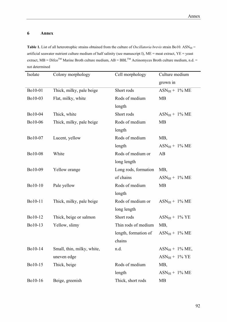
Annex
6 Annex Table 1. List of all heterotrophic strains obtained from the culture of Oscillatoria brevis strain Bo10. ASNIII =
artificial seawater nutrient culture medium of half salinity (see manuscript I), ME = meat extract, YE = yeast
extract, MB = DifcoTM Marine Broth culture medium, AB = BBLTM Actinomyces Broth culture medium, n.d. =
not determined
Isolate Colony morphology Cell morphology Culture medium
grown in
Bo10-01 Thick, milky, pale beige Short rods ASNIII + 1% ME
Bo10-03 Flat, milky, white Rods of medium
length
MB
Bo10-04 Thick, white Short rods ASNIII + 1% ME
Bo10-06 Thick, milky, pale beige Rods of medium
length
MB
Bo10-07 Lucent, yellow Rods of medium
length
MB,
ASNIII + 1% ME
Bo10-08 White Rods of medium or
long length
AB
Bo10-09 Yellow orange Long rods, formation
of chains
MB,
ASNIII + 1% ME
Bo10-10 Pale yellow Rods of medium
length
MB
Bo10-11 Thick, milky, pale beige Rods of medium or
long length
ASNIII + 1% ME
Bo10-12 Thick, beige or salmon Short rods ASNIII + 1% YE
Bo10-13 Yellow, slimy Thin rods of medium
length, formation of
chains
MB,
ASNIII + 1% ME
Bo10-14 Small, thin, milky, white,
uneven edge
n.d. ASNIII + 1% ME,
ASNIII + 1% YE
Bo10-15 Thick, beige Rods of medium
length
MB,
ASNIII + 1% ME
Bo10-16 Beige, greenish Thick, short rods MB
92
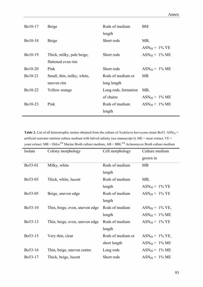
Annex
Bo10-17 Beige Rods of medium
length
BM
Bo10-18 Beige Short rods MB,
ASNIII + 1% YE
Bo10-19 Thick, milky, pale beige,
flattened even rim
Short rods ASNIII + 1% ME
Bo10-20 Pink Short rods ASNIII + 1% ME
Bo10-21 Small, thin, milky, white,
uneven rim
Rods of medium or
long length
MB
Bo10-22 Yellow orange Long rods, formation
of chains
MB,
ASNIII + 1% ME
Bo10-23 Pink Rods of medium
length
ASNIII + 1% ME
Table 2. List of all heterotrophic strains obtained from the culture of Nodularia harveyana strain Bo53. ASNIII =
artificial seawater nutrient culture medium with halved salinity (see manuscript I), ME = meat extract, YE =
yeast extract, MB = DifcoTM Marine Broth culture medium, AB = BBLTM Actinomyces Broth culture medium
Isolate Colony morphology Cell morphology Culture medium
grown in
Bo53-01 Milky, white Rods of medium
length
MB
Bo53-03 Thick, white, lucent Rods of medium
length
MB,
ASNIII + 1% YE
Bo53-05 Beige, uneven edge Rods of medium
length
ASNIII + 1% YE
Bo53-10 Thin, beige, even, uneven edge Rods of medium
length
ASNIII + 1% YE,
ASNIII + 1% ME
Bo53-13 Thin, beige, even, uneven edge Rods of medium
length
ASNIII + 1% YE
Bo53-15 Very thin, clear Rods of medium or
short length
ASNIII + 1% YE,
ASNIII + 1% ME
Bo53-16 Thin, beige, uneven centre Long rods ASNIII + 1% ME
Bo53-17 Thick, beige, lucent Short rods ASNIII + 1% ME
93
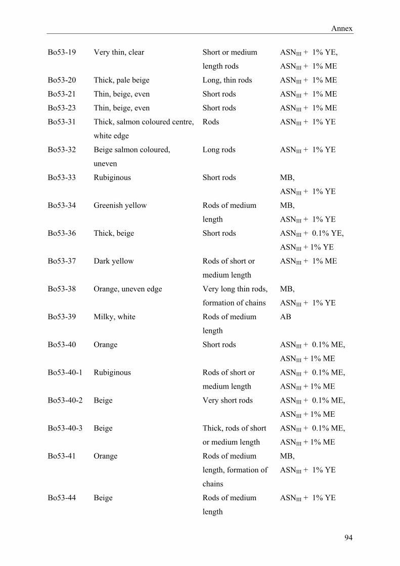
Annex
Bo53-19 Very thin, clear Short or medium
length rods
ASNIII + 1% YE,
ASNIII + 1% ME
Bo53-20 Thick, pale beige Long, thin rods ASNIII + 1% ME
Bo53-21 Thin, beige, even Short rods ASNIII + 1% ME
Bo53-23 Thin, beige, even Short rods ASNIII + 1% ME
Bo53-31 Thick, salmon coloured centre,
white edge
Rods ASNIII + 1% YE
Bo53-32 Beige salmon coloured,
uneven
Long rods ASNIII + 1% YE
Bo53-33 Rubiginous Short rods MB,
ASNIII + 1% YE
Bo53-34 Greenish yellow Rods of medium
length
MB,
ASNIII + 1% YE
Bo53-36 Thick, beige Short rods ASNIII + 0.1% YE,
ASNIII + 1% YE
Bo53-37 Dark yellow Rods of short or
medium length
ASNIII + 1% ME
Bo53-38 Orange, uneven edge Very long thin rods,
formation of chains
MB,
ASNIII + 1% YE
Bo53-39 Milky, white Rods of medium
length
AB
Bo53-40 Orange Short rods ASNIII + 0.1% ME,
ASNIII + 1% ME
Bo53-40-1 Rubiginous Rods of short or
medium length
ASNIII + 0.1% ME,
ASNIII + 1% ME
Bo53-40-2 Beige Very short rods ASNIII + 0.1% ME,
ASNIII + 1% ME
Bo53-40-3 Beige Thick, rods of short
or medium length
ASNIII + 0.1% ME,
ASNIII + 1% ME
Bo53-41 Orange Rods of medium
length, formation of
chains
MB,
ASNIII + 1% YE
Bo53-44 Beige Rods of medium
length
ASNIII + 1% YE
94

Annex
95
Bo53-45 Yellow orange, lucent Long thin rods,
formation of chains
MB,
ASNIII + 1% ME
Bo53-46 Beige Long rods ASNIII + 0.1% YE,
ASNIII + 1% YE
Bo53-47 Rubiginous Rods of medium or
short length
MB,
ASNIII + 1% ME
Bo53-49 White Thick short rods ASNIII + 1% YE

Acknowledgements
7 Acknowledgements
Several persons have contributed to the realisation of this doctoral dissertation. With these
words, I want to thank them all for their cooperation and support during the last years.
First of all, I like to thank Prof. Dr. Ulrich Fischer. I am grateful that he gave me the
opportunity to do a PhD in this interesting research field, that he provided the working space,
and for his helpfulness and support in all the years I have worked in his group.
I also thank Dr. Jens Harder for being my second reviewer and thesis committee member. He
also supported me for many years and was always receptive to all my problems.
Prof. Dr. Kai Bischof, who was member of my thesis committee and examiner, I would like to
thank as well. And I also address many thanks to the other members of my thesis defence
committee: Prof. Dr. Karl-Heinz Blotevogel, Jan Erik Rau, and Anne Schack.
Additionally, I would like to thank the committee of the International Max Planck Research
School of Marine Microbiology (MarMic) for accepting me as a member and thus giving me
the chance to enter the inspiring international and interdisciplinary environment of this
graduate school.
I particularly like to thank Dr. Birgit Heyduck-Söller for many, many hours of lively and
stimulating discussion and for patiently answering all my questions. Thanks for always being
there when I needed you.
But thanks are also due to the rest of the MMs, especially Birgit Lübben, who helped me to
retrain from working with algae to working with cyanobacteria, Tatjana Votteler, Christina
Zinnowitsch, and Clara Hoppe who helped me carrying out important parts of the practical
work for the third paper, Martina Stickan for patiently discussing all my absurd grammatical
questions, and all the others for the very pleasant working atmosphere and for many enjoyable
meetings in the “social corner”. Dr. Birgit Heyduck-Söller and Martina Stickan I would also
like to thank for proofreading my papers and my thesis.
Ein besonderer Dank gebührt meinen Eltern und Großeltern. Danke, dass Ihr immer für mich
da seid und mir Unterstützung bietet, wofür und wann immer ich sie brauche!
96
Erklärung gemäß § 6 Abs. 5 der Promotionsordnung der Universität Bremen für die
mathematischen, natur- und ingenieurwissenschaftlichen Fachbereiche
Hiermit versichere ich, dass ich die vorliegende Dissertation mit dem Titel „Molecular
Biological and Physiological Investigations of Heterotrophic Bacteria Associated with Marine
Filamentous Cyanobacteria“
1) ohne unerlaubte Hilfe angefertigt habe
2) keine anderen, als die von mir angegebenen Quellen und Hilfsmittel benutzt habe
und
3) die den benutzten Werken wörtlich oder inhaltlich entnommenen Stellen als solche
kenntlich gemacht habe.
Bremen, den 12. November 2009