Embed Size (px)
Citation preview
Molecular Neurobiology Copyright �9 The Humana Press, Inc. All rights of any nature whatsoever reserved. 0893-7648/92/6/1:75--86/$2.40
Molecular Biology of Alzheimer's Amyloid---Dutch Variant
Thomas Wisniewski 1,2," and Bias Frangione 2
Departments of 1Neurology and 2pathology, New York University Medical Center, 550 First Avenue, New York, NY 10016
Contents
Introduction A~ and Amyloid Precursor Protein Mutation in HCHWA-D and FAD AD and HCHWA-D, Two Sides of the Same Coin Effect of the Dutch Mutation on Fibfillogenesis HCHWA-D and Implications for the Origin of A[3 in AD Lessons from HCHWA-D References
Abstract
Hereditary cerebral hemorrhage with amyloidosis, Dutch type (HCHWA-D) (or familial cerebral amyloid angiopathy) and familial Alzheimer's disease (FAD) share several properties. Both are autosomal dominant forms of cerebral amyloidosis characterized by ~3-amyloid (AI3) deposition. In HCHWA-D the A~ is predominantly found in blood vessels and in early parenchymal plaques, whereas in AD parenchymal A~ deposits in the form of senile plaques and neurofibrillary tangles are a more prominent finding. Point mutations in the amyloid precursor protein (APP) have recently been described, in both conditions. A G to C transversion at codon 618 (extracellular portion of APP69s), producing a single amino acid substitution of glutamine instead of glutamine acid, occurs in HCHWA-D; whereas mutations at codon 642 in the intramembrane region of APP695 (phenyl- alanine, isoleucine, or glycine instead of valine) are associated with early onset FAD. This suggests that the site of particular mutations in the APP gene and the type of amino acid substitution in the APP holoprotein are more important in determining clincopathological phenotype and age at which A~ is deposited. Thus FAD and HCHWA-D can be regarded as two sides of the same coin.
Index Entries: Alzheimer's disease, ]3-amyloid, amyloid, familial Alzheimer's disease, Hereditary Cerebral Hemorrhage with Amyloidosis, Dutch type.
*Author to whom all correspondence and reprint requests should be addressed.
Molecular Neurobiology 75 Volume 6,1992
76 Wisniewski and Frangione
Introduction
Hereditary cerebral hemorrhage with amyl- oidosis, Dutch type (HCHWA-D) is a well char- acterized, albeit rare autosomal dominant form of cerebral amyloidosis (Wattendorf et al., I982; Luyendijk et al., 1988). Recent findings into the molecular biology and protein chemistry of this disease have provided insights into a closely re- lated and very common disorder, Alzheimer's disease (AD). HCHWA-D was first described in four large families from two coastal villages in the Netherlands. Three families (136 patients) were from Katwijk, and one family (14 patients) was from Scheveningen (Wattendorf et al., 1982; Luyendijk et al., 1988). More than 500 individu- als are at risk for developing the disease in Hol- land (Harm et al., 1989).
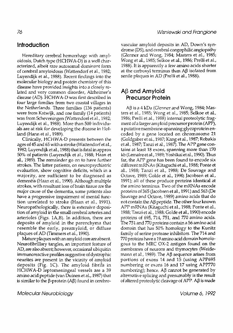
Clinically, HCHWA-D presents between the ages of 45 and 65 with a stroke (Wattendorf et al., 1982; Luyendijk et al., 1988) that is fatal in approx 50% of patients (Luyendijk et al., 1988; Haan et al., 1989). The remainder go on to have further strokes. The latter patients, on neuropsychiatric evaluation, show cognitive deficits, which in a major i ty , are suff ic ient to be d iagnosed as dementia (Haan et al., 1990). Although multiple strokes, with resultant loss of brain tissue are the major cause of the dementia, some patients also have a progressive impairment of mental func- tion unre la ted to stroke (Haan et al., 1991). Neuropathologically, there is extensive deposi- tion of amyloid in the small cerebral arteries and arterioles (Figs. 1A,B). In addition, there are deposits of amyloid in the parenchyma that resemble the early, p reamylo id , or diffuse plaques of AD (Timmers et al., 1990).
Mature plaques with an amyloid core are absent. Neurofibrillary tangles, an important feature of AD, are also absent; however, occasional ubiquitin immunoreactive profiles suggestive of dystrophic neurites are present in the vicinity of amyloid depos i t s (Fig. 1C). The amyto id fibrils in HC HWA -D leptomeningeal vessels are a 39 amino acid peptide (van Duinen et al., 1987) that is similar to the ~-protein (A~) found in cerebro-
vascular amyloid deposits in AD, Down's syn- drome (DS), and cerebral congophilic angiopathy (Glenner and Wong, 1984; Masters et al., 1985; Wong et al., 1985; Selkoe et al., 1986; PreUi et al., 1988). It is apparently a few amino acids shorter at the carboxyl terminus than A[3 isolated from senile plaques in AD (Prelli et al., 1988).
and Amyloid Precursor Protein
A[3 is a 4 kDa (Glenner and Wong, 1984; Mas- ters et al., 1985; Wong et al., 1985; Selkoe et al., 1986; Prelli et al., 1988) internal proteolytic frag- ment of a larger amyloid precursor protein (APP), a putative membrane-spanning glycoprotein en- coded by a gene located on chromosome 21 (Goldgaber et al., 1987; Kang et al., 1987; Robakis et al., 1987; Tanzi et al., 1987). The APP gene con- tains at least 18 exons, spanning more than 170 kb (Lemaire et al., 1989; Yoshikai et al., 1990). Thus far, the APP gene has been found to encode six different mRNAs (Kitaguchi et al., 1988; Ponte et al., 1988; Tanzi et al., 1988; De Sauvage and Octave, 1989; Golde et al., 1990; Jacobsen et al., 1991); all of these produce proteins identical at the amino terminus. Two of the mRNAs encode proteins of 365 (Jacobsen et al., 1991) and 563 (De Sauvage and Octave, 1989) amino acids that do not contain the A~3 peptide. The other four known APP mRNAs (Kitaguchi et al., 1988; Ponte et al., 1988; Tanzi et al., 1988; Golde et al., 1990) encode proteins of 695, 714, 751, and 770 amino acids. The 751 and 770 proteins contain a 56 amino acid domain that has 50% homology to the Kunitz family of serine protease inhibitors. The 714 and 770 proteins have a 19 amino acid domain homolo- gous to the MRC OX-2 antigen found on the membranes of neurons and thymocytes (Weide- mann et al., 1989). The AI3 sequence arises from port ions of exons 14 and 15 (using APP695 number ing or exons 16 and 17 using APP770 numbering); hence, A~ cannot be generated by alternative splicing and presumably is the result of altered proteolytic cleavage of APP. A]3 is made
Molecular Neurobiology Volume 6, 1992
Alzheimer's Amyloid--Dutch Variant 77
A o
i/-
"~ :" : t , % ,
: . : ; . . . . ,0 : Q
�9 ' ~ ~.
Fig. 1. Sections of frontal cortex from a HCHWA-D patient. A- The section was lmmunostained with polyclonal anti- bodies raised against the first 28 residues of A J3 (anti-SP28). A~ can be seen in leptomeningeal vessels (arrow), parenchymal vessel walls, parenchyma surrounding amyloid-laden vessels (large arrowhead), and preamyloid or diffuse plaques (small arrowhead). B: Higher magnification of amyloid-laden vessel wall surrounded by amyloid extending into the par- enchyma (arrow) and a diffuse plaque (arrowhead), immunostained with anti-SP28. C: Immunostaining with polyclonal antibodies to ubiquitin of amyloid-laden vessel. Ubiquitin-reactive profiles (arrowhead) suggestive of dystrophic neurite processes surround the vessel.
up of a membrane-spanning domain (11-14 resi- dues) and part of the predicted extracellular domain region adjacent to the membrane (28 residues) (Kang et al., 1987; Dyrks et al., 1988; Fig. 2).
Mutation in HCHWA-D and FAD
Recently, HCHWA-D has been found to seg- regate with a mutation in the APP gene at nucle- otide 1852 in exon 15 (APP695 numbering), where
Molecular Neurobiology Volume 6,1992
78 Wisniewski and Frangione
cDNA APP770
cDNA APP695
AMYLOID
672
597 /,
693
618 J.
2 2
717
642
" , . _ . 0
Gin Ile, Phe, Gly
STROKE DEMENTIA
HCHWA-D FAD
VASCULOTROPIC NEUROTROPIC MUTANT MUTANT
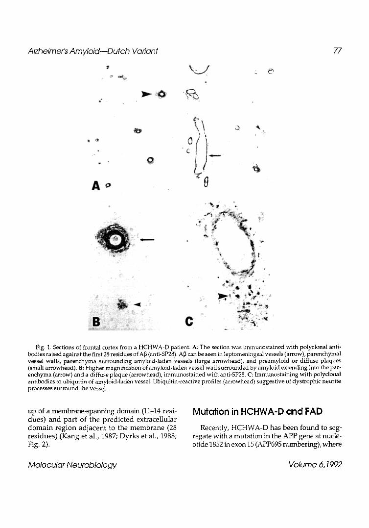
Fig. 2. Illustration of the A~ sequence of APP with its relationship to the cell membrane. Codon numbering according to both APP770 and APP695 is given on the top. At codon 22, within the ectodomain of A[~ () , the glutamic acid to glutamine substitution found in HCHWA-D is shown. At codon 642 of APP695, the FAD mutations of ( ) valine to either isoleucine, phenylalanine, or glycine is shown. Mutations in the APP at these two sites are either neurotropic with predominantly senile plaque and neurofibrillary tangle formation or vascultropic with mainly congophilic angiopathy.
guanine is replaced by cytosine (Levy et aL, 1990). The latter causes a single amino acid substitution of glutamine instead of glutamic acid at codon 618 of APP 695 and corresponding to residue 22 of the A]3 (Fig. 2). The significance of this muta- tion is corroborated by close linkage, with a Lod score of 7.59, between the APP gene and the dis- ease (Van Broeckhoven et al., 1990).
Furthermore, this mutation has been found in all Dutch amyloidosis-affected patients tested so far (Bakker et al., 1991; Fernandez-Madrid et al., 1991). The finding of a mutation in the APP gene in HCHWA-D sparked off a renewed search for a mutation in AD. Analysis of numerous familial AD (FAD) pedigrees over the last 4 yr had shown them to be genetically heterogenous. Some fami- lies with early onset symptoms had linkage to chromosome 21 (Pericak-Vance et al., 1988; St George-Hyslop and Members of the FAD Col- laborative Study Group, 1990), whereas others with late onset or early onset did not (Goate et al., 1989; St. George -Hys lop et al., 1990; Schellenberg et al., 1991).
Recently, a mutation was found in some early onset FAD families (Goate et al., 1991), where there is a C to T transition at nucleotide 1924 in exon 15 (APP69 s numbering), causing a valine to isoleucine change at codon 642 (Fig. 2). This is three to four residues away from the known car- boxyl terminus of the A]3, obtained from senile plaques. This mutation was originally found in two out of 16 families with early onset FAD and was not detected in 100 normal unrelated indi- viduals or from nine different families with late onset FAD (Goate et al., 1991). This mutation has also been found in several Japanese early onset FAD families (Yoshioka et al., 1991; Naruse et al., 1991) and one French FAD family (Lucotte et al., 1991), indicating segregation of the mutation with FAD in racially different populations. However, this mutation is relatively rare even among early onset FAD families (Schellenberg et al., 1991). Two further mutations at codon 642 have been reported in different families, where the valine is replaced by either phenytalanine (Murrell et al., 1991) or glycine (Chartier-Harlin et al., 1991).
Molecular Neurobiology Volume 6, 1992
Alzheimer's Amyloid--Dutch Variant 79
These reports of mutations in the APP gene show that mutations at different sites can result in a clinical picture that is qualitatively and quan- titatively distinctive. It appears that some muta- tions in the APP gene are neurotropic, with prominent senile plaque and neurofibrillary tangle formation, and others are vasculotropic, with deposition of amyloid predominantly in blood vessels, as in HCHWA-D. This segregation of APP mutations with two related syndromes implies that APP mutations are likely to be responsible for different ]3-amyloid-related disease phenotypes.
AD and HCHWA-D, Two Sides of the Same Coin
The finding of a mutation in the APP gene in both HCHWA-D (Levy et al., 1990) and at least some FAD cases (Goate et al., 1991; Lucotte et al., 1991; Naruse et al., 1991; Murrell et al., 1991; Chartier-Harlin et al., 1991) raises the question of whether these are really two separate disorders or different faces of a similar pathological process. The neuropathological features of AD (Probst et al., 1991) include senile plaques (Simchowicz, 1911) and neurofibri l lary tangles (NFTs) (Alzheimer, 1907), as well as in more than 90% of cases, amyloid deposits in cerebral blood vessels (Glenner et al., 1981; Joachim et al., 1986), similar to that seen in HCHWA-D. In AD, NFTs are thought by many investigators to be a second- ary change (Wisn iewski K., et al., 1979; Wisniewski H. M., et al., 1989), indicating a pathological response of neurons to A]3 deposi- tion. This is suggested by data from DS patients, where AD changes are noted very early in life. When DS brains are examined at various ages, it is the senile plaques that are the first change (Giaccone et al., 1989; Mann, 1989; Motte and Williams, 1989; Rumble et al., 1989). Neurofibril- lary tangles occur in diverse neuropathological conditions, such as subacute sclerosising panen- cephalitis, postencephalitic Parkinson's disease,
dementia pugaListica, and supranuclear palsy (Wisniewski et al., 1979); thus, they appear to be part of the neuron ' s l imi ted reper to i re of responses to injury. HCHWA-D patients often die early in life as a result of their first stroke (Wattendorf et al., 1982; Luyendijk et al., 1988; Haan et al., 1991). A majority of those that survive also become demented (Haan et al., 1990). It is not known whether these individuals would develop NFTs if they lived long enough. Indeed, immunohistochemical studies with ubiquitin an- tibodies suggest the presence of dystrophic neuritic processes, which accompany NFTs in AD, evident in the vicinity of the preamyloid and diffuse plaques of HCHWA-D patients (Fig. 1C).
Effect of the Dutch Mutation on Fibrillogenesis
The finding of a point mutation within the APP in HCHWA-D raises the question of how a sub- stitution of glutamic acid for glutamine at codon 618 could promote fibrillogenesis. Significantly, in HCHWA-Icelandic type, another autosomal dominant form of cerebral arnyloidosis (Hann et al., 1989), there is also a point mutation in the precursor protein's gene (Levy et al., 1989; Palsdottir et al., 1988) resulting in the substitution of an amino acid for glutamine (Ghiso et al., 1986). The neuropathological picture in the Icelandic arnyloidosis is similar to the Dutch (Hann et al., 1989), although the precursor molecule in the former is a variant of cystatin C (Ghiso et al., 1986; Levy et al., 1989; Palsdottir et al., 1988), an inhibi- tor of cyteine proteinase. Recent evidence indi- cates that the single amino acid substitution at residue 22 of A[3 alters its fibrillogenic proper- ties. Prior studies have shown that synthetic pep- tides corresponding to residues 1-28 of A~, form Congo Red-positive fibrils in vitro under physi- ological conditions (Castafio et al., 1986; Kirschner et al., 1987).
The presence of the glutamine at residue 22 accelerates this fibril formation (Wisniewski et al.,
Molecular Neurobiology Volume 6,1992
80 Wisniewski and Frangione
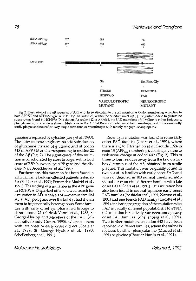
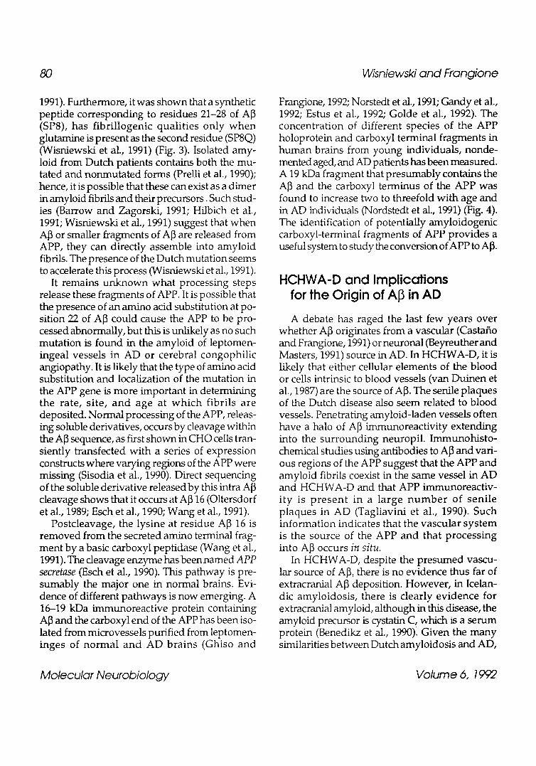
1991). Furthermore, it was shown that a synthetic peptide corresponding to residues 21-28 of A~ (SP8), has fibril logenic qualit ies only w h e n glutamine is present as the second residue (SP8Q) (Wisniewski et al., 1991) (Fig. 3). Isolated amy- loid from Dutch patients contains both the mu- tated and nonmutated forms (Prelli et al., 1990); hence, it is possible that these can exist as a dimer in amyloid fibrils and their precursors. Such stud- ies (Barrow and Zagorski, 1991; Hilbich et al., 1991; Wisniewski et al., 1991) suggest that when A]3 or smaller fragments of A[3 are released from APP, they can directly assemble into amyloid fibrils. The presence of the Dutch mutation seems to accelerate this process (Wisniewski et al., 1991).
It remains unknown what processing steps release these fragments of APP. It is possible that the presence of an amino acid substitution at po- sition 22 of A]3 could cause the APP to be pro- cessed abnormally, but this is unlikely as no such mutation is found in the amyloid of leptomen- ingeal vessels in AD or cerebral congophilic angiopathy. It is likely that the type of amino acid substitution and localization of the mutation in the APP gene is more important in determining the rate, site, and age at which fibrils are deposited. Normal processing of the APP, releas- ing soluble derivatives, occurs by cleavage within the AI3 sequence, as first shown in CHO ceils tran- siently transfected with a series of expression constructs where varying regions of the APP were missing (Sisodia et al., 1990). Direct sequencing of the soluble derivative released by this intra A[3 cleavage shows that it occurs at A~ 16 (Oltersdorf et al., 1989; Esch et al., 1990; Wang et al., 1991).
Postcleavage, the lysine at residue A[3 16 is removed from the secreted amino terminal frag- ment by a basic carboxyl peptidase (Wang et al., 1991). The cleavage enzyme has been named APP secretase (Esch et al., 1990). This pathway is pre- sumably the major one in normal brains. Evi- dence of different pathways is now emerging. A 16-19 kDa immunoreactive protein containing A]3 and the carboxyl end of the APP has been iso- lated from microvessels purified from leptomen- inges of normal and AD brains (Ghiso and
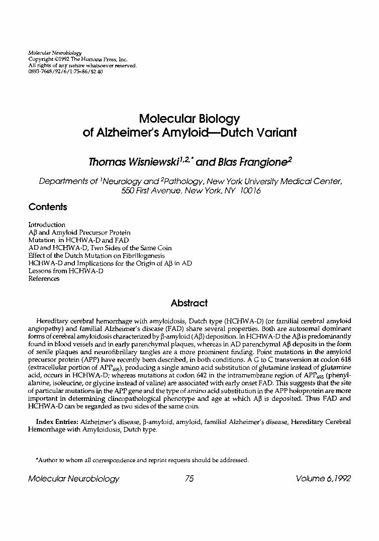
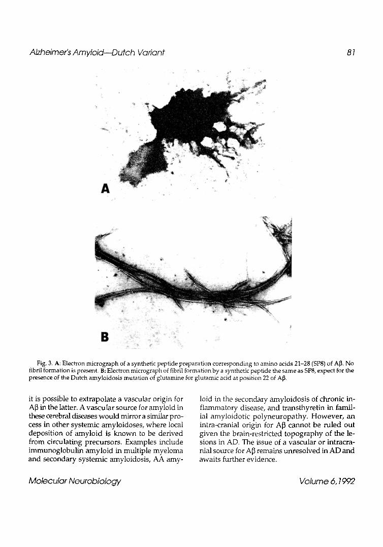
Frangione, 1992; Norstedt et al., 1991; Gandy et al., 1992; Estus et al., 1992; Golde et al., 1992). The concentration of different species of the APP holoprotein and carboxyl terminal fragments in human brains from young individuals, nonde- mented aged, and AD patients has been measured. A 19 kDa fragment that presumably contains the A]3 and the carboxyl terminus of the APP was found to increase two to threefold with age and in AD individuals (Nordstedt et al., 1991) (Fig. 4). The identification of potentially amyloidogenic carboxyl-terminal fragments of APP provides a useful system to study the conversion of APP to A]3.
HCHWA-D and Implications for the Origin of in AD
A debate has raged the last few years over whether A]3 originates from a vascular (Castafio and Frangione, 1991) or neuronal (Beyreuther and Masters, 1991) source in AD. In HCHWA-D, it is likely that either cellular elements of the blood or cells intrinsic to blood vessels (van Duinen et al., 1987) are the source of A]3. The senile plaques of the Dutch disease also seem related to blood vessels. Penetrating amyloid-laden vessels often have a halo of A]3 immunoreactivity extending into the surrounding neuropil. Immunohis to- chemical studies using antibodies to A]3 and vari- ous regions of the APP suggest that the APP and amyloid fibrils coexist in the same vessel in AD and HCHWA-D and that APP immunoreactiv- ity is p resen t in a large n u m b e r of seni le plaques in AD (Tagliavini et al., 1990). Such information indicates that the vascular system is the source of the APP and that processing into A[3 occurs in situ.
In HCHWA-D, despite the presumed vascu- lar source of A[3, there is no evidence thus far of extracranial A]3 deposition. However, in Icelan- dic amyloidosis, there is clearly evidence for extracranial amyloid, although in this disease, the amyloid precursor is cystafin C, which is a serum protein (Benedikz et al., 1990). Given the many similarities between Dutch amyloidosis and AD,
Molecular Neurobiology Volume 6, 1992
Alzheimer's Amyloid--Dutch Variant 81
2."
A
. -~ . " ~.;~' . i
B
Fig. 3. A: Electron mlcrograph of a synthetic peptide preparation corresponding to amino acids 21-28 (SP8) of AI3. No fibril formation is present. B: Electron micrograph of fibril formation by a synthetic peptide the same as SP8, expect for the presence of the Dutch amyloidosis mutation of glutamine for glutamic acid at position 22 of A~.
it is possible to extrapolate a vascular origin for A~3 in the latter. A vascuIar source for amyloid in these cerebral diseases would mirror a similar pro- cess in other systemic amyloidoses, where local deposition of amyloid is known to be derived from circulating precursors. Examples include immunoglobulin amyloid in multiple myeloma and secondary systemic amyloidosis, AA amy-
loid in the secondary amyloidosis of chronic in- flammatory disease, and transthyretin in famil- ial amyloidotic polyneuropathy. However, an intra-cranial origin for A~3 cannot be ruled out given the brain-restricted topography of the le- sions in AD. The issue of a vascular or intracra- nial source for A~3 remains unresolved in AD and awaits further evidence.
Molecular Neurobiology Volume 6,1992
82 Wisniewski and Frangione
L J
' 16.19 k_
AB
A non-A
i n l m e m b r a n e
ol
NEXIN I I
~ 0
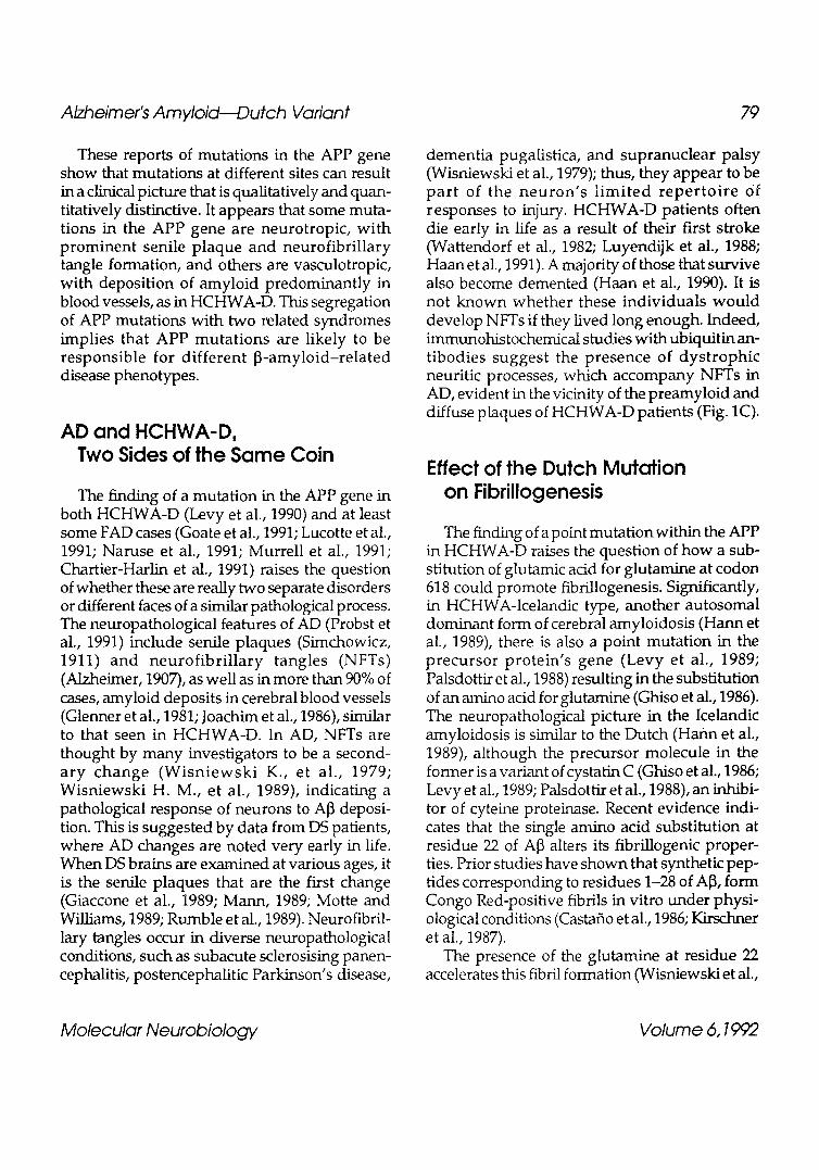
Fig. 4. Alternative processing of AD APP in vessel walls. The APP (center of figure) can be processed in at least two ways. The nonamyloidogenic pathway (non-A) is presumably the major normal route, with cleavage occurring within the A~ sequence. This normal cleavage of APP releases soluble derivatives (Nexin lJ) and leaves a small 10 kDa membrane fragment. This pathway cannot produce A~. The amyloidogenic pathway (A) is presumably very minor under normal conditions. Here cleavage occurs closer to the amino.terminal of A[~, resulting in a larger 16-19 kDa membrane fragment that contains the entire A~ sequence. Alternation of the normal processing of this fragment can release the fibrillogenic A[~.
Lessons from H C H W A - D
HCHWA-D provides a powerful example of how the study of a rare disorder provides lessons about more common disorders. The finding of a point mutation in Dutch amyloidosis (Levy et al., 1990) and in a subset of early onset FAD patients (Goate et al., 1991; Naruse et al., 1991; Yoshioka et al., 1991; Murrell et al., 1991; Chartier-Harlin et al., 1991) indicate that mutations at different sites of the APP can produce distinctive pathological presentations. However, the A~ fibril am also be deposited in leptomeningeal vessels without the presence of the Dutch mutation, albeit at a later age, in AD and sporadic cerebral amyloid angiopathy (CAA). It is likely that multiple factors can influ- ence A~ deposition, including a mutation in the APP gene, posttranslational modification and/ or other still-unknown extrinsic factors.
The recent finding of different processing path- ways for APP in AD and aged brains raises the interesting question of how the Dutch mutation influences channeling of APP metabolism down each pathway. It can be predicted that Dutch amyloid mutation containing APP wiUbe catabo- lized more rapidly, as occurs in Icelandic amy- loid, where levels in cerebrospinal fluid (CSF) of cystatin C are reduced by approx 1/2 in affected individuals (Grubb and Lofberg, 1985). Further studies are under way in transgenic mice and cells lines transfected with various transcripts contain- ing the Dutch mutation to evaluate their amy- loid fibril potential. The clear delineating of the causative mutation in HCHWA-D can illuminate the origin of amyloid in AD and its influence in the aging process (Coria et al., 1987). Interven- tions inhibiting amyloid deposition can then be tested, with resultant therapeutic benefits.
Molecular Neurobiology Volume 6, 1992
Alzheimer's Amyloid---Dutch Variant 83
References
Alzheimer A. (1907) Uber eine eigenartige Erkran- kung der Hirnrinde. Allg. Z. Psychiat. Psych. Gerichte. Med. 64, 146-148.
Bakker E., van Broeckhoven C., Haan J., Voorhoeve E., van Hul W., Levy E., Lieberburg I., Carman M. D., van Ornmen G. J. B., Frangione B., and Roos R. A. C. (1991) DNA diagnosis for hereditary cerebral hemorrhage with amyloidosis (Dutch type). Am. J. Hum. Genet. 9, 518-521.
Barrow C. J. and Zagorski M. G. (1991) Solution struc- tures of 13 peptide and its constituent frag-ments: relation to arnyloid deposition. Science 253,179-182.
Benedikz E., Blondal H., and Gudmundsson G. (1990) Skin deposits in hereditary cystatin c amyloidosis. Virchows Archly A 417, 325-331.
Beyreuther K. and Masters C. L. (1991) Amyloid precursor protein (APP) and ]3A4 amyloid in the etiology of Alzheimer's disease: Precursor-product relationships in the derangement of neuronal function. Brain Pathol. 1, 241-251.
Casta~o E., Ghiso J., Prelli F., Gorevic P., Migheli A., and Frangione B. (1986) In vitro formation of amyloid fibrils from two synthetic peptides of different lengths homologous to Alzheimer's disease ~-protein. Biochem. Biophys. Res. Commun. 141, 782-789.
Casta~o E. M. and Frangione B. (1991) Alzheimer's disease from the perspective of the systemic and localized forms of amyloidosis. Brain Pathol. 1, 263-271.
Chartier-Harlin M. C., Crawford F., Houlden H., Warren A., Hughes D., Fidani L., Goate A., Rosson M., Roques P., Hardy J., and Mullan M. (1991) Early-onset Alzheimer's disease caused by mutations at codon 717 of the [3-amyloid precursor protein gene. Nature 353, 844-846.
Coria F., Casta_6o E. M., and Frangione B. (1987) Brain amyloid in normal aging and cerebral amyloid angiopathy is antigenically related to Alzheimer's disease beta-protein. Am. J. Pathol. 129, 422-429.
De Sauvage F. and Octave J..N. (1989) A novel mRNA of the A4 amyloid precursor gene coding for a possibly secreted protein. Science 245, 651-653.
Dyrks T., Weidemann A., Multhaup G., Salbaum J. M., Lemaire H.-G., Kang J., M(iller-Hill B., Masters C. L., and Beyreuther K. (1988) Identification, transmembrane orientation and biogenesis of the amyloid A4 precursor of Alzheimer's disease. EMBO J. 7, 949-957.
Esch F. S., Keim P. S., Beattie E. C., Blacker R. W., Culwell A. K., Oltersdorf T., McClure D., and Ward P. J. (1990) Cleavage of amyloid 13 peptide during constitutive processing of its precursor. Science 248, 1122-1124.
Estus S., Golde T. E., Kunishita T., Blades D., Lowery D., Eisen M., Usiak M., Qu X., Tabira T., Greenberg B. D., and Younkin S. G. (1992) Potentially amyloid- ogenic carboxyl-terminal derivates of the amyloid protein precursor. Science 255, 726-728.
Fernandez-Madrid I., Levy E., Marder K., and Frangione B. (1991) Codon 618 variant of Alzheimer amyloid gene associated with inherited cerebral hemorrhage. Ann. Neurol. 30, 730-733.
Gandy S. E., Bhasin R., Ramabhadran T. V., Koo E. H., Price D. L., Goldgaber D., and Greengard P. (1992) Alzheimer 13/A4-amyloid precursor protein: evidence for putative amyloidogenic fragment. J. Neurochem. 58, 383-386.
Ghiso J., Jensson O., and Frangione B. (1986) Amyloid fibrils in hereditary cerebral hemorrhage with amyloidosis of Icelandic type is a variant of gamma-trace basic protein (cystatin c). Proc. Natl. Acad. Sci. USA 83, 2974-2978.
Ghiso J. and Frangione B. (1992) Alternative pro- cessing pathway of Alzheimer's amyloid precursor protein in cerebral vessel walls (unpublished observations).
Giaccone G., Tagliavini F., Linoli G., Bouras C., Frigerior L., Frangione B., and Bugiani O. (1989) Down patients: extracellular preamyloid deposits precede neuritic degeneration and senile plaques. Neurosci. Lett. 97, 232-238.
Glenner G. G., Henry J. H., and Fujihara S. (1981) Congophilic angiopathy in the pathogenesis of Alzheimer's degeneration. Ann. Pathol. 1,120-129.
Glenner G. G. and Wong C. W. (1984) Alzheimer's disease and Down's syndrome: sharing of a unique cerebrovascular amyloid fibril protein. Biochem. Biophys. Res. Commun. 122, 1131-1135.
Goate A., Chartier-Harlin M.-C., Mullan M., Brown J., Crawford F., Fidani L., Giuffra L., Haynes A., Irving N., James L., Mant R., Newton P., Rooke K., Roques P., Talbot C., Pericak-Vance M., Roses A., Williamson R., Rossor M., Owen M., and Hardy J. (1991) Segregation of a missense mutation in the amyloid precursor protein gene with familial Alzheimer's disease. Nature 349, 704-706.
Goate A., Owen M. J., James L. A., Mullan M. J., Rossor M. N., Haynes A. R., Farrall M., Lai L. Y. C., Roques P., Williamson R., and Hardy J. A. (1989)
Molecular Neurobiology Volume 6,1992
84 Wisniewski and Frangione
Predisposing locus for Alzheimer's disease on chromosome 21. Lancet 1, 352-355.
Golde T. E., Estus S., Usiak M., Younkin L. H., and Younkin S. Go (1990) Expression of ]3 amyloid protein precursor mRNAs: recognition of a novel alternatively spliced form and quantitation in Alzheimer's disease using PCR. Neuron 4, 253-267.
Golde T. E., Estus S., Younkin L. H., Selkoe D. J., and Younkin S. G. (1992) Processing of the amyloid protein precursor to potentially amyloidogenic derivatives. Science 255, 728-730.
Goldgaber D., Lerman M. J., McBride O. W., Saffiotti U., and Gajdusek D. C. (1987) Characterization and chromosomal localization of a cDNA encoding brain amyloid of Alzheimer's disease. Science 235, 877--88O.
Grubb A. and Lofberg H. (1985) Human gamma trace structure, function and clinical use of concentration measurements . Scand. J. Clin. Lab. Invest. 45 (Suppl. 177), 7-13.
Gudmundsson G., Hallgrimsson J., Jonasson T. A., and Bjarnason O. (1972) Hereditary cerebral hemor- rhage with amyloidosis. Brain 95, 387-404.
Haan J., Roos R. A. C., Briet P. E., Herpers M. H. J. M., Luyendijk W., Bots G. T. A. M., and the Research Group on Hereditary Cerebral Amyloid-Angio- pathy (1989) Hereditary cerebral hemorrhage with amyloidosis--Dutch type. Clin. Neurol. Neurosurg. 91, 285-290.
Haan J., Lauser J. B. K., Zijderveld I., vander Does I. G. F., and Roos R. A. C. (1990) Dementia in her- editary cerebral hemorrhage with amyloidosis- Dutch type. Arch. Neurol. 47, 965-967.
Haan J., Hardy J. A., and Roos R. A. C. (1991) Hereditary cerebral hemorrhage with amyloidosis- Dutch type: its importance for Alzheimer research. Trends in Neuroscience 14(No. 6), 231-234.
Hilbich C., Kisters-Weike B., Reed J., Masters C. L., and Beyreuther K. (1991) Aggregation and secondary structure of synthetic amyloid [3A4 peptides of Alzheimer's disease. I. Mol. Biol. 218, 149-163.
Jacobsen J. S., Muerkel H: A., Blume A. J., and Vitek M. P. (1991) A novel species specific RNA related to alternatively spliced amyloid precursor protein mRNAs. Neurobiol. Aging 12, 575-583.
Joachim C. L., Morris J., and Selkoe D. J. (1986) Autopsy neuropathology in 76 cases of clinically diagnosed Alzheirner's disease. Neurology 36 (Suppl. 1), 226.
Kang J., Lemaire H. G., Unterbeck A., Salbaum J. M., Masters C. L~ Grzeschik K. H., Malthaup G., Beyreuther K., and Mtiller-Hill B. (1987) The pre- cursor of Alzheimer's disease amyloid A4 protein resembles cell surface receptor. Nature 325, 733-736.
Kirschner D. A., Inouye H., Duffy L., Sinclair A., Lind M., and Selkoe D. J. (1987) Synthetic peptide homologous to [3 protein from Alzheimer disease forms amyloid-like fibrils in vitro. Proc. Natl. Acad. Sci. USA 84, 6953-6957.
Kitaguchi N., Takahashi Y., Tokushima Y., Shiojuiri S., and Ito H. (19~) Novel precursor of Alzheimer's disease arnyloid protein shows protease inhibitory activity. Nature 331, 530-532.
Lernaire H. G., Salbaum J. M., Multhaup G., Kang J., Bayney R. M., Unterbeck A., Beyreuther K., and M6ller-Hill B. (1989) The Pre A4695 precursor protein of Alzheirr~Vs disease A4 amyloid is encoded by 16 exons. Nucleic Acids Res. 17, 517-522.
Levy E., Lopez-Otin C., Ghiso J., Geltner D., and Frangione B. (1989) Stroke in Icelandic patients with hereditary amyloid angiopathy is related to a mutation in the cystatin C gene, an inhibitor of cysteine proteases. J. Exp. Med. 169, 1771-1778.
Levy E., Carman M. D., Fernandez-Madrid I. J., Power M. D., Lieberburg I., van Duinen S. G., Bots G. Th. A. M., Luyendijk W., and Frangione B. (1990) Mutation of the Alzheimer's disease amyloid gene in hereditary cerebral hemorrhage, Dutch type. Science 248, 1124-1126.
Lucotte G., Berriche S., and David F. (1991) Alzheimer's mutation. Nature 351, 530.
Luyendijk W., Bots G. T. A. M., Vegter-van der Vlis M., Went L. N., and Frangione B. (1988) Hereditary cerebral haemorrhage caused by cortical amyloid angiopathy. J. Neurol. Sci. 85, 267-280.
Mann D. M. A. (1989) Cerebral amyloidosis, aging and Alzheimer's disease: a contribution from studies on Down's syndrome. Neurobiol. Aging 10, 397-399.
Masters C. L., Simms G., Weinman N. A., Multhaup G., McDonald B. L., and Beyreuther K. (1985) Amyloid plaque core protein in Alzheimer disease and Down syndrome. Proc. Natl. Acad. Sci. USA 82, 4245-4249.
Motte J. and Williams R. S. (1989) Age-related changes in the density and morphology of plaques and neurofibrillary tangles in Down syndrome brain. Acta Neuropathol. 77, 535-546.
Murrell J., Farlow M., Ghetti B., and Benson M. D. (1991) A mutation in the amyloid precursor protein
Molecular Neurobiology Volume 6, 1992
Alzheimer's Amyloid--Dutch Variant 85
associated with hereditary Alzheimer's disease. Science 254, 97-99.
Naruse S., Igarashi S., Kobayashi H., Aoki K., Inuzuka T., Kaneko K., Shimizu T., Iihara K., Kojima T., Miyatake T., and Tsuji S. (1991) Mis-sense mutation Val -4 Ile in exon 17 of amyloid precursor gene in Japanese familial Alzheimer's disease. Lancet 337, 978,979.
Nordstedt C., Gandy S. E., Mafuzoff I., Caporaso G. L., Iverfeldt K., Grebb J. A., Winblad B., and Greengard P. (1991) Alzheimer /A4 amyloid precursor protein in human brain: aging asso- ciated increases in holoprotein and in a proteolytic fragment. Proc. Natl. Acad. Sci. USA 88, 8910- 8914.
Oltersdorf T., Ward P. J., Henrikkson T., Beattie E. C., Neve R., Lieberburg I., and Fritz L. C. (1989) The Alzheimer amyloid precursor protein: Identi- fication of a stable intermediate in the biosynthetic / degradative pathway. J. Biol. Chem. 265, 4492-4497.
Palsdottir A., Abrahamson M., Thorsteinsson L., Amason A., Olafsson I., Grubb A., and Jensson O. (1988) Mutation in cystatin C gene causes hered- itary brain haemorrhage. Lancet 2, 603,604.
Pericak-Vance M., Yamaoka L. H., Haynes C. S., Speer M. C., Haines J. L., Gaskell P. C., Hung W.-Y., Clark C. M., Heyman A. L., Trofatter J. A., Eisenmenber J. P., Gilbert J. R., Lee J. E., Alberts M. J., Dawson D. V., Bartlett R. J., Earl N. L., Siddique T., Vance J. M., Conneally P. M., and Roses A. D. (1988) Genetic linkage studies in Alzheimer's disease families. Exp. Neurol. 102, 271-279.
Ponte P., Gonzales-DeWhitt P., Schilling J., Miller J., Hsu D., Greenberg B., Davis K., Wallace W., Leiberburg I., Fuller F., and Cordell B. (1988) A new A4 amy lo id m R N A contains a d o m a i n homologous to serine protease inhibitors. Nature 331, 525-527.
Prelli F., Castano E. M., van Duinen S. G., Bots G. T., Luyendijk W., and Frangione B. (1988) Different processing of Alzheimer's beta-protein precursor in the vessel wall of patients with hereditary cerebral hemorrhage with amyloidosis, Dutch type. Biochem. Biophys. Res. Commun. 151, 1150-1155.
Prelli F., Levy E., van Duinen S. G., Bots G. Th. A. M., Luyendijk W., and Frangione B. (1990) Expression of a normal and variant Alzheimer's beta-protein gene in amyloid of hereditary cerebral hemorrhage, Dutch type, DNA and protein diagnostic assays. Biochem. Biophys. Res. Commun. 170, 301-307.
Prelli F., Castano E. M., Glermer G. G., and Frangione B. (1988) Differences between vascular and plaque core amyloid in Alzheimer's disease. I. Neurochem. 51, 648--651.
Probst A., Langui D., and Ulrich J. (1991) Alzheimer's disease: a description of the structural lesions. Brain Pathol. 1, 229-239.
Robakis N. K., Wisniewski H. M., Jenkins E. C., Devine-Gage E. A., Houck G .E., Yao X.-L., Ramakrishna N., Wolfe G., Silverrnan W. P., and Brown W. T. (1987) Chromosome 21q21 sublocaliz- ation of gene encoding ]3-amyloid peptide in cere- bral vessels and neuritic (senile) plaques of people with Alzheimer's disease and Down syndrome. Lancet 1, 384,385.
Rumble B., Retallack R., Hilbich C., S imms G., Multhaup G., Martins R., Hockey A., Montgo- mery P., Beyreuther K., and Masters C. L. (1989) Amyloid A4 protein and its precursor in Down's syndrome and Alzheimer's disease. N. Engl. J. Med. 320, 1446-1452.
Schellenberg G. D., Anderson L., O'Dahl S., Wisjman E. M., Sadovnick A. D., Ball M. J., Larson E. B., Kukull W. A., Martin G. M., Roses A. D., and Bird T. D. (1991) APP717, APP693 and PRIP gene muta- tions are rare in Alzheimer's disease. Am. J. Hum. Genet. 49, 511-517.
Schellenberg G. D., Bird T. D., Wijsman E. M., Moore D. K., Boehnke M., Bryant E. M., Lampe T. H., Nochlin D., Sumi S. M., Deeb S. S., Beyreuther K., and Martin G.M. (1988) Absence of linkage of chromosome 21q21 markers to familial Alzheimer's disease. Science 241, 1507-1510.
Selkoe D. J., Abraham C. R., Podlisny M. B., and Duffy L. K. (1986) Isolation of low-molecular-weight proteins from amyloid plaque fibers in Alzheimer's disease. J. Neurochem. 146, 1820-1834.
Simchowicz T. (1911) Histologische studien fiber die senile demenz. Histologische und Histopatholoaische Arbeiten fdber die Grosshirnrinde (Nissl F. and Alzheimer A., eds.), 4, 267-444.
Sisodia S. S. Koo E. H., Beyreuther K., Unterbeck A., and Price D. L. (1990) Evidence that amyloid protein in Alzheimer's disease is not derived by normal processing. Science 248, 492-495.
St. George-Hyslop P. and Members of the FAD Collaborative Study Group (1990) Genetic linkage s tudies sugges t that A l z h e i m e r ' s d i sease is no t a single homogeneous disorder. Nature 347, 194-197.
Molecular Neurobiology Volume 6,1992
86 Wisniewski and Frangione
Tagliavini F., Ghiso J., Timmers W. F., Giaccone G., Bugiani O., and Frangione B. (1990) Coexistence of Alzheimer 's amyloid precursor protein and amyloid protein in cerebral vessel walls. Lab. Invest. 62, 761-767.
Tanzi R. E., McClatchy A. I., Lamperti E. D., Villa- Komaroff L., Gusella J. F., and Neve R. L. (1988) Protease inhibitor domain encoded by an amyloid precursor mRNA associated with Alzheimer's disease. Nature 331, 528-530.
Tanzi R. E., Gusella J. F., Watkins P. C., Bruns G. A. P., St. George-Hyslop P., Van Keuren M. C., Patterson D., Pajan S., Kurnit D. M., and Neve R. L. (1987) Amyloid [3-protein gene: cDNA, mRNA distribution and genetic linkage near the Alzheimer locus. Science 235, 880-884.
Timmers W. F., Tagliavini F., Haan J., and Frangione B. (1990) Parenchymal preamyloid and amyloid deposits in the brains of patients with hereditary cerebral hemorrhage with amyloidosis Dutch type. Neurosci. Lett. 118, 223-226.
Van Broeckhoven C., Haan J., Bakker F., Hardy J. A., Van Hul W., Wehnert A., Vegter-Van der Vlis M., and Roos R. A. C. (1990) Amyloid 13 protein precursor gene and hereditary cerebral hemorrhage with amyloidosis (Dutch). Science 248, 1120-1122.
van Duinen S. G., Castano E. M., Prelli F~ Bots G. Th. A. M., Luyendijk W. and Frangione B. (1987) Hereditary cerebral hemorrhage with amyloidosis in patients of Dutch origin is related to Alzheimer disease. Proc. Natl. Acad. Sci. USA 8, 5991-5994.
Wang R., Meschia J. F., Lotter R. J., and Sisodia S. S. (1991) Secretion of the ~/A4 amyloid precursor protein. J. Biol. Chem. 266, 16,960--16,964.
Wattendorff A. R., Bots G. T. A. M., Went L. N., and Endtz L. J. (1982) Familial cerebral amyloid angiopathy present ing as recurrent cerebral hemorrhage. J. Neurol. Sci. 55, 121-135.
Weidemann A., Konig G., Bunke D., Fischer P., Salbaum J. M., Masters C. L., and Beyreuther K. (1989) Identification, biogenesis, and localization of precursors of Alzheimer's disease A4 amyloid protein. Cell 57, 115-126.
Wisniewski H. M., Bancher C., Barcikowska M., Wen G. Y., and Currie J. (1989) Spectrum of morpho- logical appearance of amyloid deposits in Alzheimer's disease. Acta Neuropathol. 78, 337-347.
Wisniewski K., Jervis G. A., Moretz R. C., and Wisniewski H. M. (1979) Alzheimer neurofibrillary
tangles in diseases other than senile and presenile dementia. Ann. Neurol. 5, 228-294.
Wisniewski T., Ghiso J., and Frangione B. (1991) Pep- tides homologous to the amyloid protein of Alzheimer's disease containing a glutamine for glu- tamic acid substitution have accelerated amyloid fibril formation. Biochem. Biophys. Res. Commun. 179, 1247-1254.
Wong C. W., Quaranta V~ and Glenner G. G. (1985) Neuritic plaques and cerebrovascular amyloid in Alzheimer disease are antigenically related. Proc. Natl. Acad. Sci. USA 82, 8729-8732.
Yoshikai S., Sasaki H., Doh-ura K., Furuya H~ and Sakaki Y. (1990) Genomic organization of the human amyloid -protein precursor gene~ Gene 87, 257-263.
Yoshioka K., Miki T., Katsuya T., Ogihara T., and Sakaki Y. (1991) The 717 Val --~ Ile substitution in amyloid precursor protein is associated with familial Alzheimer's disease regardless of ethnic groups. Biochem. Biophys. Res. Commun. 178, 1141-1146.
Molecular Neurobiology Volume 6, 1992