Embed Size (px)
Citation preview
Molecular Biology ofHead and Neck Cancer :Risks and Pathways
Michael E. Stadler, MDa, Mihir R. Patel, MDa,Marion E. Couch, MD, PhD, FACSa,b, David Neil Hayes, MD, MPHb,c,*
KEYWORDS
� Head and neck neoplasms � Cancer � Review� Molecular biology � Pathway
There is a remarkably diverse array of anatomy and tumor morphologies, with at least10 anatomic subsites of the head and neck, challenging all members of the multidis-ciplinary team to precisely define the extent of a patient’s disease (Fig. 1). Althoughmost of the histopathology consists of squamous cell carcinoma (SCC), there aredozens of other pathologic diagnoses. Accordingly, a broad spectrum of treatmentmodalities is offered, frequently in combination, including chemotherapy, radiation (in-cluding intensity-modulated radiation therapy), and surgery with and without recon-struction. Historically, the treating teams have labored to join the anatomic andmorphologic considerations of a patient’s disease (and patient preference and comor-bidities) to select the appropriate range of treatment options. Increasingly, cliniciansare also required to consider a new set of issues—the so-called ‘‘molecular determi-nants’’ of head and neck cancer. In the sections that follow, the authors attempt tohighlight the spectrum of these targets that are most likely to impact clinicians and pa-tients in the coming years. The authors touch briefly on inherited and somatic aberra-tions that predispose to tumorigenesis (genetic and epigenetic) and on a number ofspecific cancer pathways as targets of tumorigenesis and therapeutics. Finally, therole of new and developing molecular diagnostics in the management of patientcare is considered.
a Department of Otolaryngology–Head & Neck Surgery, University of North Carolina at ChapelHill, CB #7070, Chapel Hill, NC 27599, USAb Lineberger Comprehensive Cancer Center, Room 11-130, Chapel Hill, NC 27599, USAc Department of Medicine, Division of Hematology/Oncology, University of North Carolina atChapel Hill, 450 West Drive, CB #7295, Chapel Hill, NC 27599, USA* Corresponding author. Department of Medicine, Division of Hematology/Oncology,University of North Carolina at Chapel Hill, 450 West Drive, CB #7295, Chapel Hill, NC 27599.E-mail address: [email protected] (D.N. Hayes).
Hematol Oncol Clin N Am 22 (2008) 1099–1124doi:10.1016/j.hoc.2008.08.007 hemonc.theclinics.com0889-8588/08/$ – see front matter ª 2008 Elsevier Inc. All rights reserved.
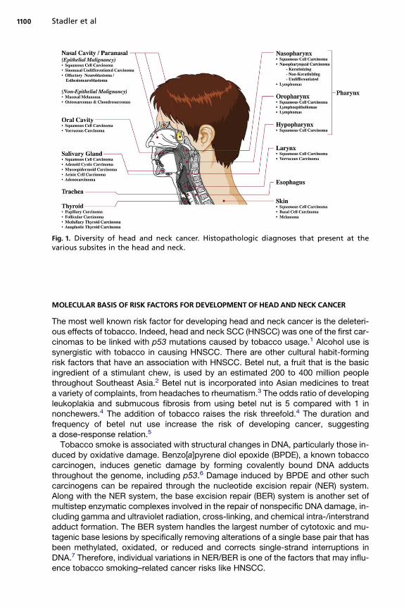
Fig. 1. Diversity of head and neck cancer. Histopathologic diagnoses that present at thevarious subsites in the head and neck.
Stadler et al1100
MOLECULAR BASIS OF RISK FACTORS FOR DEVELOPMENT OF HEADAND NECK CANCER
The most well known risk factor for developing head and neck cancer is the deleteri-ous effects of tobacco. Indeed, head and neck SCC (HNSCC) was one of the first car-cinomas to be linked with p53 mutations caused by tobacco usage.1 Alcohol use issynergistic with tobacco in causing HNSCC. There are other cultural habit-formingrisk factors that have an association with HNSCC. Betel nut, a fruit that is the basicingredient of a stimulant chew, is used by an estimated 200 to 400 million peoplethroughout Southeast Asia.2 Betel nut is incorporated into Asian medicines to treata variety of complaints, from headaches to rheumatism.3 The odds ratio of developingleukoplakia and submucous fibrosis from using betel nut is 5 compared with 1 innonchewers.4 The addition of tobacco raises the risk threefold.4 The duration andfrequency of betel nut use increase the risk of developing cancer, suggestinga dose-response relation.5
Tobacco smoke is associated with structural changes in DNA, particularly those in-duced by oxidative damage. Benzo[a]pyrene diol epoxide (BPDE), a known tobaccocarcinogen, induces genetic damage by forming covalently bound DNA adductsthroughout the genome, including p53.6 Damage induced by BPDE and other suchcarcinogens can be repaired through the nucleotide excision repair (NER) system.Along with the NER system, the base excision repair (BER) system is another set ofmultistep enzymatic complexes involved in the repair of nonspecific DNA damage, in-cluding gamma and ultraviolet radiation, cross-linking, and chemical intra-/interstrandadduct formation. The BER system handles the largest number of cytotoxic and mu-tagenic base lesions by specifically removing alterations of a single base pair that hasbeen methylated, oxidated, or reduced and corrects single-strand interruptions inDNA.7 Therefore, individual variations in NER/BER is one of the factors that may influ-ence tobacco smoking–related cancer risks like HNSCC.

Molecular Biology of Head and Neck Cancer 1101
Several studies have demonstrated that sequence variations in NER/BER genescontribute to HNSCC susceptibility.8–11 The ERCC1 gene product is a key enzymein the NER system, and one particular polymorphism at the ERCC1 gene (C8092A)may affect its mRNA stability, resulting in impaired DNA repair capacity.12 Two sin-gle-nucleotide polymorphisms (SNPs) in the XPD gene (Asp312Asn and Lys751Gln),also part of the NER cascade, have been associated with suboptimal DNA repaircapacity.13 There are conflicting data regarding SNPs in the BER system and thepredilection for developing HNSCC. Li and colleagues,9 in one of the largest case-control studies of 830 patients who had HNSCC and 854 cancer-free control sub-jects, evaluated the progression to HNSCC based on polymorphisms in three BERnonsynonymous SNPs. Although the BER system enzyme XRCC1 (Arg399Gln) in-consistently increases the risk of HNSCC in Caucasians,13–16 Li and colleagues9
concluded that polymorphisms in the ADPRT enzyme of the BER system are asso-ciated with HNSCC and demonstrated that individuals who have the ADPRT 762Ala/Ala and Ala/Ala1Val/Ala genotypes are at lower risk of developing HNSCC comparedwith individuals who have the Val/Val genotypes. Further studies to elucidate thegenetic predisposition of developing HNSCC in the face of total tobacco burdenmay provide preventative health benefits in the future to those who have susceptiblepolymorphisms.
Marijuana is the most commonly used illegal drug in the United States and the sec-ond most commonly smoked substance after tobacco.17 Habitual marijuana smokingmanifests with similar signs and symptoms associated with chronic tobacco use.18,19
Furthermore, the carcinogenic properties of marijuana smoke are similar to those oftobacco, and numerous studies parallel the use of cannabinoids to cancer develop-ment.20–22 Marijuana has been shown to induce cytogenic changes consisting of chro-mosomal breaks, deletions, and translocations in mammalian cells in vivo.23 Untilrecently, there was not enough evidence to suggest a causative relation with oropha-ryngeal HNSCC, especially those cases caused by tobacco use;24 however, HNSCCcaused by human papilloma virus (HPV) may be associated with marijuana (see laterdiscussion).
Clearly, normal variation in patient genotype for genes in DNA repair pathways ap-pears to modify baseline risk for cancer development, especially when impacted byenvironmental toxins such as smoking. In parallel, there is a range of germline variantsthat are more rare than SNPs and accordingly called mutations. These rare heritableevents can be sporadic or conserved in families and are frequently recognized due tothe high penetrance of one of a number of recognized familial cancer syndromes. Fan-coni’s anemia, known for the risk of developing lymphoreticular malignancies dueto germline mutations in the caretaker genes FAA, FAD, and FCC, carries a risk fordeveloping second primary cancers in the tongue, pyriform sinus, and postcricoid re-gion.25 Patients who have Bloom syndrome are characterized to have mutations inthe helicase genes and are predisposed to developing solid tumors in a number ofanatomic sites, 6% and 8% of which arise from the tongue and larynx, respectively.26
Homozygotes who have ataxia telangiectasia who survive into their 20s and 30s areat increased risk of developing chronic T-cell leukemia and solid malignancies ofthe oral cavity, breast, stomach, pancreas, ovary, and bladder.27 Xeroderma pigmen-tosum, an autosomal recessive disorder of one or more of the XP genes in the NERsystem, manifests second primaries within the oral cavity in addition to the knownrisks of skin malignancies.26,28 Other such syndromes and affected genes (indicatedin parentheses) with primary manifestations in the head and neck include Cowden dis-ease (PTEN), multiple endocrine neoplasia type I (MEN I), multiple endocrine neoplasiatype II (MEN II), neurofibromatosis type II (NF-2), and retinoblastoma (Rb).

Stadler et al1102
Genetic cancer syndromes are generally recognized by the early age of onset of ma-lignancies in impacted individuals and by specific or unusual patterns of tumors. Can-cer syndrome tumors are of scientific importance out of proportion to their incidence inthat they point clearly at specific pathways and targets that are key to the develop-ment of malignancy, in contrast to sporadic tumors in which the causative lesionmay be difficult to identify. For example, a relatively rare cancer syndrome, multipleendocrine neoplasia, type IIA, is well known for its association with germline mutationof the RET proto-oncogene. Although MEN IIa is rare, the RET gene has relevance ina wide variety of tumors and is targeted by anti-cancer drugs, such as vandetanib.29,30
VIRAL ASSOCIATIONS AND NEW EPIDEMIC OF HEADAND NECK SQUAMOUSCELL CARCINOMACAUSED BY HUMAN PAPILLOMAVIRUS
Recently, HPV infection has been identified as an etiologic agent for oropharyngealcarcinoma, a subset of SCCs that comprises the tongue base and tonsil. Patientswho have oropharyngeal SCCs and have the HPV genes incorporated in their tumorgenome are younger in age (by 3–5 years) and are less likely to have a history oftobacco and alcohol use.31
What is most disconcerting is that although the overall incidence of HNSCC (1973–2004) has steadily declined according to the Surveillance Epidemiology and EndResults database, the incidence of oropharyngeal cancer is increasing among youn-ger age groups.32–35 The unsettling implication is that the incidence of HPV-relatedHNSCC of the oropharynx could overtake HPV-unrelated HNSCC, which is thoughtto be associated more with traditional risk factors.
There is substantial evidence that infection with high-risk HPV subtypes, in particu-lar HPV-16, is a risk factor for the development of oropharyngeal cancers.36–47 In fact,Gillison and colleagues48 claimed that HPV-positive and HPV-negative HNSCC of theoropharynx should be classified as two distinct cancers based on the clinical andmolecular risk factors and etiology. According to their case-controlled study, patientsat risk for HPV-16–positive oropharyngeal SCC were more likely to be white (P 5 .06),married (P<.001), college educated (P 5 .03), and have an annual income over$50,000 (P<.01); intensity or duration of tobacco smoking or alcohol consumptiondid not increase the odds ratio of HPV-16–positive HNSCC.48 Although case-controlstudies have not linked marijuana use to HPV-negative HNSCC, Gillison andcolleagues48 demonstrated a strong association of marijuana use and HPV-16–positive HNSCC and further theorized plausible mechanisms of cannabinoid modula-tion of the immune system.
High-risk HPV strains (HPV-16 and HPV-18) associated with oropharyngeal SCC(and cervical cancer) manipulate cellular pathways within affected cells to activatecell growth and suppress apoptosis. Malignant transformation begins with inactivationof the p53 tumor suppressor gene by E6, whereas a second HPV protein, E7, inacti-vates the retinoblastoma tumor suppressor protein (Rb). The HPV E6 and E7 proteins,encoded in the HPV-16 genome, functionally disrupt regulatory cell-cycle and DNA-repair pathways that drive genetic or epigenetic changes during molecular progres-sion of HNSCC.49 E6 targets the cellular ubiquitin-protein ligase E6-AP, which thentargets p53 for ubiquitination and degradation, leaving cell growth unregulated. E7associates with Rb and p21, blocking the interaction of Rb with E2F and initiatinguncontrolled cell division.50
Not only is the nascent HPV-positive tumor subject to inactivation of tumor sup-pression genes from the viral genome, but several genes involved in transcriptionand cell cycle regulation are among the most prominent up-regulated genes in these

Molecular Biology of Head and Neck Cancer 1103
tumors. One such cell cycle inhibitor is CDKN2A, which encodes the p16INK4A tumorsuppressor protein that functions as a cyclin-dependent kinase inhibitor in the Rbtumor suppressor pathway. Increased expression of p16INK4A may potentially reflectloss of a negative feedback loop associated with inactivation of Rb by HPV E7.51
Overexpression of p16INK4A is strongly correlated with HPV infection in head andneck carcinomas and has been used as a surrogate marker for HPV.52
Detecting high-risk HPV using in situ hybridization from ethanol-fixed and Papanico-laou-stained smears after fine-needle aspiration has a 93% correlation to correspond-ing tissue sections positive for HPV-16 with polymerase chain reaction.53 Of interest,HPV-positive tumors are associated with nonkeratinizing cytomorphology.53 A recentmeta-analysis showed that 26% of HNSCCs from all subsites contain HPV genomicDNA,54 and it is now estimated that over 50% of oropharyngeal HNSCCs are relatedto HPV infection.55 Although in situ hybridization and polymerase chain reaction tech-niques are available, there are no standardized clinical tests approved by the Foodand Drug Administration for HPV-positive HNSCC tumors.
TUMORIGENESIS/CARCINOGENESIS
In the case of HPV, a virus can usurp normal cellular processes, whereas in the case ofmost patients, the development of carcinoma is the result of a stepwise accumulationof genetic alterations.56 Three main steps include initiation, promotion, and progres-sion. For this multiple-step process to succeed, numerous cellular processes and de-rangements must occur. The creation of an initial, critical, early genetic change helpsset into motion the carcinogenic process.57 Exposure to carcinogenic factors maylead to the abnormal expression of tumor suppressor genes, proto-oncogenes, orboth, which in turn activate pathways that lead to the malignant transformation ofcells. Oftentimes, this abnormal expression may include a sporadic mutation, deletion,loss of heterozygosity, overexpression, or epigenetic modification such as hyperme-thylation. For example, telomerase, an enzyme involved in immortalization, hasbeen shown to be reactivated in roughly 90% of HNSCCs, whereas a deletion of9p21 is found in 70% to 80% of these cases. Various point mutations in TP53 andthe loss of heterozygosity of 17p are shown to exist in over 50% of HNSCC lesions.58
When this occurs, secondary genetic changes create greater genetic instability, shift-ing the cell toward a more malignant phenotype (Fig. 2). Specifically, inactivation oftumor-suppressor genes allows for cellular proliferation to continue with unregulatedand autonomous, self-sufficient growth. Proto-oncogenes also play a key role intumorigenesis by helping the cell attain a malignant phenotype.
Six hallmarks of cancer cells have been described that distinguish them from theirnormal counterparts: (1) self-sufficiency in growth signals, (2) insensitivity to growth-inhibitory signals, (3) evasion of programmed cell death, (4) immortality or unlimitedreplicative potential, (5) sustained angiogenesis, and (6) tissue invasion and metasta-sis.59 In the following sections, the authors discuss major pathways, receptors, andproteins implicated in the initiation or progression of HNSCC as they relate to aspectsof all six of these hallmarks. It is the accumulation of specific abnormalities such asthose described, likely along with other genetic events and alterations, that accountsfor the process of carcinogenesis in HNSCC.
FIELD CANCERIZATION
Although it is unclear exactly why HPV appears to target certain subsites in the headand neck, the pattern is clear. In contrast, there seems to be a more general phenom-enon seen in smokers in whom broad regions of tissue appear to be damaged, giving

NormalMucosa
Dysplasia Carcinomain situ
InvasionOR9p loss3p,
17p loss11q, 13q14q loss
6p, 8, 4qloss
AlternatePrecursor
Lesion
BenignSquamous
Hyperplasia
Fig. 2. Genetic progression of HNSCC. Genetic changes associated with the histologic pro-gression of HNSCC based on loss of chromosomal material (allelic loss). Genetic alterationshave been placed before the lesion where the frequency of the particular event plateaus.It is the accumulation and not necessarily the order of genetic events that determines theprogression. A small fraction of benign squamous hyperplastic lesions contain 9p21 or3p21 loss, suggesting that an unidentified precursor lesion (or cells) may also give rise todysplasia. Candidate tumor suppressor genes include p16 (9p21), p53 (17p), and Rb (13q),and a candidate proto-oncogene includes cyclin D1 (11q13). (Courtesy of Joseph A. Califano,MD, Baltimore, MD.)
Stadler et al1104
rise to multiple premalignant and frankly invasive tumors. In 1953, Slaughter and col-leagues60 first hypothesized that primary tumors emerge from a layer of precanceroustissue and coined the term ‘‘field cancerization’’ after demonstrating histopathologicchanges consistent with genetic aberration from normal mucosa. Forty years afterSlaughter and colleagues proposed field cancerization, Califano and colleagues49
demonstrated the molecular basis for histopathologic changes. Samples of dysplasticmucosa and benign hyperplastic lesions displayed loss of heterozygosity at specificloci (9p21 [20%], 3p21 [16%], 17p13 [11%]). In particular, loss of 9p21 or 3p21 isone of the earliest detectable events leading to the progression to dysplasia. Fromdysplasia, further genetic alteration in 11q, 13q, and 14q creates carcinoma in situ(see Fig. 2).
The high rate of recurrence in the location of the primary tumor is thought to bea result of the fact that 30% of histopathologically benign squamous cell epitheliumconsists of a clonal population, with genetic alterations seen in HNSCC.61 Studiesusing microsatellite analysis and X chromosome inactivation have verified that meta-chronous and synchronous lesions from distinct anatomic sites in HNSCC often orig-inate from a common clone.62 This evidence confirms that genetically altered mucosais difficult to cure in the HNSCC patient because it is on the path to tumorigenesis.Indeed, second primaries are common in patients who have HNSCC.
EPITHELIAL-TO-MESENCHYMALTRANSITION
There is evidence that suggests that fundamental changes to the programming ofcells, including stem cells, may also be involved in tumorigenesis. One program thatis particularly dangerous is the epithelial-to-mesenchymal transition (EMT), a pheno-typic change in cells that provides them with the ability to escape from the constraintsof surrounding tissue architecture. It has been postulated that EMT is the means bywhich epithelial tumors invade and metastasize to other tissues. As defined byHugo and colleagues,63 EMT is a culmination of protein modifications and transcrip-tional events in response to extracellular stimuli. These changes lead to long-termyet sometimes reversible cellular changes. Abnormalities in cadherins, tight junctions,

Molecular Biology of Head and Neck Cancer 1105
and desmosomes lead to a decrease in cell–cell adherence and loss of polarity in thecells, increasing the mobility of these cells. More specifically, epithelial cells disassem-ble their junctional structures, undergo extracellular matrix remodeling, begin to ex-press proteins of mesenchymal origin, and subsequently become migratory.64 Thisprocess has been postulated to be a part of normal embryogenesis, the inflammatoryprocess, and wound healing.63 When the process of EMT becomes pathologic, it lacksthe tight coordination and regulatory checkpoints that are normally present. Specifi-cally, in the carcinogenic process, EMT causes changes in tumor cell propertiesthat contribute to tumor invasion and metastasis, enabling cancer cell disseminationand self-renewal capabilities.65 In HNSCC, EMT has been found to play a role, espe-cially in high-risk tumor subtypes. Chung and colleagues66 showed that genes in-volved in EMT and nuclear factor–kappaB (NF-kB) signaling deregulation were themost prominent molecular characteristics of the high-risk tumors in the subset theyexamined. Although it is clear that EMT plays a role in tumorigenesis in many cancers,the complete clinical significance of this process is yet to be fully defined.
EPIGENETIC MODIFICATION
Many programs (such as those discussed in Fig. 2) are the result of direct damage tothe genome; however, there are other mechanisms of heritable somatic changes ingene expression that do not require direct alteration of the DNA sequence itself.The DNA molecule can be modified, such as by the addition or subtraction of methylgroups, without a change in the base composition. Similarly, histones, the structuralproteins found in close association with DNA, can be modified by acetylation, meth-ylation, or ubiquitylation. These non-DNA encoded modifications can result in herita-ble changes in gene expression that are clinically significant, including in the setting ofcancer. Different cancers display varying behaviors, likely due to the multiple epige-netic changes and genetic mutations that occur within a tumor environment. Hyper-methylation is one such type of epigenetic modification that is increasingly wellcharacterized. Recently identified as a probable component in the development ofcarcinoma, hypermethylation in certain promoter regions of a gene can lead to repres-sion of transcription.67 Numerous studies have implicated this process of aberrantmethylation in many tumor suppressor genes, causing them to become inactive.67
MOLECULAR PATHWAYS INVOLVED IN HEADAND NECK SQUAMOUS CELL CARCINOMA
Increasingly, model systems and other research techniques have helped to decipherpathways of importance for patients (Fig. 3). Knowledge of these pathways has led in-vestigators to interrogate key pathway components for tumor-specific gene muta-tions, and many have been reported in head and neck tumors (Table 1). Initialclarity in the activated pathways and mutated genes of head and neck tumors resultedin clinical trials of a host of targeted therapies, such as those documented in Table 2.The most promising pathways and agents from this inventory are discussed in the fol-lowing paragraphs.
Epidermal Growth Factor Receptor
Epidermal growth factor receptor (EGFR) signaling has been strongly implicated incarcinogenesis, tumor progression, and response to therapy in HNSCC (reviewedby Thariat and colleagues).68 The ErbB family of proteins, a family of four structurallyrelated receptor tyrosine kinases, comprises four receptors (ErbB 1–4, also knownas HER 1–4) and 13 polypeptide extracellular ligands.69 In the literature, ErbB2 issynonymous with HER2/neu, whereas ErbB1 is commonly referred to as EGFR.

Fig. 3. Molecular pathways contributing to the promotion and progression of tumorigenesisin head and neck cancer. Akt, protein kinase B; EGFR, epidermal growth factor receptor;ERK, extracellular signal–related kinase; Hsp90, heat shock protein 90; IGF-1R, insulin-likegrowth factor-1 receptor; IKKa, inhibitor kB kinase alpha; IKKb, inhibitor kB kinase beta;IKKg, inhibitor kB kinase gamma; JAK, Janus kinase; MEK, mitogen-activated protein kinase;mTOR, mammalian target of rapamycin; PI3-K, phosphatidylinositol-3-kinase; STAT, signaltransducers and activators of transcription; VEGF, vascular endothelial growth factor.
Stadler et al1106
When ligands bind to one of the ErbB receptors, a dimer forms and the receptor’sintracellular tyrosine residues then undergo ATP-dependent autophosphorylation.Currently, there are 12 different ligands that are known to activate four knownErbB receptors.
When phosphorylated, the receptor has the potential to trigger a number of differentintracellular downstream pathways that can eventually arrest apoptosis, promote cel-lular proliferation, stimulate tumor-induced neovascularization, and activatecarcinoma invasion and metastasis.69 The Ras/mitogen-activated protein kinase/ex-tracellular signal–related kinase (Ras-MAPK-ERK) pathway is known to control genetranscription, cell proliferation, and cell-cycle progression, whereas the phosphatidy-linositol-3-kinase/protein kinase B (PI3K/Akt) pathway has been shown to stimulatenumerous antiapoptotic signals within the cell. The Janus kinase/signal transducersand activators of transcription (STAT) and phospholipase-Cg/protein kinase C path-ways are also activated in association with EGFR phosphorylation.70 Thus, EGFRplays a role in carcinoma growth and survival through a multitude of oncogenic down-stream signaling pathways.
EGFR mRNA and protein are known to be preferentially expressed in HNSCC com-pared with surrounding normal tissues, suggesting a significant role in carcinogenesis.Similarly, most epithelial carcinomas overexpress and possess functional activation ofthe EGFR family of receptors.71 In HNSCC, EGFR is overexpressed in up to 80% to

Table 1Limited listing of themost common somatic mutations of various head and neck subsites
Head and Neck Subsitea Gene Sample Positive/Total (% Positive)Larynx CDKN2A 45/262 (17)
PTEN 10/43 (23)
EGFR 5/82 (6)
KRAS 4/166 (2)
HRAS 2/96 (2)
Oral cavity CDKN2A 98/508 (19)
HRAS 67/494 (13)
FGFR3 44/136 (32)
PIK3CA 18/145 (12)
KRAS 14/497 (2)
Oropharynx MET 33/156 (21)
CDKN2A 19/173 (10)
PTEN 7/27 (25)
KRAS 3/105 (2)
BRAF 3/52 (5)
Tonsil EGFR 7/45 (15)
CDKN2A 0/3 (0)
HRAS 0/3 (0)
KRAS 0/3 (0)
NRAS 0/3 (0)
Sinonasal cavity KRAS 4/121 (3)
HRAS 2/11 (18)
NRAS 2/11 (18)
STK11 2/7 (28)
EGFR 1/5 (20)
Esophagus (upper one third) TP53 4/4 (100)
KRAS 1/4 (25)
CDKN2A 1/3 (33)
PIK3CA 1/3 (33)
CTNNB1 0/9 (0)
Thyroid BRAF 2013/4793 (41)
RET 274/706 (38)
NRAS 132/1962 (6)
KRAS 80/1878 (4)
HRAS 56/1844 (3)
Salivary gland HRAS 17/90 (18)
PTEN 5/13 (38)
DTNNB1 2/44 (4)
KRAS 1/40 (2)
CDKN2A 1/8 (12)
Although the capability exists to detect these mutations within tumor samples, their full clinicarelevance has yet to be fully realized.
a SCC was the only histology tested for in the larynx, oral cavity, oropharynx, tonsil, sinonasacavity, and esophagus. A wider range of histologic variants was included for the analysis of thyroidand salivary gland subsites.
Molecular Biology of Head and Neck Cancer 1107
l
l

Table 2Limited listing of selected targeted agents that are currently undergoing clinical trialsfor the treatment of head and neck cancer
Drug Name (Trade Name) Target Phase of Study in Head and Neck CancerCetuximab (Erbitux) EGFR III
Gefitinib (Iressa) EGFR I/II/III
Erlotinib (Tarceva) EGFR I/II/III
Panitumumab (Vectibix) EGFR I/II/III
BIBW 2992 (Tovok) EGFR, HER-2/neu II
Zalutumumab (HuMax-EGFr) EGFR I/II/III
Trastuzumab (Herceptin) HER-2/neu II
Lapatinib (Tykerb) EGFR, HER-2/neu I/II/III
Cediranib (Recentin) VEGF I/II
Sorafenib (Nexavar) Raf, VEGF I/II
Semaxanib VEGF I/II
Pazopanib VEGF II
Sunitinib (Sutent) VEGF I/II
Bevacizumab (Avastin) VEGF I/II/III
Romidepsin Histone deacetylase I/II
Vorinostat (Zolinza) Histone deacetylase I/II
Dasatinib (Sprycel) Tyrosine kinases II
Imatinib (Gleevec) Tyrosine kinases II
Pazopanib VEGF, tyrosine kinases II
Vandetanib (Zactima) VEGF, EGFR I/II
XL880 VEGF, tyrosine kinases II
Perifosine (KRX-0401) AKT II
Bortezomib (Velcade) NF-kB, tyrosine kinases I/II
Lonafarnib (Serasar) Farnesyl transferase I/II
Tanespimycin (KOS-953) Hsp90 I/II
AZD0530 Src/Abl kinase II
This partial list was obtained through an extensive and comprehensive search on www.clinicaltrial.gov.
Abbreviations: Akt, protein kinase B; EGFR, epidermal growth factor receptor; Hsp90, heat shockprotein 90; VEGF, vascular endothelial growth factor.
Stadler et al1108
100% of tumors, some of the highest rates of any human carcinoma.72,73 There areregional differences among tissues in the head and neck that express EGFR, with rel-atively lower levels associated with laryngeal tumors compared with those of the oralcavity and oropharynx.74
EGFR demonstrates increased overexpression in the more advanced-stage carci-nomas and in those carcinomas found to be poorly differentiated.70 In addition,EGFR overexpression is associated with decreased patient survival rates and hasbeen demonstrated by some groups to confer resistance to various therapeutic mo-dalities, including targeted therapy.70,75–79 Although the association with poor patientprognosis has not been as clearly established, specific mutations of the EGFR recep-tor have also been studied. The most common mutation of EGFR is likely EGFRvIII,occurring in up to 40% of HNSCC.80 This mutant receptor is found only in cancer cells

Molecular Biology of Head and Neck Cancer 1109
and manifests from an in-frame deletion of exons 2 to 7, which encodes the receptor’sextracellular domain, thus resulting in a constitutively active receptor that is com-pletely independent of any activation by way of ligand binding.80 The fact that EGFRvIIIis not found in normal tissues makes this a very intriguing, highly specific target fortherapy, given that it would not interfere with the normal EGFR signaling in noncancer-ous tissues (see Table 1).
In addition to overexpression, other pathologic manifestations of EGFR can be per-formed through mutational activation, amplification, and transactivation by other tyro-sine kinases.81 The potential, constitutive activation of several different oncogenicpathways, by way of EGFR-independent mechanisms, likely explains the lack of re-sponse that is commonly appreciated in patients being treated with EGFR inhibitortherapy.70
With the prominent role that EGFR is known to play in tumorigenesis, this family ofproteins was a logical choice in pursuing a new class of targeted cancer therapy. Cur-rently, there are several EGFR antagonists available for clinical use in the treatment offour metastatic epithelial carcinomas, including non–small cell lung cancer, colorectalcancer, pancreatic cancer, and HNSCC. The two classes of therapies that exist todate are monoclonal antibodies to EGFR receptor subunits and small-moleculeEGFR tyrosine kinase inhibitors. In simplest terms, the monoclonal antibodies proba-bly act by binding the conserved extracellular domain of EGFR and by blocking theligand-binding region by competitive inhibition, which in turn blocks ligand-inducedautophosphorylation through the inability to stimulate tyrosine kinase. The EGFR tyro-sine kinase inhibitors function by way of a separate mechanism. They act by reversiblycompeting with ATP in its binding site to the intracellular catalytic domain of tyrosinekinase, therefore inhibiting autophosphorylation of EGFR and its subsequent down-stream signaling.71
Akimoto and colleagues82 were the first to provide evidence that EGFR expressionmay have an effect on radiation sensitivity, a result that has been validated clini-cally.75,83 EGFR overexpression in head and neck cancer cell lines was found tohave greater radioresistence compared with cell lines that had relatively lower levelsof EGFR expression. It was also found that following radiation, EGFR becomes up-regulated within the tumor, leading to increased activation of its downstream signalingpathways.84,85 This work culminated in the landmark 2006 publication of the random-ized trial by Bonner and colleagues86 that showed an overall and progression-freesurvival advantage with the addition of cetuximab to standard radiation therapy.
At least 40 trials involving patients who have HNSCC are currently investigating var-ious ‘‘targeted agents’’ including tyrosine kinase inhibitors and antibody therapy.87
More than 10 different EGFR-targeting agents are in development for the treatmentof various carcinomas. Although a great deal of effort has gone into the developmentand validation of predictive biomarkers, it remains difficult to determine a priori whowill benefit from these therapies. In some studies, EGFR expression is an independentpredictor of response, whereas in others, no relationship is appreciated.75,88–91 Thesearch for molecular predictors of clinical outcome that would potentially optimize pa-tient selection and therapeutic efficacy continues to be an area of intense ongoinginvestigation.
Insulin-Like Growth Factor-1 Receptor
An emerging potential target for directed, molecular-based cancer therapy is the insu-lin-like growth factor (IGF) signaling axis. Numerous preclinical and clinical studieshave implicated the IGF-1 receptor (IGF-1R) and its ligands, IGF-1 and IGF-2, in thedevelopment and progression of a number of human cancers.92–94 IGF-1R is

Stadler et al1110
a transmembrane heterotetramer receptor that consists of two a and two b subunits.Like the insulin receptor, IGF1-R possesses tyrosine kinase activity. With activation ofthe receptor, downstream signaling events include phosphorylation of insulin receptorsubstrate-1, activation of MAPKs, and stimulation of the PI3K pathway.95 This activa-tion of the Ras-MAPK-ERK and PI3K/Akt pathways is similar to the downstream sig-naling seen with EGFR autophosphorylation and activation. Six IGF binding proteins(IGFBPs) are known to exist in humans. These proteins have been shown to help mod-ulate the effects of IGF-1 by way of multiple unique mechanisms. In humans, IGF-1 isbound to one of the IGFBPs over 95% of the time, with IGFBP-3 accounting forroughly 85% of this binding.96
In vitro and in vivo studies have shown that IGF-1R encourages cellular growth andprotects cells from apoptosis.97 This phosphorylation cascade leads to the activationof various transcription factors involved in cellular proliferation and transforma-tion.98–102 Constitutive activation and overexpression of IGF-1R has been associatedwith malignant transformation, involving glioblastoma, melanoma, pancreatic, breast,colon, and ovarian carcinoma models.103–105 The receptor has been closely linked tothe metastatic properties of tumor cells, with studies showing that the receptor tosignal pathways are linked to tumor invasiveness and angiogenesis.106,107 IGF-1R sig-naling has also been shown to influence and promote focal adhesion stability, cell-to-cell contact, and cellular motility.108 Numerous studies have also shown that enhancedIGF-1R activation is associated with resistance to certain cytotoxic chemotherapy reg-imens, hormonal agents, biologic anticancer therapies, and radiation therapy.109–117
Although Ouban and colleagues118 showed only 13% immunostaining positivity forIGF-1R in 31 different human HNSCC tissue samples, other studies have shown muchhigher levels of expression. IGF-1R is ubiquitously expressed at varying levels in can-cerous tissues, and plays an intricate role in the regulation of cellular proliferation anddifferentiation even at very low levels of expression.95,118
As mentioned previously, the IGF-1R and the EGFR signaling pathways are intri-cately associated with one another, regulating overlapping downstream signalingpathways. Increased IGF-1R expression has been reported to mediate resistance toanti-EGFR–based therapies in certain solid tumors, including glioblastoma, pancreatic,and breast carcinoma.119–122 It has been found that the use of both antibodies in com-bination was more effective in reducing cancer cell growth than the use of either singleagent alone.123 There may be a potential benefit in the use of combined anti–tyrosinekinase receptor–directed therapies to treat HNSCC. Slomiany and colleagues124
also demonstrated the potential for the cotargeting of IGF-1R and EGFR signalingpathways in HNSCC.
Design of IGF-1R inhibitors has proved to be somewhat problematic, due to the closehomology (60%–70% amino acid homology) with the insulin receptor;125 however, spe-cific inhibitors have recently been developed. Several approaches to tumor growth in-hibition have been undertaken, including IGF-1R dominant mutants, IGF-1R blockingantibodies, and oligonucleotides aimed at down-regulating IGF-1R expression, but lit-tle success has been achieved in terms of improved clinical outcomes. The first IGF-1Rtyrosine kinase inhibitor demonstrating in vivo therapeutic potential (NVP-AEW541 [No-vartis]), however, has been shown to enhance tumor cell chemosensitivity and inhibittumor growth in human fibrosarcoma, myeloma, and Ewing’s sarcoma models.125,126
Studies with the inhibitor have yet to be undertaken in an HNSCC model.
Phosphatidylinositol-3-Kinase/Protein Kinase B Pathway
The PI3-K/Akt signal transduction pathway has been shown to regulate numerous cel-lular processes, including apoptosis, proliferation, cell cycle progression, cytoskeletal
Molecular Biology of Head and Neck Cancer 1111
stability and motility, and energy metabolism.127,128 Activated Akt induces increasedexpression of numerous proliferative and antiapoptotic proteins, including Bcl-2,Bcl-x, and NF-kB.127 The pathway has been shown to be activated in up to 50% to80% of HNSCCs.78 The PI3-K/Akt pathway is one of the main downstream signalingpathways activated by the ErbB/tyrosine kinase receptor family of receptors. Afterligand binding, the cytoplasmic domain of the EGFR undergoes tyrosine phosphoryla-tion and subsequently activates PI3-K.129 Activation of the PI3-K/Akt pathway, how-ever, is not entirely dependent on the tyrosine kinase family of receptors. In certaincarcinoma models, it has been shown to be activated through direct mutation or am-plification of PI3-K, amplification of Akt, activation of the RAS oncogene, or decreasedexpression of the tumor-suppressor protein phosphatase and tensin homolog (PTEN),a known inhibitor of the PI3-K/Akt pathway.129 Loss of PTEN expression, along withAkt activation, correlates with worse clinical outcomes in patients who have SCC ofthe tongue.130,131 This pathway has also been found to be overexpressed and acti-vated in a number of different carcinomas, including HNSCC. In a study conductedby Massarelli and colleagues,132 it was shown that disease-free survival was signifi-cantly decreased in cases of SCC of the tongue that stained positive for activatedAkt (p-Akt). The comparatively poor outcome that was associated with p-Akt expres-sion was also found to be independent of cancer stage and nodal status. Akt activationhas also been correlated with the squamous cell progression and transformation—from normal epithelial tissue, to dysplasia, and even to invasive SCC.133
The PI3kK/Akt pathway has also been shown to be up-regulated following radiationtherapy. Bussink and colleagues129 described how the pathway is intricately involvedwith resistance to radiation therapy by way of multiple mechanisms. The RAS oncogeneis a well-known contributor to the intrinsic radioresistance of tumor cells, mediated atleast partially through its downstream signaling through the PI3-K/Akt pathway.129 Inaddition, this pathway is involved in DNA repair by way of EGFR signaling, with multiplestudies showing that with EGFR blockade, the PI3-K/Akt pathway–mediated DNArepair process is altered. When combined with radiation therapy, this decreasedDNA repair leads to greater levels of tumor cell apoptosis and subsequent improvedlocoregional control compared with radiation therapy alone.129
Although this pathway is most often activated through EGFR, EGFR-independentactivation is also common and of clinical relevance.129 Using tissue microarray tech-nology, Molino and colleagues134 showed that the PI3-K/Akt/mammalian target of ra-pamycin (mTOR) pathway was frequently activated in HNSCC samples and that thiswas often independent of any associated EGFR activation. There has also beena strong and independent correlation between expression of activated Akt (pAkt)and treatment outcome in laryngeal and oropharyngeal HNSCC.132,135 Similarly, en-hanced Akt activity has been independently associated with more-advanced tumorstage and progression in a number of different malignancies.136 Knowing this informa-tion, further study of this pathway (independent of EGFR) is needed so that it can beused in regard to treatment and prognostic markers for HNSCC.
A recent phase II trial conducted by Karamouzis and colleagues137 involved the use ofan Akt phosphorylation/activation inhibitor (perifosine) in a small group of patients whohad incurable recurrent HNSCC, metastatic HNSCC, or both. Although the inhibitorhad some preclinical antitumor activity in vivo, no objective clinical responses were ap-preciated,with18 of19patients having disease thatprogressed at8 weeks’ follow-up.137
Akt activation, however, has also been associated with resistance to EGFR inhibition ina non–small cell lung cancer model,138 and therefore, benefits from combined targetedmolecular therapy are still of potential interest. Thepotential clinical usefulness of the PI3-K/Akt pathway as a therapeutic target and a prognostic marker in HNSCC is promising.

Stadler et al1112
Mammalian Target of Rapamycin
One of the downstream cell-growth regulators associated with the PI3-K/Akt pathwayis an atypical serine/threonine kinase named mTOR. Although Akt helps control cellu-lar proliferation and growth through the coordination of mitogenic signaling with en-ergy- and nutrient-sensing pathways that control protein synthesis, it requiresmTOR to fully exert these effects.139 mTOR is involved in modulation of the cell cycleand ribosomal function to subsequently participate in cell growth and apoptosis.140
When activated through Akt, mTOR phosphorylates the translation regulator p70-S6kinase, which in turn activates the ribosomal S6 protein, a protein involved in transla-tion and one of the most downstream targets of the PI3-K/Akt/mTOR pathway. Thisactivation of S6 ribosomal protein thus adds to the control of cell growth throughincreased manipulation of mRNA translation.141
Amornphimoltham and colleagues139 found that in clinical specimens from patientswho had HNSCC and from HNSCC-derived cell lines, aberrant accumulation of acti-vated S6 (p-S6) was a frequent occurrence in early dysplastic lesions and carcinomas.These investigators also showed that the activated ribosomal protein was decreasedwhen HNSCC cell lines were treated with rapamycin, a macrolide antibiotic anda known inhibitor of mTOR.139 Similar findings were seen when rapamycin wasused in an HNSCC xenograft model. In vivo, rapamycin’s effects included inductionof apoptosis and inhibition of cellular growth of the HNSCC cells, with subsequent tu-mor regression.139 It has also been shown that rapamycin sensitivity of tumor cells is atleast partially dependent on the dysregulation of the tumor suppressor gene PTEN, thewell-known inhibitor of the PI3-K/Akt pathway.142,143 In some of the HNSCC cell lines,EGFR inhibition had no effect on the activity of the mTOR pathway, indicating a poten-tial clinical and therapeutic benefit when EGFR inhibitors and mTOR inhibitors areused in combination.139 The study revealed the Akt/mTOR pathway to be a potentialtherapeutic target for the treatment of HNSCC, particularly in terms of development ofanalogs to rapamycin.
Nathan and colleagues144 showed that one of the main downstream effectors of theAkt/mTOR pathway, eIF4E, was overexpressed in histologically ‘‘tumor-free’’ surgicalmargins of resected HNSCC samples and was an independent predictor of tumorrecurrence. In a later study, the same group used an eIF4E-overexpressing, PTEN-mutant HNSCC cell line, FaDu, in a minimal residual disease murine model, andshowed that the rapamycin analog temsirolimus (CCI-779) was effective in prolongingsurvival, including improved tumor-free survival.145 It was concluded that CCI-779represented a new potential targeted therapy for the treatment of HNSCC, becauseoverexpression and activation of mTOR occurs in many of these tumors.139,146
Nuclear Factor–Kappa B
NF-kB transcription factors are the final downstream mediators of many of the earlier-mentioned pathways and therefore play a key role in head and neck cancers. It hasbecome clear that NF-kB–mediated inflammatory signaling is overexpressed inmany HNSCCs. Expression has also been shown to be associated with tumorigenesisand metastasis.147,148 Constitutive NF-kB activation has been shown to be commonwhen induced by carcinogen exposure or stimulation with oncogenic viruses, with in-duced levels of activation being seen in tissue specimens of HNSCC.149–151 Increasesin nuclear localization of NF-kB have been detected in roughly 85% of patients whohave HNSCC, with increased immunostaining correlating with worse prognosis ofthe disease.151
Molecular Biology of Head and Neck Cancer 1113
NF-kB is known to be an important factor in regulating the expression of genes as-sociated with angiogenesis (interleukin [IL]-8), apoptosis (Bcl-xL), cellular proliferation(cyclin D1), and proinflammatory cytokine cascades (IL-6, IL-1a).148,150,152–156 In termsof cell survival and apoptosis, NF-kB, in close association with STAT3 and p53, hasbeen found to directly affect the balance of proapoptotic and antiapoptotic proteinsin SCC cell lines.157 NF-kB is also known be involved in attenuated sensitivity to cyto-toxic anticancer therapy.155,158 Similarly, the reactive oxygen species that ionizing ra-diation creates in the body have been shown to induce NF-kB as a cytoprotectivemechanism within tumor cells, rendering the radiation therapy less effective.159 Thereis even evidence that NF-kB has direct carcinogenic effects that give tumor cells theability to evade the regulatory functions of the immune system.148 It has also beenshown that tobacco smoke condensate and betel nut extract activate NF-kB indirectlyby way of degradation of inhibitor kB (IkB) proteins.160,161
With its constitutive activation and proven role in radiation therapy resistance, itsinhibition is a logical target for therapy. Salicylates, antioxidants, nonsteroidal anti-inflammatory drugs, prostaglandins, and glucocorticoids have shown effectivenessin various settings to inhibit NF-kB.162 IkB kinase beta inhibitors have also shownsome effectiveness in preclinical studies in various carcinomas, yet no head andneck carcinoma studies are currently underway.163 Further development of thesenovel therapies continues in the hopes that through inhibition of NF-kB, a decreasein tumor invasion, aggressiveness, and metastasis will be realized.
Heat Shock Protein 90
Heat shock protein 90 (Hsp90) is a molecular chaperone that induces conformationalchanges in numerous protein substrates including transcription factors and protein ki-nases.164,165 Hsp90 has been shown to be involved with proteins in many signalingpathways important for tumor cell survival, proliferation, and metastasis. Specifically,Hsp90 is associated with the EGFR pathway, IGF-R, Akt, ERK, Ikb kinases, p53, andSTAT3.165 It has been shown to be constitutively expressed at up to 10 times higherlevels in tumor cells compared with normal cells.166 Therefore, therapeutic inhibitionof Hsp90 offers the potential to simultaneously disrupt numerous pathways knownto be involved in the progression toward malignant phenotypes.
A geldanamycin derivative and semisynthetic analog, 17-AAG (Tanespimycin), be-came the first Hsp90 inhibitor to enter clinical testing.167 Recently, Shintani and col-leagues168 investigated the effects of 17-AAG on radiation sensitivity on oral SCCcell lines in vitro. They found that the radiation response was enhanced in 17-AAG–treated cells, but only in cell lines with wild-type p53 expression. Yin and colleagues169
recently used a novel Hsp90 inhibitor, EC5, in their study of eight HNSCC cell lines.EC5, a benzoquinone ansamycin antibiotic derivative, was shown to have antitumoreffects in xenograft models.169 These investigators also compared these resultswith 17-AAG, and it was shown that EC5 caused more potent antitumor effects inthese models.169
PROGNOSTIC AND DIAGNOSTIC MARKERS, GENETIC PROFILING
With the advent of increasingly sophisticated molecular detection techniques andtechnologies such as DNA microarrays, large numbers of genetic markers are ableto be tested with greater ease. In a study by Roepman and colleagues,170 predictorgene sets were found to have greater predictive power in the detection of local nodalmetastases from primary tumor samples than the current clinical diagnosis and stag-ing systems. Although this technology offers excellent opportunities to further dissect

Stadler et al1114
the molecular and genetic interactions that participate in carcinogenesis, it is clear thatconflicting data and findings will persist as continued improvement in analysis and in-terpretation occurs.
Early in the molecular study of head and neck cancer, several studies demonstratedan association between p53 abnormalities and poor outcome. Yet, other large follow-up studies failed to demonstrate such an association.78 The prognostic significance ofp16 aberrations has also been variable. Most studies conducted on the significance ofEGFR and cyclin D1 overexpression have shown them to be associated with a worseprognosis in patients who have HNSCC.78 Similarly, genetic alterations of Cox-2,p27, p53, CCDN1, and vascular endothelial growth factor, among others, have beenshown in various studies to be associated with an increased risk of metastasis, diseaserecurrence, or overall worse prognosis.78,171 Like many other markers, however, therehave been other studies that have failed to confirm these significant findings.
As single molecular markers, most that have been studied to date have failed toshow sufficient predictive potential in terms of the course of disease, prognosis,and survival. Although single markers may not prove to have the clinical applicabilitythat many had hoped for, combinations of different molecular markers and genetic ex-pression patterns may offer more promising diagnostic and prognostic value. UsingcDNA microarray technology, Chung and colleagues172 looked at 60 HNSCC tumorsamples and categorized them into four distinct subtypes of HNSCC, each showingclinically unique behavior. Not only did they show that these subtypes showed distinctbehavior clinically but they also concluded that the status of possible regional metas-tases could be predicted by genetic microarray analysis of the primary lesion.172 Othercombinatorial approaches using biomarkers and traditional clinical markers have beenproposed and will likely gain increasing interest.173 Selection from among the compet-ing markers and their incorporation into clinical practice will be one of the major chal-lenges on the horizon for physicians.
SUMMARY
Although cancer is generally thought of as a disease of DNA, the risk for any individualof developing a tumor based on his or her genomic makeup cannot be explicitly de-termined given our current scope of knowledge. The risk, however, is certainly a func-tion of germline DNA composition, interaction with the environment (especially toxinsand infections), and perhaps an arbitrary component of statistical probability appliedover the years of a normal lifespan. The types of genetic damage incurred are probablyprimarily to DNA in the form of deletions, amplifications, or focal damaging mutations,although other mechanisms are increasingly noted to be important, including epige-netic changes to chromatin. No matter what the mechanism, the ultimate event isa perturbation of normal cellular biomolecules and homeostasis leading to the hall-mark processes of cancer characterized by pathways such as those described inthe preceding sections.
Certainly, most scientists and clinicians emphasize models of carcinogenesis sim-ilar to the ones just described—heavily focused on molecular events and cancer path-ways. Yet, even though our research colleagues rely heavily on an increasinglysophisticated set of molecular assays and reagents, the clinical care of the headand neck cancer patient is currently almost devoid of any molecular diagnostic. Like-wise, the set of targeted cancer therapies for our patients remains limited, and prog-nosis or response to therapy has rarely if ever been assigned based on moleculartesting. The landscape, however, appears likely to change in the near future. Onehas only to look at the therapies listed in Table 2 to witness the increasing need to

Molecular Biology of Head and Neck Cancer 1115
identify which specific genes and pathways are altered in a particular tumor. It is likelythat patients at differential risk of developing cancer are increasingly likely to be iden-tified from germline DNA, in the form of mutations or SNPs. Tumors that are primarilyattributable to viral etiologies are likely to be identified with potentially altered treat-ment paradigms, or perhaps averted entirely through vaccines. The integration of mo-lecular diagnostics and the molecular risk–based approach to cancer treatment that ispathway driven is likely to be not only a major success but also a major challenge thatour generation faces over the coming years.
REFERENCES
1. Brennan JA, Boyle JO, Koch WM, et al. Association between cigarette smokingand mutation of the p53 gene in squamous-cell carcinoma of the head and neck.N Engl J Med 1995;332(11):712–7.
2. Zain RB, Gupta PC, Warnakulasuriya S, et al. Oral lesions associated with betelquid and tobacco chewing habits. Oral Dis 1997;3(3):204–5.
3. Norton SA. Betel: consumption and consequences. J Am Acad Dermatol 1998;38(1):81–8.
4. Warnakulasuriya S, Trivedy C, Peters TJ. Areca nut use: an independent risk fac-tor for oral cancer. BMJ 2002;324(7341):799–800.
5. Lu CT, Yen YY, Ho CS, et al. A case-control study of oral cancer in ChanghuaCounty, Taiwan. J Oral Pathol Med 1996;25(5):245–8.
6. Denissenko MF, Pao A, Tang M, et al. Preferential formation of benzo[a]pyreneadducts at lung cancer mutational hotspots in P53. Science 1996;274(5286):430–2.
7. Wilson DM 3rd, Bohr VA. The mechanics of base excision repair, and its relation-ship to aging and disease. DNA Repair (Amst) 2007;6(4):544–59.
8. Hung RJ, Hall J, Brennan P, et al. Genetic polymorphisms in the base excisionrepair pathway and cancer risk: a HuGE review. Am J Epidemiol 2005;162(10):925–42.
9. Li C, Hu Z, Lu J, et al. Genetic polymorphisms in DNA base-excision repairgenes ADPRT, XRCC1, and APE1 and the risk of squamous cell carcinoma ofthe head and neck. Cancer 2007;110(4):867–75.
10. Cheng L, Sturgis EM, Eicher SA, et al. Expression of nucleotide excision repairgenes and the risk for squamous cell carcinoma of the head and neck. Cancer2002;94(2):393–7.
11. Handra-Luca A, Hernandez J, Mountzios G, et al. Excision repair cross comple-mentation group 1 immunohistochemical expression predicts objective re-sponse and cancer-specific survival in patients treated by cisplatin-basedinduction chemotherapy for locally advanced head and neck squamous cell car-cinoma. Clin Cancer Res 2007;13(13):3855–9.
12. Shen MR, Jones IM, Mohrenweiser H. Nonconservative amino acid substitutionvariants exist at polymorphic frequency in DNA repair genes in healthy humans.Cancer Res 1998;58(4):604–8.
13. Lunn RM, Helzlsouer KJ, Parshad R, et al. XPD polymorphisms: effects on DNArepair proficiency. Carcinogenesis 2000;21(4):551–5.
14. Duell EJ, Wiencke JK, Cheng TJ, et al. Polymorphisms in the DNA repair genesXRCC1 and ERCC2 and biomarkers of DNA damage in human blood mononu-clear cells. Carcinogenesis 2000;21(5):965–71.

Stadler et al1116
15. Lunn RM, Langlois RG, Hsieh LL, et al. XRCC1 polymorphisms: effects on afla-toxin B1-DNA adducts and glycophorin A variant frequency. Cancer Res 1999;59(11):2557–61.
16. Lei YC, Hwang SJ, Chang CC, et al. Effects on sister chromatid exchange fre-quency of polymorphisms in DNA repair gene XRCC1 in smokers. Mutat Res2002;519(1–2):93–101.
17. Johnston LD, O’Malley PM. The recanting of earlier reported drug use by youngadults. NIDA Res Monogr 1997;167:59–80.
18. Tashkin DP. Pulmonary complications of smoked substance abuse. West J Med1990;152(5):525–30.
19. Tashkin DP. Is frequent marijuana smoking harmful to health? West J Med 1993;158(6):635–7.
20. Hashibe M, Ford DE, Zhang ZF. Marijuana smoking and head and neck cancer.J Clin Pharmacol 2002;42(11 Suppl):103S–7S.
21. Zhang ZF, Morgenstern H, Spitz MR, et al. Marijuana use and increased risk ofsquamous cell carcinoma of the head and neck. Cancer Epidemiol BiomarkersPrev 1999;8(12):1071–8.
22. Donald PJ. Marijuana smoking—possible cause of head and neck carcinoma inyoung patients. Otolaryngol Head Neck Surg 1986;94(4):517–21.
23. Zimmerman S, Zimmerman AM. Genetic effects of marijuana. Int J Addict 1990;25(1A):19–33.
24. Rosenblatt KA, Daling JR, Chen C, et al. Marijuana use and risk of oral squa-mous cell carcinoma. Cancer Res 2004;64(11):4049–54.
25. Lustig JP, Lugassy G, Neder A, et al. Head and neck carcinoma in Fanconi’sanaemia—report of a case and review of the literature. Eur J Cancer B OralOncol 1995;31B(1):68–72.
26. Prime SS, Thakker NS, Pring M, et al. A review of inherited cancer syndromesand their relevance to oral squamous cell carcinoma. Oral Oncol 2001;37(1):1–16.
27. Hecht F, Hecht BK. Cancer in ataxia-telangiectasia patients. Cancer GenetCytogenet 1990;46(1):9–19.
28. Keukens F, van Voorst Vader PC, Panders AK, et al. Xeroderma pigmentosum:squamous cell carcinoma of the tongue. Acta Derm Venereol 1989;69(6):530–1.
29. Cohen EE, Rosen LS, Vokes EE, et al. Axitinib is an active treatment for all his-tologic subtypes of advanced thyroid cancer: results from a phase II study.J Clin Oncol 2008.
30. Wells SA Jr, GJ, Gagel RF, et al. Vandetanib in metastatic hereditary medullarythyroid cancer: follow-up results of an open-label phase II trial. J Clin Oncol2007;25:303s.
31. Gillison ML. Current topics in the epidemiology of oral cavity and oropharyngealcancers. Head Neck 2007;29(8):779–92.
32. Canto MT, Devesa SS. Oral cavity and pharynx cancer incidence rates in theUnited States, 1975–1998. Oral Oncol 2002;38(6):610–7.
33. Shiboski CH, Schmidt BL, Jordan RC. Tongue and tonsil carcinoma: increasingtrends in the U.S. population ages 20-44 years. Cancer 2005;103(9):1843–9.
34. Schantz SP, Yu GP. Head and neck cancer incidence trends in young Ameri-cans, 1973–1997, with a special analysis for tongue cancer. Arch OtolaryngolHead Neck Surg 2002;128(3):268–74.
35. Chaturvedi AK, Engels EA, Anderson WF, et al. Incidence trends for human pap-illomavirus-related and -unrelated oral squamous cell carcinomas in the UnitedStates. J Clin Oncol 2008;26(4):612–9.

Molecular Biology of Head and Neck Cancer 1117
36. Syrjanen S. HPV infections and tonsillar carcinoma. J Clin Pathol 2004;57(5):449–55.
37. Woods KV, Shillitoe EJ, Spitz MR, et al. Analysis of human papillomavirus DNA inoral squamous cell carcinomas. J Oral Pathol Med 1993;22(3):101–8.
38. Mork J, Lie AK, Glattre E, et al. Human papillomavirus infection as a risk factorfor squamous-cell carcinoma of the head and neck. N Engl J Med 2001;344(15):1125–31.
39. Miller CS, Johnstone BM. Human papillomavirus as a risk factor for oral squa-mous cell carcinoma: a meta-analysis, 1982–1997. Oral Surg Oral Med OralPathol Oral Radiol Endod 2001;91(6):622–35.
40. Luo CW, Roan CH, Liu CJ. Human papillomaviruses in oral squamous cell car-cinoma and pre-cancerous lesions detected by PCR-based gene-chip array.Int J Oral Maxillofac Surg 2007;36(2):153–8.
41. Herrero R, Castellsague X, Pawlita M, et al. Human papillomavirus and oral can-cer: the International agency for research on cancer multicenter study. J NatlCancer Inst 2003;95(23):1772–83.
42. Gillison ML, Koch WM, Capone RB, et al. Evidence for a causal associationbetween human papillomavirus and a subset of head and neck cancers.J Natl Cancer Inst 2000;92(9):709–20.
43. El-Mofty SK, Patil S. Human papillomavirus (HPV)-related oropharyngealnonkeratinizing squamous cell carcinoma: characterization of a distinct phenotype.Oral Surg Oral Med Oral Pathol Oral Radiol Endod 2006;101(3):339–45.
44. Ragin CC, Modugno F, Gollin SM. The epidemiology and risk factors of head andneck cancer: a focus on human papillomavirus. J Dent Res 2007;86(2):104–14.
45. Kreimer AR, Clifford GM, Snijders PJ, et al. HPV16 semiquantitative viral loadand serologic biomarkers in oral and oropharyngeal squamous cell carcinomas.Int J Cancer 2005;115(2):329–32.
46. Klussmann JP, Weissenborn SJ, Wieland U, et al. Prevalence, distribution, andviral load of human papillomavirus 16 DNA in tonsillar carcinomas. Cancer2001;92(11):2875–84.
47. D’Souza G, Kreimer AR, Viscidi R, et al. Case-control study of human papilloma-virus and oropharyngeal cancer. N Engl J Med 2007;356(19):1944–56.
48. Gillison ML, D’Souza G, Westra W, et al. Distinct risk factor profiles for humanpapillomavirus type 16-positive and human papillomavirus type 16-negativehead and neck cancers. J Natl Cancer Inst 2008;100(6):407–20.
49. Califano J, van der Riet P, Westra W, et al. Genetic progression model for headand neck cancer: implications for field cancerization. Cancer Res 1996;56(11):2488–92.
50. Tran N, Rose BR, O’Brien CJ. Role of human papillomavirus in the etiology ofhead and neck cancer. Head Neck 2007;29(1):64–70.
51. Slebos RJ, Yi Y, Ely K, et al. Gene expression differences associated with humanpapillomavirus status in head and neck squamous cell carcinoma. Clin CancerRes 2006;12(3 Pt 1):701–9.
52. Ansari-Lari MA, Staebler A, Zaino RJ, et al. Distinction of endocervical andendometrial adenocarcinomas: immunohistochemical p16 expression corre-lated with human papillomavirus (HPV) DNA detection. Am J Surg Pathol2004;28(2):160–7.
53. Zhang MQ, El-Mofty SK, Davila RM. Detection of human papillomavirus-relatedsquamous cell carcinoma cytologically and by in situ hybridization in fine-needleaspiration biopsies of cervical metastasis: a tool for identifying the site of anoccult head and neck primary. Cancer 2008;114(2):118–23.

Stadler et al1118
54. Kreimer AR, Clifford GM, Boyle P, et al. Human papillomavirus types in head andneck squamous cell carcinomas worldwide: a systematic review. Cancer Epide-miol Biomarkers Prev 2005;14(2):467–75.
55. Fakhry C, Gillison ML. Clinical implications of human papillomavirus in head andneck cancers. J Clin Oncol 2006;24(17):2606–11.
56. Fearon ER, Vogelstein B. A genetic model for colorectal tumorigenesis. Cell1990;61(5):759–67.
57. Weinberg R. Oncogenes, antioncogenes, and the molecular bases of multistepcarcinogenesis. Cancer Res 1989;49:3713–21.
58. Argiris A, Karamouzis MV, Raben D, et al. Head and neck cancer. Lancet 2008;371(9625):1695–709.
59. Hanahan D, Weinberg RA. The hallmarks of cancer. Cell 2000;100(1):57–70.60. Slaughter DP, Southwick HW, Smejkal W. Field cancerization in oral stratified
squamous epithelium; clinical implications of multicentric origin. Cancer 1953;6(5):963–8.
61. Sidransky D, Mikkelsen T, Schwechheimer K, et al. Clonal expansion of p53 mutantcells is associated with brain tumour progression. Nature 1992;355(6363):846–7.
62. Bedi GC, Westra WH, Gabrielson E, et al. Multiple head and neck tumors:evidence for a common clonal origin. Cancer Res 1996;56(11):2484–7.
63. Hugo H, Ackland ML, Blick T, et al. Epithelial–mesenchymal and mesenchymal–epithelial transitions in carcinoma progression. J Cell Physiol 2007;213(2):374–83.
64. Moustakas A, Heldin CH. Signaling networks guiding epithelial-mesenchymaltransitions during embryogenesis and cancer progression. Cancer Sci 2007;98(10):1512–20.
65. Mani SA, Guo W, Liao MJ, et al. The epithelial-mesenchymal transition generatescells with properties of stem cells. Cell 2008;133(4):704–15.
66. Chung CH, Parker JS, Ely K, et al. Gene expression profiles identify epithelial-to-mesenchymal transition and activation of nuclear factor-kappaB signalingas characteristics of a high-risk head and neck squamous cell carcinoma. Can-cer Res 2006;66(16):8210–8.
67. Worsham MJ,Chen KM, Meduri V, et al.Epigenetic eventsof disease progression inhead and neck squamous cell carcinoma. Arch Otolaryngol Head Neck Surg 2006;132(6):668–77.
68. Thariat J, Milas L, Ang KK. Integrating radiotherapy with epidermal growth factorreceptor antagonists and other molecular therapeutics for the treatment of headand neck cancer. Int J Radiat Oncol Biol Phys 2007;69(4):974–84.
69. Citri A, Yarden Y. EGF-ERBB signalling: towards the systems level. Nat Rev MolCell Biol 2006;7(7):505–16.
70. Kalyankrishna S, Grandis JR. Epidermal growth factor receptor biology in headand neck cancer. J Clin Oncol 2006;24(17):2666–72.
71. Ciardiello F, Tortora G. EGFR antagonists in cancer treatment. N Engl J Med2008;358(11):1160–74.
72. Herbst RS, Giaccone G, Schiller JH, et al. Gefitinib in combination with paclitaxeland carboplatin in advanced non-small-cell lung cancer: a phase III trial—IN-TACT 2. J Clin Oncol 2004;22(5):785–94.
73. Grandis JR, Tweardy DJ. TGF-alpha and EGFR in head and neck cancer. J CellBiochem Suppl 1993;17F:188–91.
74. Takes RP, Baatenburg de Jong RJ, Schuuring E, et al. Differences in expressionof oncogenes and tumor suppressor genes in different sites of head and necksquamous cell. Anticancer Res 1998;18(6B):4793–800.

Molecular Biology of Head and Neck Cancer 1119
75. Ang KK, Berkey BA, Tu X, et al. Impact of epidermal growth factor receptorexpression on survival and pattern of relapse in patients with advanced headand neck carcinoma. Cancer Res 2002;62(24):7350–6.
76. Kim S, Grandis JR, Rinaldo A, et al. Emerging perspectives in epidermal growthfactor receptor targeting in head and neck cancer. Head Neck 2008;30(5):667–74.
77. Ford AC, Grandis JR. Targeting epidermal growth factor receptor in head andneck cancer. Head Neck 2003;25(1):67–73.
78. Lothaire P, de Azambiya E, Dequanter D, et al. Molecular markers of head andneck squamous cell carcinoma: promising signs in need of prospective evalua-tion Head Neck 2006;28(3):256–69.
79. Reuter CW, Morgan MA, Eckardt A. Targeting EGF-receptor-signalling in squa-mous cell carcinomas of the head and neck. Br J Cancer 2007;96(3):408–16.
80. Sok JC, Coppeti FM, Thomas SM, et al. Mutant epidermal growth factor receptor(EGFRvIII) contributes to head and neck cancer growth and resistance to EGFRtargeting. Clin Cancer Res 2006;12(17):5064–73.
81. Karamouzis MV, Grandis JR, Argiris A. Therapies directed against epidermalgrowth factor receptor in aerodigestive carcinomas. J Am Med Assoc 2007;298(1):70–82.
82. Akimoto T, Hunter NR, Buchmiller L, et al. Inverse relationship between epider-mal growth factor receptor expression and radiocurability of murine carcinomas.Clin Cancer Res 1999;5(10):2884–90.
83. Liang K, Ang KK, Milas L, et al. The epidermal growth factor receptor mediatesradioresistance. Int J Radiat Oncol Biol Phys 2003;57(1):246–54.
84. Schmidt-Ullrich RK, Valerie KC, Chan W, et al. Altered expression of epidermalgrowth factor receptor and estrogen receptor in MCF-7 cells after single andrepeated radiation exposures. Int J Radiat Oncol Biol Phys 1994;29(4):813–9.
85. Schmidt-Ullrich RK, Contessa JN, Lammering G, et al. ERBB receptor tyrosinekinases and cellular radiation responses. Oncogene 2003;22(37):5855–65.
86. Bonner JA, Harari PM, Giralt J, et al. Radiotherapy plus cetuximab for squa-mous-cell carcinoma of the head and neck. N Engl J Med 2006;354(6):567–78.
87. Clinical trials.gov [cited 2007 September 21]. Available at: http://www.clinicaltrials.gov/. Accessed April 28, 2008.
88. Burtness B, Goldwasser MA, Flood W, et al. Phase III randomized trial of cis-platin plus placebo compared with cisplatin plus cetuximab in metastatic/recur-rent head and neck cancer: an eastern cooperative oncology group study. J ClinOncol 2005;23(34):8646–54.
89. Soulieres D, Senzer NN, Vokes EE, et al. Multicenter phase II study of erlotinib,an oral epidermal growth factor receptor tyrosine kinase inhibitor, in patients withrecurrent or metastatic squamous cell cancer of the head and neck. J Clin Oncol2004;22(1):77–85.
90. Eriksen JG, Steiniche T, Askaa J, et al. The prognostic value of epidermal growthfactor receptor is related to tumor differentiation and the overall treatment time ofradiotherapy in squamous cell carcinomas of the head and neck. Int J RadiatOncol Biol Phys 2004;58(2):561–6.
91. Bentzen SM, Atasoy BM, Daley FM, et al. Epidermal growth factor receptorexpression in pretreatment biopsies from head and neck squamous cell carci-noma as a predictive factor for a benefit from accelerated radiation therapy ina randomized controlled trial. J Clin Oncol 2005;23(24):5560–7.
92. Pollak MN, Schernhammer ES, Hankinson SE. Insulin-like growth factors andneoplasia. Nat Rev Cancer 2004;4(7):505–18.
Stadler et al1120
93. Mitsiades CS, Mitsiades N. Treatment of hematologic malignancies and solidtumors by inhibiting IGF receptor signaling. Expert Rev Anticancer Ther 2005;5(3):487–99.
94. Larsson O, Girnita A, Girnita L. Role of insulin-like growth factor 1 receptor sig-nalling in cancer. Br J Cancer 2005;92(12):2097–101.
95. Khandwala HM, McCutcheon IE, Flyvbjerg A, et al. The effects of insulin-likegrowth factors on tumorigenesis and neoplastic growth. Endocr Rev 2000;21(3):215–44.
96. Baxter RC. The role of insulin-like growth factors and their binding proteins intumor hypoglycemia. Horm Res 1996;46(4–5):195–201.
97. Resnicoff M, Abraham D, Yutanawiboonchai W, et al. The insulin-like growth fac-tor I receptor protects tumor cells from apoptosis in vivo. Cancer Res 1995;55(11):2463–9.
98. DeAngelis T, Ferber A, Baserga R. Insulin-like growth factor I receptor isrequired for the mitogenic and transforming activities of the platelet-derivedgrowth factor receptor. J Cell Physiol 1995;164(1):214–21.
99. Sell C, Dumenil G, Deveaud C, et al. Effect of a null mutation of the insulin-likegrowth factor I receptor gene on growth and transformation of mouse embryofibroblasts. Mol Cell Biol 1994;14(6):3604–12.
100. Resnicoff M, Ambrose D, Coppola D, et al. Insulin-like growth factor-1 and itsreceptor mediate the autocrine proliferation of human ovarian carcinoma celllines. Lab Invest 1993;69(6):756–60.
101. Trojan J, Johnson TR, Rudin SD, et al. Treatment and prevention of rat glioblas-toma by immunogenic C6 cells expressing antisense insulin-like growth factor IRNA. Science 1993;259(5091):94–7.
102. Sell C, Rubini M, Rubin R, et al. Simian virus 40 large tumor antigen is unable totransform mouse embryonic fibroblasts lacking type 1 insulin-like growth factorreceptor. Proc Natl Acad Sci U S A 1993;90(23):11217–21.
103. Reinmuth N, Fan F, Liu W, et al. Impact of insulin-like growth factor receptor-Ifunction on angiogenesis, growth, and metastasis of colon cancer. Lab Invest2002;82(10):1377–89.
104. Zeng H, Datta K, Neid M, et al. Requirement of different signaling pathways me-diated by insulin-like growth factor-I receptor for proliferation, invasion, and VPF/VEGF expression in a pancreatic carcinoma cell line. Biochem Biophys ResCommun 2003;302(1):46–55.
105. Zhang X, Lin M, van Golen KL, et al. Multiple signaling pathways are activatedduring insulin-like growth factor-I (IGF-I) stimulated breast cancer cell migration.Breast Cancer Res Treat 2005;93(2):159–68.
106. Long L, Rubin R, Baserga R, et al. Loss of the metastatic phenotype in murinecarcinoma cells expressing an antisense RNA to the insulin-like growth factorreceptor. Cancer Res 1995;55(5):1006–9.
107. Zeigler ME, Dutcheshen NT, Gibbs DF, et al. Growth factor-induced epidermalinvasion of the dermis in human skin organ culture: expression and role of matrixmetalloproteinases. Invasion Metastasis 1996;16(1):11–8.
108. Goel HL, Breen M, Zhang J, et al. Beta1A integrin expression is required for type1 insulin-like growth factor receptor mitogenic and transforming activities andlocalization to focal contacts. Cancer Res 2005;65(15):6692–700.
109. Abe S, Funato T, Takahashi S, et al. Increased expression of insulin-like growthfactor I is associated with Ara-C resistance in leukemia. Tohoku J Exp Med 2006;209(3):217–28.

Molecular Biology of Head and Neck Cancer 1121
110. Allen GW, Saba C, Armstrong EA, et al. Insulin-like growth factor-I receptor sig-naling blockade combined with radiation. Cancer Res 2007;67(3):1155–62.
111. Camirand A, Lu Y, Pollak M. Co-targeting HER2/ErbB2 and insulin-like growthfactor-1 receptors causes synergistic inhibition of growth in HER2-overexpress-ing breast cancer cells. Med Sci Monit 2002;8(12):BR521–6.
112. Desbois-Mouthon C, Cacheux W, Blivet-Van Eggelpoel MJ, et al. Impact of IGF-1R/EGFR cross-talks on hepatoma cell sensitivity to gefitinib. Int J Cancer 2006;119(11):2557–66.
113. Gee JM, Robertson JF, Gutteridge E, et al. Epidermal growth factor receptor/HER2/insulin-like growth factor receptor signalling and oestrogen receptor activ-ity in clinical breast cancer. Endocr Relat Cancer 2005;12(Suppl 1):S99–111.
114. Knowlden JM, Hutcheson IR, Barrow D, et al. Insulin-like growth factor-I receptorsignaling in tamoxifen-resistant breast cancer: a supporting role to the epider-mal growth factor receptor. Endocrinology 2005;146(11):4609–18.
115. Wan X, Helman LJ. Effect of insulin-like growth factor II on protecting myoblastcells against cisplatin-induced apoptosis through p70 S6 kinase pathway.Neoplasia 2002;4(5):400–8.
116. Wiseman LR, Johnson MD, Wakeling AE, et al. Type I IGF receptor and acquiredtamoxifen resistance in oestrogen-responsive human breast cancer cells. Eur JCancer 1993;29A(16):2256–64.
117. Yin D, Tamaki N, Parent AD, et al. Insulin-like growth factor-I decreased etopo-side-induced apoptosis in glioma cells by increasing bcl-2 expression and de-creasing CPP32 activity. Neurol Res 2005;27(1):27–35.
118. Ouban A, Muraca P, Yeatman T, et al. Expression and distribution of insulin-likegrowth factor-1 receptor in human carcinomas. Hum Pathol 2003;34(8):803–8.
119. Jones HE, Goddard L, Gee JM, et al. Insulin-like growth factor-I receptor signal-ling and acquired resistance to gefitinib (ZD1839; Iressa) in human breast andprostate cancer cells. Endocr Relat Cancer 2004;11(4):793–814.
120. Ueda S, Hatsuse K, Tsuda H, et al. Potential crosstalk between insulin-likegrowth factor receptor type 1 and epidermal growth factor receptor in progres-sion and metastasis of pancreatic cancer. Mod Pathol 2006;19(6):788–96.
121. Chakravarti A, Loeffler JS, Dyson NJ. Insulin-like growth factor receptorI mediates resistance to anti-epidermal growth factor receptor therapy inprimary human glioblastoma cells through continued activation of phosphoino-sitide 3-kinase signaling. Cancer Res 2002;62(1):200–7.
122. Tao Y, Pinzi V, Bourhis J, et al. Mechanisms of disease: signaling of the insulin-like growth factor 1 receptor pathway—therapeutic perspectives in cancer. NatClin Pract Oncol 2007;4(10):591–602.
123. Barnes CJ, Ohshiro K, Rayala SK, et al. Insulin-like growth factor receptor as a ther-apeutic target in head and neck cancer. Clin Cancer Res 2007;13(14):4291–9.
124. Slomiany MG, Black LA, Kibbey MM, et al. Insulin-like growth factor-1 receptorand ligand targeting in head and neck squamous cell carcinoma. Cancer Lett2007;248(2):269–79.
125. Riedemann J, Macaulay VM. IGF1R signalling and its inhibition. Endocr RelatCancer 2006;13(Suppl. 1):S33–43.
126. Garcia-Echeverria C, Pearson MA, Marti A, et al. In vivo antitumor activity ofNVP-AEW541—a novel, potent, and selective inhibitor of the IGF-IR kinase.Cancer Cell 2004;5(3):231–9.
127. Brazil DP, Yang ZZ, Hemmings BA. Advances in protein kinase B signalling:AKTion on multiple fronts. Trends Biochem Sci 2004;29(5):233–42.

Stadler et al1122
128. Kada F, Saji M, Ringel MD. Akt: a potential target for thyroid cancer therapy. CurrDrug Targets Immune Endocr Metabol Disord 2004;4(3):181–5.
129. Bussink J, van der Kogel AJ, Kaanders JH. Activation of the PI3-K/AKT pathwayand implications for radioresistance mechanisms in head and neck cancer. Lan-cet Oncol 2008;9(3):288–96.
130. P Oc, Rhys-Evans P, Modjtahedi H, et al. Vascular endothelial growth factor fam-ily members are differentially regulated by c-erbB signaling in head and necksquamous carcinoma cells. Clin Exp Metastasis 2000;18(2):155–61.
131. Vokes EE, Cohen EE, Mauer AM, et al. A phase I study of erlotinib and bevaci-zumab for recurrent or metastatic squamous cell carcinoma of the head andneck (HNC). J Clin Oncol 2005;23:5504.
132. Massarelli E, Liu DD, Lee JJ, et al. Akt activation correlates with adverse out-come in tongue cancer. Cancer 2005;104(11):2430–6.
133. Amornphimoltham P, Sriuranpong V, Patel V, et al. Persistent activation of theAkt pathway in head and neck squamous cell carcinoma: a potential targetfor UCN-01. Clin Cancer Res 2004;10(12 Pt 1):4029–37.
134. Molinolo AA, Hewitt SM, Amornphimoltham P, et al. Dissecting the Akt/mamma-lian target of rapamycin signaling network: emerging results from the head andneck cancer tissue array initiative. Clin Cancer Res 2007;13(17):4964–73.
135. Lim J, Kim JH, Paeng JY, et al. Prognostic value of activated Akt expression inoral squamous cell carcinoma. J Clin Pathol 2005;58(11):1199–205.
136. Kim D, Dan HC, Park S, et al. AKT/PKB signaling mechanisms in cancer andchemoresistance. Front Biosci 2005;10:975–87.
137. Karamouzis MV FD, Johnson R, et al. Phase II trial of pemetrexed (P) and bev-acizumab (B) in patients with recurrent metastatic head and neck squamous cellcarcinoma (HNSCC): an interim analysis [abstract]. J Clin Oncol 2007;25:6049.
138. Williamson Sk, Moon J, Huang CH, et al. A phase II trial of BAY 43-9006 in pa-tients with recurrent and/or metastatic head and neck squmaous cell carcinoma(HNSCC): a Southwest Oncology Group (SWOG) trial. J Clin Oncol 2007;25:3766–73.
139. Amornphimoltham P, Sodhi A, Patel V, et al. Mammalian target of rapamycin,a molecular target in squamous cell carcinomas of the head and neck. CancerRes 2005;65(21):9953–61.
140. Elser C, Siu LL, Winquist E, et al. Phase II trial of sorafenib in patients with re-current or metastatic squamous cell carcinoma of the head and neck or naso-pharyngeal carcinoma. J Clin Oncol 2007;25(24):3766–73.
141. Wendel HG, De Stanchina E, Fridman JS, et al. Survival signalling by Akt andeIF4E in oncogenesis and cancer therapy. Nature 2004;428(6980):332–7.
142. Neshat MS, Mellinghoff IK, Tran C, et al. Enhanced sensitivity of PTEN-deficienttumors to inhibition of FRAP/mTOR. Proc Natl Acad Sci U S A 2001;98(18):10314–9.
143. Sansal I, Sellers WR. The biology and clinical relevance of the PTEN tumor sup-pressor pathway. J Clin Oncol 2004;22(14):2954–63.
144. Nathan CO, Franklin S, Abreo FW, et al. Analysis of surgical margins with themolecular marker eIF4E: a prognostic factor in patients with head and neck can-cer. J Clin Oncol 1999;17(9):2909–14.
145. Nathan CO, Amirghahari N, Rong X, et al. Mammalian target of rapamycin inhib-itors as possible adjuvant therapy for microscopic residual disease in head andneck squamous cell cancer. Cancer Res 2007;67(5):2160–8.
146. Nathan CO, Amirghahari N, Abreo F, et al. Overexpressed eIF4E is functionallyactive in surgical margins of head and neck cancer patients via activation of the

Molecular Biology of Head and Neck Cancer 1123
Akt/mammalian target of rapamycin pathway. Clin Cancer Res 2004;10(17):5820–7.
147. Dong G, Chen Z, Kato T, et al. The host environment promotes the constitutiveactivation of nuclear factor-kappaB and proinflammatory cytokine expressionduring metastatic tumor progression of murine squamous cell carcinoma.Cancer Res 1999;59(14):3495–504.
148. Loercher A, Lee TL, Ricker JL, et al. Nuclear factor-kappaB is an important mod-ulator of the altered gene expression profile and malignant phenotype in squa-mous cell carcinoma. Cancer Res 2004;64(18):6511–23.
149. Chen Z, Malhotra PS, Thomas GR, et al. Expression of proinflammatoryand proangiogenic cytokines in patients with head and neck cancer. ClinCancer Res 1999;5(6):1369–79.
150. Ondrey FG, Dong G, Sunwoo J, et al. Constitutive activation of transcription fac-tors NF-(kappa)B, AP-1, and NF-IL6 in human head and neck squamous cellcarcinoma cell lines that express pro-inflammatory and pro-angiogenic cyto-kines. Mol Carcinog 1999;26(2):119–29.
151. Zhang PL, Pellitteri PK, Law A, et al. Overexpression of phosphorylated nuclearfactor-kappa B in tonsillar squamous cell carcinoma and high-grade dysplasiais associated with poor prognosis. Mod Pathol 2005;18(7):924–32.
152. Dong G, Loukinova E, Chen Z, et al. Molecular profiling of transformed andmetastatic murine squamous carcinoma cells by differential display andcDNA microarray reveals altered expression of multiple genes related togrowth, apoptosis, angiogenesis, and the NF-kappaB signal pathway. CancerRes 2001;61(12):4797–808.
153. Duan J, Friedman J, Nottingham L, et al. Nuclear factor-kappaB p65 small inter-fering RNA or proteasome inhibitor bortezomib sensitizes head and neck squa-mous cell carcinomas to classic histone deacetylase inhibitors and novelhistone deacetylase inhibitor PXD101. Mol Cancer Ther 2007;6(1):37–50.
154. Duffey DC, Chen Z, Dong G, et al. Expression of a dominant-negative mutantinhibitor-kappaBalpha of nuclear factor-kappaB in human head and neck squa-mous cell carcinoma inhibits survival, proinflammatory cytokine expression, andtumor growth in vivo. Cancer Res 1999;59(14):3468–74.
155. Kato T, Duffey DC, Ondrey FG, et al. Cisplatin and radiation sensitivity in humanhead and neck squamous carcinomas are independently modulated by gluta-thione and transcription factor NF-kappaB. Head Neck 2000;22(8):748–59.
156. Van Waes C, Chang AA, Lebowitz PF, et al. Inhibition of nuclear factor-kappaBand target genes during combined therapy with proteasome inhibitor bortezo-mib and reirradiation in patients with recurrent head-and-neck squamous cellcarcinoma. Int J Radiat Oncol Biol Phys 2005;63(5):1400–12.
157. Lee TL, Yeh J, Friedman J, et al. A signal network involving coactivated NF-kap-paB and STAT3 and altered p53 modulates BAX/BCL-XL expression and pro-motes cell survival of head and neck squamous cell carcinomas. Int J Cancer2008;122(9):1987–98.
158. Wang CY, Cusack JC Jr, Liu R, et al. Control of inducible chemoresistance:enhanced anti-tumor therapy through increased apoptosis by inhibition ofNF-kappaB. Nat Med 1999;5(4):412–7.
159. Li N, Karin M. Ionizing radiation and short wavelength UV activate NF-kappaBthrough two distinct mechanisms. Proc Natl Acad Sci U S A 1998;95(22):13012–7.
160. Anto RJ, Mukhopadhyay A, Shishodia S, et al. Cigarette smoke condensate ac-tivates nuclear transcription factor-kappaB through phosphorylation and

Stadler et al1124
degradation of IkappaB(alpha): correlation with induction of cyclooxygenase-2.Carcinogenesis 2002;23(9):1511–8.
161. Lin SC, Lu SY, Lee SY, et al. Areca (betel) nut extract activates mitogen-acti-vated protein kinases and NF-kappaB in oral keratinocytes. Int J Cancer2005;116(4):526–35.
162. Allen CT, Ricker JL, Chen Z, et al. Role of activated nuclear factor-kappaB in thepathogenesis and therapy of squamous cell carcinoma of the head and neck.Head Neck 2007;29(10):959–71.
163. Van Waes C. Nuclear factor-kappaB in development, prevention, and therapy ofcancer. Clin Cancer Res 2007;13(4):1076–82.
164. Morimoto RI, Kline MP, Bimston DN, et al. The heat-shock response: regulationand function of heat-shock proteins and molecular chaperones. EssaysBiochem 1997;32:17–29.
165. Stravopodis DJ, Margaritis LH, Voutsinas GE. Drug-mediated targeteddisruption of multiple protein activities through functional inhibition of theHsp90 chaperone complex. Curr Med Chem 2007;14(29):3122–38.
166. Ferrarini M, Heltai S, Zocchi MR, et al. Unusual expression and localization ofheat-shock proteins in human tumor cells. Int J Cancer 1992;51(4):613–9.
167. Solit DB, Rosen N. Hsp90: a novel target for cancer therapy. Curr Top MedChem 2006;6(11):1205–14.
168. Shintani S, Zhang T, Aslam A, et al. p53-dependent radiosensitizing effects ofHsp90 inhibitor 17-allylamino-17-demethoxygeldanamycin on human oralsquamous cell carcinoma cell lines. Int J Oncol 2006;29(5):1111–7.
169. Yin X, Zhang H, Burrows F, et al. Potent activity of a novel dimeric heat shockprotein 90 inhibitor against head and neck squamous cell carcinoma in vitroand in vivo. Clin Cancer Res 2005;11(10):3889–96.
170. Roepman P, Wessels LF, Kettelarij N, et al. An expression profile for diagnosis oflymph node metastases from primary head and neck squamous cell carcino-mas. Nat Genet 2005;37(2):182–6.
171. Thomas GR, Nadiminti H, Regalado J. Molecular predictors of clinical outcomein patients with head and neck squamous cell carcinoma. Int J Exp Pathol 2005;86(6):347–63.
172. Chung CH, Parker JS, Karaca G, et al. Molecular classification of head and necksquamous cell carcinomas using patterns of gene expression. Cancer Cell2004;5(5):489–500.
173. Kumar B, Cordell KG, Lee JS, et al. EGFR, p16, HPV Titer, Bcl-xL and p53, sex,and smoking as indicators of response to therapy and survival in oropharyngealcancer. J Clin Oncol 2008;26(19):3128–37.