Embed Size (px)
Citation preview

1
MOLECULAR CARDIOLOGY-THE FUTURE
STEVEN MARX, MD
The goal of molecular medicine is to find treatments for human diseases by clever and effective application of tools of molecular and cellular biology.
Set of animal models of the disease is devised, investigated andcharacterized. Novel therapies are conceived and tested on the animal models until a rescue from the pathology is achieved. The strategy is then developed for human trial.
Not simple… The level of complexity of most diseases is great and our present knowledge of physiology and pathology is inadequate to undertake comprehensive repair.
Sobie, E. A. et al. J. Clin. Invest. 2003;111:801-803

2
DEFINE UNMET CLINICAL PROBLEMS
• ACUTE CORONARY SYNDROMES• HEART FAILURE• ARRHYTHMIAS
DEFINE CLINICAL PROBLEMS
• ACUTE CORONARY SYNDROMES- lecture by Dr. Rabbani– UNSTABLE ANGINA– MYOCARDIAL INFARCTION
• TREATMENTS DESIGNED BASED UPON MOLECULAR UNDERSTANDING OF PATHOPHYSIOLOGY – DRUG ELUTING STENTS- IDENTIFICATION OF ANTI-
PROLIFERATIVE AGENTS.ENDOTHELIAL PROGENITOR CELL CAPTURE MECHANISM
– BETTER ANTI-THROMBOTICS/ANTI-PLATELET AGENTS

3
DEFINE CLINICAL PROBLEMS
• Angioplasty of coronary arteries for anginaClinical problem- abnormal smooth muscle cell proliferation, following balloon injury. Occurred in ~30% of cases.
Solution: use of anti-proliferative agent to inhibit smooth muscle proliferation
Molecular Regulators of the Cell Cycle
G1
SG2
MG0
6-12 hrs
6-8 hrs
3-4 hrs
1 hr
CyclinsCyclin-dependent kinases-CDKCDK-inhibitors
INK4 familyKIP/CIP family
Major regulators of VSMC proliferationpRb phosphorylationp27KIP1
Rapamycin (Sirolimus)Paclitaxel (Taxol)

4
Numerous trials have demonstrated that the sirolimus (rapamycin)-eluting and the paclitaxel-eluting stents are much more effective than bare-metal stents in preventing restenosis.
A meta-analysis has compared rapamycin and paclitaxel-eluting stents. Target lesion revascularization was needed in 95 of 1845 (5.1%) patients assigned to sirolimus-eluting stent group and 142 (7.8%) of the 1824 patients assigned to the paclitaxel-eluting stent group.

5
• Other uses of rapamycin in cardiology– Transplant arteriopathy--
Mancini et al Circulation 2003 Jul 8;108(1):48-53
46 patients--treatment with rapamycin slowed disease progression
FUTURE USES OF A STENT, AS A DELIVERY PLATFORM:
Gershlick, A H Heart 2005;91:iii24-31iii
The Conor drug eluting stent design.
6
Gershlick, A H Heart 2005;91:iii24-31iii
Endothelial progenitor cell technology.
Heart failure: Deficiency in ventricular pump functionResults from numerous initial causesCauses of heart failure:(1)defects intrinsic to cardiac muscle contractility due
to altered expression or operation of Ca2+ -cycling proteins, components of the sarcomere and enzymes for cardiac energy production.
(2) defects extrinsic to cardiac muscle cells, such as interstitial fibrosis (compliance), myocyte loss.

7
Bodi, I. et al. J. Clin. Invest. 2005;115:3306-3317
Ca2+ is the link in excitation-contraction (EC) coupling. Ca2+ influx is required for contraction in cardiac muscle, which requires Ca2+ entry with each beat and triggers Ca2+ release from the sarcoplasmic reticulum (SR) via Ca2+-release channels- ryanodine receptor 2 (RyR2). Rapid increase in intracellular Ca2+ concentration from 100 nM to 1 µM-a level required for optimal binding of Ca2+ to troponin C. Contraction is followed by Ca2+ release from troponin C and its reuptake by the SR via activation of the SR Ca2+-ATPase 2a (SERCA2a) Ca2+ pump in addition to extrusion across the sarcolemma via the Na+/Ca2+ exchanger (NCX).
Bodi, I. et al. J. Clin. Invest. 2005;115:3306-3317
In the human heart under resting conditions, the time required for cardiac myocyte depolarization, Ca2+-induced Ca2+ release, contraction, relaxation, and recovery is 600 ms. This process occurs approximately 70 times a minute or over 2 billion times in the average lifespan. Ca2+ is also required for maintenance of cell integrity and gene expression relevant to the growth and development of the embryonic heart.

8
Heart failure:Phosphorylation of RyR2 by PKA-- release of regulatory protein FKBP12.6, leads to RyR leakiness,especially during diastoleHypophosphorylation of phospholamban, the regulator of SERCA. Leads to decreased Ca2+ uptake
Proposed therapy: Enhancing SR Ca2+ loadingIncreasing reuptake of Ca2+ into the SR by stimulating SERCA2a has been proposed as an approach to improving systolic and diastolic function.
Proof of principle experiments have been performed using adenovirus-mediated gene transfer of SERCA2a, which normalized Ca2+ handling and cardiac contractility in a rat model of heart failure. Overexpression of SERCA2a was shown to improve myocardial performance in the senescent heart, to prevent heart failure due to aortic banding and to improve the contractile performance of human heart cells taken from the explanted hearts of patients with heart failure.
SERCA-activating drugs not yet available.

9
Minamisawa et al. (1999)
Enhancing SR Ca2+ loading- knockout of PLB leads to normalization ofheart function
Mechanistically, not clear why the problems caused by breaking one thing (genetic ablation of the muscle LIM protein), a structural protein involved in muscle development) could be completely repaired by knocking out another protein, phospholamban.
Increase SR Ca2+ reuptake by increasing levels of phospholambanphosphorylation, because PLB inhibits SERCA2a function in its unphosphorylated form.
Antisense PLB gene transfer into cardiomyocytes isolated from failing human hearts could normalize contractile function.

10
Copyright ©2004 American Society for Clinical Investigation
Iwanaga, Y. et al. J. Clin. Invest. 2004;113:727-736
Serial changes of echocardiographic variables before and after S16EPLN gene transfer
Gene therapy using adenoviral transfer of pseudo-phosphorylated PLB after MI in rat
These studies provided hope that a relatively simple solution could overcome the complexity inherent in heart failure.
However…
PLN ablation failed to rescue two mouse models of hypertrophiccardiomyopathy caused by either overexpression of Gαq or expression of a mutant myosin binding protein C. At the cellular level, crossing of these mice with PLN -/- mice appeared to rescue heart failure phenotype in that contraction strength was increased and Ca2+ signaling defects were reversed. However, the molecular therapy of PLN ablation did not reverse the hypertrophy or prevent cardiac dysfunction at the organ level.

11
Studies on PLN -/- mice indicated that these mice are super-healthy with improved cardiac function relative to control mice and no apparent deleterious effects. Surprising, given important role of PLN in regulating Ca2+ signaling.
Two families carrying a point mutation in PLN that produces a stop codon at Leu39. Heterozygous individuals develop hypertrophy without reduced contractilityHomozygous individuals develop dilated cardiomyopathy and heart failure.
Are there differences between human and mice?Is the truncation mutant a true null or have other consequences?
Two findings challenges conventional wisdom and suggest that simple moleuclar therapeutic strategies may not be optimal in heart failure.

12
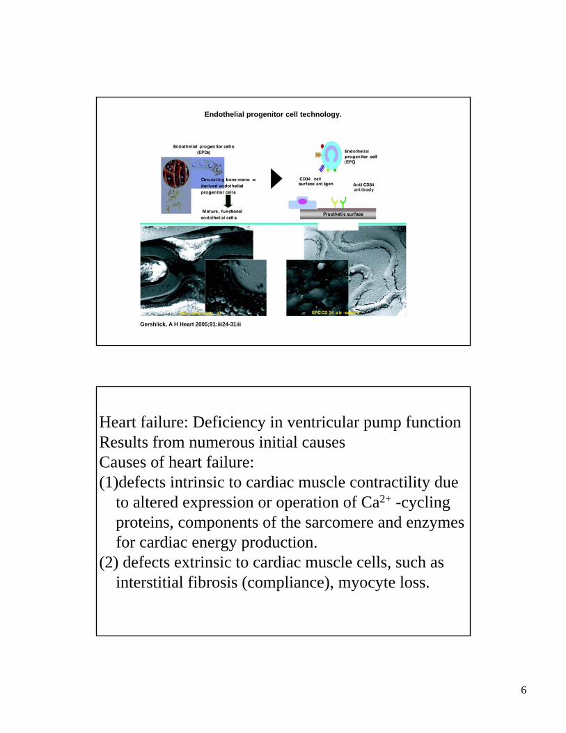
RyR2/calcium release channel is PKA hyperphosphorylated in failing hearts: reversal with Left Ventricular Assist Device
13
Farr and Basson, NEJM, July 8 2004 351: 185-187

14
The Long QT Syndrome: Dysfunction in Ventricular Repolarization
Keating and Sanguinetti, Cell (2001): 104: 569-580

15
Voltage at the cellular level
Surface potential –Entire heart
Membrane potential –Individual cells

16
Triggering Events for Syncope or SCD
• 3 main factors contributing to syncope or SCD– Exercise, especially swimming– Emotions or emotional stress– Events occurring during sleep or at rest, with or
without arousal
Circ 2001;103:89-95Mayo Clin Proc. 1999;74:1088-1094
Occurrence of Gene-Specific Triggers
Circ 2001;103:89-95

17
Beta Blocker Therapy is Effective
Moss, et al., Circulation. 2000 Feb 15;101(6):616-23
SCN5A Mutations: Invalid Targets for Beta Blockers:
Bradycardia and excessive AP prolongation

18
Control: QTc= 524 ms
Lidocaine: QTc= 442 ms
Figure 5. Lidocaine shortens Qtc intervalin patient carrying KPQ deletion mutationof SCN5A gene. Note minimal effects on QRS interval. Courtesy Dr. A. Moss.
Lidocaine Controls LQT-3 Prolongation
Catecholaminergic Ventricular Tachycardia: Mutations in RyR2

19
Timothy syndrome; loss of voltage dependentinactivation of Cav1.2

20
Conclusions for Inherited LQTS
• Inherited structural defects in ion channel architecture can cause arrhythmias;
• Triggers for arrhythmias are gene (mutation)-specific
• Therapeutic strategy is gene-specific
Cardiac regenerationCardiac injury in mammals and amphibians typically leads to scarring, with minimal regeneration of heart muscle.Cardiac regeneration is robust for certain organisms such as newt and zebrafish.Regeneration occurs through robust proliferation ofcardiomyocytes localized at the leading epicardial edge of the new myocardium. Poss et al Science 298:2188

21
Cardiac regeneration
Restorative growth dependent upon the retention of proliferativepotential in a subset of adult cardiomyocytes and is impossible in mammals under normal, unassisted biological circumstances.
Strategies to overcome restrictions:
(1) Overriding cell cycle checkpoints
(2) Supplementing cytoprotective mechanisms or inhibiting pro-death pathways
(3) Supplementing angiogenic mechanisms that occur naturally using defined growth factors or vessel-forming cells.
(4) Providing exogenous cells as a surrogate or precursor for cardiac muscle.
Dimmeler, S. et al. J. Clin. Invest. 2005;115:572-583
First strategy to be translated from bench to bedside: Cell implantation
Variety of adult progenitor cells, all autologousto avoid tissue rejection.First clinically relevant cells proposed were skeletal muscle myoblasts- undifferentiated, proliferation-competent cells that serve as precursors to skeletal muscle. Isolated from muscle biopsies, propagated and expanded ex vivo for a few days or weeks and then injected directly into the ventricular wall.
22
Dimmeler, S. et al. J. Clin. Invest. 2005;115:572-583
Cell implantation
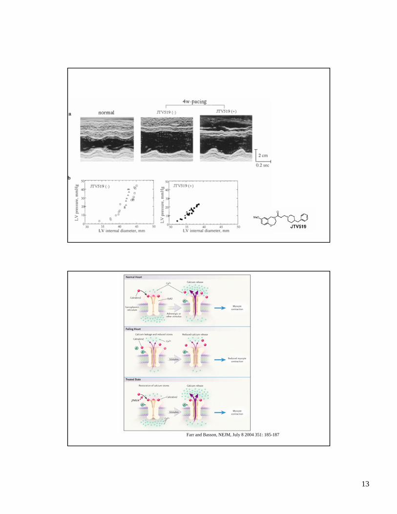
Bone marrow, at present, is the most frequent source of cells used for clincial cardiac repair.Consists of complex assortment of progenitor cells, including HSCs; side population (SP) cells, defined by their ability to expel aHoescht dye, mesenchymal stem cells (MSC) or stromal cells and multipotential adult progenitor cells (MAPCs), a subset of MSC.
Bone marrow aspirated under local or general anesthesia, the entire mononuclear cell fraction or specific subpopulation purified and isolated cells are injected into the heart without need for further ex vivo expansion.
Dimmeler, S. et al. J. Clin. Invest. 2005;115:572-583
Cell implantation
Peripheral blood-derived progenitor cells are used for clinical cardiac repair and neovascularization in peripheral arterial occlusive disease. These circulating cells (endothelial progenitor cells; EPC) are bone marrow derived. EPCs are isolated from mononuclear blood cells and selected ex vivo by culturing in endothelium specific medium for 3 days, prior to reinjection into the heart.Hypothesis is that these cells might transdifferentiate to create new cardiomyocytes.

23
Dimmeler, S. et al. J. Clin. Invest. 2005;115:572-583
Routes of applicationDelivered three ways:intracoronary arterial routeinjection into ventricular wall via percutaneous endocardial or surgical epicardial approach.
Intracoronary infusion- cells travel directly into myocardial regions in which nutrient blood flow and oxygen are preserved, ensuring a favorable environment for cells’ survival. Unperfused areas are targeted far less efficiently.Whereas bone marrow- and blood-derived progenitors cells are known to extravasate and migrate to ischemic areas, skeletal myoblastsdo not and may even obstruct the microcirculation leading to embolic myocardial damage.
Dimmeler, S. et al. J. Clin. Invest. 2005;115:572-583
Direct delivery of progenitor cells into scar tissue or areas of hibernating myocardium by catheter-based needle injection, direct injection during open-heart surgey, and minimally invasive thorascopic procedures are not limited by cell uptake from the circulation or embolic risk. Offsetting consideration is risk of ventricular perforation, which may limit use of direct needle injection into freshly infarcted hearts. Regions may lack syncytium of live muscle cells that may furnish instructive signals and lack blood flow for delivery of oxygen and nutrients. Electromechanical mapping of viable but hibernating myocardium may be useful.Diffuse disease- focal areas of injected cells might be poorly matched to underlying anatomy

24
Dimmeler, S. et al. J. Clin. Invest. 2005;115:572-583
Patient’s individual pathobiology will influence the source and route chosen. Not yet possible to assert an optimal cell type or best mode of delivery

25
Clinical trials:Distinguish between patients with acute myocardial infarction and chronic heart failure due to prior myocardial infarction.
In patients with acute myocardial infarction, progenitor cell transplantation is predicted to modify postinfarction LV remodeling through enhanced neovascularization and reduced cardiomyocyte apoptosis, irrespective of long-term engraftment and transdifferentiation. Enhanced neovascularization and reduced cardiomyocyte apoptosis may have little effect in long-established scars.
Myocardial ischemia acutely and potently upregulates the chemoattractants for neoangiogenesis- logical to test intracoronary infusion of bone marrow- or blood-derived progenitor cells in patients with acute MI.
Dimmeler, S. et al. J. Clin. Invest. 2005;115:572-583
Clinical trials of intracoronary progenitor cells for acute MI
All published trials reported nearly identical results; 7-9% improvement in global LV ejection fraction, significantly reduced end-systolic LV volumes and improved perfusion in the infarcted area, 4-6 months after cell transplantation.

26
Dimmeler, S. et al. J. Clin. Invest. 2005;115:572-583
Clinical trials of intracoronary progenitor cells for acute MI
In BOOST study (prospectively randomized), global LV function was significantly improved compared to nontreated control group.
In TOPCARE-AMI, magnetic resonance showed improvement of LV function and absence of reactive hypertrophy preserved after 1 year
Dimmeler, S. et al. J. Clin. Invest. 2005;115:572-583
Clinical trials of intracoronary progenitor cells for acute MI
In all 4 trials- totaling >100 patients- observed complications did not exceed controls. No arrhythmic complications.

27
First trial used skeletal muscle-derived progenitor cells directly injected in LV during open heart surgery for CABG. Global and regional LV function were improved, but may be due concomitant revascularization, complicating the assessment of benefit.
Transcatheter injection of myoblasts in myocardial scar reduced symptoms of heart failure but without objective evidence of improved global LV function.
Patients experienced life-threatening arrhythmias; lack of electrical coupling of skeletal muscle to neighboring cardiomyocytes, or coupling by the few hybrid cells formed by fusion with adjacent cardiomyocytes, which generate spatially heterogeneous calcium transients. Requires implantation of an cardioverter/defibrillator.
Dimmeler, S. et al. J. Clin. Invest. 2005;115:572-583
Clinical trials of catheter-based progenitor cell delivery for chronic coronary heart disease
Initial attempts in patients with chronic ischemic heart disease and old MI more heterogeneous in outcome. Patient population more heterogeneous.
28
Summary:For acute MI patients, the safety and suggestive efficacy of intracoronary progenitor cell transplantations provide rationale for randomized double blinded trials--- ongoing in USA and Europe.
For chronic ischemic heart failure--- is identification of hibernating myocyardium to direct cell therapy essential for effective outcome? Is delivery of skeletal myoblasts safe?
Does cellular therapy improve morbidity and mortality?
Do marrow derived cells implanted in the heart form cardiomyocytes? To date there is no direct clinical evidence that cellular cardiomyogenesis occurs in the human heart after transplantation of progenitor cells.
Cardiac myogenesis by noncardiac cells- human embryonic stem cells, if politically accceptable, pose the clinical challenge of immunological barriers.
In culture, embryonic stem cells form nodes of pulsing cells, presumably immature heart muscle cells, that beat in unison. But might these stem cells differentiate into different tissue (i.e. bone)
G-CSF- a number of investigators are using to force bone marrow to release stem cells, which are then collected from the blood and reinfused- could it promote cardiac inflammation? Boost blood vessel development in undetected tumors? 7/10 people receiving G-CSF in a trial in South Korea expereinced a renarrowing of the previously blocked artery, requiring treatment.

29
Cardiac myogenesis by adult cardiac progenitor cells
Several rationales: inability of skeletal myocytes to transdifferentiate; challenges to the claims of bone marrow-derived cells’ far-ranging plasticity and findings of tissue-resident progenitor cells, showing some evidence for stemness, yet predisposed to differentiate into lineages of the organ in which they reside.
?persistence as undifferentiated remnants of heart-forming tissue in the early embryo;
? Hematogenous origin
?ingrowth of the developing coronary vasculature
How many new cardiomyocytes, if any, are generated in the normal heart after birth? Do they replace dead or dying cells?
Conclusions:Molecular cardiology: lessons learned(1) Novel approaches to treat coronary artery disease- the identification of
smooth muscle proliferation after stent implantation-- targeted approaches using anti-proliferative drugs
(2) Heart failure-- SERCA2a/PLB; are cellular studies equivalent to whole animal studies? Animal models vs. man?
(3) Ryanodine receptor phosphorylation-- potential use of novel pharmaceuticals such as JTV519
(4) Cell transplantation-- potential uses for acute MI; chronic use unclear(5) Identifying genetic abnormalities for arrhythmias.