Embed Size (px)
Citation preview
Molecular Cell
Short Article
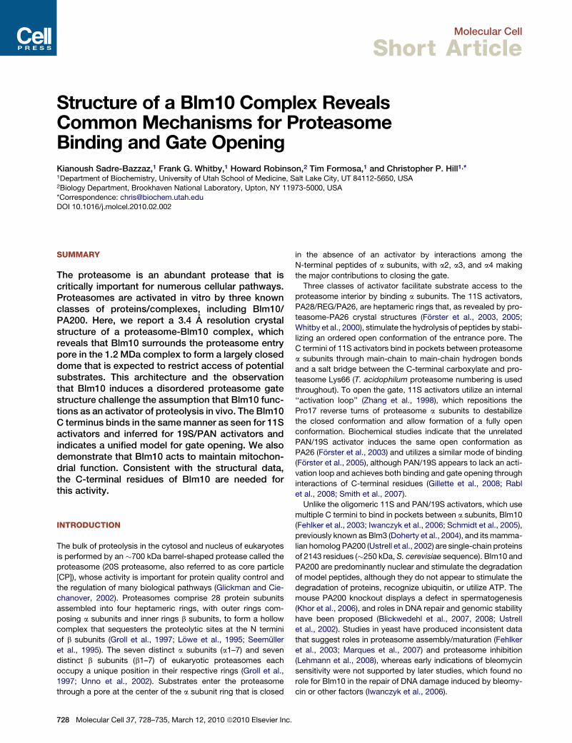
Structure of a Blm10 Complex RevealsCommon Mechanisms for ProteasomeBinding and Gate OpeningKianoush Sadre-Bazzaz,1 Frank G. Whitby,1 Howard Robinson,2 Tim Formosa,1 and Christopher P. Hill1,*1Department of Biochemistry, University of Utah School of Medicine, Salt Lake City, UT 84112-5650, USA2Biology Department, Brookhaven National Laboratory, Upton, NY 11973-5000, USA
*Correspondence: [email protected] 10.1016/j.molcel.2010.02.002
SUMMARY
The proteasome is an abundant protease that iscritically important for numerous cellular pathways.Proteasomes are activated in vitro by three knownclasses of proteins/complexes, including Blm10/PA200. Here, we report a 3.4 A resolution crystalstructure of a proteasome-Blm10 complex, whichreveals that Blm10 surrounds the proteasome entrypore in the 1.2 MDa complex to form a largely closeddome that is expected to restrict access of potentialsubstrates. This architecture and the observationthat Blm10 induces a disordered proteasome gatestructure challenge the assumption that Blm10 func-tions as an activator of proteolysis in vivo. The Blm10C terminus binds in the same manner as seen for 11Sactivators and inferred for 19S/PAN activators andindicates a unified model for gate opening. We alsodemonstrate that Blm10 acts to maintain mitochon-drial function. Consistent with the structural data,the C-terminal residues of Blm10 are needed forthis activity.
INTRODUCTION
The bulk of proteolysis in the cytosol and nucleus of eukaryotes
is performed by an �700 kDa barrel-shaped protease called the
proteasome (20S proteasome, also referred to as core particle
[CP]), whose activity is important for protein quality control and
the regulation of many biological pathways (Glickman and Cie-
chanover, 2002). Proteasomes comprise 28 protein subunits
assembled into four heptameric rings, with outer rings com-
posing a subunits and inner rings b subunits, to form a hollow
complex that sequesters the proteolytic sites at the N termini
of b subunits (Groll et al., 1997; Lowe et al., 1995; Seemuller
et al., 1995). The seven distinct a subunits (a1–7) and seven
distinct b subunits (b1–7) of eukaryotic proteasomes each
occupy a unique position in their respective rings (Groll et al.,
1997; Unno et al., 2002). Substrates enter the proteasome
through a pore at the center of the a subunit ring that is closed
728 Molecular Cell 37, 728–735, March 12, 2010 ª2010 Elsevier Inc.
in the absence of an activator by interactions among the
N-terminal peptides of a subunits, with a2, a3, and a4 making
the major contributions to closing the gate.
Three classes of activator facilitate substrate access to the
proteasome interior by binding a subunits. The 11S activators,
PA28/REG/PA26, are heptameric rings that, as revealed by pro-
teasome-PA26 crystal structures (Forster et al., 2003, 2005;
Whitby et al., 2000), stimulate the hydrolysis of peptides by stabi-
lizing an ordered open conformation of the entrance pore. The
C termini of 11S activators bind in pockets between proteasome
a subunits through main-chain to main-chain hydrogen bonds
and a salt bridge between the C-terminal carboxylate and pro-
teasome Lys66 (T. acidophilum proteasome numbering is used
throughout). To open the gate, 11S activators utilize an internal
‘‘activation loop’’ (Zhang et al., 1998), which repositions the
Pro17 reverse turns of proteasome a subunits to destabilize
the closed conformation and allow formation of a fully open
conformation. Biochemical studies indicate that the unrelated
PAN/19S activator induces the same open conformation as
PA26 (Forster et al., 2003) and utilizes a similar mode of binding
(Forster et al., 2005), although PAN/19S appears to lack an acti-
vation loop and achieves both binding and gate opening through
interactions of C-terminal residues (Gillette et al., 2008; Rabl
et al., 2008; Smith et al., 2007).
Unlike the oligomeric 11S and PAN/19S activators, which use
multiple C termini to bind in pockets between a subunits, Blm10
(Fehlker et al., 2003; Iwanczyk et al., 2006; Schmidt et al., 2005),
previously known as Blm3 (Doherty et al., 2004), and its mamma-
lian homolog PA200 (Ustrell et al., 2002) are single-chain proteins
of 2143 residues (�250 kDa, S. cerevisiae sequence). Blm10 and
PA200 are predominantly nuclear and stimulate the degradation
of model peptides, although they do not appear to stimulate the
degradation of proteins, recognize ubiquitin, or utilize ATP. The
mouse PA200 knockout displays a defect in spermatogenesis
(Khor et al., 2006), and roles in DNA repair and genomic stability
have been proposed (Blickwedehl et al., 2007, 2008; Ustrell
et al., 2002). Studies in yeast have produced inconsistent data
that suggest roles in proteasome assembly/maturation (Fehlker
et al., 2003; Marques et al., 2007) and proteasome inhibition
(Lehmann et al., 2008), whereas early indications of bleomycin
sensitivity were not supported by later studies, which found no
role for Blm10 in the repair of DNA damage induced by bleomy-
cin or other factors (Iwanczyk et al., 2006).
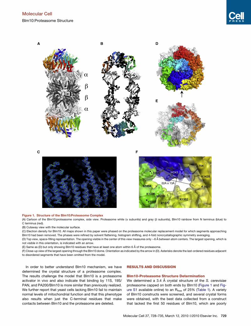
Figure 1. Structure of the Blm10:Proteasome Complex
(A) Cartoon of the Blm10:proteasome complex, side view. Proteasome white (a subunits) and gray (b subunits), Blm10 rainbow from N terminus (blue) to
C terminus (red).
(B) Cutaway view with the molecular surface.
(C) Electron density for Blm10. All maps shown in this paper were phased on the proteasome molecular replacement model for which segments approaching
Blm10 had been removed. The phases were refined by solvent flattening, histogram shifting, and 4-fold noncrystallographic symmetry averaging.
(D) Top view, space-filling representation. The opening visible in the center of this view measures only�6 A between atom centers. The largest opening, which is
not visible in this orientation, is indicated with an arrow.
(E) Same as (D) but only showing Blm10 residues that have at least one atom within 6 A of the proteasome.
(F) Close-up view of the largest opening through the Blm10 dome. Orientation as indicated by the arrow in (D). Asterisks denote the last-ordered residues adjacent
to disordered segments that have been omitted from the model.
Molecular Cell
Blm10:Proteasome Structure
In order to better understand Blm10 mechanism, we have
determined the crystal structure of a proteasome complex.
The results challenge the model that Blm10 is a proteasome
activator in vivo and also indicate that binding by 11S, 19S/
PAN, and PA200/Blm10 is more similar than previously realized.
We further report that yeast cells lacking Blm10 fail to maintain
normal levels of mitochondrial function and that this phenotype
also results when just the C-terminal residues that make
contacts between Blm10 and the proteasome are deleted.
M
RESULTS AND DISCUSSION
Blm10-Proteasome Structure DeterminationWe determined a 3.4 A crystal structure of the S. cerevisiae
proteasome capped on both ends by Blm10 (Figure 1 and Fig-
ure S1 available online) to an Rfree of 25% (Table 1). A variety
of Blm10 constructs were screened, and several crystal forms
were obtained, with the best data collected from a construct
that lacked the first 50 residues of Blm10, which are poorly
olecular Cell 37, 728–735, March 12, 2010 ª2010 Elsevier Inc. 729
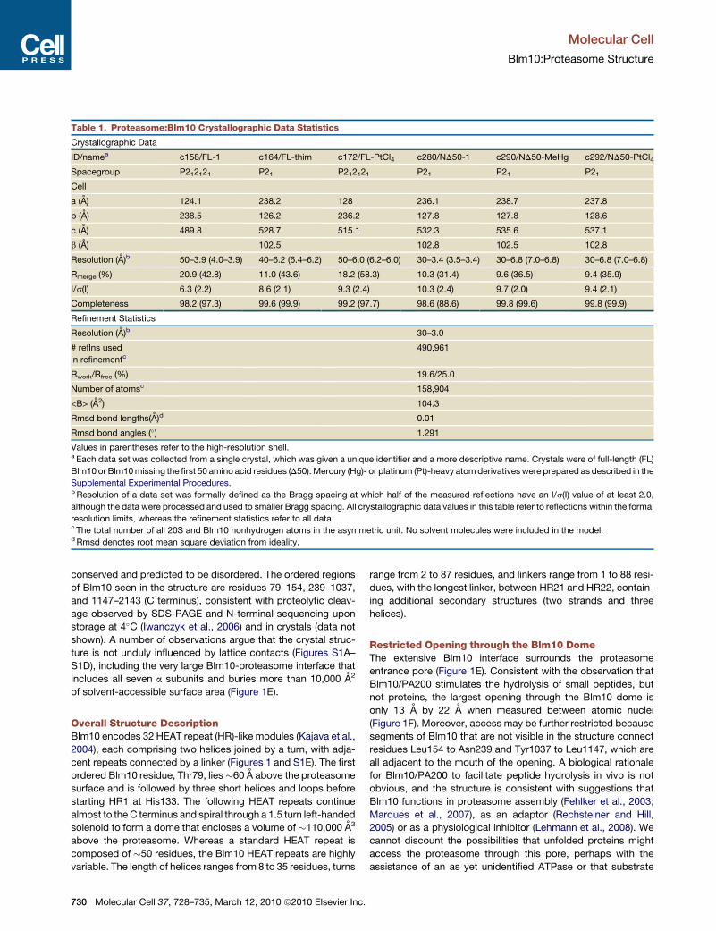
Table 1. Proteasome:Blm10 Crystallographic Data Statistics
Crystallographic Data
ID/namea c158/FL-1 c164/FL-thim c172/FL-PtCl4 c280/ND50-1 c290/ND50-MeHg c292/ND50-PtCl4
Spacegroup P212121 P21 P212121 P21 P21 P21
Cell
a (A) 124.1 238.2 128 236.1 238.7 237.8
b (A) 238.5 126.2 236.2 127.8 127.8 128.6
c (A) 489.8 528.7 515.1 532.3 535.6 537.1
b (A) 102.5 102.8 102.5 102.8
Resolution (A)b 50–3.9 (4.0–3.9) 40–6.2 (6.4–6.2) 50–6.0 (6.2–6.0) 30–3.4 (3.5–3.4) 30–6.8 (7.0–6.8) 30–6.8 (7.0–6.8)
Rmerge (%) 20.9 (42.8) 11.0 (43.6) 18.2 (58.3) 10.3 (31.4) 9.6 (36.5) 9.4 (35.9)
I/s(I) 6.3 (2.2) 8.6 (2.1) 9.3 (2.4) 10.3 (2.4) 9.7 (2.0) 9.4 (2.1)
Completeness 98.2 (97.3) 99.6 (99.9) 99.2 (97.7) 98.6 (88.6) 99.8 (99.6) 99.8 (99.9)
Refinement Statistics
Resolution (A)b 30–3.0
# reflns used
in refinementc490,961
Rwork/Rfree (%) 19.6/25.0
Number of atomsc 158,904
<B> (A2) 104.3
Rmsd bond lengths(A)d 0.01
Rmsd bond angles (�) 1.291
Values in parentheses refer to the high-resolution shell.a Each data set was collected from a single crystal, which was given a unique identifier and a more descriptive name. Crystals were of full-length (FL)
Blm10 or Blm10 missing the first 50 amino acid residues (D50). Mercury (Hg)- or platinum (Pt)-heavy atom derivatives were prepared as described in the
Supplemental Experimental Procedures.b Resolution of a data set was formally defined as the Bragg spacing at which half of the measured reflections have an I/s(I) value of at least 2.0,
although the data were processed and used to smaller Bragg spacing. All crystallographic data values in this table refer to reflections within the formal
resolution limits, whereas the refinement statistics refer to all data.c The total number of all 20S and Blm10 nonhydrogen atoms in the asymmetric unit. No solvent molecules were included in the model.d Rmsd denotes root mean square deviation from ideality.
Molecular Cell
Blm10:Proteasome Structure
conserved and predicted to be disordered. The ordered regions
of Blm10 seen in the structure are residues 79–154, 239–1037,
and 1147–2143 (C terminus), consistent with proteolytic cleav-
age observed by SDS-PAGE and N-terminal sequencing upon
storage at 4�C (Iwanczyk et al., 2006) and in crystals (data not
shown). A number of observations argue that the crystal struc-
ture is not unduly influenced by lattice contacts (Figures S1A–
S1D), including the very large Blm10-proteasome interface that
includes all seven a subunits and buries more than 10,000 A2
of solvent-accessible surface area (Figure 1E).
Overall Structure DescriptionBlm10 encodes 32 HEAT repeat (HR)-like modules (Kajava et al.,
2004), each comprising two helices joined by a turn, with adja-
cent repeats connected by a linker (Figures 1 and S1E). The first
ordered Blm10 residue, Thr79, lies�60 A above the proteasome
surface and is followed by three short helices and loops before
starting HR1 at His133. The following HEAT repeats continue
almost to the C terminus and spiral through a 1.5 turn left-handed
solenoid to form a dome that encloses a volume of �110,000 A3
above the proteasome. Whereas a standard HEAT repeat is
composed of �50 residues, the Blm10 HEAT repeats are highly
variable. The length of helices ranges from 8 to 35 residues, turns
730 Molecular Cell 37, 728–735, March 12, 2010 ª2010 Elsevier Inc.
range from 2 to 87 residues, and linkers range from 1 to 88 resi-
dues, with the longest linker, between HR21 and HR22, contain-
ing additional secondary structures (two strands and three
helices).
Restricted Opening through the Blm10 DomeThe extensive Blm10 interface surrounds the proteasome
entrance pore (Figure 1E). Consistent with the observation that
Blm10/PA200 stimulates the hydrolysis of small peptides, but
not proteins, the largest opening through the Blm10 dome is
only 13 A by 22 A when measured between atomic nuclei
(Figure 1F). Moreover, access may be further restricted because
segments of Blm10 that are not visible in the structure connect
residues Leu154 to Asn239 and Tyr1037 to Leu1147, which are
all adjacent to the mouth of the opening. A biological rationale
for Blm10/PA200 to facilitate peptide hydrolysis in vivo is not
obvious, and the structure is consistent with suggestions that
Blm10 functions in proteasome assembly (Fehlker et al., 2003;
Marques et al., 2007), as an adaptor (Rechsteiner and Hill,
2005) or as a physiological inhibitor (Lehmann et al., 2008). We
cannot discount the possibilities that unfolded proteins might
access the proteasome through this pore, perhaps with the
assistance of an as yet unidentified ATPase or that substrate
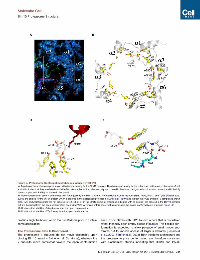
Figure 2. Proteasome Conformational Changes Induced by Blm10(A) Top view of the proteasome pore region with electron density for the Blm10 complex. The absence of density for the N-terminal residues of proteasome a2, a3,
and a4 indicates that they are disordered in the Blm10 complex (white), whereas they are ordered in the closed, unliganded conformation (colors) and in the fully
open complex with PA26 (not shown in this panel).
(B) Open conformation seen in complexes with PA26 (yellow) and Blm10 (white). The stabilizing cluster residues (Tyr8, Asp9, Pro17, and Tyr26 [Forster et al.,
2003]) are labeled for the a6/a7 cluster, which is ordered in the unliganded proteasome (Groll et al., 1997) and in both the PA26 and Blm10 complexes shown
here. Tyr8 and Asp9 residues are not ordered for a2, a3, or a4 in the Blm10 complex. Residues indicated with an asterisk are ordered in the Blm10 complex
but are displaced from the open conformation seen with PA26. A version of this panel that also includes the closed conformation is shown in Figure S2.
(C) Contacts that stabilize a5Asp9 away from the open conformation.
(D) Contacts that stabilize a7Tyr8 away from the open conformation.
Molecular Cell
Blm10:Proteasome Structure
proteins might be bound within the Blm10 dome prior to protea-
some association.
The Proteasome Gate Is DisorderedThe proteasome b subunits do not move discernibly upon
binding Blm10 (rmsd = 0.4 A on all Ca atoms), whereas the
a subunits move somewhat toward the open conformation
M
seen in complexes with PA26 to form a pore that is disordered
rather than fully open or fully closed (Figure 2). This flexible con-
formation is expected to allow passage of small model sub-
strates but to impede access of larger substrates (Benaroudj
et al., 2003; Forster et al., 2003). Both the dome architecture and
the proteasome pore conformation are therefore consistent
with biochemical studies indicating that Blm10 and PA200
olecular Cell 37, 728–735, March 12, 2010 ª2010 Elsevier Inc. 731

Figure 3. Interactions of the Blm10 C-Terminal Residues
(A) Side view with Blm10 C terminus labeled ‘‘C.’’
(B) The electron density map is well defined for the Blm10 penultimate
tyrosine (Tyr2142) and surrounding residues.
(C) The last three residues of PA26 (green) and Blm10 (red) are shown after over-
lap of the two complexes on surrounding proteasome residues. Unliganded
proteasome (Groll et al., 1997), cyan. Blm10 Tyr2142 stabilizes the open posi-
Molecular Cell
Blm10:Proteasome Structure
732 Molecular Cell 37, 728–735, March 12, 2010 ª2010 Elsevier Inc.
stimulate the hydrolysis of peptides, but not proteins (Iwanczyk
et al., 2006; Schmidt et al., 2005; Ustrell et al., 2002).
It is instructive to compare the proteasome complexes with
Blm10 and PA26. The fully open conformation results from repo-
sitioning of the seven proteasome a subunit Pro17 turns by the
PA26 activation loop to form a wider, more circular arrangement,
with the largest Pro17 Ca movement (3.6 A) seen for a4 and the
smallest Pro17 Ca movement (0.4 A) seen for a1 (Forster et al.,
2003). Repositioning of the Pro17 turns induces ordering of the
Tyr8 and Asp9 residues of all seven proteasome a subunits to
form a continuous belt around the pore circumference that is
stabilized by conserved clusters of Tyr8, Asp9, Pro17, and Tyr26
proteasome residues (Figure 2B) (notwithstanding the nonca-
nonical a1/a2 cluster [Forster et al., 2003]). Blm10 stabilizes
the same Pro17 transition for proteasome a5 as seen with PA26,
although it does so primarily by interactions of its C-terminal resi-
dues rather than by an internal activation loop. In contrast, the
Pro17 turns of a2, a3, and a4 lack direct contacts with Blm10
and become disordered. Moreover, Blm10 blocks the fully open
conformation by displacing a5Asp9 from a position where it
could bind a4Tyr8 (Figure 2C) and by displacing a7Tyr8 from
a position where it could bind a1Asp9 (Figure 2D). This explains
why a1 and a4, and hence their contacting a2 and a3 subunits,
do not form the same open conformation as seen with PA26.
Implications for Binding and Gate Opening by 19S/PANThe C-terminal residues of Blm10 bind in the pocket between
proteasome a5 and a6 in a conformation that superimposes
with the C-terminal residues of PA26 (Figure 3). PA26 is hepta-
meric and binds to all seven pockets of the 7-fold symmetric
archaeal T. acidophilum proteasome and to four (a2/a3, a3/a4,
a4/a5, a5/a6) pockets of the S. cerevisiae proteasome (Forster
et al., 2005). Like PA26, the Blm10 C-terminal residues form
b sheet-like hydrogen bonds with the proteasome, and the
Blm10 C-terminal carboxylate forms a salt bridge with a6Lys66.
Of interest, biochemical (Forster et al., 2005; Smith et al., 2007)
and electron microscopic (Rabl et al., 2008) data indicate that
the C termini of some of the 19S/RC activator ATPases and their
archaeal homolog PAN also bind to the same site, presumably
using the same interactions.
In contrast to the apparently shared mode of binding by 11S,
Blm10/PA200, and 19S/PAN, an important difference is that
peptides corresponding to the seven C-terminal residues of
PAN and some 19S subunits are able to both bind proteasomes
and stabilize the open gate conformation (Gillette et al., 2008;
Rabl et al., 2008; Smith et al., 2007), whereas PA26/11S use their
C-terminal sequences for binding but rely upon a distantly
located activation loop to induce gate opening. Furthermore,
a critical interaction for proteasome gate opening has been map-
ped to the penultimate tyrosine of PAN/19S ATPases, with some
of the homologs containing a phenylalanine at this position
(Gillette et al., 2008; Smith et al., 2007). Remarkably, the penul-
timate residue of Blm10, Tyr2142, is also invariably conserved
as tyrosine or phenylalanine in an alignment of 46 sequences
(Figure S1E). This residue possesses well-defined electron
tion of a5 by hydrogen bonding with Gly19 O. PA26 stabilizes the same transi-
tion by hydrogen bonding interactions of its activation loop residue Glu102.
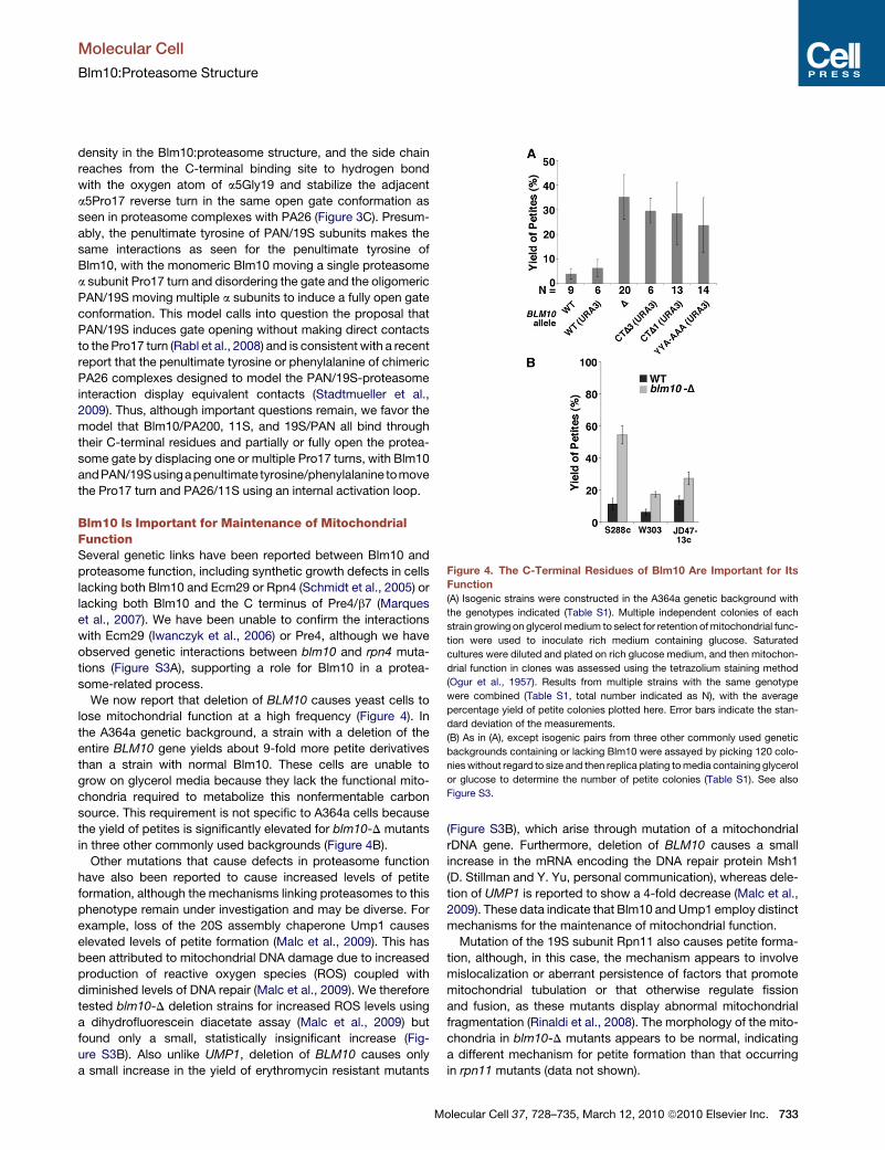
Figure 4. The C-Terminal Residues of Blm10 Are Important for Its
Function
(A) Isogenic strains were constructed in the A364a genetic background with
the genotypes indicated (Table S1). Multiple independent colonies of each
strain growing on glycerol medium to select for retention of mitochondrial func-
tion were used to inoculate rich medium containing glucose. Saturated
cultures were diluted and plated on rich glucose medium, and then mitochon-
drial function in clones was assessed using the tetrazolium staining method
(Ogur et al., 1957). Results from multiple strains with the same genotype
were combined (Table S1, total number indicated as N), with the average
percentage yield of petite colonies plotted here. Error bars indicate the stan-
dard deviation of the measurements.
(B) As in (A), except isogenic pairs from three other commonly used genetic
backgrounds containing or lacking Blm10 were assayed by picking 120 colo-
nies without regard to size and then replica plating to media containing glycerol
or glucose to determine the number of petite colonies (Table S1). See also
Figure S3.
Molecular Cell
Blm10:Proteasome Structure
density in the Blm10:proteasome structure, and the side chain
reaches from the C-terminal binding site to hydrogen bond
with the oxygen atom of a5Gly19 and stabilize the adjacent
a5Pro17 reverse turn in the same open gate conformation as
seen in proteasome complexes with PA26 (Figure 3C). Presum-
ably, the penultimate tyrosine of PAN/19S subunits makes the
same interactions as seen for the penultimate tyrosine of
Blm10, with the monomeric Blm10 moving a single proteasome
a subunit Pro17 turn and disordering the gate and the oligomeric
PAN/19S moving multiple a subunits to induce a fully open gate
conformation. This model calls into question the proposal that
PAN/19S induces gate opening without making direct contacts
to the Pro17 turn (Rabl et al., 2008) and is consistent with a recent
report that the penultimate tyrosine or phenylalanine of chimeric
PA26 complexes designed to model the PAN/19S-proteasome
interaction display equivalent contacts (Stadtmueller et al.,
2009). Thus, although important questions remain, we favor the
model that Blm10/PA200, 11S, and 19S/PAN all bind through
their C-terminal residues and partially or fully open the protea-
some gate by displacing one or multiple Pro17 turns, with Blm10
andPAN/19Susingapenultimate tyrosine/phenylalanine tomove
the Pro17 turn and PA26/11S using an internal activation loop.
Blm10 Is Important for Maintenance of MitochondrialFunctionSeveral genetic links have been reported between Blm10 and
proteasome function, including synthetic growth defects in cells
lacking both Blm10 and Ecm29 or Rpn4 (Schmidt et al., 2005) or
lacking both Blm10 and the C terminus of Pre4/b7 (Marques
et al., 2007). We have been unable to confirm the interactions
with Ecm29 (Iwanczyk et al., 2006) or Pre4, although we have
observed genetic interactions between blm10 and rpn4 muta-
tions (Figure S3A), supporting a role for Blm10 in a protea-
some-related process.
We now report that deletion of BLM10 causes yeast cells to
lose mitochondrial function at a high frequency (Figure 4). In
the A364a genetic background, a strain with a deletion of the
entire BLM10 gene yields about 9-fold more petite derivatives
than a strain with normal Blm10. These cells are unable to
grow on glycerol media because they lack the functional mito-
chondria required to metabolize this nonfermentable carbon
source. This requirement is not specific to A364a cells because
the yield of petites is significantly elevated for blm10-D mutants
in three other commonly used backgrounds (Figure 4B).
Other mutations that cause defects in proteasome function
have also been reported to cause increased levels of petite
formation, although the mechanisms linking proteasomes to this
phenotype remain under investigation and may be diverse. For
example, loss of the 20S assembly chaperone Ump1 causes
elevated levels of petite formation (Malc et al., 2009). This has
been attributed to mitochondrial DNA damage due to increased
production of reactive oxygen species (ROS) coupled with
diminished levels of DNA repair (Malc et al., 2009). We therefore
tested blm10-D deletion strains for increased ROS levels using
a dihydrofluorescein diacetate assay (Malc et al., 2009) but
found only a small, statistically insignificant increase (Fig-
ure S3B). Also unlike UMP1, deletion of BLM10 causes only
a small increase in the yield of erythromycin resistant mutants
M
(Figure S3B), which arise through mutation of a mitochondrial
rDNA gene. Furthermore, deletion of BLM10 causes a small
increase in the mRNA encoding the DNA repair protein Msh1
(D. Stillman and Y. Yu, personal communication), whereas dele-
tion of UMP1 is reported to show a 4-fold decrease (Malc et al.,
2009). These data indicate that Blm10 and Ump1 employ distinct
mechanisms for the maintenance of mitochondrial function.
Mutation of the 19S subunit Rpn11 also causes petite forma-
tion, although, in this case, the mechanism appears to involve
mislocalization or aberrant persistence of factors that promote
mitochondrial tubulation or that otherwise regulate fission
and fusion, as these mutants display abnormal mitochondrial
fragmentation (Rinaldi et al., 2008). The morphology of the mito-
chondria in blm10-D mutants appears to be normal, indicating
a different mechanism for petite formation than that occurring
in rpn11 mutants (data not shown).
olecular Cell 37, 728–735, March 12, 2010 ª2010 Elsevier Inc. 733
Molecular Cell
Blm10:Proteasome Structure
The C Terminus of Blm10 Is Importantfor Its Physiological FunctionTo test the importance of the conserved C terminus of Blm10, we
deleted the last three codons (TyrTyrAla) in the normal genomic
context. Consistent with the observation that these residues
make intimate contact with the proteasome but do not contact
other Blm10 residues, this mutant was as stable as the intact
protein (Figure S3C) and localized to the nucleus in a manner
indistinguishable from WT (data not shown). The truncated
protein failed to maintain normal levels of mitochondrial func-
tion, and the yield of petites was similar to that obtained with
a complete deletion of BLM10 (Figure 4A). Other perturbations
of the C terminus, including deletion of just the last residue or
mutation of the YYA sequence to AAA, also caused severe
impairment of Blm10 function (Figure 4A). These demonstrations
that the C terminus of Blm10 performs a physiologically impor-
tant function are consistent with our structural finding that these
residues play a specific role in proteasome binding and definition
of the gate conformation.
ConclusionsWe have verified that Blm10 functions in a proteasome-related
process and have demonstrated that it is required for the
maintenance of mitochondrial function by a mechanism that
is distinct from that of previously reported genes. We have
also determined the structure of Blm10 in complex with the pro-
teasome and found an unexpected similarity in binding by
C-terminal residues, which indicates common modes of protea-
some binding for Blm10, 11S, and 19S/PAN. Of note, the penul-
timate tyrosine residue makes specific interactions that suggest
a refinement of current models for binding and gate opening
by 19S/PAN activators. Consistent with the structure, genetic
analysis indicates that the C-terminal residues of Blm10 are
required for its physiological function. Despite these advances,
a number of important questions remain. For example, we do
not yet understand the role of Blm10 in maintaining mitochon-
drial vitality, although this phenotype provides both a conceptual
rationale for the conservation of Blm10 among eukaryotes and
an assay to probe the effect of mutating the BLM10 gene on
a physiologically important function of Blm10. We also note
that, in view of the high abundance of mitochondria in sperma-
tozoa, it is attractive to speculate that a role in maintaining mito-
chondrial function underlies the requirement of the mammalian
Blm10 homolog PA200 for spermatogenesis (Khor et al., 2006).
The structure appears somewhat at odds with Blm10s charac-
terization as a proteasome activator in vitro and seems more
consistent with possible functions as an adaptor, assembly
factor, or inhibitor. Thus, whereas fundamentally important
questions of Blm10 biology remain to be answered, the structure
and genetic demonstration of a role in maintaining mitochondrial
function provide tools and insight that can guide future studies.
EXPERIMENTAL PROCEDURES
See the Supplemental Information for a more complete description of the
methods. Double-capped S. cerevisiae proteasome:Blm10 and D50Blm10
complexes were prepared largely as described (Iwanczyk et al., 2006). Protein
was concentrated to 20–25 mg/ml for crystallization by vapor diffusion.
734 Molecular Cell 37, 728–735, March 12, 2010 ª2010 Elsevier Inc.
Diffraction data were collected at 100K at the National Synchrotron Light
Source beamline X29 and were phased by molecular replacement using the
unliganded proteasome (Groll et al., 1997) (PDB code: 1ryp) as the search
model.
ACCESSION NUMBERS
Coordinates and diffraction data have been deposited at the Protein Data
Bank with accession code 3L5Q.
SUPPLEMENTAL INFORMATION
Supplemental Data include Supplemental Experimental Procedures,
three figures, and one table and can be found with this article online at
doi:10.1016/j.molcel.2010.02.002.
ACKNOWLEDGMENTS
We thank Charisse Kettelkamp and Hua Xin for technical assistance, Pavel
Afonine for running Phenix calculations, Heidi Schubert for assistance and
advice on aspects of the crystallography, Daniel Finley and David Legett for
providing the yeast strain sDL135 that expresses the protein A-tagged Pre1
proteasome subunit, David Stillman and Yaxin Yu for MSH1 mRNA data,
and Martin Rechsteiner and Beth Stadtmueller for critical comments on the
manuscript. We gratefully acknowledge the DNA Synthesis and Sequencing
Core Facilities at the University of Utah. X-ray diffraction data for this study
were measured at beamline X29 of the National Synchrotron Light Source
(NSLS). Financial support for NSLS comes principally from the Offices of Bio-
logical and Environmental Research and of Basic Energy Sciences of the US
Department of Energy and from the National Center for Research Resources
of the National Institutes of Health. This work was supported by National Insti-
tutes of Health Grant RO1 GM059135.
Received: June 29, 2009
Revised: November 9, 2009
Accepted: December 29, 2009
Published: March 11, 2010
REFERENCES
Benaroudj, N., Zwickl, P., Seemuller, E., Baumeister, W., and Goldberg, A.L.
(2003). ATP hydrolysis by the proteasome regulatory complex PAN serves
multiple functions in protein degradation. Mol. Cell 11, 69–78.
Blickwedehl, J., McEvoy, S., Wong, I., Kousis, P., Clements, J., Elliott, R.,
Cresswell, P., Liang, P., and Bangia, N. (2007). Proteasomes and proteasome
activator 200 kDa (PA200) accumulate on chromatin in response to ionizing
radiation. Radiat. Res. 167, 663–674.
Blickwedehl, J., Agarwal, M., Seong, C., Pandita, R.K., Melendy, T., Sung, P.,
Pandita, T.K., and Bangia, N. (2008). Role for proteasome activator PA200 and
postglutamyl proteasome activity in genomic stability. Proc. Natl. Acad. Sci.
USA 105, 16165–16170.
Doherty, K., Pramanik, A., Pride, L., Lukose, J., and Moore, C.W. (2004).
Expression of the expanded YFL007w ORF and assignment of the gene
name BLM10. Yeast 21, 1021–1023.
Fehlker, M., Wendler, P., Lehmann, A., and Enenkel, C. (2003). Blm3 is part of
nascent proteasomes and is involved in a late stage of nuclear proteasome
assembly. EMBO Rep. 4, 959–963.
Forster, A., Whitby, F.G., and Hill, C.P. (2003). The pore of activated 20S pro-
teasomes has an ordered 7-fold symmetric conformation. EMBO J. 22, 4356–
4364.
Forster, A., Masters, E.I., Whitby, F.G., Robinson, H., and Hill, C.P. (2005). The
1.9 A structure of a proteasome-11S activator complex and implications for
proteasome-PAN/PA700 interactions. Mol. Cell 18, 589–599.
Gillette, T.G., Kumar, B., Thompson, D., Slaughter, C.A., and DeMartino, G.N.
(2008). Differential roles of the COOH termini of AAA subunits of PA700 (19 S
Molecular Cell
Blm10:Proteasome Structure
regulator) in asymmetric assembly and activation of the 26 S proteasome.
J. Biol. Chem. 283, 31813–31822.
Glickman, M.H., and Ciechanover, A. (2002). The ubiquitin-proteasome
proteolytic pathway: destruction for the sake of construction. Physiol. Rev.
82, 373–428.
Groll, M., Ditzel, L., Lowe, J., Stock, D., Bochtler, M., Bartunik, H.D., and
Huber, R. (1997). Structure of 20S proteasome from yeast at 2.4 A resolution.
Nature 386, 463–471.
Iwanczyk, J., Sadre-Bazzaz, K., Ferrell, K., Kondrashkina, E., Formosa, T., Hill,
C.P., and Ortega, J. (2006). Structure of the Blm10-20 S proteasome complex
by cryo-electron microscopy. Insights into the mechanism of activation of
mature yeast proteasomes. J. Mol. Biol. 363, 648–659.
Kajava, A.V., Gorbea, C., Ortega, J., Rechsteiner, M., and Steven, A.C. (2004).
New HEAT-like repeat motifs in proteins regulating proteasome structure and
function. J. Struct. Biol. 146, 425–430.
Khor, B., Bredemeyer, A.L., Huang, C.Y., Turnbull, I.R., Evans, R., Maggi, L.B.,
Jr., White, J.M., Walker, L.M., Carnes, K., Hess, R.A., and Sleckman, B.P.
(2006). Proteasome activator PA200 is required for normal spermatogenesis.
Mol. Cell. Biol. 26, 2999–3007.
Lehmann, A., Jechow, K., and Enenkel, C. (2008). Blm10 binds to pre-acti-
vated proteasome core particles with open gate conformation. EMBO Rep.
9, 1237–1243.
Lowe, J., Stock, D., Jap, B., Zwickl, P., Baumeister, W., and Huber, R. (1995).
Crystal structure of the 20S proteasome from the archaeon T. acidophilum at
3.4 A resolution. Science 268, 533–539.
Malc, E., Dzierzbicki, P., Kaniak, A., Skoneczna, A., and Ciesla, Z. (2009). Inac-
tivation of the 20S proteasome maturase, Ump1p, leads to the instability of
mtDNA in Saccharomyces cerevisiae. Mutat. Res. 669, 95–103.
Marques, A.J., Glanemann, C., Ramos, P.C., and Dohmen, R.J. (2007). The
C-terminal extension of the beta7 subunit and activator complexes stabilize
nascent 20 S proteasomes and promote their maturation. J. Biol. Chem.
282, 34869–34876.
Ogur, M., St. John, R., and Nagai, S. (1957). Tetrazolium overlay technique for
population studies of respiration deficiency in yeast. Science 125, 928–929.
M
Rabl, J., Smith, D.M., Yu, Y., Chang, S.C., Goldberg, A.L., and Cheng, Y.
(2008). Mechanism of gate opening in the 20S proteasome by the proteasomal
ATPases. Mol. Cell 30, 360–368.
Rechsteiner, M., and Hill, C.P. (2005). Mobilizing the proteolytic machine: cell
biological roles of proteasome activators and inhibitors. Trends Cell Biol. 15,
27–33.
Rinaldi, T., Hofmann, L., Gambadoro, A., Cossard, R., Livnat-Levanon, N.,
Glickman, M.H., Frontali, L., and Delahodde, A. (2008). Dissection of the
carboxyl-terminal domain of the proteasomal subunit Rpn11 in maintenance
of mitochondrial structure and function. Mol. Biol. Cell 19, 1022–1031.
Schmidt, M., Haas, W., Crosas, B., Santamaria, P.G., Gygi, S.P., Walz, T., and
Finley, D. (2005). The HEAT repeat protein Blm10 regulates the yeast protea-
some by capping the core particle. Nat. Struct. Mol. Biol. 12, 294–303.
Seemuller, E., Lupas, A., Stock, D., Lowe, J., Huber, R., and Baumeister, W.
(1995). Proteasome from Thermoplasma acidophilum: a threonine protease.
Science 268, 579–582.
Smith, D.M., Chang, S.C., Park, S., Finley, D., Cheng, Y., and Goldberg, A.L.
(2007). Docking of the proteasomal ATPases’ carboxyl termini in the 20S pro-
teasome’s alpha ring opens the gate for substrate entry. Mol. Cell 27, 731–744.
Stadtmueller, B.M., Ferrell, K., Whitby, F.G., Heroux, A., Robinson, H., Myszka,
D.G., and Hill, C.P. (2009). Structural models for interactions between the 20S
proteasome and its PAN/19S activators. J. Biol. Chem. 285, 13–17.
Unno, M., Mizushima, T., Morimoto, Y., Tomisugi, Y., Tanaka, K., Yasuoka, N.,
and Tsukihara, T. (2002). The structure of the mammalian 20S proteasome at
2.75 A resolution. Structure 10, 609–618.
Ustrell, V., Hoffman, L., Pratt, G., and Rechsteiner, M. (2002). PA200, a nuclear
proteasome activator involved in DNA repair. EMBO J. 21, 3516–3525.
Whitby, F.G., Masters, E.I., Kramer, L., Knowlton, J.R., Yao, Y., Wang, C.C.,
and Hill, C.P. (2000). Structural basis for the activation of 20S proteasomes
by 11S regulators. Nature 408, 115–120.
Zhang, Z., Clawson, A., Realini, C., Jensen, C.C., Knowlton, J.R., Hill, C.P., and
Rechsteiner, M. (1998). Identification of an activation region in the proteasome
activator REGalpha. Proc. Natl. Acad. Sci. USA 95, 2807–2811.
olecular Cell 37, 728–735, March 12, 2010 ª2010 Elsevier Inc. 735


![Research Paper Proteasome activator PA200 maintains ...the activator promotes the ubiquitin-dependent protein degradation [13]. PA200 and its yeast ortholog, Blm10, bind to the ends](https://img.pdfslide.net/doc/110x75/6119ed7d9e36c015a772f0c5/research-paper-proteasome-activator-pa200-maintains-the-activator-promotes-the.jpg)