Embed Size (px)
Citation preview
RESEARCH ARTICLE Open Access
Molecular characterization of immuneresponses of Helicoverpa armigera toinfection with the mermithid nematodeOvomermis sinensisGui-Jie Wang, Xiao-Rong Zhuo, Wen-Wen Wang, Xu-Sheng Liu, Guo-Xiu Wang and Jia-Lin Wang*
Abstract
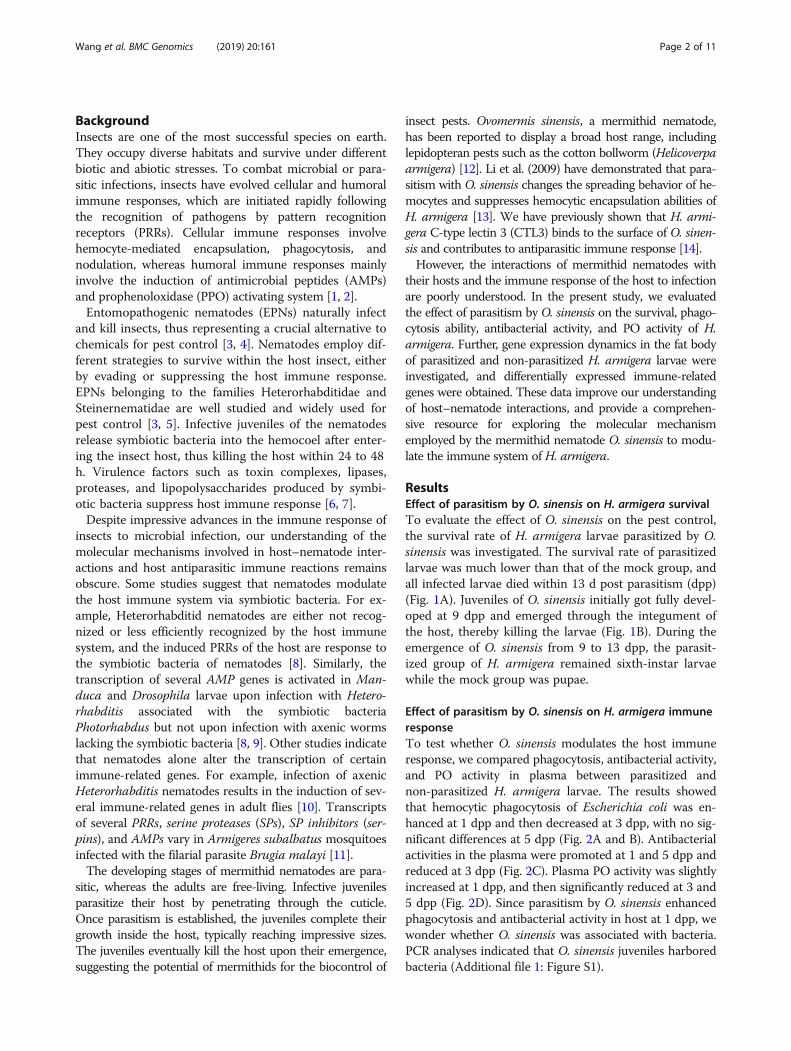
Background: Mermithid nematodes, such as Ovomermis sinensis, display a broad host range including somelepidopteran pests. Infective juveniles penetrate their host through the cuticle, complete their growth within thehemocoel and eventually kill the host upon their emergence. Hence, mermithid nematodes are consideredpotential biological control agents of insect pests. Our previous data indicate that the infection rate of O. sinensis oncotton bollworm (Helicoverpa armigera) is low, which may be largely due to the strong immune system of the host.However, current knowledge on the interactions of mermithid nematodes with their hosts and the mechanismsemployed by hosts to defend themselves against mermithid nematodes is limited.
Results: Here, we investigated the response of H. armigera to O. sinensis infection. Parasitism by O. sinensis caused asharp decline in the survival rate of H. armigera. The hemocytic phagocytosis ability, antibacterial activity, andphenoloxidase (PO) activity in plasma of H. armigera increased at 1 d post parasitism (dpp) but decreased at 3 dpp.Further, we investigated gene expression in the fat body of parasitized and non-parasitized H. armigera larvae at 1,3, and 5 dpp using a digital gene expression system. In total, 41, 60 and 68 immune-related differentially expressedgenes were identified at 1, 3, and 5 dpp, respectively. These genes encoded pattern recognition receptors (PRRs),antimicrobial peptides (AMPs), serine proteases (SPs), SP inhibitors, mucins and other immune-related proteins. Theexpression of most PRRs, AMPs, SPs, and mucins was upregulated in the fat body of larvae at 1 dpp, downregulatedat 3 dpp, and then again upregulated at 5 dpp by O. sinensis. The increased expression of SP inhibitors maycontribute to the inhibited PO activity at 5 dpp.
Conclusions: This study demonstrates that parasitism by O. sinensis modulates the immune reaction of the host H.armigera by altering the expression of immune-related genes. Our data provide a basis for future investigation of themolecular mechanisms employed by the mermithid nematode O. sinensis to modulate the immunity of the host H.armigera. These data will also likely facilitate the improvement of success in parasitism of H. armigera by O. sinensis.
Keywords: Ovomermis sinensis, Helicoverpa armigera, Immunity, Parasitism, Fat body
* Correspondence: [email protected] Key Laboratory of Genetic Regulation and Integrative Biology, Schoolof Life Sciences, Central China Normal University, Wuhan 430079, China
© The Author(s). 2019 Open Access This article is distributed under the terms of the Creative Commons Attribution 4.0International License (http://creativecommons.org/licenses/by/4.0/), which permits unrestricted use, distribution, andreproduction in any medium, provided you give appropriate credit to the original author(s) and the source, provide a link tothe Creative Commons license, and indicate if changes were made. The Creative Commons Public Domain Dedication waiver(http://creativecommons.org/publicdomain/zero/1.0/) applies to the data made available in this article, unless otherwise stated.
Wang et al. BMC Genomics (2019) 20:161 https://doi.org/10.1186/s12864-019-5544-1
BackgroundInsects are one of the most successful species on earth.They occupy diverse habitats and survive under differentbiotic and abiotic stresses. To combat microbial or para-sitic infections, insects have evolved cellular and humoralimmune responses, which are initiated rapidly followingthe recognition of pathogens by pattern recognitionreceptors (PRRs). Cellular immune responses involvehemocyte-mediated encapsulation, phagocytosis, andnodulation, whereas humoral immune responses mainlyinvolve the induction of antimicrobial peptides (AMPs)and prophenoloxidase (PPO) activating system [1, 2].Entomopathogenic nematodes (EPNs) naturally infect
and kill insects, thus representing a crucial alternative tochemicals for pest control [3, 4]. Nematodes employ dif-ferent strategies to survive within the host insect, eitherby evading or suppressing the host immune response.EPNs belonging to the families Heterorhabditidae andSteinernematidae are well studied and widely used forpest control [3, 5]. Infective juveniles of the nematodesrelease symbiotic bacteria into the hemocoel after enter-ing the insect host, thus killing the host within 24 to 48h. Virulence factors such as toxin complexes, lipases,proteases, and lipopolysaccharides produced by symbi-otic bacteria suppress host immune response [6, 7].Despite impressive advances in the immune response of
insects to microbial infection, our understanding of themolecular mechanisms involved in host–nematode inter-actions and host antiparasitic immune reactions remainsobscure. Some studies suggest that nematodes modulatethe host immune system via symbiotic bacteria. For ex-ample, Heterorhabditid nematodes are either not recog-nized or less efficiently recognized by the host immunesystem, and the induced PRRs of the host are response tothe symbiotic bacteria of nematodes [8]. Similarly, thetranscription of several AMP genes is activated in Man-duca and Drosophila larvae upon infection with Hetero-rhabditis associated with the symbiotic bacteriaPhotorhabdus but not upon infection with axenic wormslacking the symbiotic bacteria [8, 9]. Other studies indicatethat nematodes alone alter the transcription of certainimmune-related genes. For example, infection of axenicHeterorhabditis nematodes results in the induction of sev-eral immune-related genes in adult flies [10]. Transcriptsof several PRRs, serine proteases (SPs), SP inhibitors (ser-pins), and AMPs vary in Armigeres subalbatus mosquitoesinfected with the filarial parasite Brugia malayi [11].The developing stages of mermithid nematodes are para-
sitic, whereas the adults are free-living. Infective juvenilesparasitize their host by penetrating through the cuticle.Once parasitism is established, the juveniles complete theirgrowth inside the host, typically reaching impressive sizes.The juveniles eventually kill the host upon their emergence,suggesting the potential of mermithids for the biocontrol of
insect pests. Ovomermis sinensis, a mermithid nematode,has been reported to display a broad host range, includinglepidopteran pests such as the cotton bollworm (Helicoverpaarmigera) [12]. Li et al. (2009) have demonstrated that para-sitism with O. sinensis changes the spreading behavior of he-mocytes and suppresses hemocytic encapsulation abilities ofH. armigera [13]. We have previously shown that H. armi-gera C-type lectin 3 (CTL3) binds to the surface of O. sinen-sis and contributes to antiparasitic immune response [14].However, the interactions of mermithid nematodes with
their hosts and the immune response of the host to infectionare poorly understood. In the present study, we evaluatedthe effect of parasitism by O. sinensis on the survival, phago-cytosis ability, antibacterial activity, and PO activity of H.armigera. Further, gene expression dynamics in the fat bodyof parasitized and non-parasitized H. armigera larvae wereinvestigated, and differentially expressed immune-relatedgenes were obtained. These data improve our understandingof host–nematode interactions, and provide a comprehen-sive resource for exploring the molecular mechanismemployed by the mermithid nematode O. sinensis to modu-late the immune system of H. armigera.
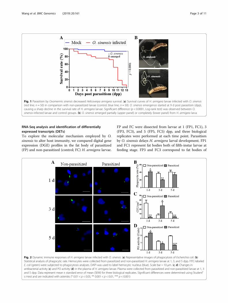
ResultsEffect of parasitism by O. sinensis on H. armigera survivalTo evaluate the effect of O. sinensis on the pest control,the survival rate of H. armigera larvae parasitized by O.sinensis was investigated. The survival rate of parasitizedlarvae was much lower than that of the mock group, andall infected larvae died within 13 d post parasitism (dpp)(Fig. 1A). Juveniles of O. sinensis initially got fully devel-oped at 9 dpp and emerged through the integument ofthe host, thereby killing the larvae (Fig. 1B). During theemergence of O. sinensis from 9 to 13 dpp, the parasit-ized group of H. armigera remained sixth-instar larvaewhile the mock group was pupae.
Effect of parasitism by O. sinensis on H. armigera immuneresponseTo test whether O. sinensis modulates the host immuneresponse, we compared phagocytosis, antibacterial activity,and PO activity in plasma between parasitized andnon-parasitized H. armigera larvae. The results showedthat hemocytic phagocytosis of Escherichia coli was en-hanced at 1 dpp and then decreased at 3 dpp, with no sig-nificant differences at 5 dpp (Fig. 2A and B). Antibacterialactivities in the plasma were promoted at 1 and 5 dpp andreduced at 3 dpp (Fig. 2C). Plasma PO activity was slightlyincreased at 1 dpp, and then significantly reduced at 3 and5 dpp (Fig. 2D). Since parasitism by O. sinensis enhancedphagocytosis and antibacterial activity in host at 1 dpp, wewonder whether O. sinensis was associated with bacteria.PCR analyses indicated that O. sinensis juveniles harboredbacteria (Additional file 1: Figure S1).
Wang et al. BMC Genomics (2019) 20:161 Page 2 of 11
RNA-Seq analysis and identification of differentiallyexpressed transcripts (DETs)To explore the molecular mechanism employed by O.sinensis to alter host immunity, we compared digital geneexpression (DGE) profiles in the fat body of parasitized(FP) and non-parasitized (control; FC) H. armigera larvae.
FP and FC were dissected from larvae at 1 (FP1, FC1), 3(FP3, FC3), and 5 (FP5, FC5) dpp, and three biologicalreplicates were performed at each time point. Parasitismby O. sinensis delays H. armigera larval development. FP1and FC1 represent fat bodies both of fifth-instar larvae atfeeding stage. FP3 and FC3 correspond to fat bodies of
Fig. 1 Parasitism by Ovomermis sinensis decreased Helicoverpa armigera survival. (a) Survival curves of H. armigera larvae infected with O. sinensis(red line; n = 50) in comparison with non-parasitized larvae (control; blue line; n = 50). O. sinensis emergence started at 9 d post parasitism (dpp),causing a sharp decline in the survival rate of H. armigera larvae. Significant difference (p < 0.0001, Log-rank test) was observed between O.sinensis-infected larvae and control groups. (b) O. sinensis emerged partially (upper panel) or completely (lower panel) from H. armigera larva
Fig. 2 Dynamic immune responses of H. armigera larvae infected with O. sinensis. (a) Representative images of phagocytosis of Escherichia coli. (b)Statistical analysis of phagocytic rate. Hemocytes were collected from parasitized and non-parasitized H. armigera larvae at 1, 3, and 5 dpp. FITC-labeledE. coli (green) were subjected to phagocytosis analyses. DAPI was used to label hemocytic nucleus (blue). Scale bar = 10 μm. (c, d) Changes inantibacterial activity (c) and PO activity (d) in the plasma of H. armigera larvae. Plasma were collected from parasitized and non-parasitized larvae at 1, 3and 5 dpp. Data represent mean ± standard error of mean (SEM) for three biological replicates. Significant differences were determined using Student′s t-test and are indicated with asterisks (* 0.01 < p < 0.05, ** 0.001 < p < 0.01, *** p < 0.001)
Wang et al. BMC Genomics (2019) 20:161 Page 3 of 11
fifth-instar larvae at head capsule slippage (HCS) andsixth-instar larvae at feeding stage, respectively. FP5 andFC5 represent fat bodies of sixth-instar larvae at feedingstage and metamorphic stage, respectively. In total, 18RNA-Seq libraries (FP1–1, − 2, − 3; FC1–1, − 2, − 3; FP3–1, − 2, − 3; FC3–1, − 2, − 3; FP5–1, − 2, − 3; FC5–1, − 2, −3) were constructed and sequenced. The number of rawreads generated from each library ranged from 23.96 to27.92 million, whereas the number of cleaned readsranged from 21.01 to 23.62 million. Cleaned reads fromeach library were mapped to previously assembledunigenes of H. armigera fat body [15], with a mappingratio ranging from 49.36 to 58.76% (average 52.91%)(Additional file 2: Table S1).To compare the differential gene expression profiles be-
tween FP and FC larvae at different time points (FP1 vs.FC1; FP3 vs. FC3; FP5 vs. FC5), fragments per kilobase oftranscript per million mapped fragments (FPKM) werequantified. Heatmap indicated that the square of correl-ation coefficient ranged from 0.909–0.99, 0.975–0.993,0.994–0.996, 0.871–0.977, 0.978–0.99, and 0.912–0.99 inFC1, FC3, FC5, FP1, FP3, and FP5 groups, respectively(Additional file 3: Figure S2). These high Pearson valuessuggest the reliable replications and the reasonable samples.NOISeq-sim was adopted to screen DETs with cut-off
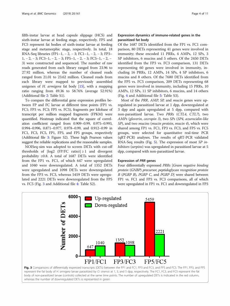
thresholds of |log2 (FP/FC ratio)| ≥ 1 and divergentprobability ≥0.8. A total of 1687 DETs were identifiedfrom the FP1 vs. FC1, of which 647 were upregulatedand 1040 were downregulated. A total of 1352 DETswere upregulated and 1098 DETs were downregulatedfrom the FP3 vs. FC3, whereas 5459 DETs were upregu-lated and 2221 DETs were downregulated from the FP5vs. FC5 (Fig. 3 and Additional file 4: Table S2).
Expression dynamics of immune-related genes in theparasitized fat bodyOf the 1687 DETs identified from the FP1 vs. FC1 com-parison, 80 DETs representing 41 genes were involved inimmunity; these encoded 11 PRRs, 4 AMPs, 12 SPs, 3SP inhibitors, 6 mucins and 5 others. Of the 2450 DETsidentified from the FP3 vs. FC3 comparison, 131 DETsrepresenting 60 genes were involved in immunity, in-cluding 16 PRRs, 12 AMPs, 14 SPs, 4 SP inhibitors, 6mucins and 8 others. Of the 7680 DETs identified fromthe FP5 vs. FC5 comparison, 209 DETs representing 68genes were involved in immunity, including 15 PRRs, 10AMPs, 12 SPs, 11 SP inhibitors, 6 mucins, and 14 others(Fig. 4 and Additional file 5: Table S3).Most of the PRR, AMP, SP, and mucin genes were up-
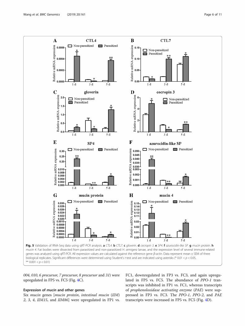
regulated in parasitized larvae at 1 dpp, downregulated at3 dpp and again upregulated at 5 dpp, compared withnon-parasitized larvae. Two PRRs (CTL4, CTL7), twoAMPs (gloverin, cecropin 3), two SPs (SP4, azurocidin-likeSP), and two mucins (mucin protein, mucin 4), which wereshared among FP1 vs. FC1, FP3 vs. FC3, and FP5 vs. FC5groups, were selected for quantitative real-time PCR(qRT-PCR) analyses. The results of qRT-PCR validatedRNA-Seq results (Fig. 5). The expression of most SP in-hibitors (serpins) was upregulated in parasitized larvae at 5dpp, compared with non-parasitized larvae.
Expression of PRR genesFour differentially expressed PRRs [Gram negative bindingprotein (GNBP) precursor, peptidoglycan recognition proteinB (PGRP B), PGRP C, and PGRP D] were shared betweenFP1 vs. FC1 and FP3 vs. FC3 comparisons, all of whichwere upregulated in FP1 vs. FC1 and downregulated in FP3
Fig. 3 Comparisons of differentially expressed transcripts (DETs) between the FP1 and FC1, FP3 and FC3, and FP5 and FC5. The FP1, FP3, and FP5represent the fat body of H. armigera larvae parasitized by O. sinensis at 1, 3, and 5 dpp, respectively. The FC1, FC3, and FC5 represent the fatbody of non-parasitized larvae (controls) collected at the same time points. The number of upregulated DETs is indicated in the red column,whereas the number of downregulated DETs is represented in green
Wang et al. BMC Genomics (2019) 20:161 Page 4 of 11
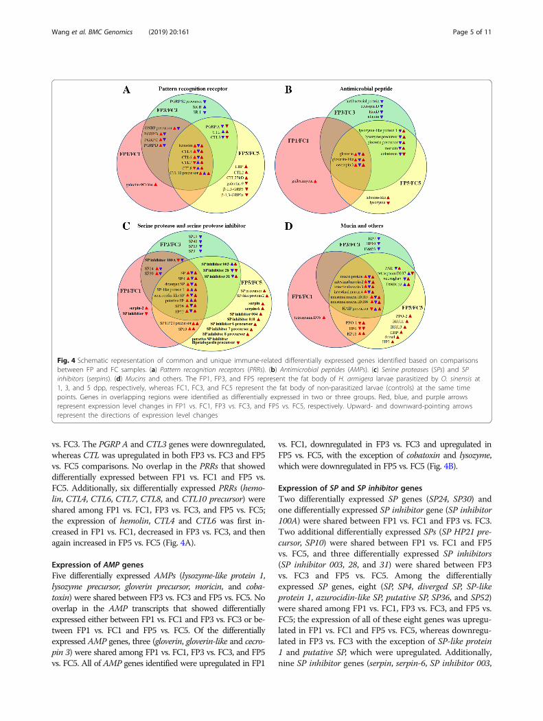
vs. FC3. The PGRP A and CTL3 genes were downregulated,whereas CTL was upregulated in both FP3 vs. FC3 and FP5vs. FC5 comparisons. No overlap in the PRRs that showeddifferentially expressed between FP1 vs. FC1 and FP5 vs.FC5. Additionally, six differentially expressed PRRs (hemo-lin, CTL4, CTL6, CTL7, CTL8, and CTL10 precursor) wereshared among FP1 vs. FC1, FP3 vs. FC3, and FP5 vs. FC5;the expression of hemolin, CTL4 and CTL6 was first in-creased in FP1 vs. FC1, decreased in FP3 vs. FC3, and thenagain increased in FP5 vs. FC5 (Fig. 4A).
Expression of AMP genesFive differentially expressed AMPs (lysozyme-like protein 1,lysozyme precursor, gloverin precursor, moricin, and coba-toxin) were shared between FP3 vs. FC3 and FP5 vs. FC5. Nooverlap in the AMP transcripts that showed differentiallyexpressed either between FP1 vs. FC1 and FP3 vs. FC3 or be-tween FP1 vs. FC1 and FP5 vs. FC5. Of the differentiallyexpressed AMP genes, three (gloverin, gloverin-like and cecro-pin 3) were shared among FP1 vs. FC1, FP3 vs. FC3, and FP5vs. FC5. All of AMP genes identified were upregulated in FP1
vs. FC1, downregulated in FP3 vs. FC3 and upregulated inFP5 vs. FC5, with the exception of cobatoxin and lysozyme,which were downregulated in FP5 vs. FC5 (Fig. 4B).
Expression of SP and SP inhibitor genesTwo differentially expressed SP genes (SP24, SP30) andone differentially expressed SP inhibitor gene (SP inhibitor100A) were shared between FP1 vs. FC1 and FP3 vs. FC3.Two additional differentially expressed SPs (SP HP21 pre-cursor, SP10) were shared between FP1 vs. FC1 and FP5vs. FC5, and three differentially expressed SP inhibitors(SP inhibitor 003, 28, and 31) were shared between FP3vs. FC3 and FP5 vs. FC5. Among the differentiallyexpressed SP genes, eight (SP, SP4, diverged SP, SP-likeprotein 1, azurocidin-like SP, putative SP, SP36, and SP52)were shared among FP1 vs. FC1, FP3 vs. FC3, and FP5 vs.FC5; the expression of all of these eight genes was upregu-lated in FP1 vs. FC1 and FP5 vs. FC5, whereas downregu-lated in FP3 vs. FC3 with the exception of SP-like protein1 and putative SP, which were upregulated. Additionally,nine SP inhibitor genes (serpin, serpin-6, SP inhibitor 003,
Fig. 4 Schematic representation of common and unique immune-related differentially expressed genes identified based on comparisonsbetween FP and FC samples. (a) Pattern recognition receptors (PRRs). (b) Antimicrobial peptides (AMPs). (c) Serine proteases (SPs) and SPinhibitors (serpins). (d) Mucins and others. The FP1, FP3, and FP5 represent the fat body of H. armigera larvae parasitized by O. sinensis at1, 3, and 5 dpp, respectively, whereas FC1, FC3, and FC5 represent the fat body of non-parasitized larvae (controls) at the same timepoints. Genes in overlapping regions were identified as differentially expressed in two or three groups. Red, blue, and purple arrowsrepresent expression level changes in FP1 vs. FC1, FP3 vs. FC3, and FP5 vs. FC5, respectively. Upward- and downward-pointing arrowsrepresent the directions of expression level changes
Wang et al. BMC Genomics (2019) 20:161 Page 5 of 11
004, 010, 6 precursor, 7 precursor, 8 precursor and 31) wereupregulated in FP5 vs. FC5 (Fig. 4C).
Expression of mucin and other genesSix mucin genes [mucin protein, intestinal mucin (IIM)2, 3, 4, IIM15, and IIM86] were upregulated in FP1 vs.
FC1, downregulated in FP3 vs. FC3, and again upregu-lated in FP5 vs. FC5. The abundance of PPO-1 tran-scripts was inhibited in FP1 vs. FC1, whereas transcriptsof prophenoloxidase activating enzyme (PAE) were sup-pressed in FP3 vs. FC3. The PPO-1, PPO-2, and PAEtranscripts were increased in FP5 vs. FC5 (Fig. 4D).
Fig. 5 Validation of RNA-Seq data using qRT-PCR analysis. a CTL4. b CTL7. c gloverin. d cecropin 3. e SP4. f azurocidin-like SP. g mucin protein. hmucin 4. Fat bodies were dissected from parasitized and non-parasitized H. armigera larvae, and the expression level of several immune-relatedgenes was analyzed using qRT-PCR. All expression values are calculated against the reference gene β-actin. Data represent mean ± SEM of threebiological replicates. Significant differences were determined using Student’s t-test and are indicated using asterisks (* 0.01 < p < 0.05,** 0.001 < p < 0.01)
Wang et al. BMC Genomics (2019) 20:161 Page 6 of 11
DiscussionUnlike EPNs (Heterorhabditidae and Steinernematidae)that kill their host within 24–48 h post infection, themermithid nematode O. sinensis killed most of the H.armigera larvae within 9–13 dpp in this study. The effi-cient insecticidal effect of Heterorhabditidae and Stei-nernematidae is due to their symbiotic bacteria, whichare released within the insect hemocoel and secrete viru-lence factors [6, 7]. Hence, this nematode-bacteria sym-biosis is crucial for efficient action of EPNs in pestcontrol [16]. The mermithid nematode Strelkovimermisspiculatus serves as a vector of the iridescent virus [17];however, association of mermithid nematode with sym-biotic bacteria has been rarely detected [18]. AlthoughO. sinensis was associated with bacteria, we cannot con-clude whether the bacteria was symbiotic or came fromoutside contamination. Considering that O. sinensis killsthe host mostly by emerging through the host integu-ment, which takes a relatively long time, it is likely thatO. sinensis does not harbor pathogenic bacteria.O. sinensis not only takes a long time to kill its hosts,
but also exhibits a relatively low infection rate in the hostH. armigera, as shown in our laboratory [14]. The low in-fection rate of O. sinensis on H. armigera may be largelydue to the highly developed immune system of the host.Hence, clarification of the interactions between O. sinensisand its host, especially the immune response of H. armi-gera to O. sinensis is of vital importance. The relativelylow mapping ratio of the cleaned reads to previously as-sembled unigenes of peptidoglycan (PGN)-challenged fatbody was observed. This may reflect the differences in thetranscriptional profiles between the O. sinensis-challengedfat body and the PGN-challenged one, or the insufficientsequencing depth of the previous fat body transcriptome[15]. The number of DETs between FP and FC increasedalong with larval development. These DETs represent abroad range of pathways, including metabolic pathways,fat digestion and absorption, fatty acid biosynthesis, ster-oid biosynthesis and Ribosome (data not shown). Here,we focused on molecular characterization of immuneresponses.Insects employ hemocytic phagocytosis against bacteria,
which requires the participation and coordination of bothcellular and humoral factors. Several humoral PRRs suchas opsonic factors, secreted mainly from fat body, functionin non-self recognition and triggering phagocytosis [19,20]. Since the phagocytosis ability of H. armigera is largelydue to PRRs, which attach to hemocytes, variation in theexpression levels of genes encoding PRRs at different timepoints post parasitism by O. sinensis would have import-ant consequences. The expression of many PRR genes isconsistent with the phagocytosis ability of H. armigera, assome PRR proteins are involved in phagocytosis. For ex-ample, PGRP D has previously been demonstrated to
promote phagocytosis of E. coli in H. armigera [21]. In thisstudy, the upregulation and downregulation of PGRP Dexpression at 1 and 3 dpp by O. sinensis, respectively, wereconsistent with the phagocytosis ability of H. armigera atthese time points. Similar dynamic expression of PRRs hasalso been detected in A. subalbatus in response to B.malayi infection, with a PGRP transcript upregulated at 1h post infection and a CTL transcript downregulated at 12or 24 h post infection [11].The EPN Steinernema feltiae modulates hemocytic
phagocytosis by removing opsonic factors from the hosthemolymph, thus supporting its symbiotic bacteria [22].Since O. sinensis most likely does not need to supportsymbiotic bacteria, initial infection of O. sinensis is pre-sumed to activate the host immune system, for example byactivating phagocytosis. In addition to the role of PRRs inphagocytosis, some PRRs also participate in encapsulation.For example, infection of A. subalbatus with the filarialworm Dirofilaria immitis leads to induction of β1,3-glucanrecognition protein, a PRR involved in encapsulation [23].Although the expression of most PRR genes was upregu-lated in H. armigera at 1 dpp by O. sinensis, encapsulationwas suppressed possibly because of the destruction of he-mocyte cytoskeleton, as demonstrated previously [13].Parasitization also suppresses host cellular encapsulationby inhibiting the expression of PRRs such as CTL andscavenger receptor [24, 25]. Given that PGRP D and CTL3have been previously demonstrated to promote encapsula-tion [14, 21], we speculate that the decreased expression ofcertain PRRs at 3 dpp would facilitate the survival of O.sinensis within the homocoel of H. armigera.Antimicrobial effectors such as AMPs are mainly syn-
thesized in fat body and subsequently secreted into thehemolymph, and play important roles in the restrictionor elimination of the invading pathogen [26]. Infectionof Aedes aegypti with Wuchereria bancrofti filarial nema-todes has been shown to increase mRNA levels of defen-sin, cecropin and transferrin [27]. Some researchersargue that the upregulation of AMP transcripts may beattributed to symbiotic bacteria, considering it is symbi-otic nematodes but not axenic nematodes that inducedthe transcription of AMP genes [8, 9]. Other studieshave raised the possibility that the increased expressionof immune-related genes (including AMPs) reflects strat-egies for tolerating tissue damage caused by nematodes[28]. Cecropin was reported to attenuate the motility ofthe filarial nematode Brugia pahangi in vitro and reducethe number of B. pahangi in vivo [29]. Hence, we specu-late that the expression of AMP genes is increased in re-sponse to either O. sinensis or bacteria associated withO. sinensis, which needs further investigation.To establish infection, nematodes must be capable of
suppressing the immunity of their host. Nematodesmodulate the humoral and cellular immune responses of
Wang et al. BMC Genomics (2019) 20:161 Page 7 of 11

the host by producing molecules such as proteases. Forexample, symbiotic Heterorhabditis bacteriophora se-crete a proteinase into the bodies of greater wax mothlarvae and inhibit the expression of cecropin [30]. In thisstudy, we showed upregulation of AMP genes in H.armigera at 1 dpp by O. sinensis and downregulation at3 dpp; this was consistent with the antibacterial activityin plasma. The AMP genes exhibit similar expressionprofiles in A. subalbatus at different time points post in-fection by filarial worm. For example, the expression oftwo cecropin genes is increased at 6 h post infection andthat of two defensin and two lysozyme genes is decreasedat 12 h post infection [11]. Since molecules produced byO. sinensis have not yet been characterized, the mecha-nisms employed by O. sinensis to suppress the immunityof the host remain unclear. Interestingly, we observedupregulation of AMP genes and enhanced antibacterialactivity in H. armigera larvae at 5 dpp by O. sinensis,possibly because O. sinensis may have developed suffi-ciently during 5 d to endure higher immune stress.Mucin is reported to be involved in the entrapment ofbacteria [31]. In this study, mucin genes exhibited asimilar expression profile as the AMP genes; thus, it islikely that mucins function in antiparasitic immunityand contribute toward antibacterial activity in plasma.SPs and SP inhibitors (serpins) play important roles in
melanin biosynthesis, and are most likely involved in anti-parasitic immunity [32, 33]. Global transcriptional re-sponse of A. aegypti to B. malayi infection has revealed anincrease in the abundance of several SP transcripts [28].Transcriptional levels of 30 SP or serpin genes vary in A.subalbatus at different time points following infection byB. malayi [11], suggesting a potential role of melanizationin antiparasitic immunity. In this study, the abundance ofmost SP transcripts in H. armigera larvae parasitized byO. sinensis increased at 1 dpp and decreased at 3 dpp.Given that SP functions in the activation of PPO and is re-quired for parasite melanization [34, 35], it is reasonablethat PO activity in plasma slightly increased at 1 dpp anddecreased at 3 dpp. However, at 5 dpp, the abundance ofmost of the SP transcripts increased, whereas PO activitydecreased. This may be because of the increased abun-dance of SP inhibitor transcripts, as serpin inhibits the ac-tivation of PPO by SP [36].Some virulence factors produced by parasites during
infection protect them against the potent effects of theimmune system of the host and improve the success rateof parasitism. For example, a trypsin-like serine proteaseand a chymotrypsin serine protease secreted during theparasitic phase of Steinernema carpocapsae exhibit PPOinhibitory activity [37, 38]. Here we showed that O.sinensis parasitism modulated PO activity of H. armigeralarvae by altering the expression of SPs and serpins, al-though the underlying mechanisms remain unclear.
ConclusionsOverall, our data revealed dynamic immune responses ofH. armigera to O. sinensis infection. The initial infectionof H. armigera by O. sinensis activated the expression ofmany PRR, AMP, SP, and mucin genes, which is consistentwith the enhanced immune reactions of the host (phago-cytosis, antibacterial activity, and PO activity). Subse-quently, O. sinensis suppressed these immune reactions byinhibiting the expression of most PRR, AMP, SP, andmucin genes to facilitate its survival within the host. Afterthe successful establishment of O. sinensis parasitism, wespeculate that O. sinensis was able to endure the relativelyhigher immune-related stress (such as antibacterial activ-ity) presumably because it has grown to a sufficiently largesize within 5 dpp. However, the inhibited PO activity at5 dpp may be attributed to the increased expressionof serpins, suggesting that O. sinensis is moresensitive to melanization. We provide a comprehen-sive resource for exploring the complex molecularmechanisms underlying the interaction between themermithid nematode O. sinensis and its host H. armi-gera. Further investigation of DEGs between FP andFC would provide critical target genes for improve-ment of infection rate. Characterization of virulencefactors produced by O. sinensis and elucidation of themechanism employed by O. sinensis to suppress hostimmunity will also be our future study. Our findingswill likely facilitate the development of O. sinensis asan effective and eco-friendly biological control agent.
MethodsCulture conditions, PCR amplification and infection of H.armigera larvaeH. armigera larvae were maintained in the laboratoryat 28 ± 1 °C, 70% relative humidity and 14 h light/10 hdark photoperiod. Larvae were reared on an artificialdiet mainly made from wheat germ and soybean pow-der [39].A colony of adult O. sinensis nematodes was collected
from a wheat field in Shangcai, Henan, China. Thesenematodes were maintained in the laboratory until in-fective juveniles were obtained. Approximately one thou-sand juveniles were applied for genomic DNA extractionusing a bacterial DNA kit (OMEGA, USA). The primers(Additional file 6: Table S4) were designed to amplify theV4-V5 region of 16S rRNA. Then PCR reactions wereperformed with 30 ng of genomic DNA as a template.Fifteen juvenile nematodes were used to infect a
fourth-instar larva of H. armigera for 3 h, as describedpreviously [14]. After successful parasitization by O.sinensis, H. armigera larvae continued to feed on artifi-cial diet until the emergence of fully developed nema-todes. The number of dead larvae was recorded everyday. Infected larvae, which died before the emergence of
Wang et al. BMC Genomics (2019) 20:161 Page 8 of 11
fully developed nematodes, were dissected to confirmwhether the juveniles succeeded in penetrating. Survivalcurves were created and analyzed using GraphPad soft-ware. Log-rank (Mantel-Cox) test was performed to cal-culate statistical significance.
Phagocytosis assayFluorescein isothiocyanate (FITC; Sigma) labeling ofE. coli was conducted at 37 °C for 1 h. Samples werewashed five times with PBS, and FITC-labeled bac-teria were resuspended in PBS to a final concentra-tion of 2 × 10⁸ cells/ml. Phagocytosis analyses wereperformed in triplicate, as described previously [40],with minor modifications. Briefly, hemocytes werecollected from parasitized and non-parasitized larvaeat various time points and suspended in PBS. Subse-quently, PBS containing FITC-labeled bacteria wasadded to the PBS containing hemocytes. After incuba-tion for 1 h, aliquots of the mixture were dispensedonto glass slides, and hemocytes were allowed to set-tle down for 30 min. Hemocytes were then fixed with4% formaldehyde for 10 min, washed and then ob-served under a fluorescence microscope. The phago-cytic rate was calculated as follows:
Phagocytic rate %ð Þ ¼ ½Number of bacteria‐ingesting hemocytes
=Total number of hemocytes� � 100
Measurement of antibacterial activityHemolymph was collected from parasitized andnon-parasitized larvae at various time points, and diluted3-fold in sterile anticoagulant buffer. After centrifugationat 1000 rpm for 10 min, cell-free plasma was obtained,and antibacterial activity was determined as describedpreviously [15]. Briefly, 90 μl of plasma was mixed with10 μl of E. coli suspension. After incubation for 1 h atroom temperature, the plasma–bacteria mixture wasplated onto lysogeny broth agar plates and incubated at37 °C overnight. Subsequently, the number of colonyforming units (CFU) was counted in each plate.
Measurement of PO activityTo evaluate the effect of parasitism by O. sinensis onPO activity of H. armigera, plasma was collected fromparasitized and non-parasitized larvae at various timepoints. PO activity was measured as described previ-ously [41]. Briefly, plasma (50 μl) was incubated with50 μl trypsin (2 mg/ml) for 20 min at room temperature,followed by addition of 50 μl substrate solution contain-ing dopamine (3 mg/ml). Initial absorbance was mea-sured at 490 nm, and one unit of PO activity wasdefined as the amount of enzyme yielding an increaseof 0.001 absorbance units per min.
RNA extraction, DGE library preparation and RNA-SeqanalysisTo prepare DGE libraries, three biological replicates ofFP and FC samples were collected at 1, 3 and 5 dpp.Total RNA was extracted from these samples using TRI-zol Reagent (Invitrogen), and used with oligo dT mag-netic beads to enrich mRNAs. The mRNA samples werethen fragmented into short sequences and reverse tran-scribed using N6 random primer. Subsequently, theresulting double-stranded complimentary DNA wasend-repaired to generate blunt ends and ligated withtwo blunt end adaptors. Following PCR amplification,the PCR products were denatured, and single-strandedDNA was cyclized using splint oligo. The prepared li-braries were subjected to SE50 sequencing at the BeijingGenomics Institute.
Mapping reads to the reference unigenes and analysis ofDETsThe high number of unknown bases, adaptor sequencesand low quality reads were filtered from raw sequencereads to generate clean reads. Clean reads were thenmapped onto reference sequences of H. armigera fat bodytranscriptome generated previously [15] using Bowtie2[42]. To eliminate the influence of gene length and se-quencing discrepancy, gene transcripts were quantified asFPKM values for comparing expression levels of DETsamong samples. Correction for false positive and falsenegative errors were performed using false discovery rates(FDR), with FDR ≤ 0.001 as the default threshold to judgethe significance of gene expression differences. Definitionof divergence probability of each transcript differentiallyexpressing was following the formula described previously[43]. Values of |log2 fold-change| ≥ 1 and divergence prob-ability ≥0.8 were used as cut-off thresholds for identifyingDETs between the different experimental conditions,based on a non-parametric algorithm NOISeq-sim [44].
Validation of gene expression using qRT-PCRResults of RNA-Seq analysis were validated usingqRT-PCR. Total RNA (2 μg) prepared for RNA-Seq wasused for the synthesis of first-strand cDNA. Subse-quently, qRT-PCR was conducted with TransStart TopGreen qPCR SuperMix (TransGen Bio-tech, Beijing,China) using a CF × 96 system (Bio-Rad, Hercules, CA,USA). A total of eight differentially expressed genes, in-cluding PRRs (CTL4, CTL7), AMPs (gloverin, cecropin 3),SPs (SP4, azurocidin-like SP), and mucins (mucin pro-tein, mucin 4), were selected for qRT-PCR analyses. Theexpression level of each gene was normalized relative tothat of the reference gene β-actin using the 2-ΔCT
method (ΔCT = CT[test gene] - CT[β-actin]). Gene-specificprimers used for qRT-PCR analysis are listed inAdditional file 6: Table S4.
Wang et al. BMC Genomics (2019) 20:161 Page 9 of 11

Additional files
Additional file 1: Figure S1. PCR analyses confirming the association ofO. sinensis with bacteria. M, DL2000 DNA marker. Lane 1, PCR amplicon ofV4-V5 region of 16S rRNA. (TIF 38 kb)
Additional file 2: Table S1. Statistics of DGE library sequencing andreads mapping. (XLS 20 kb)
Additional file 3: Figure S2. Heatmap indicating the square ofcorrelation value from three biological replicates. The correlation valueswere assessed by using the Pearson method. (TIF 476 kb)
Additional file 4: Table S2. Transcripts differentially expressed betweenthe fat body of parasitized and non-parasitized larva. (XLS 3723 kb)
Additional file 5: Table S3. Immune-related DETs between the fat bodyof parasitized and non-parasitized larva. (XLS 191 kb)
Additional file 6: Table S4. List of primers used. (XLS 21 kb)
AbbreviationsAMPs: antimicrobial peptides; CFU: colony forming units; CTL: C-type lectin;DETs: differentially expressed transcripts; DGE: digital gene expression; dpp: dpost parasitism; EPNs: entomopathogenic nematodes; FC: fat body of non-parasitized; FDR: false discovery rates; FITC: Fluorescein isothiocyanate; FP: fatbody of parasitized; FPKM: fragments per kilobase of transcript per millionmapped fragments; GNBP: Gram negative binding protein; HCS: headcapsule slippage; PAE: prophenoloxidase activating enzyme;PGRP: peptidoglycan recognition protein; PO: phenoloxidase;PPO: prophenoloxidase; PRRs: pattern recognition receptors; qRT-PCR: quantitative real-time PCR; SPs: serine proteases
AcknowledgementsNot applicable.
FundingThis work was supported by Grants from the National Natural ScienceFoundation of China (Grant Nos. 31872301 and 31772220), and NaturalScience Foundation of Hubei Province (Grant No. 2016CFB560). The fundingbodies played no roles in the study design and data collection, analysis,interpretation of data and in writing the manuscript.
Availability of data and materialsThe data sets have been deposited at the NCBI Sequence Read Archive (SRA)database (http://www.ncbi.nlm.nih.gov/sra/) under the accession numberPRJNA497980. Other data supporting the results are available in theadditional files.
Authors’ contributionsThe study was conceived and designed by JLW. GXW and XSL participatedin the design and coordination of the work. GJW and XRZ performed theinfection, RNA extraction and qRT-PCR. GJW and WWW conducted phagocyt-osis assays, measurement of antibacterial and PO activities. JLW, GJW andXSL analyzed the RNA-seq data and prepared the manuscript. All authorshave read and approved the final version of the manuscript.
Ethics approval and consent to participateNot applicable.
Consent for publicationNot applicable.
Competing interestsThe authors declare that they have no competing interests.
Publisher’s NoteSpringer Nature remains neutral with regard to jurisdictional claims inpublished maps and institutional affiliations.
Received: 4 October 2018 Accepted: 18 February 2019
References1. Lemaitre B, Hoffmann J. The host defense of Drosophila melanogaster. Annu
Rev Immunol. 2007;25:697–743.2. Williams MJ. Drosophila hemopoiesis and cellular immunity. J Immunol.
2007;178(8):4711–6.3. Castillo JC, Reynolds SE, Eleftherianos I. Insect immune responses to
nematode parasites. Trends Parasitol. 2011;27(12):537–47.4. Dillman AR, Sternberg PW. Entomopathogenic nematodes. Curr Biol. 2012;
22(11):R430–1.5. Gaugler R, Lewis E, Stuart RJ. Ecology in the service of biological control: the
case of entomopathogenic nematodes. Oecologia. 1997;109(4):483–9.6. Forst S, Dowds B, Boemare N, Stackebrandt E. Xenorhabdus and
Photorhabdus spp.: bugs that kill bugs. Annu Rev Microbiol. 1997;51:47–72.7. Owuama CI. Entomopathogenic symbiotic bacteria, Xenorhabdus and
Photorhabdus of nematodes. World J Microbiol Biotechnol. 2001;17:505–15.8. Eleftherianos I, Joyce S, Ffrench-Constant RH, Clarke DJ, Reynolds SE.
Probing the tri-trophic interaction between insects, nematodes andPhotorhabdus. Parasitology. 2010;137(11):1695–706.
9. Hallem EA, Rengarajan M, Ciche TA, Sternberg PW. Nematodes, bacteria, andflies: a tripartite model for nematode parasitism. Curr Biol. 2007;17(10):898–904.
10. Castillo JC, Shokal U, Eleftherianos I. Immune gene transcription inDrosophila adult flies infected by entomopathogenic nematodes and theirmutualistic bacteria. J Insect Physiol. 2013;59(2):179–85.
11. Aliota MT, Fuchs JF, Mayhew GF, Chen CC, Christensen BM. Mosquitotranscriptome changes and filarial worm resistance in Armigeres subalbatus.BMC Genomics. 2007;8:463.
12. Wang G, Chen Q, Chen G. In vitro cultivation of the entomogenousnematode Ovomermis sinensis. Acta Zool Sin. 2001;47(2):235–9.
13. Li Q, Sun Y, Wang G, Liu X. Effects of the mermithid nematode Ovomermissinensis on the hemocytes of its host Helicoverpa armigera. J Insect Physiol.2009;55(1):47–50.
14. Wang P, Zhuo XR, Tang L, Liu XS, Wang YF, Wang GX, Yu XQ, Wang JL. C-type lectin interacting with β-integrin enhances hemocytic encapsulation inthe cotton bollworm, Helicoverpa armigera. Insect Biochem Mol Biol. 2017;86:29–40.
15. Wang JL, Chen L, Tang L, Zhao HB, Liu XS, Wang YF. 20-hydroxyecdysonetranscriptionally regulates humoral immunity in the fat body of Helicoverpaarmigera. Insect Mol Biol. 2014;23(6):842–56.
16. Dunphy GB, Thurston GS. Insect immunity. In: Gaugler R, Kaya HK, editors.Entomopathogenic nematodes in biological control, vol. 30. Boca Raton, FL:CRC press; 1990. p. 326.
17. Muttis E, Micieli MV, Urrutia MI, García JJ. Transmission of a pathogenic virus(Iridoviridae) of Culex pipiens larvae mediated by the mermithidStrelkovimermis spiculatus (Nematoda). J Invertebr Pathol. 2015;129:40–4.
18. Noda H, Miyoshi T, Zhang Q, Watanabe K, Deng K, Hoshizaki S. Wolbachiainfection shared among planthoppers (Homoptera: Delphacidae) and theirendoparasite (Strepsiptera: Elenchidae): a probable case of interspeciestransmission. Mol Ecol. 2001;10(8):2101–6.
19. Kim CH, Shin YP, Noh MY, Jo YH, Han YS, Seong YS, Lee IH. An insectmultigand recognition protein functions as an opsonin for the phagocytosisof microorganisms. J Biol Chem. 2010;285(33):25243–50.
20. Cheng Y, Lin Z, Wang JM, Xing LS, Xiong GH, Zou Z. CTL14, a recognitionreceptor induced in late stage larvae, modulates anti-fungal immunity incotton bollworm Helicoverpa armigera. Dev Comp Immunol. 2018;84:142–52.
21. Li L, Li YP, Song CX, Xiao M, Wang JL, Liu XS. Identification and functionalcharacterization of a peptidoglycan recognition protein from the cottonbollworm, Helicoverpa armigera. Arch Insect Biochem Physiol. 2014;86(4):240–58.
22. Brivio MF, Mastore M, Nappi AJ. A pathogenic parasite interferes withphagocytosis of insect immunocompetent cells. Dev Comp Immunol. 2010;34(9):991–8.
23. Wang X, Fuchs JF, Infanger LC, Rocheleau TA, Hillyer JF, Chen CC,Christensen BM. Mosquito innate immunity: involvement of beta 1,3-glucanrecognition protein in melanotic encapsulation immune responses inArmigeres subalbatus. Mol Biochem Parasitol. 2005;139(1):65–73.
24. Fang Q, Wang F, Gatehouse JA, Gatehouse AM, Chen XX, Hu C, Ye GY.Venom of parasitoid, Pteromalus puparum, suppresses host, Pieris rapae,immune promotion by decreasing host C-type lectin gene expression. PLoSOne. 2011;6(10):e26888.
Wang et al. BMC Genomics (2019) 20:161 Page 10 of 11

25. Wang L, Fang Q, Qian C, Wang F, Yu XQ, Ye G. Inhibition of host cellencapsulation through inhibiting immune gene expression by the parasiticwasp venom calreticulin. Insect Biochem Mol Biol. 2013;43(10):936–46.
26. Ferrandon D, Imler JL, Hetru C, Hoffmann JA. The Drosophila systemicimmune response: sensing and signalling during bacterial and fungalinfections. Nat Rev Immunol. 2007;7(11):862–74.
27. Magalhaes T, Oliveira IF, Melo-Santos MA, Oliveira CM, Lima CA, Ayres CF.Expression of defensin, cecropin, and transferrin in Aedes aegypti (Diptera:Culicidae) infected with Wuchereria bancrofti (Spirurida: Onchocercidae), andthe abnormal development of nematodes in the mosquito. Exp Parasitol.2008;120(4):364–71.
28. Erickson SM, Xi Z, Mayhew GF, Ramirez JL, Aliota MT, Christensen BM,Dimopoulos G. Mosquito infection responses to developing filarial worms.PLoS Negl Trop Dis. 2009;3(10):e529.
29. Chalk R, Townson H, Ham PJ. Brugia pahangi: the effects of cecropins onmicrofilariae in vitro and in Aedes aegypti. Exp Parasitol. 1995;80(3):401–6.
30. Jarosz J. Active resistance of entomophagous rhabditid Heterorhabditisbacteriophora to insect immunity. Parasitology. 1998; 117 (Pt 3):201–208.
31. Korayem AM, Fabbri M, Takahashi K, Scherfer C, Lindgren M, Schmidt O,Ueda R, Dushay MS, Theopold U. A Drosophila salivary gland mucin is alsoexpressed in immune tissues: evidence for a function in coagulation andthe entrapment of bacteria. Insect Biochem Mol Biol. 2004;34(12):1297–304.
32. Tang H, Kambris Z, Lemaitre B, Hashimoto C. Two proteases defining amelanization cascade in the immune system of Drosophila. J Biol Chem.2006;281(38):28097–104.
33. Zou Z, Shin SW, Alvarez KS, Kokoza V, Raikhel AS. Distinct melanizationpathways in the mosquito Aedes aegypti. Immunity. 2010;32(1):41–53.
34. Lee SY, Kwon TH, Hyun JH, Choi JS, Kawabata SI, Iwanaga S, Lee BL. In vitroactivation of pro-phenol-oxidase by two kinds of pro-phenol-oxidase-activating factors isolated from hemolymph of coleopteran, Holotrichiadiomphalia larvae. Eur J Biochem. 1998;254(1):50–7.
35. Volz J, Müller HM, Zdanowicz A, Kafatos FC, Osta MA. A genetic moduleregulates the melanisation response of Anopheles to Plasmodium. CellMicrobiol. 2006;8(9):1392–405.
36. Chu Y, Hong F, Liu Q, An C. Serine protease SP105 activatesprophenoloxidase in Asian corn borer melanization, and is regulated byserpin-3. Sci Rep. 2017;7:45256.
37. Balasubramanian N, Hao YJ, Toubarro D, Nascimento G, Simões N.Purification, biochemical and molecular analysis of a chymotrypsin proteasewith prophenoloxidase suppression activity from the entomopathogenicnematode Steinernema carpocapsae. Int J Parasitol. 2009;39(9):975–84.
38. Balasubramanian N, Toubarro D, Simões N. Biochemical study and in vitroinsect immune suppression by a trypsin-like secreted protease from thenematode Steinernema carpocapsae. Parasite Immunol. 2010;32(3):165–75.
39. Zhao XF, An XM, Wang JX, Dong DJ, Du XJ, Sueda S, Kondo H. Expressionof the Helicoverpa cathepsin B-like proteinase during embryonicdevelopment. Arch Insect Biochem Physiol. 2005;58(1):39–46.
40. Li J, Song CX, Li YP, Li L, Wei XH, Wang JL, Liu XS. Rab3 is involved incellular immune responses of the cotton bollworm, Helicoverpa armigera.Dev Comp Immunol. 2015;50(2):78–86.
41. Qiao C, Li J, Wei XH, Wang JL, Wang YF, Liu XS. SRP gene is required forHelicoverpa armigera prophenoloxidase activation and nodulation response.Dev Comp Immunol. 2014;44(1):94–9.
42. Langmead B, Trapnell C, Pop M, Salzberg SL. Ultrafast and memory-efficientalignment of short DNA sequences to the human genome. Genome Biol.2009;10(3):R25.
43. Zhang Y, Wang P, Xia H, Zhao C, Hou L, Li C, Gao C, Wang X, Zhao S.Comparative transcriptome analysis of basal and zygote-located tip regionsof peanut ovaries provides insight into the mechanism of light regulation inpeanut embryo and pod development. BMC Genomics. 2016;17(1):606.
44. Tarazona S, García-Alcalde F, Dopazo J, Ferrer A, Conesa A. Differentialexpression in RNA-seq: a matter of depth. Genome Res. 2011;21(12):2213–23.
Wang et al. BMC Genomics (2019) 20:161 Page 11 of 11