Embed Size (px)
Citation preview

Vol.58, No.1, 2016J Fac Med Baghdad 85
Molecular Characterization of Malassezia furfur isolated from patients with pityriasis versicolor compared to healthy
control in Baghdad, Iraq
Abbas M. Al-Ammari* PhD MycologyAzhar A. F. Al-Attraqhchi** PhD Medical MicrobiologySaife D. Al-Ahmer*** PhD Genetic Engineering
Abstract:Background: Humans skin, is the largest organ of the integumentary system, it has multiple layers of ectodermal tissue and guards the underlying muscles, bones, ligaments and internal organs. Pityriasis versicolor is the prototypical skin disease etiologically connected to Malassezia species. Malassezia furfur is the primary causative agent of pityriasis versicolor which causes either hyperpigmentation or hypopigmentation of the skin.Objective: To identify of Malassezia furfur associated with pityriasis versicolor patients and healthy control by using molecular detection methods.Material and Methods: Sixty patients suffering from pityriasis versicolor disease who attended Medical Imammaine Kadhmain City from beginning of 1st December 2013 to the 30th of April 2014. Clinical diagnoses were done by consultant dermatologist. Forty were males and twenty were females with a mean age of (28.63 ± 11.83) years old. Control group includes skin swabs collected from 120 healthy volunteers, with ages ranging from 1 to 70 years with a mean of (30.03 ± 14.58 years). Both groups were investigated for M. furfur using phenotypic and molecular characterization.Results: According to the gender, pityriasis versicolor was more infection in males than females with (68.3%). Malassezia furfur had a high percentage with male of pityriasis versicolor patients and healthy volunteers (65.0% and 73.1%, respectively). According to the site of lesions, Malassezia furfur was most isolated from chest site with percentage (35.0%) while in healthy volunteers, Upper limbs and chest were most site lesions associated with M. furfur with a percentage (23.1%).Conclusions: It was concluded that pityriasis versicolor was more common in male than female. Also the chest site was most the lesions associated with Malassezia furfur in pityriasis versicolor patients. Key words: Malassezia furfur, Pityriasis versicolor.
Original Article
*Dept. of Biology, College of Science, Baghdad University [email protected] ** Dept. of Medical Microbiology, College of Medicine, Al-Nahrain University*** Institute of Genetic Engineering and Biotechnology, Baghdad University.
Fac Med Baghdad2016; Vol.58, No.1Received: Nov, 2015Accepted: Mar.2016
Introduction:
In Humans, the skin is the largest organ of the integumentary system, it has multiple layers of ectodermal tissue and guards the underlying muscles, bones, ligaments and internal organs. Human skin is similar to that of most other mammals, except that it is not protected by a pelt, though nearly all human skin is covered with hair follicles, it appears hairless, there are two general types of skin, hairy and glabrous skin (1).Pityriasis versicolor is the prototypical skin disease etiologically connected to Malassezia species. Malassezia furfur is a coccal, and their cells contain a plasma membrane, a thick and multi-laminar cell wall composed of chitin with an invagination characteristic of Malassezia (2), mitochondria, a nucleus, and all of the other vital organelles (3). Malassezia furfur is the primary causative agent of pityriasis versicolor
which causes either hyperpigmentation or hypopigmentation of the skin, scaly, slow-growing skin, and itchiness. It’s caused when Malassezia furfur population grows over the control levels (4). The Internal transcribed spacer (ITS) gene is now perhaps the most widely sequenced DNA region in fungi. It has typically been most useful for molecular systematic at the species level, and even within species (e.g., to identify geographic races). RFLP stands for ‘restriction fragment length polymorphism,’ which is a term that refers to a variation in a sequence of DNA that is detectable through gel electrophoresis. (5).
Materials and Methods:Sample collections: Sixty patients suffering from pityriasis versicolor disease who attended Medical Immamaine Kadhumaine City and one hundred twenty control individuals were randomly selected from (entities, primary and secondary schools) from 1st of December 2013 to the 30th of April 2014. Clinical diagnosis was done by consultant dermatologist. Forceps, surgical blades and swabs were used for skin sampling.

Vol.58, No.1, 2016J Fac Med Baghdad 86
Molecular Characterization of Malassezia furfur isolated from patients Abbas M. Al-Ammariwith pityriasis versicolor compared to healthy control in Baghdad, Iraq
Direct and in direct methods were applied for diagnosis (6). Skin specimens were subjected for direct examination by placing on a clean slide mounted with a drop of 10 % KOH (to dissolved keratinized material), covered with a cover slip. The slides were warmed gently (but not boiled to prevent crystallization of KOH) and examined under microscope (40X) (7). To microscopic observation of yeast cells, loopful of culture were stained with lacto phenol cotton blue on sterile glass slide.Phenotypic characterization: Scales and swabs were inoculated into Sabouraud′s dextrose agar containing 0.05gm\L chloramphenicol, Penicillin procaine at a concentration of 0.4 ml\L and Streptomycin at a concentration of 2 ml\L overlaid with olive oil. The vials were incubated at 37°C for 1-2 weeks. (8). The suspension was obtained by inoculating 5 ml of sterile distilled water with a loopful of actively growing yeast and the concentration was adjusted to about 105 cell/ml (9). Yeast cells were cultured on pigment production medium. After sterilization and cooling at room temperature, the suspension was smeared on the agar medium using sterile swab. The plates were incubated at 32°C for 2 to 4 weeks, production of brown pigment was considered as a positive result for Malassezia furfur (10). All yeast cells were streaked on CHROM Malassezia agar at 32°C for 48 hours. Large, pale pink and wrinkled colonies as positive culture of Malassezia furfur well developed (11).Molecular characterization: (I) DNA extraction. Brain heart infusion broth (100 ml) was inoculated with one loop full fungal growth and left for overnight incubation at 37°C, broth was collected 4.5ml of a culture grown for 20 hours in brain heart infusion broth were added to a1.5ml micro centrifuge tube. The cells were harvested by centrifugation at 13000 rpm for 3 minutes. DNA was extracted as described (12).(ii) Conventional PCR technique. The primers were selected to allow the amplification of ITS gene in M. furfur and their sequences were: forward 5’- GCATCGATGAAGAACGCAGC-3’ and reverse 5’- TCCTCCGCTTATTGATATGC-3’ (Alpha DNA, Canada). PCR amplification was carried out in a final volume of 50 µl. each reaction contained 4 µl of template DNA, 2 µl of each primer, 20 µl of Go Tag Green Master Mix and 22 µl of Nuclease free water. An initial denaturation step at 94°C for 3 min. was followed by 30 cycles of denaturation at 94°C for 30S, annealing at 50°C for 30s, and extension at 72°C for 30 sec. with final extension step at 72°C for 10 min. Amplified product was visualized by 1%(w\v) agarose gel electrophoresis in TBE buffer, stained with ethidium bromide (0.5 µg \ml) and photographed under UV transillumination (13).(iii) PCR-RFLP technique. The amplified product of ITS gene was digested with 10 U of the restriction enzyme Hinf1 (Biolabs, England) by incubation for 2-16 hours at 37°C.The product was visualized by 1% agarose gel electrophoresis in TBE buffer, stained with ethidium bromide (0.5 µg \ml) and photographed under UV transillumination (14).Statistical analysis: Statistical analysis was performed with the statistical Package for Social Sciences (SPSS) 21.0 and
Excell 2013. Descriptive statistics for categorical data were formulated as frequency and percentage. While numerical data were formulated as mean, standard errors (SE) and standard deviation (SD). Data analysis was done using Chi-square for comparison of categorical data, while ANOVA for comparison of numerical data. P-value of ≤ 0.05 was used as the level of significant.
Results: A total of sixty patients had been included in the present study with ages ranging from 1 to 70 years, with a mean age of (28.63 ± 11.83) years, consisting of 40 males and 20 females (66.7% and 33.3% respectively) with the most frequent among (21-30 years old) age groups. The control group includes skin swabs sites collected from 120 apparently healthy volunteers, with ages ranging from 1 to 70 years with a mean of (30.03 ± 14.58 years). Males were 83 and females were 37 (69.20% and 30.80%, respectively) (Table 1).
Table (1): Age of persons involved in the study.Study groups Healthy control Pityriasis versicolor
Mean 30.03 28.63
St. Deviation 14.58 11.83
Median 29.50 28.00
Maximum 60.00 55.00
Minimum 9.00 6.00
P value <0.001** Highly statistical significant difference
Phenotypic identification of Malassezia furfur: Based on the gross morphology of the colonies, Pigment induction medium and CHROM agar Malassezia medium allowed the differentiation of M. furfur in this study population. The phenomenon resulted in color of colonies was brown and pink, respectively (15). Macroscopical appearance: Skin samples were collected from different sites of pityriasis versicolor patients and healthy control, with different characteristics features (Fig.1).
Vol.58, No.1, 2016J Fac Med Baghdad 87
Molecular Characterization of Malassezia furfur isolated from patients Abbas M. Al-Ammariwith pityriasis versicolor compared to healthy control in Baghdad, Iraq
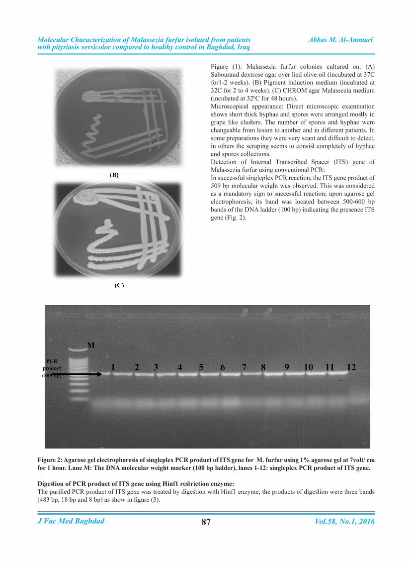
Figure (1): Malassezia furfur colonies cultured on: (A) Sabouraud dextrose agar over lied olive oil (incubated at 37C for1-2 weeks). (B) Pigment induction medium (incubated at 32C for 2 to 4 weeks). (C) CHROM agar Malassezia medium (incubated at 320C for 48 hours).Microscopical appearance: Direct microscopic examination shows short thick hyphae and spores were arranged mostly in grape like clusters. The number of spores and hyphae were changeable from lesion to another and in different patients. In some preparations they were very scant and difficult to detect, in others the scraping seems to consist completely of hyphae and spores collections.Detection of Internal Transcribed Spacer (ITS) gene of Malassezia furfur using conventional PCR:In successful singleplex PCR reaction, the ITS gene product of 509 bp molecular weight was observed. This was considered as a mandatory sign to successful reaction; upon agarose gel electrophoresis, its band was located between 500-600 bp bands of the DNA ladder (100 bp) indicating the presence ITS gene (Fig. 2).
Figure 2: Agarose gel electrophoresis of singleplex PCR product of ITS gene for M. furfur using 1% agarose gel at 7volt/ cm for 1 hour. Lane M: The DNA molecular weight marker (100 bp ladder), lanes 1-12: singleplex PCR product of ITS gene.
Digestion of PCR product of ITS gene using Hinf1 restriction enzyme:The purified PCR product of ITS gene was treated by digestion with Hinf1 enzyme; the products of digestion were three bands (483 bp, 18 bp and 8 bp) as show in figure (3).

Vol.58, No.1, 2016J Fac Med Baghdad 88
Molecular Characterization of Malassezia furfur isolated from patients Abbas M. Al-Ammariwith pityriasis versicolor compared to healthy control in Baghdad, Iraq
Figure 3: Agarose gel electrophoresis of digested PCR product of ITS gene for M. furfur isolates by Hinf 1 restriction enzyme (483 bp) (18 bp and 8 bp, not visible fragment) using 1% agarose gel at 7 volt/ cm for 1 hour. Lane M: The DNA molecular weight marker (100 bp ladder), lanes 1-9: digested PCR product of ITS gene.Upon stratification of the M. furfur isolated according to the gender in pityriasis versicolor patients and healthy control, M. furfur was the most frequently isolated in males, with a percentage of 65.0% and 73.1%, respectively (Table 2). No statistically significant difference was observed between the patient and control groups concerning gender and presence of Malassezia furfur (p>0.05).
Table (2): Rate of Malassezia furfur in patients with pityriasis versicolor in comparison with control group concerning gender.
Study groupsResults
Total p valueNeg. M. furfur Malassezia spp.
Healthy control
Type of gender
FemaleCo. 18 7 12 37
0.884 NS
% 31.6% 26.9% 32.4% 30.8%
MaleCo. 39 19 25 83
% 68.4% 73.1% 67.6% 69.2%
TotalCo. 57 26 37 120
% 100.% 100.% 100.0% 100.0%
PV. patients
Type of gender
FemaleCo. 7 7 6 20
0.459 NS
% 43.8% 35.0% 25.0% 33.3%
MaleCo. 9 13 18 40
% 56.3% 65.0% 75.0% 66.7%
TotalCo. 16 20 24 60
% 100.% 100.% 100.0% 100.0%
Upon stratification of the isolated M. furfur according to site of lesions in the pityriasis versicolor patients. M. furfur was most frequently isolated in chest comparing with neck, back and upper limbs sites, with a percentage of 35.0%,. While, among control groups, M. furfur and was the most predominant isolated in upper limbs and chest, with a percentage of 23.1%. No statistically significant differences were detected among site of lesions in both of patients with pityriasis versicolor and healthy control (P>0.05).
Discussion:Accurate identifications of the species are needed to obtain a better understanding of the role of each individual species in the etiology of disease, and to facilitate adequate treatment, this can be determined based on species-specific susceptibilities to antifungal agents (16). Many factors play role in M. furfur pathogencity such as increased sebum production, hormonal fluctuations, stress, illness, infrequent shampooing, food

Vol.58, No.1, 2016J Fac Med Baghdad 89
Molecular Characterization of Malassezia furfur isolated from patients Abbas M. Al-Ammariwith pityriasis versicolor compared to healthy control in Baghdad, Iraq
allergies, vitamin B deficiency, hair curlers and blow dryers, cold weather, use of hair sprays, gels and hair coloring chemicals (17). Molecular methods may resolve the time consuming and the difficulties in interpretation of some morphological and physiological patterns, RFLP method was used in this study enable us to examine genetic variations through cleaving the amplified DNA with Hinf1 restriction enzyme and analyzing the patterns of the fragments. As showed in table (2), the significantly higher infection rate among males compared to females obtained in the current study is in agreement with the results of (18, 19, and 20). The reason of this rate in males may due to high production of androgens than females, the body begins producing more androgen-type hormones, which cause the sebaceous glands to enlarge and produce more sebum, they tend to have more severe colonize of M. furfur, so military personnel, athletes, and those doing hard works that usually associated with hyper sweating were more vulnerable to pityriasis versicolor infection (21). Furthermore, sebaceous glands secretion is increased in males 15 years and older compared to females, and that may also promote pityriasis versicolor infection in males (19, 22). The high frequency of chest as sites of infection may be attributed to the fact that scalp, upper trunk and the face are rich in sebaceous glands compared to other parts of the body, the differences in the number of enrolled cases, racial factor, habits of patients all may play roles in these differences in results. (23). However, our results in this regard are consistent with those reported by (20).
Author contributions:Abbas Al-Ammari: (samples collection, practical part of mycology, data collection, analysis design and interpretation of results).Azhar Al-Attraqhchi: (study conception, data analysis and critical revision)Saif Al-Ahmer: ( data analysis, practical part of molecular biology and critical revision)
References:1. Marks, G. and Miller, J. Lookingbill and Marks’ Principles of Dermatology. (4th Ed.). Elsevier Inc. 2006.4160-3185.2. Marcon, M., Powell, D. “Human Infections Due to Malassezia spp”. Clinical Microbiology Reviews. 1992. 5.2: 101-119.3. Slonczewski, J., Foster, J. “Microbiology: An Evolving Science 2 ed.”. Norton. 2011. 761-769;A-21-A-22.4. Seabury, S. “The generalized rash: part II. Diagnostic approach”. Am Fam Physician . 2010. 81 (6): 735–9.5. Pinchus, H.; Oreans, S. and Chatellier, S. Yeast identification past, present, and future methods. Medical Mycology . 2007. 45: 97-121.6. Khosravi, R.; Eidi, S.; Katiraee, F.; Ziglari, T.; Bayat, M. and Nissiani, M. Identification of Different Malassezia species Isolated from Patients with Malassezia Infections. World Journal of Zoology. 2009. 4 (2): 85-89.
7. Al-Ammari, A. Relation of Malassezia species with some skin diseases. M.Sc. Thesis. Biology. Dept. College of Science. Baghdad University. 2012.8. Shokohi, T.; Afshar, P. and Barzgar, A. Distribution of Malassezia species in patients with pityriasis versicolor in northern Iran. Indian Journal of Medical Microbiology. 2009. 27(4): 321-324.9. Kindo, A.; Sophia, S.; Kalyani, J. and Anandan, S. identification of Malassezia spp. Indian J Med Microbiol. 2004.22 (3):179-81.10. Gouda, A. Malassezia species isolated from lesional and non lesional skin in patients with pityriasis versicolor. M. Sc. thesis. College of Medicine , University of Ain Shams. 2008.11. Kaneko, T.; Koichi, M.; Michiko, A.; Ryoko, S.; Yuka, N.; Rui, K.; Atsuhiko, H.; Takashi, S.; Shuichi, S.; Shinichi, W.; Hideyo, Y.; Shigeru, A. and Noboru, O. Revised Culture-Based System for identification of Malassezia species. Clin Microbiol. 2007. 53 (11): 3737–3742.12. Ali. S. Molecular study of some virulence factors of Candida albicans isolated from Iraqi Candidiasis patients. M.Sc. College of medicine. Al-Nahrain University. 2014.13. Mirhendi, H.; Makimura, K.; Zomorodian, K.; Yamada, T.; Sugita, T. and Yamaguchi, H. A simple PCR-RFLP method for identification and differentiation of 11 Malassezia species. J. Microbiol. 2005. 61:281-284.14. Gonzalez, A. Sierra, A. Cardenas, M. and Celis, A. Physiological and Molecular Characterization of A typical Isolates of Malassezia furfur. Clin Microbiol. 2008. 55 (9): 3930–3939.15. Ahmed, W. Isolation and identification of Malassezia species in pityriasis versicolor in Baghdad, Iraq. A thesis submitted to Iraqi board for medical specialization. 2004.16. Batra, R.; Boekhout, T.; Gueho, E.; Cabanes, F.; Dawson, T. and Gupta, A. Malassezia Baillon, emerging clinical yeasts. FEMS. Yeast Res. 2005. 5, 1101–1113.17. Anonymous. Condition and diseases: skin diseases (Dandruff). Omani Medical search. (2010).18. DiSilverio, A.; Mosca, M.; Gatti, M. and Brandozzi, G. Pityriasis versicolor in the aged: A clinical investigation and epidemiological survey in 190 elderly hospitalized patients. Mycopathologia. 1989.105(3): 187-90.19. Al-Rubaie, M.G. Clinic-epidemiological study of pityriasis versicolor in Baghdad. M.Sc. thesis. College of Medicine, University of Al- Mustansiriya. 1991.20. Al-Duboon, A.; Al-Rubaie, K. and Muhsin, T. A study of pityriasis versicolor in Basrah (Iraq). Med. J. Basrah, 2000.19(1):31-34.21. Ellabid, M. and Khalifa, Z. Dermatophytes and other fungi associated with skin mycosis in Tripoli, Libyia. Ann. Saudi. Med. 2001. 21(3-4): 193-5.22. Cotterill, J.A. Cunliffe, W.J.; Williamson, B. and Bulusu, L. Age and sex variation in skin surface lipid composition and sebum excretion rate. Brit. J. Dermatol.1972. 87:333-40.23. Faergemann J. Pityriasis versicolor infections. J. Am. Acad. Dermatol. 1994.31(3):18-20.