Embed Size (px)
Citation preview

JOURNAL OF BACTERIOLOGY, Aug. 2007, p. 5749–5761 Vol. 189, No. 150021-9193/07/$08.00�0 doi:10.1128/JB.00194-07Copyright © 2007, American Society for Microbiology. All Rights Reserved.
Molecular Characterization of Membrane-Associated Soluble SerinePalmitoyltransferases from Sphingobacterium multivorum
and Bdellovibrio stolpii�
Hiroko Ikushiro,1* Mohammad Mainul Islam,1† Hiromasa Tojo,2 and Hideyuki Hayashi1*Department of Biochemistry, Osaka Medical College, Takatsuki, Osaka 569-8686, Japan,1 and Department of Biochemistry and
Molecular Biology, Osaka University, Graduate School of Medicine, 2-2 Yamadaoka, Suita, Osaka 565-0871, Japan2
Received 5 February 2007/Accepted 10 May 2007
Serine palmitoyltransferase (SPT) is a key enzyme in sphingolipid biosynthesis and catalyzes the decar-boxylative condensation of L-serine and palmitoyl coenzyme A (CoA) to form 3-ketodihydrosphingosine (KDS).Eukaryotic SPTs comprise tightly membrane-associated heterodimers belonging to the pyridoxal 5�-phosphate(PLP)-dependent �-oxamine synthase family. Sphingomonas paucimobilis, a sphingolipid-containing bacterium,contains an abundant water-soluble homodimeric SPT of the same family (H. Ikushiro et al., J. Biol. Chem.276:18249–18256, 2001). This enzyme is suitable for the detailed mechanistic studies of SPT, although singlecrystals appropriate for high-resolution crystallography have not yet been obtained. We have now isolated threenovel SPT genes from Sphingobacterium multivorum, Sphingobacterium spiritivorum, and Bdellovibrio stolpii,respectively. Each gene product exhibits an �30% sequence identity to both eukaryotic subunits, and theputative catalytic amino acid residues are conserved. All bacterial SPTs were successfully overproduced inEscherichia coli and purified as water-soluble active homodimers. The spectroscopic properties of the purifiedSPTs are characteristic of PLP-dependent enzymes. The KDS formation by the bacterial SPTs was confirmedby high-performance liquid chromatography/mass spectrometry. The Sphingobacterium SPTs obeyed normalsteady-state ordered Bi-Bi kinetics, while the Bdellovibrio SPT underwent a remarkable substrate inhibition atpalmitoyl CoA concentrations higher than 100 �M, as does the eukaryotic enzyme. Immunoelectron micros-copy showed that unlike the cytosolic Sphingomonas SPT, S. multivorum and Bdellovibrio SPTs were bound tothe inner membrane of cells as peripheral membrane proteins, indicating that these enzymes can be aprokaryotic model mimicking the membrane-associated eukaryotic SPT.
Sphingolipids are ubiquitous membrane components of theeukaryotic plasma membrane (32) and are known to be essen-tial lipidic signaling molecules required for various cellularevents, such as proliferation, differentiation, and apoptosis (22,38, 54). In addition, sphingolipids together with cholesterol arethe major components of the membrane microdomains called“lipid rafts,” which serve as platforms for signal transduction orthe transport of various bioactive molecules via membranetrafficking (14, 25, 52).
Serine palmitoyltransferase (SPT) (EC 2.3.1.50) catalyzesthe pyridoxal 5�-phosphate (PLP)-dependent condensation re-action of L-serine with palmitoyl coenzyme A (CoA) to gener-ate 3-ketodihydrosphingosine (KDS). This reaction is the firstcommitted step in the de novo biosynthetic pathway of allsphingolipids, producing long-chain bases (LCBs), the back-bone structure of sphingolipids. SPT is thought to be the keyenzyme regulating the cellular sphingolipid content (21). Eu-karyotic SPTs are enriched in the endoplasmic reticulum, withtheir catalytic sites facing the cytosol (36), and function asheterodimers comprising two tightly membrane-bound sub-
units, called LCB1 and LCB2, which share a sequence similar-ity (�25% identity) (10, 19, 20, 41, 42, 64). Recently a newsubunit protein of the human SPT, SPTLC3, was found (24).Due to the high sequence similarity (68% identity) betweenSPTLC2 (LCB2 subunit of human SPT) and SPTLC3, SPTLC3is thought to form a dimer with SPTLC1. LCB2 (SPTLC2) andSPTLC3 are the putative catalytic subunits carrying a lysineresidue that forms the Schiff base with PLP. In contrast, LCB1does not have such a motif (10, 19) and does not seem tofunction as the catalytic center. Nevertheless, LCB1 is re-garded to be essential for the catalytic action of SPT (20), andmutations in the LCB1 gene are known to cause human he-reditary sensory neuropathy type I (HSN1) (6, 11, 61). Theroles of SPT activity in the pathogenesis of HSN1, however, areelusive at present (7, 17, 40). Elucidation of the structure-activity relationship of SPT is essential for understanding therole of the rate-limiting enzyme, SPT, in regulating the cellularsphingolipid homeostasis and for clarifying the underlyingcauses of HSNI. There is, however, little structural and mech-anistic information on the mammalian SPT currently available,because the instability and the hydrophobic nature of eachsubunit have hindered the successful purification of recombi-nant SPT for crystallization and structural analysis (26).
Previously we found and isolated a water-soluble ho-modimeric SPT from Sphingomonas paucimobilis EY2395T
(27). The Sphingomonas enzyme was successfully overpro-duced in Escherichia coli (27, 28). This bacterial prototype ofthe eukaryotic SPT provided a simple model system for study-
* Corresponding author. Mailing address: Department of Biochem-istry, Osaka Medical College, Takatsuki, Osaka 569-8686, Japan.Phone: 81-72-684-7291. Fax: 81-72-684-6516. E-mail for H. Ikushiro:[email protected]. E-mail for H. Hayashi: [email protected].
† Present address: Department of Biochemistry, Wake Forest Uni-versity School of Medicine, Winston-Salem, NC 27157.
� Published ahead of print on 8 June 2007.
5749
on May 21, 2018 by guest
http://jb.asm.org/
Dow
nloaded from
ing the enzyme reaction without detergent micelles or lipidmembranes. However, despite the successful elucidation of theenzymological properties of the Sphingomonas SPT (29), wewere unable to obtain crystals appropriate for a high-resolu-tion X-ray analysis, which is essential for further clarification ofthe detailed catalytic mechanism of the enzyme. Therefore, wesearched for SPT proteins that are suitable for crystallizationin other sphingolipid-containing bacteria. One such candidatefor the enzyme source is the genus Sphingobacterium, which isa deep-orange-pigmented rod belonging to the class Sphingo-bacteria of the phylum Bacterioidetes and is isolated from theenvironment (51) or from patients with opportunistic infec-tions (8, 16, 23, 37, 60). Sphingobacterium has a high concen-tration of sphingophospholipids with unique branched LCBs,including ceramide phosphorylethanolamines, ceramide phos-phoryl-myo-inositols, and ceramide phosphoryl-1-�-mannoseas the major components (43, 44, 71, 72). Another candidate,Bdellovibrio, is a small curved rod belonging to the delta sub-class of the phylum Proteobacteria that can be found in diverseenvironments, such as marine and fresh waters, sewage, andsoil (47). Bdellovibrio is characterized by the unique predatorybehavior by which it invades various other larger gram-nega-tive bacteria and grows as a parasite in the intraperiplasmicspace of the prey (46, 47, 56, 57). Bdellovibrio contains a phos-phono ceramide, which carries the characteristic head group1-hydroxy-2-aminoethyl phosphonate (62). The bacteria listedabove are exceptions in gram-negative bacteria in that theylack lipopolysaccharides and instead contain a large amount ofsphingolipids, including glycosphingolipids (33, 67–70, 72);most gram-negative bacteria contain lipopolysaccharides, themajor pathogenic glycolipids of the outer membrane. glyco-sphingolipidss, such as �-D-glucuronosyl-ceramide and �-D-galacturonosyl-ceramide of Sphingomonas, were reported toactivate CD1d-restricted natural killer T (NKT) cells (34, 39,55, 66). The action of the bacterial sphingolipids in innateimmunity is attracting much attention in the field of infectiousdiseases.
In this context, studying the SPT proteins in these bacteria isimportant not only for obtaining the ideal source for crystalli-zation of the enzyme but also for providing the structural basisfor the elucidation of the biosynthetic mechanism of theunique glycosphingolipids in these bacteria. We now report themolecular cloning of three novel SPTs from sphingolipid-con-taining bacteria, Sphingobacterium spiritivorum EY3101T,Sphingobacterium multivorum GTC97, and Bdellovibrio stolpiiATCC 27052. All of these bacterial enzymes were successfullyoverproduced in E. coli and enzymatically characterized. Theirproperties resembled those of the eukaryotic enzyme moreclosely than those of the Sphingomonas enzyme. Thus, theseenzymes can be useful models for mammalian SPT and candi-dates for high-resolution crystallographic analyses.
MATERIALS AND METHODS
Chemicals. L-Serine and the other natural L-amino acids were obtained fromNacalai Tesque (Kyoto, Japan). Palmitoyl CoA and lauroyl CoA were fromFunakoshi (Tokyo, Japan). Isopropyl 1-thio-�-D-galactoside, myristoyl CoA,n-heptadecanoyl CoA, stearoyl CoA, arachidoyl CoA, palmitoleoyl CoA, andoleoyl CoA were from Sigma. The LMW gel filtration calibration kit, gel filtra-tion calibration kit PD-10, and Sephacryl S-200 HR were from Amersham Bio-science/GE Healthcare. DEAE-Toyopearl 650 M and butyl Toyopearl 650 Mwere from Tosoh (Tokyo, Japan). The competent E. coli JM109 was purchased
from Nippon Gene (Tokyo, Japan). E. coli BL21(DE3)(pLysS) and plasmidspET21b and pET28b were from Novagen. Plasmid pUC118 was from Takara Bio(Kyoto, Japan). All other chemicals were of the highest grade available fromcommercial sources.
Bacterial strains and growth conditions. Sphingomonas paucimobilis EY2395T
and S. spiritivorum EY3101T were gifts from Eiko Yabuuchi (Gifu UniversitySchool of Medicine, Gifu, Japan). S. multivorum GTC97 was from the Gifu TypeCulture Collection of Gifu University, Japan. These three strains were aerobi-cally grown in Bacto heart infusion broth (Difco, Becton Dickinson) at 30°C. B.stolpii (ATCC 27052) was purchased twice from ATCC, but it did not grow fromthe freeze-dried stock. A living culture was a gift from Yoko Watanabe (NiigataUniversity, Niigata, Japan). This strain was grown in culture medium containing1% Bacto tryptone and 0.3% Bacto yeast extract (Difco, Becton Dickinson, MD)at 30°C. The cells were collected by centrifugation and stored at �20°Cbefore use.
Isolation and sequencing of genomic DNA clones encoding S. multivorum SPT.The genomic DNA of S. multivorum was prepared according to a standardmethod (5). Based on the amino acid sequences of the Sphingomonas SPT andeukaryotic LCB1/LCB2 proteins, we synthesized degenerate oligonucleotides toobtain partial DNA fragments encoding the S. multivorum SPT gene by PCRwith genomic DNA from S. multivorum. The oligonucleotides 5�-GG(TCAG)(T A)(C G)(TC AG)TA(T C)AA(T C)T A(T C)(TA)T(AG)GG(TCAG)(TA)T-3� and 5�-CC(TCAG)AT(TCAG)G(TA)(AG)TG(TCAG)GC(TC)TC(AG)TC-3� corresponded to the amino acid sequences GSYNYLMGF and DEAHSMG, respectively, of the Sphingomonas SPT. PCR was performed using the LATaq DNA polymerase (Takara Bio, Kyoto, Japan) under the following condi-tions: 30 cycles of 94°C for 30 s, 40°C for 30 s, and 72°C for 1 min and then 72°Cfor 10 min. The PCR product was directly cloned into a pCRII vector (Invitro-gen) and sequenced using a DYEnamic ET Dye Terminator cycle sequencing kit(Amersham Biosciences) and an ABI 310 DNA sequencer (Perkin-Elmer). Toobtain the full-length SPT gene, a genomic DNA library (3 � 104 recombinants)was screened with the 32P-labeled PCR product (500 bp) as a probe. The librarywas constructed as follows: genomic DNA from S. multivorum was partiallydigested with Sau3AI, and fragments of between 2.5 and 3.5 kb were purified byagarose gel electrophoresis and ligated into the BamHI-digested pUC118; theseconstructs were used to transform the E. coli JM109. Labeling of the probe anddetection of the hybridizing fragments were performed using the BcaBESTlabeling kit (Takara Bio, Kyoto, Japan) and Quick-Hyb hybridization solution(Stratagene), respectively. Three positive clones were isolated in the first screen-ing, and the complete DNA sequence was determined for both strands of allthree clones.
Isolation and sequencing of genomic DNA clones encoding the S. spiritivorumSPT. Genomic DNA from S. spiritivorum was prepared by ISOPLANT (Wako,Osaka, Japan) according to the manufacturer’s specifications. Based on theamino acid sequences of Sphingomonas paucimobilis and S. multivorum SPTs, wesynthesized degenerate oligonucleotides to obtain partial DNA fragments en-coding the S. spiritivorum SPT gene by PCR with genomic DNA from S. spiri-tivorum. The oligonucleotides, 5�-CCA(TC)GC(TCAG)TC(AG)AT(TC)(TA)(TA)(TC)GA(TC)G-3� and 5�-CC(AG)CC(GT)A(TCAG)(TA)G(TA)(GT)(CG)C(TCAG)A(CA)CGA(TC)TT-3�, corresponded to the amino acid sequencesHASIID and KSLASLGG, respectively, of the S. multivorum SPT. PCR wasperformed using the LA Taq DNA polymerase under the following conditions:30 cycles of 94°C for 30 s, (40 � t)°C for 30 s, and 72°C for 1 min and then 72°Cfor 10 min, where t denotes that the annealing temperature was successivelyincreased by 0.25°C for each cycle. The PCR product was cloned and sequenced.To obtain the full-length SPT gene, a genomic DNA library (4 � 104 recombi-nants) was screened with the 32P-labeled PCR product (342 bp) as a probe. Theconstruction of the genomic DNA library, labeling of the probe, and detection ofthe hybridizing fragments were carried out in the same way as for the S. mul-tivorum. Two positive clones were isolated in the first screening, and the com-plete DNA sequences were determined for both strands of both clones.
Isolation and sequencing of genomic DNA clones encoding the B. stolpii SPT.Genomic DNA from B. stolpii was prepared by using ISOPLANT according tothe manufacturer’s specifications. Based on the amino acid sequences of thebacterial SPTs, we synthesized degenerate oligonucleotides to obtain partialDNA fragments encoding the B. stolpii SPT gene by PCR with genomic DNAfrom B. stolpii. The oligonucleotides, 5�-TGG(CA)TCACG(GT)(AT)T(CG)(TC)T(CA)AACGG(TC)AC(CG)TT-3� and 5�-CGAC(AG)AA(GT)CC(AG)CC(GT)A(CA)TG(AT)(GT)(CG)C(GT)A(CA)CGATTTTGA-3�, correspondedto the amino acid sequences GSRFLNGTLD and SKSLASLGGFVA, respec-tively, of the S. multivorum SPT. The PCR conditions were the same as those forthe S. spiritivorum SPT gene. To obtain the full-length SPT gene, a genomic DNAlibrary (1 � 105 recombinants) was screened with the 32P-labeled PCR product
5750 IKUSHIRO ET AL. J. BACTERIOL.
on May 21, 2018 by guest
http://jb.asm.org/
Dow
nloaded from

(536 bp) as a probe. Other methods to isolate the SPT gene were the same asdescribed above. Twenty-eight positive clones were isolated in the first screening,and the complete DNA sequences were determined for both strands of the threelongest clones.
Expression of S. multivorum, S. spiritivorum, and B. stolpii SPT genes in E. coli.In order to construct an expression system for the bacterial SPTs in E. coli, newrestriction sites, NdeI and EcoRI (for the S. multivorum SPT) or NdeI and SalI(for the S. spiritivorum and B. stolpii SPTs) were introduced into each SPT geneat the translation initiation and termination sites, respectively, by PCR. Theinternal NdeI restriction site (889CATATG) of the S. spiritivorum SPT gene waschanged to CACATG without changing the codons by site-directed mutagenesisusing the QuikChange site-directed mutagenesis kit (Stratagene). Each modifiedDNA fragment was ligated into the pET21b or pET28a vector using the LigaFastRapid DNA ligation system (Promega), and each recombinant plasmid was usedto transform the E. coli BL21(DE3)(pLysS) cells. Protein expression was inducedwith 0.1 mM isopropyl 1-thio-�-D-galactoside and continued for 6 h at 37°C.
Purification of recombinant SPTs. All purification procedures were performedat 4°C. All buffers used for the purification contained 0.1 mM EDTA, 5 mMdithiothreitol, and 20 �M PLP unless otherwise indicated. For the purification ofB. stolpii SPT, 20% (wt/vol) glycerol was added to the purification buffers. Thecells (10 to 25 g [wet weight]) were suspended in 150 ml of 20 mM potassiumphosphate buffer (pH 7.6) and disrupted by sonication (Branson Sonic Power,Sonifier model 450) at 20 kHz for 3 min, three times. The intact cells and debriswere removed by centrifugation (100,000 � g, 60 min). The supernatant solutionwas applied to a DEAE-Toyopearl 650 M column (2.5 by 20 cm) equilibratedwith the same buffer. The proteins were eluted with a linear gradient of 0 to 500mM NaCl in 1 liter of 20 mM potassium phosphate. The fractions containing theSPT were collected. (NH4)2SO4 was added to 30% saturation, and the solutionwas applied onto a Butyl-Toyopearl 650 M column (2.5 by 20 cm) equilibratedwith the same buffer containing 30%-saturated (NH4)2SO4. For S. spiritivorumSPT, the condition of 20%-saturated (NH4)2SO4 was adapted. SPT was elutedwith a decreasing linear gradient of (NH4)2SO4 concentrations (30 to 0% or 20to 0%) in 1 liter of 20 mM potassium phosphate. The pooled fractions wereconcentrated and then applied to a hydroxyapatite column (1.6 by 20 cm) equil-ibrated with 10 mM potassium phosphate buffer (pH 7.6). The proteins wereeluted with a linear gradient of 10 to 250 mM potassium phosphate in 1 liter. TheSPT fractions were concentrated and then applied to a Sephacryl S-200 HRcolumn (1.6 by 80 cm) equilibrated with 50 mM potassium phosphate buffer (pH7.6) containing 0.1 mM EDTA and 150 mM NaCl. The active fractions werecombined, concentrated to 2 to 5 ml, filtered, and stored at 4°C.
Mass spectrometric analyses of reaction products. To identify the reactionproducts of the bacterial SPT, the reaction was carried out in the presence of 1.6mg purified SPT, 20 mM L-serine, 5 mM acyl CoA, 50 mM EDTA, 50 mMHEPES, and 0.15 M KCl (pH 7.5) in a final volume of 0.1 ml. The reaction wasstopped by the addition of 0.1 ml of �2 M ammonia, and then the total lipidswere extracted by the method of Bligh and Dyer (9), followed by hexane-2-propanol (3:2 [vol/vol]) and a salt solution partition (45). The lipid products wereidentified by electrospray ionization (ESI)/ion-trap mass spectrometry connectedonline to the normal-phase high-performance liquid chromatography (HPLC)(30, 58). LCBs can be analyzed by a method similar to the ceramide analysispreviously reported (30), with minor modifications. A trap column (1 by 20 mm)fitted to a switching valve (Valco Instruments Co., Houston, Texas) was pre-equilibrated with solvent A (hexane containing 0.1% formic acid). An aliquot ofthe extracted lipids was applied onto the trap and then washed with the samesolvent. Immediately after connecting the trap by valve switching to a separationsilica column (1 by 150 mm; OmniSeparo-TJ, Hyogo, JAPAN) equilibrated withsolvent A, the LCBs bound to the trap were eluted with the following gradientsequence: from 100% solvent A to 70% solvent A and 30% solvent B (hexane:2-propanol, 4:6 by volume) in 6 min, to 95% solvent B and 5% solvent C(hexane:2-propanol:1 M ammonium formate:water, 40:60:2.24:9.76 by volume)in 5 min, and then to 50% solvent B and 50% solvent C in 15 min. The effluentwas monitored by a ThermoElectron LCQdeca mass spectrometer equipped withan ESI tip, FortisTip (20-mm inside diameter and 150-mm outside diameter;OmniSeparo-TJ, Hyogo, JAPAN) on an xyz stage (AMR, Tokyo, Japan) inpositive-ion full scan and data-dependent positive-ion tandem mass spectrometry(MS/MS) modes on a single run. An ESI voltage of 1.6 kV was used.
Spectrophotometric measurements. The absorption spectra of the SPTs wererecorded by a Hitachi U-3300 spectrophotometer at 25°C. The circular dichroism(CD) spectra of the SPTs were recorded by a Jasco J720-WI spectropolarimeterat 25°C. The buffer solution for the spectrometric measurements contained 50mM HEPES-NaOH (pH 7.5), 150 mM KCl, and 0.1 mM EDTA. The purifiedenzyme was equilibrated with this buffer by gel filtration using a PD-10 (Seph-adex G-25) column prior to the measurements.
Antibody preparation against each bacterial SPT. The antiserum againstSphingomonas paucimobilis, S. multivorum, or B. stolpii SPT was prepared byimmunization of rabbits with the purified SPT proteins. Each anti-SPT immu-noglobulin G (IgG) was affinity purified with the corresponding SPT-immobi-lized Sepharose 4B from the total IgG fraction of the rabbit serum by a standardmethod.
Immunoblot analysis. sodium dodecyl sulfate (SDS)-polyacrylamide gel elec-trophoresis was performed as described by Laemmli (35) with the SDS-Trissystem using a 3% (wt/vol) stacking and a 10% (wt/vol) separating gel. The gelswere blotted onto Immobilon-PSQ polyvinylidene difluoride membranes (Milli-pore, MA) using a semidry blotting method. The membranes were blocked atroom temperature for 2 h in phosphate-buffered saline (PBS) containing 1.5%(wt/vol) bovine serum albumin (BSA), followed by incubation with diluted anti-SPT rabbit antibodies at room temperature for 3 h. The membranes were washedand incubated with a 1:5,000 dilution of the horseradish peroxidase-conjugatedgoat antirabbit antibody solution at room temperature for 3 h. Visualization ofthe immunoreactive bands was performed using chemical luminescence (ECLdetection kit; Amersham Biosciences Inc., Piscataway, NJ).
Electron microscopic analysis by the thin-section (postembedding) method.The cells were first fixed in a 0.1 M sodium cacodylate buffer (pH 7.4) containing4% paraformaldehyde and 0.1% glutaraldehyde for 2 h at 4°C, dehydrated in agraded series of ethanol solutions (50% to 100%), and embedded in LR Whiteresin (London Resin Co., Ltd., Hampshire, United Kingdom) overnight at 60°C.Ultrathin sections (90 nm in thickness) were prepared by an ultramicrotome(2088-V, LKB; Bromma, Sweden). The sections were transferred onto fine nickelgrids and incubated in PBS containing 1% BSA and 1.5% normal goat serum for15 min at room temperature and in PBS containing 1% BSA, 1.5% normal goatserum, and anti-SPT IgG overnight at 4°C. Subsequently, the grid was incubatedin PBS containing 1% BSA, 1.5% normal goat serum, and goat anti-rabbit IgGconjugated to 10-nm gold particles for 30 min at room temperature. All sectionswere finally stained with uranyl acetate and lead citrate. The preparation wasexamined using an electron microscope, JEM2000EX (JEOL, Tokyo, Japan).
Other methods. SPT activity was measured according to previously describedmethods (27). The protein concentration during the purification procedure wasdetermined using a BCA protein assay kit (Pierce Chemical) with bovine serumalbumin as the standard. The protein concentration of the purified SPT wasspectrophotometrically determined using the following molar extinction coeffi-cients at 280 nm for the PLP form of each enzyme: 2.83 � 104 M�1 � cm�1 forS. paucimobilis SPT (27); 2.68 � 104 M�1 � cm�1 for S. multivorum SPT; 2.68 �104 M�1 � cm�1 for S. spiritivorum SPT; and 2.68 � 104 M�1 � cm�1 for B.stolpii SPT.
Nucleotide sequence accession numbers. The nucleotide and protein se-quences of the spt genes have been submitted to the GenBank database (S.multivorum, AB259214; S. spiritivorum, AB259215; B. stolpii, AB259216).
RESULTS
SPT activity in S. multivorum and B. stolpii. We previouslyreported that both Sphingomonas paucimobilis SPT and S.spiritivorum SPT are water-soluble enzymes (27). The SPTactivities in S. multivorum and B. stolpii were examined in asimilar way (27), and the results are presented in Fig. 1 to-gether with those for Sphingomonas paucimobilis, S. spiritivo-rum, and the mouse liver microsome. For all of the bacterialstrains, SPT activity was found in both the supernatant andprecipitate fractions, which were prepared by ultracentrifuga-tion. The distribution profile of the activity in the supernatantand the precipitate seems to vary depending on the species.However, since the precipitate fraction contains a nonnegli-gible amount of unlysed cells, it is hard to precisely estimatehow much of the SPT activity is associated with the membrane.This issue was morphologically assessed using immunoelectronmicroscopy, as described later in detail.
Cloning of SPT genes from S. multivorum, S. spiritivorum,and B. stolpii. The three SPT genes (spt) from S. multivorum, S.spiritivorum, and B. stolpii were cloned by degenerate PCR andgenome library screening, as described in Materials and Meth-
VOL. 189, 2007 MEMBRANE-LOCALIZED BACTERIAL SERINE PALMITOYLTRANSFERASES 5751
on May 21, 2018 by guest
http://jb.asm.org/
Dow
nloaded from

ods. Properties of the gene products of the bacterial SPTs aresummarized in Table 1.
Sequence comparisons. The amino acid sequence alignmentof the human SPT subunits, SPTLC1/SPTLC2/SPTLC3, prod-ucts of isolated bacterial SPT genes and the putative SPT geneof Zymomonas mobilis ZM4 (50), and two other enzymes ofthe �-oxamine synthase family, i.e., E. coli 8-amino-7-oxonano-ate synthase (AONS) and human 5-aminolevulinate synthase(ALAS), is shown in Fig. 2A. AONS and ALAS were selectedas the proteins most and second most homologous to SPT,respectively. An overall sequence similarity was found betweenthese proteins. Bacterial SPTs are highly similar to each other,and conserved amino acids are distributed throughout the en-tire sequences of the polypeptides. The S. multivorum SPTshared the highest amino acid sequence identity (87.7%) withthe S. spiritivorum SPT. The Z. mobilis SPT also showed a highidentity (73.1%) with the Sphingomonas SPT. The amino acidsequence identities among other bacterial SPTs were 35.0 to48.2%. The conservation of the amino acid sequences betweeneach bacterial enzyme was higher than that between the bac-terial SPTs and human SPTLC proteins. S. multivorum SPT is25%, 31%, and 32% identical, S. spiritivorum SPT is 27%, 32%,and 33% identical, and B. stolpii SPT is 24%, 33%, and 34%identical to human SPTLC1, SPTLC2, and SPTLC3, respec-tively. Several small hydrophobic stretches of amino acids weredistributed throughout the bacterial SPT protein; however, noobvious transmembrane region(s) were found. The SPT-spe-cific PLP-binding motif (GTFSKSXXXXGG) is completelyconserved among all the bacterial SPTs.
A phylogenetic tree of the SPTs was constructed by theneighbor-joining method using the E. coli AONS protein as an
outgroup (Fig. 2B). The selection of this protein as the out-group was done because E. coli AONS is the protein appar-ently distinct from SPT but has the highest similarity to SPTamong the �-oxamine synthase family enzymes. The bootstrapvalues at the nodes, except for the leftmost two nodes, were all100%. The reason for the relatively low values of 74% and 82%of the two nodes is not clear, but it may be partially attributedto the use of the evolutionarily distant (i.e., functionally dif-ferent) outgroup. The divergence of the bacterial SPTs reflectsthe phylogeny of the bacteria; Sphingomonas paucimobilis andZ. mobilis belong to the same family of bacteria, called Sphin-gomonadaceae, and S. multivorum and S. spiritivorum belong tothe family Sphingobacteriaceae (50, 69). The branch lengths ofthe LCB1 proteins are significantly greater than those of otherproteins, including the LCB2, SPTLC3, and bacterial SPTs,suggesting relatively higher evolution rates of the LCB1 pro-teins. S. spiritivorum SPT is the nearest relative to the mam-malian LCB2 proteins, followed by the SPTs from S. multivo-rum, B. stolpii, Z. mobilis, and Sphingomonas paucimobilis.
Overproduction and purification of recombinant SPTs.Each bacterial SPT has been stably overexpressed as a solubleprotein in E. coli. The expression levels of the recombinantproteins reached approximately 10 to 20% of the total proteinof E. coli cells without growth inhibition of the expression host.While the B. stolpii SPT was most abundantly expressed, half ofthe expressed protein formed inclusion bodies. In order toincrease the solubility of the recombinant enzyme, we made adeletion variant of the B. stolpii SPT lacking the N-terminal 13amino acid residues. The addition of 20% (wt/vol) glycerol wasnecessary to prevent the precipitation of the B. stolpii SPTduring purification and storage. All of the recombinant en-zymes were purified to homogeneity by column chromatogra-phy in three steps, and the purified SPTs showed a singleprotein band with an apparent Mr of approximately 45,000 forthe S. multivorum and S. spiritivorum SPTs and 50,000 for theB. stolpii SPT, respectively, by SDS-polyacrylamide gel electro-phoresis (data not shown). About 20 mg of purified enzymewas routinely obtained from 1-liter cultures in each case andcould be stored at 4°C for more than 6 months.
Physicochemical characterizations. The Mr values of allthree bacterial SPTs were estimated to be 90,000 by gel filtra-tion. Matrix-assisted laser desorption ionization–time-of-flightMS analyses gave a signal at m/z 43,645 for the S. multivorumSPT, 43,780 for the S. spiritivorum SPT, and 44,397 for the B.stolpii SPT lacking the N-terminal 13 amino acid residues.These values were in good agreement with the values of 43,640,43,797 and 44,522, which were calculated from the deducedamino acid sequences of each recombinant enzyme without thefirst methionine within experimental error. These results show
TABLE 1. Bacterial SPTs
Bacterial speciesGenBank
accession no.of SPT
ORF size(bp)a
G�C(%) Mr pI
S. paucimobilis AB055142 1,263 65.00 45,041 5.66S. multivorum AB259214 1,200 38.33 43,771 5.05S. spiritivorum AB259215 1,200 41.08 43,929 4.97B. stolpii AB259216 1,263 43.31 46,172 6.25
a ORF, open reading frame.
FIG. 1. Thin-layer chromatography of radiolabeled products ob-tained by SPT assay reactions of mouse liver microsomes, S. paucimo-bilis, S. multivorum, S. spiritivorum, and B. stolpii. The assay reactionsand thin-layer chromatography were carried out as described in refer-ence 27. Reaction products formed in the presence of the crude ex-tract, the precipitate (resuspended), or the supernatant were spotted.The volume of the crude extract used for the reaction was 100 �l (140mg protein/ml), and the amounts of the precipitate and the superna-tant were those obtained from the same volume (100 �l) of the crudeextract. Lanes 1 and 8, mouse liver microsomes as a reference; lanes 2,5, 9, and 12, crude extracts after sonication of S. paucimobilis (lane 2),S. multivorum (lane 5), S. spiritivorum (lane 9), and B. stolpii (lane 12);lanes 3, 6, 10, and 13, the precipitates after centrifugation at1,000,000 � g of the crude extracts of S. paucimobilis (lane 3), S.multivorum (lane 6), S. spiritivorum (lane 10), and B. stolpii (lane 13);lanes 4, 7, 11, and 14, the supernatants after centrifugation at100,000 � g of the crude extracts of S. paucimobilis (lane 4), S. mul-tivorum (lane 7), S. spiritivorum (lane 11), and B. stolpii (lane 14).
5752 IKUSHIRO ET AL. J. BACTERIOL.
on May 21, 2018 by guest
http://jb.asm.org/
Dow
nloaded from
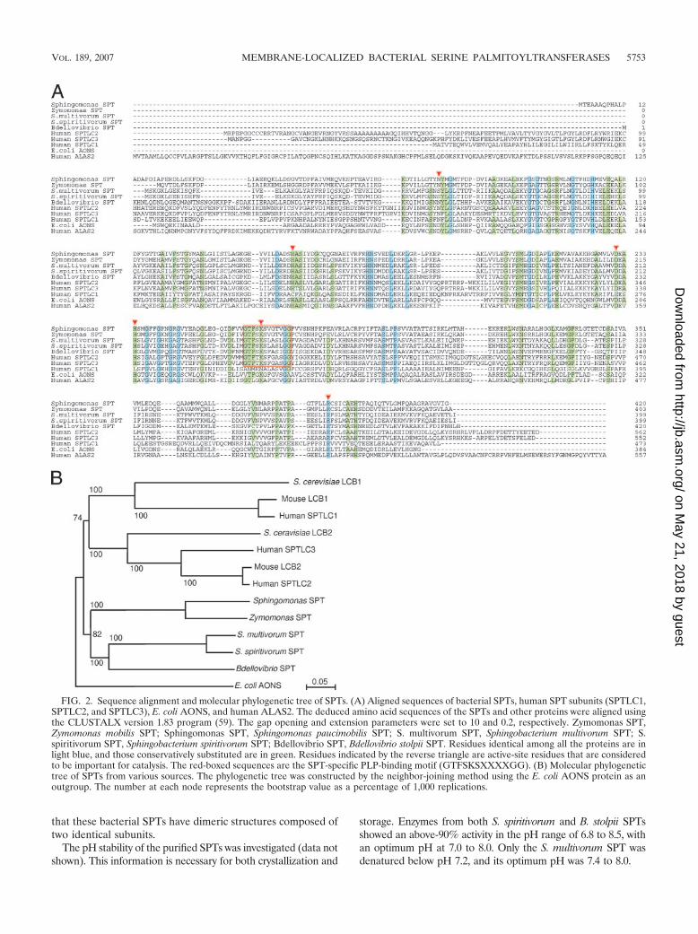
that these bacterial SPTs have dimeric structures composed oftwo identical subunits.
The pH stability of the purified SPTs was investigated (data notshown). This information is necessary for both crystallization and
storage. Enzymes from both S. spiritivorum and B. stolpii SPTsshowed an above-90% activity in the pH range of 6.8 to 8.5, withan optimum pH at 7.0 to 8.0. Only the S. multivorum SPT wasdenatured below pH 7.2, and its optimum pH was 7.4 to 8.0.
FIG. 2. Sequence alignment and molecular phylogenetic tree of SPTs. (A) Aligned sequences of bacterial SPTs, human SPT subunits (SPTLC1,SPTLC2, and SPTLC3), E. coli AONS, and human ALAS2. The deduced amino acid sequences of the SPTs and other proteins were aligned usingthe CLUSTALX version 1.83 program (59). The gap opening and extension parameters were set to 10 and 0.2, respectively. Zymomonas SPT,Zymomonas mobilis SPT; Sphingomonas SPT, Sphingomonas paucimobilis SPT; S. multivorum SPT, Sphingobacterium multivorum SPT; S.spiritivorum SPT, Sphingobacterium spiritivorum SPT; Bdellovibrio SPT, Bdellovibrio stolpii SPT. Residues identical among all the proteins are inlight blue, and those conservatively substituted are in green. Residues indicated by the reverse triangle are active-site residues that are consideredto be important for catalysis. The red-boxed sequences are the SPT-specific PLP-binding motif (GTFSKSXXXXGG). (B) Molecular phylogenetictree of SPTs from various sources. The phylogenetic tree was constructed by the neighbor-joining method using the E. coli AONS protein as anoutgroup. The number at each node represents the bootstrap value as a percentage of 1,000 replications.
VOL. 189, 2007 MEMBRANE-LOCALIZED BACTERIAL SERINE PALMITOYLTRANSFERASES 5753
on May 21, 2018 by guest
http://jb.asm.org/
Dow
nloaded from

All purified recombinant SPTs showed characteristic ab-sorption spectra of PLP-dependent enzymes (Fig. 3). For allof the bacterial SPTs, the intensities of the absorption peakswere not changed by the pH. The shapes of the spectra ofthe two Sphingobacterium SPTs were different from that ofthe Sphingomonas enzyme (Fig. 3A, B, and C). They hadonly a single peak at 426 nm in addition to the proteinabsorption peak at 278 nm. On the other hand, the B. stolpiiSPT showed two peaks, at 338 and 426 nm (Fig. 3D), and inthis respect was similar to the Sphingomonas enzyme, al-though the relative intensities of the two peaks are different.These absorption peaks, respectively, correspond to theenolimine and ketoenamine forms of the internal Schiff base(aldimine formed between the aldehyde group of PLP andthe ε-amino group of a lysine residue in the active site) ofSPT. The addition of L-serine to the purified SPTs gave riseto an intense absorption band at 426 nm for all of the SPTsand a weak band at 338 nm for the Sphingomonas and B.stolpii SPTs. These spectral changes showed hyperbolic de-pendencies on the concentrations of L-serine, and the ap-parent dissociation constants (KD) for L-serine were calcu-lated to be 0.47, 1.20, and 2.55 mM, respectively, for the S.multivorum, S. spiritivorum, and B. stolpii SPTs (Table 2).The CD spectra of these bacterial SPTs showed positivebands at 426 nm (and additionally at 338 nm for B. stolpiiSPT), corresponding to the absorption spectra of each en-zyme (data not shown). The CD spectra in the presence ofa saturating amount of L-serine showed a negative band at426 nm (data not shown). These results indicate that theadded L-serine formed the external Schiff base (aldimine
formed between PLP and extraneously added amino acid)with PLP. The addition of the second substrate, palmitoylCoA, to the B. stolpii SPT, which is saturated with L-serine,resulted in a slight decrease in the 426-nm peak and theappearance of an absorption band at 515 nm (Fig. 3C). Theformation of the 515-nm peak was transient, and the peakvanished within a few minutes. Such spectral changes werenot observed for the Sphingobacterium SPTs or the Sphin-gomonas SPT.
Identification of reaction products of bacterial SPTs. Theformation of KDS by bacterial enzymes was confirmed byHPLC/ESI–ion-trap mass spectrometry (Fig. 4). Figure 4Ashows the ion chromatograms (m/z 300) of the SPT reactionproduct in the positive-ion mode. The most abundant ions atm/z 300 (Fig. 4C) corresponded to the protonated molecularions [M � H]� of C18:0 KDS formed from L-serine and palmi-toyl CoA, the structure of which is shown in Fig. 4B. This LCB
FIG. 3. Absorption spectra of purified SPTs. The purified SPTs (10 �M) were dissolved in 50 mM HEPES-NaOH (pH 7.5) containing 150 mMKCl and 0.1 mM EDTA, and their absorption spectra were measured at 25°C. (A to C) Absorption spectra of the Sphingomonas paucimobilis, S.multivorum, and S. spiritivorum SPTs in the absence (solid line) or presence (dashed line) of 45 mM L-serine. (D) Absorption spectra of the B. stolpiiSPT in the absence (solid line) or presence (dashed line) of 100 mM L-serine. The dotted line shows the spectrum in the presence of 100 mML-serine and 90 �M palmitoyl CoA.
TABLE 2. Kinetic parameters of bacterial SPTs
Bacterial species
Value for SPT parameter
KD (Ser)(mM)
Km (Ser)(mM)
Km(palmitoyl
CoA) (mM)kcat (s�1)
S. paucimobilis 1.40 � 0.10 4.7 � 0.6 0.69 � 0.09 2.3 � 0.11S. multivorum 0.47 � 0.10 4.8 � 0.6 0.10 � 0.01 0.12 � 0.01S. spiritivorum 1.20 � 0.03 5.0 � 0.8 0.39 � 0.04 0.15 � 0.01B. stolpii 2.55 � 0.12 3.7 � 0.4 NDa 0.03 � 0.002b
a ND, not determined.b v/Et value in the presence of 100 �M of palmitoyl CoA.
5754 IKUSHIRO ET AL. J. BACTERIOL.
on May 21, 2018 by guest
http://jb.asm.org/
Dow
nloaded from
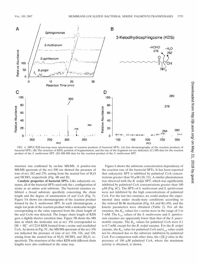
structure was confirmed by on-line MS/MS. A positive-ionMS/MS spectrum of the m/z-300 ion showed the presence ofions of m/z 282 and 270, arising from the neutral loss of H2Oand HCHO, respectively (Fig. 4B and D).
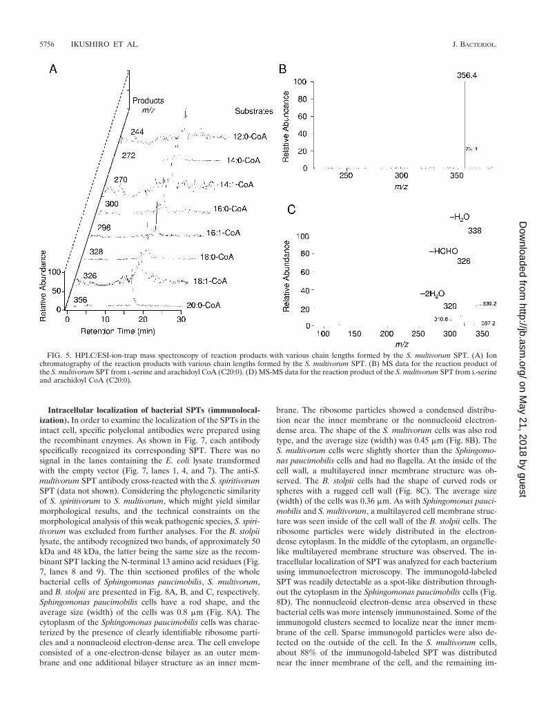
Catalytic properties of bacterial SPTs. Like eukaryotic en-zymes, all of the bacterial SPTs used only the L configuration ofserine as an amino acid substrate. The bacterial enzymes ex-hibited a broad substrate specificity concerning the chainlength and the degree of unsaturation of acyl CoA (Fig. 5).Figure 5A shows ion chromatograms of the reaction productformed by the S. multivorum SPT. In each chromatogram, asingle ion peak of the reaction product with a molecular weightcorresponding to the value expected from the chain length ofthe acyl CoAs was detected. The longer chain length of KDSgave a slightly shorter retention time. Figure 5B shows the MSdata in which the molecular ion at m/z 356 corresponded to[M � H]� of C22:0 KDS formed from L-serine and arachidoylCoA. As shown in Fig. 5C, the MS/MS spectrum of the m/z-356ion indicated the presence of ions of m/z 338, 326, and 320,arising from the neutral loss of H2O, HCHO, and 2H2O, re-spectively. The structures of the other KDS with different chainlengths were also confirmed in the same way.
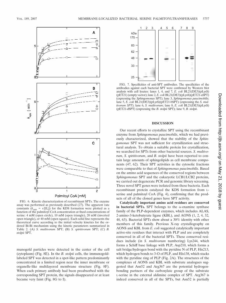
Figure 6 shows the substrate concentration dependency ofthe reaction rate of the bacterial SPTs. It has been reportedthat eukaryotic SPT is inhibited by palmitoyl CoA concen-trations greater than 50 �M (20, 53). A similar phenomenonwas observed with the B. stolpii SPT, which was significantlyinhibited by palmitoyl CoA concentrations greater than 100�M (Fig. 6C). The SPTs of S. multivorum and S. spiritivorumwere not inhibited by the high concentrations of palmitoylCoA. For the last two enzymes, we could analyze the exper-imental data under steady-state conditions according tothe ordered Bi-Bi mechanism (Fig. 6A and B) (49), and thekinetic parameters were obtained (Table 2). For all theenzymes, the Km values for L-serine were in the range of 3 to5 mM. The kcat values of the S. multivorum and S. spiritivo-rum enzymes are apparently lower than that of the S. pauci-mobilis enzyme. The Km values for palmitoyl CoA were 0.1to 0.7 mM, except for the B. stolpii enzyme. For the B. stolpiienzyme, the Km value for palmitoyl CoA and kcat value couldnot be obtained due to the substrate inhibition by palmitoylCoA. For comparison with other data, the v/[Et] value in thepresence of 100 �M palmitoyl CoA, where the maximumactivity is obtained, is shown.
FIG. 4. HPLC/ESI-ion-trap mass spectroscopy of reaction products of bacterial SPTs. (A) Ion chromatography of the reaction products ofbacterial SPTs. (B) The structure of KDS, position of fragmentation, and the size of the fragment ion are indicated. (C) MS data for the reactionproduct of the S. multivorum SPT. (D) MS-MS data for the reaction product of the S. multivorum SPT.
VOL. 189, 2007 MEMBRANE-LOCALIZED BACTERIAL SERINE PALMITOYLTRANSFERASES 5755
on May 21, 2018 by guest
http://jb.asm.org/
Dow
nloaded from
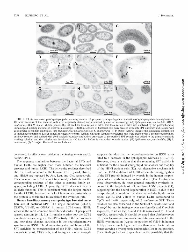
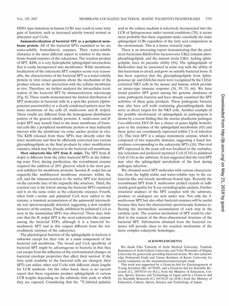
Intracellular localization of bacterial SPTs (immunolocal-ization). In order to examine the localization of the SPTs in theintact cell, specific polyclonal antibodies were prepared usingthe recombinant enzymes. As shown in Fig. 7, each antibodyspecifically recognized its corresponding SPT. There was nosignal in the lanes containing the E. coli lysate transformedwith the empty vector (Fig. 7, lanes 1, 4, and 7). The anti-S.multivorum SPT antibody cross-reacted with the S. spiritivorumSPT (data not shown). Considering the phylogenetic similarityof S. spiritivorum to S. multivorum, which might yield similarmorphological results, and the technical constraints on themorphological analysis of this weak pathogenic species, S. spiri-tivorum was excluded from further analyses. For the B. stolpiilysate, the antibody recognized two bands, of approximately 50kDa and 48 kDa, the latter being the same size as the recom-binant SPT lacking the N-terminal 13 amino acid residues (Fig.7, lanes 8 and 9). The thin sectioned profiles of the wholebacterial cells of Sphingomonas paucimobilis, S. multivorum,and B. stolpii are presented in Fig. 8A, B, and C, respectively.Sphingomonas paucimobilis cells have a rod shape, and theaverage size (width) of the cells was 0.8 �m (Fig. 8A). Thecytoplasm of the Sphingomonas paucimobilis cells was charac-terized by the presence of clearly identifiable ribosome parti-cles and a nonnucleoid electron-dense area. The cell envelopeconsisted of a one-electron-dense bilayer as an outer mem-brane and one additional bilayer structure as an inner mem-
brane. The ribosome particles showed a condensed distribu-tion near the inner membrane or the nonnucleoid electron-dense area. The shape of the S. multivorum cells was also rodtype, and the average size (width) was 0.45 �m (Fig. 8B). TheS. multivorum cells were slightly shorter than the Sphingomo-nas paucimobilis cells and had no flagella. At the inside of thecell wall, a multilayered inner membrane structure was ob-served. The B. stolpii cells had the shape of curved rods orspheres with a rugged cell wall (Fig. 8C). The average size(width) of the cells was 0.36 �m. As with Sphingomonas pauci-mobilis and S. multivorum, a multilayered cell membrane struc-ture was seen inside of the cell wall of the B. stolpii cells. Theribosome particles were widely distributed in the electron-dense cytoplasm. In the middle of the cytoplasm, an organelle-like multilayered membrane structure was observed. The in-tracellular localization of SPT was analyzed for each bacteriumusing immunoelectron microscopy. The immunogold-labeledSPT was readily detectable as a spot-like distribution through-out the cytoplasm in the Sphingomonas paucimobilis cells (Fig.8D). The nonnucleoid electron-dense area observed in thesebacterial cells was more intensely immunostained. Some of theimmunogold clusters seemed to localize near the inner mem-brane of the cell. Sparse immunogold particles were also de-tected on the outside of the cell. In the S. multivorum cells,about 88% of the immunogold-labeled SPT was distributednear the inner membrane of the cell, and the remaining im-
FIG. 5. HPLC/ESI-ion-trap mass spectroscopy of reaction products with various chain lengths formed by the S. multivorum SPT. (A) Ionchromatography of the reaction products with various chain lengths formed by the S. multivorum SPT. (B) MS data for the reaction product ofthe S. multivorum SPT from L-serine and arachidoyl CoA (C20:0). (D) MS-MS data for the reaction product of the S. multivorum SPT from L-serineand arachidoyl CoA (C20:0).
5756 IKUSHIRO ET AL. J. BACTERIOL.
on May 21, 2018 by guest
http://jb.asm.org/
Dow
nloaded from
munogold particles were detected in the center of the cell(cytoplasm) (Fig. 8E). In the B. stolpii cells, the immunogold-labeled SPT was detected in a spot-like pattern predominantlyconcentrated in a limited region near the inner membrane ororganelle-like multilayered membrane structure (Fig. 8F).When each primary antibody had been preabsorbed with thecorresponding SPT protein, the signals disappeared or at leastbecame very faint (Fig. 8G to I).
DISCUSSION
Our recent efforts to crystallize SPT using the recombinantenzyme from Sphingomonas paucimobilis, which we had previ-ously characterized, showed that the stability of the Sphin-gomonas SPT was not sufficient for crystallization and struc-tural analysis. To obtain a suitable protein for crystallization,we searched for SPTs from other bacterial sources. S. multivo-rum, S. spiritivorum, and B. stolpii have been reported to con-tain large amounts of sphingolipids as cell membrane compo-nents (47, 62). Their SPT activities in the cytosolic fractionswere comparable to that of Sphingomonas paucimobilis. Basedon the amino acid sequences of the conserved regions betweenSphingomonas SPT and the eukaryotic LCB1/LCB2 proteins,we carried out degenerate PCR and genomic library screening.Three novel SPT genes were isolated from these bacteria. Eachrecombinant protein catalyzed the KDS formation from L-serine and palmitoyl CoA (Fig. 4), confirming that the prod-ucts of all of the cloned genes have SPT activity.
Catalytically important amino acid residues are conservedin bacterial SPTs. SPT belongs to the �-oxamine synthasefamily of the PLP-dependent enzymes, which includes ALAS,2-amino-3-ketobutyrate ligase (KBL), and AONS (1, 2, 4, 32,48, 63). Bacterial SPTs show about a 30% identity with othermembers of this family. Previous X-ray crystallography onAONS and KBL from E. coli suggested catalytically importantactive-site residues that interact with PLP and are completelyconserved in all of the bacterial SPTs. These conserved resi-dues include (in S. multivorum numbering) Lys244, whichforms a Schiff base linkage with PLP, Asp210, which forms asalt bridge/hydrogen bond with the pyridine N of PLP, His213,which hydrogen bonds to 3-O of PLP, and His138, which stackswith the pyridine ring of PLP (Fig. 2A). The structures of thecomplexes of AONS and KBL with substrate analogues sug-gested that Asn52 and Arg367 are the potential hydrogen-bonding partners of the carboxylate group of the substrateL-serine in the external aldimine complex of SPT. Arg367 isindeed conserved in all of the SPTs, but Asn52 is partially
FIG. 6. Kinetic characterization of recombinant SPTs. The enzymeassay was performed as previously described (27). The apparent rateconstants (kapp � v/[Et]) for the KDS formation were plotted as afunction of the palmitoyl CoA concentration at fixed concentrations ofserine: 4 mM (open circle), 10 mM (open triangle), 20 mM (invertedopen triangle), or 40 mM (open square). Each solid line represents thetheoretical curve according to the initial velocity kinetics for the or-dered Bi-Bi mechanism using the kinetic parameters summarized inTable 2. (A) S. multivorum SPT; (B) S. spiritivorum SPT; (C) B.stolpii SPT.
FIG. 7. Specificities of anti-SPT antibodies. The specificities of theantibodies against each bacterial SPT were confirmed by Western blotanalysis with cell lysates: lanes 1, 4, and 7, E. coli BL21(DE3)(pLysS)(pET21) (empty vector); lane 2, E. coli BL21(DE3)(pLysS)(pET21-aSPT)(expressing the Sphingomonas SPT); lane 3, Sphingomonas paucimobilis;lane 5, E. coli BL21(DE3)(pLysS)(pET21-bSPT) (expressing the S. mul-tivorum SPT); lane 6, S. multivorum; lane 8, E. coli BL21(DE3)(pLysS)(pET21-dSPT) (expressing the B. stolpii SPT); lane 9, B. stolpii.
VOL. 189, 2007 MEMBRANE-LOCALIZED BACTERIAL SERINE PALMITOYLTRANSFERASES 5757
on May 21, 2018 by guest
http://jb.asm.org/
Dow
nloaded from
conserved; it shifts by one residue in the Sphingomonas and Z.mobilis SPTs.
The sequence similarities between the bacterial SPTs andhuman LCB2 are higher than those between the bacterialenzymes and human LCB1. The active-site residues describedabove are not conserved in the human LCB1; Lys244, His213,and His138 are replaced by Asn, Leu, and Cys, respectively.These residues in LCB1 cannot functionally substitute for thecorresponding residues of the other �-oxamine family en-zymes, including LCB2. Apparently, LCB1 does not have acatalytic function. This is consistent with the longer branchlength of LCB1, because the lack of functional constraints onthe protein is considered to accelerate the evolution rate.
Human hereditary sensory neuropathy type I-related muta-tion site of bacterial SPT. The single mutations (C133Y,C133W, V144D, or G387A) in human LCB1 cause HSN1,which is the most common hereditary disorder of peripheralsensory neurons (6, 11, 61). It remains elusive how the LCB1mutations cause changes in the SPT activity of the heterodimerand how these changes participate in the neurodegenerativesymptoms in HSN1. The dominant-negative inhibition of theSPT activities by overexpression of the HSN1-related LCB1mutants in yeast, CHO cells, and transgenic mouse strongly
supports the idea that the neurodegeneration in HSN1 is re-lated to a decrease in the sphingolipid synthesis (7, 17, 40).However, there is a claim that the remaining SPT activity issufficient for the normal sphingolipid metabolism and viabilityof the HSN1 patient cells (12). An alternative mechanism isthat the HSN1 mutations of LCB1 accelerate the aggregationof the SPT protein induced by hypoxia in the human lympho-cytes, which leads to nonapoptotic death (13). Contrary tothese observations, de novo glucosyl ceramide synthesis in-creased in the lymphoblast cell lines from HSN1 patients (11),suggesting that the neural degeneration in HSN1 is due to theoverproduced ceramide or the abnormal cellular lipid compo-sition. Cys133 and Val144 of human LCB1 correspond toCys78 and Ile90, respectively, of S. multivorum SPT. Theseresidues are also conserved in the SPTs of S. spiritivorum andB. stolpii but not in Sphingomonas paucimobilis and Z. mobilissequences, in which Cys133 and Val144 correspond to Thr andAsp/Gly, respectively. It should be noted that SphingomonasSPT, which carries an amino acid substitution equivalent to theHSN1-type mutation (Asp at the position of Val144 in humanLcb1p), has a much higher activity than other bacterial en-zymes carrying a hydrophobic amino acid (Ile) at that position.These findings lead us to speculate on the possibility that the
FIG. 8. Electron microscopy of sphingolipid-containing bacteria. Upper panels, morphological examination of sphingolipid-containing bacteria.Ultrathin sections of the bacterial cells were negatively stained and examined by electron microscopy. (A) Sphingomonas paucimobilis; (B) S.multivorum; (C) B. stolpii. Middle panels, the intracellular localization of SPT. The localization of SPT was analyzed by the postembeddingimmunogold-labeling method of electron microscopy. Ultrathin sections of bacterial cells were treated with anti-SPT antibody and stained withgold-labeled secondary antibodies. (D) Sphingomonas paucimobilis; (E) S. multivorum; (F) B. stolpii. Arrows indicate the condensed distributionof immunogold particles. Lower panels, the negative control section. Ultrathin sections of bacterial cells were treated with a preabsorbed primaryantibody solution and stained with gold-labeled secondary antibodies. An excess of the purified SPT protein was added to the primary antibodyworking solution, and the solution was incubated at 4°C for 48 h before it was added to each section. (G) Sphingomonas paucimobilis; (H) S.multivorum; (I) B. stolpii. Size markers are indicated.
5758 IKUSHIRO ET AL. J. BACTERIOL.
on May 21, 2018 by guest
http://jb.asm.org/
Dow
nloaded from
HSN1-type mutations in human LCB1 may result in some toxicgain of function, such as increased activity toward normal orabnormal acyl CoAs.
Immunolocalization of bacterial SPT as a peripheral mem-brane protein. All of the bacterial SPTs examined so far arewater-soluble homodimeric enzymes. Their water-solublecharacter is the most different aspect in relation to the mem-brane-bound enzymes of the eukaryotes. The reaction productof SPT, KDS, is a very hydrophobic sphingolipid intermediatethat is easily incorporated into membranes. While membranelocalization of the eukaryotic SPT complex seems very reason-able, the characteristics of the bacterial SPT as a water-solubleprotein in vitro raised questions about the mechanism of theproduct release or the interaction with the cellular membranein vivo. Therefore, we further analyzed the intracellular local-ization of the bacterial SPT by immunoelectron microscopy(Fig. 8). These results demonstrated the limited distribution ofSPT molecules in bacterial cells in a spot-like pattern (Sphin-gomonas paucimobilis) or a clearly condensed pattern near theinner membrane of the cells (S. multivorum and B. stolpii).These results are different from the homogenous distributionpattern of the general soluble proteins. S. multivorum and B.stolpii SPT may loosely bind to the inner membrane of bacte-rial cells like a peripheral membrane protein or may indirectlyinteract with the membrane via some anchor protein in vivo.The KDS released from these SPTs may directly enter theinner membrane and then be efficiently converted into variousglycosphingolipids as the final products by other modificationenzymes, which may be present in the bacterial cell membrane.
Most eukaryotic-like SPT from B. stolpii. The SPT from B.stolpii is different from the other bacterial SPTs in the follow-ing ways. First, during purification, the recombinant enzymerequired the addition of 20% glycerol, which is the most gen-eral stabilizer for membrane proteins. Second, B. stolpii has anorganelle-like multilayered membrane structure within thecell, and the immunoreaction to the native SPT was detectedon the cytosolic face of the organelle-like structure. Third, thereaction rate is the lowest among the bacterial SPTs examinedand is on the same order as the eukaryotic enzymes. Fourth,when both L-serine and palmitoyl CoA were added to theenzyme, a transient accumulation of the quinonoid intermedi-ate was spectroscopically detected, suggesting a slow catalyticturnover of the enzyme. Finally, inhibition by palmitoyl CoA asseen in the mammalian SPTs was observed. These data indi-cate that the B. stolpii SPT is the most eukaryotic-like enzymeamong the bacterial STPs, although it is a bacterial ho-modimeric SPT and in this respect different from the het-erodimeric enzymes of the eukaryotes.
The physiological function of the sphingolipids in bacteria isunknown except for their role as a main component of thebacterial cell membrane. The broad acyl CoA specificity ofbacterial SPT might be advantageous to bacteria in that theycan escape from the influence of the environmental changes onbacterial envelope properties that affect their survival. If thefatty acids available to the bacterial cells are changed, theirSPTs can utilize other acyl CoAs with different chain lengthsfor LCB synthesis. On the other hand, there is no currentreport that these organisms produce sphingolipids of variousLCB lengths depending on the substrate fatty acids to whichthey are exposed. Considering that the 14C-labeled palmitic
acid in the culture medium is selectively incorporated into theLCB of Sphingomonas under normal conditions (70), it seemsmore probable that these organisms make essentially the samesphingolipid LCBs regardless of the fatty acid composition inthe environment. This is a future research topic.
There is an interesting report demonstrating that the pred-atory bacterium Bdellovibrio bacteriovorus UKi2 contains phos-phosphingolipids, and the mutant strain UKi1, lacking sphin-golipids, loses its parasitic ability (56). The sphingolipids ofBdellovibrio may be associated in some way with the ability ofthis bacterium to attack and grow on suitable bacterial hosts. Ithas been reported that the glycosphingolipids from Sphin-gomonas sp. and Ehrlichia muris were recognized by the CD1d-restricted NKT cells in the mouse and human, which providean innate-type immune response (34, 39, 55, 66). We havefound putative SPT genes among the genome databases ofsome pathogenic bacteria and have already examined the SPTactivities of these gene products. These pathogenic bacteriamay also have cell walls containing glycosphingolipids thatserve as direct targets for the NKT cells. Another example ofthe possible involvement of sphingolipids in pathogenesis isshown by a recent finding that the marine planktonic pathogenCoccolithovirus EhV-86 has a cluster of genes highly homolo-gous to the enzymes of the sphingolipid metabolism (65) andthese genes are coordinately expressed within 2 h of infection(3). This viral SPT is a unique monomeric enzyme, which iscomposed of two separable domains, suggesting a fused het-erodimer corresponding to the eukaryotic SPTs (18). This viralSPT expressed in the yeast cell was localized to the endoplas-mic reticulum and preferred myristoyl CoA (C14) to palmitoylCoA (C16) as the substrate. It was suggested that the viral SPTmay alter the sphingolipid metabolism of the host duringpathogen infection.
We obtained novel SPT molecules with various characteris-tics, from the highly stable and water-soluble type to the eu-karyotic-like and loosely membrane-bound type. Recently therecombinant SPT from S. multivorum yielded crystals of suffi-ciently good quality for X-ray crystallographic analysis. Furtherstructural analyses of the SPT complex with the substrate,product, or analogues are now under way. Not only the S.multivorum SPT but also other bacterial enzymes will be usefulbecause they have the characteristic spectroscopic features re-flecting the intermediate accumulation of each step in thecatalytic cycle. The reaction mechanism of SPT could be clar-ified in the context of the three-dimensional structure of thebacterial SPT. Information obtained from the bacterial en-zymes will provide clues to the reaction mechanism of themore complex eukaryotic homologue.
ACKNOWLEDGMENTS
We thank Eiko Yabuichi of Aichi Medical University, YoshiakiKawamura of Aichi Gakuin University, and Yoko Watanabe of NiigataUniversity for generously providing bacterial strains. We also acknowl-edge Nobuyoshi Esaki and Tatsuo Kurihara of Kyoto University foruseful comments on the immunoelecromicroscopic study.
This work was supported by a Grant-in-Aid for Encouragement ofYoung Scientists (B), 16770103, and a Grant-in-Aid for Scientific Re-search (C), 18570114 (to H.I.), from the Ministry of Education, Cul-ture, Sports, Science and Technology of Japan and by a Grant-in-Aidfor Scientific Research (C) 16570125 (to H.H.) from the Ministry ofEducation, Culture, Sports, Science and Technology of Japan.
VOL. 189, 2007 MEMBRANE-LOCALIZED BACTERIAL SERINE PALMITOYLTRANSFERASES 5759
on May 21, 2018 by guest
http://jb.asm.org/
Dow
nloaded from

REFERENCES
1. Alexander, F. W., E. Sandmeier, P. K. Mehta, and P. Christen. 1994. Evo-lutionary relationships among pyridoxal-5�-phosphate-dependent enzymes.Regio-specific alpha, beta and gamma families. Eur. J. Biochem. 219:953–960.
2. Alexeev, D., M. Alexeeva, R. L. Baxter, D. J. Campopiano, S. P. Webster, andL. Sawyer. 1998. The crystal structure of 8-amino-7-oxononanoate synthase:a bacterial PLP-dependent, acyl-CoA-condensing enzyme. J. Mol. Biol. 284:401–419.
3. Allen, M. J., T. Forster, D. C. Schroeder, M. Hall, D. Roy, P. Ghazal, andW. H. Wilson. 2006. Locus-specific gene expression pattern suggests a uniquepropagation strategy for a giant algal virus. J. Virol. 80:7699–7705.
4. Astner, I., J. O. Schulze, J. van den Heuvel, D. Jahn, W. D. Schubert, andD. W. Heinz. 2005. Crystal structure of 5-aminolevulinate synthase, the firstenzyme of heme biosynthesis, and its link to XLSA in humans. EMBO J.24:3166–3177.
5. Ausubel, F. M., R. Brent, R. E. Kingston, D. D. Moore, J. G. Seidman, J. A.Smith, and K. Struhl. 1994. Current protocols in molecular biology, Suppl.27, unit 2.4. John Wiley & Sons, Inc., New York, NY.
6. Bejaoui, K., C. Wu, M. D. Scheffler, G. Haan, P. Ashby, L. Wu, P. de Jong,and R. H. Brown, Jr. 2001. SPTLC1 is mutated in hereditary sensory neu-ropathy, type 1. Nat. Genet. 27:261–262.
7. Bejaoui, K., Y. Uchida, S. Yasuda, M. Ho, M. Nishijima, R. H. Brown, Jr.,W. M. Holleran, and K. Hanada. 2002. Hereditary sensory neuropathy type1 mutations confer dominant negative effects on serine palmitoyltransferase,critical for sphingolipid synthesis. J. Clin. Investig. 110:1301–1308.
8. Blahova, J., K. Kralikova, V. Krcmery, Sr., and K. Kubonova. 1997. Hydro-lysis of imipenem, meropenem, ceftazidime, and cefepime by multiresistantnosocomial strains of Sphingobacterium multivorum. Eur. J. Clin. Microbiol.Infect. Dis. 16:178–180.
9. Bligh, E. G., and W. J. Dyer. 1959. A rapid method of total lipid extractionand purification. Can. J. Biochem. Physiol. 37:911–917.
10. Buede, R., S. C. Rinker, W. J. Pinto, R. L. Lester, and R. C. Dickson. 1991.Cloning and characterization of LCB1, a Saccharomyces gene required forbiosynthesis of the long-chain base component of sphingolipids. J. Bacteriol.173:4325–4332.
11. Dawkins, J. L., D. J. Hulme, S. B. Brahmbhatt, M. Auer-Grumbach, andG. A. Nicholson. 2001. Mutations in SPTLC1, encoding serine palmitoyl-transferase, long chain base subunit-1, cause hereditary sensory neuropathytype I. Nat. Genet. 27:309–312.
12. Dedov, V. N., I. V. Dedova, A. H. Merrill, Jr., and G. A. Nicholson. 2004.Activity of partially inhibited serine palmitoyltransferase is sufficient fornormal sphingolipid metabolism and viability of HSN1 patient cells. Bio-chim. Biophys. Acta 1688:168–175.
13. Dedov, V. N., I. V. Dedova, and G. A. Nicholson. 2004. Hypoxia causesaggregation of serine palmitoyltransferase followed by non-apoptotic deathof human lymphocytes. Cell Cycle 3:1271–1277.
14. Dykstra, M., A. Cherukuri, H. W. Sohn, S. J. Tzeng, and S. K. Pierce. 2003.Location is everything: lipid rafts and immune cell signaling. Annu. Rev.Immunol. 21:457–481.
15. Ferreira, G. C., and J. S. Zhang. 2002. Mechanism of 5-aminolevulinatesynthase and the role of the protein environment in controlling the cofactorchemistry. Cell Mol. Biol. 48:827–833.
16. Freney, J., W. Hansen, C. Ploton, H. Meugnier, S. Madier, N. Bornstein, andJ. Fleurette. 1987. Septicemia caused by Sphingobacterium multivorum.J. Clin. Microbiol. 25:1126–1128.
17. Gable, K., G. Han, E. Monaghan, D. Bacikova, M. Natarajan, R. Williams,and T. M. Dunn. 2002. Mutations in the yeast LCB1 and LCB2 genes,including those corresponding to the hereditary sensory neuropathy type Imutations, dominantly inactivate serine palmitoyltransferase. J. Biol. Chem.277:10194–101200.
18. Han, G., K. Gable, L. Yan, M. J. Allen, W. H. Wilson, P. Moitra, J. M.Harmon, and T. M. Dunn. 2006. Expression of a novel marine viral single-chain serine palmitoyltransferase and construction of yeast and mammaliansingle-chain chimera. J. Biol. Chem. 281:39935–39942.
19. Hanada, K., T. Hara, M. Nishijima, O. Kuge, R. C. Dickson, and M. M.Nagiec. 1997. A mammalian homolog of the yeast LCB1 encodes a compo-nent of serine palmitoyltransferase, the enzyme catalyzing the first step insphingolipid synthesis. J. Biol. Chem. 272:32108–32114.
20. Hanada, K., T. Hara, and M. Nishijima. 2000. Purification of the serinepalmitoyltransferase complex responsible for sphingoid base synthesis byusing affinity peptide chromatography techniques. J. Biol. Chem. 275:8409–8415.
21. Hanada, K. 2003. Serine palmitoyltransferase, a key enzyme of sphingolipidmetabolism. Biochim. Biophys. Acta 1632:16–30.
22. Hannun, Y. A., and C. Luberto. 2000. Ceramide in the eukaryotic stressresponse. Trends Cell Biol. 10:73–80.
23. Holmes, B., R. J. Owen, and D. G. Hollis. 1982. Flavobacterium spiritivorum,a new species isolated From human clinical specimens. Int. J. Syst. Bacteriol.32:157–165.
24. Hornemann, T., S. Richard, M. F. Rutti, Y. Wei, and A. von Eckardstein.
2006. Cloning and initial characterization of a new subunit for mammalianserine-palmitoyltransferase. J. Biol. Chem. 281:37275–37281.
25. Huwiler, A., T. Kolter, J. Pfeilschifter, and K. Sandhoff. 2000. Physiology andpathophysiology of sphingolipid metabolism and signaling. Biochim. Bio-phys. Acta 1485:63–99.
26. Ikushiro, H., H. Hayashi, and H. Kagamiyama. 2000. Expression and puri-fication of serine palmitoyltransferase, p. 251–254. In A. Iriarte, H. M.Kagan, and M. Martinez-Carrion (ed.), Biochemistry and molecular biologyof vitamin B6 and PQQ-dependent proteins. Birkhaeuser Verlag, Basel,Switzerland.
27. Ikushiro, H., H. Hayashi, and H. Kagamiyama. 2001. A water-soluble ho-modimeric serine palmitoyltransferase from Sphingomonas paucimobilisEY2395T strain. Purification, characterization, cloning, and overproduction.J. Biol. Chem. 276:18249–18256.
28. Ikushiro, H., H. Hayashi, and H. Kagamiyama. 2003. Bacterial serine palmi-toyltransferase: a water-soluble homodimeric prototype of the eukaryoticenzyme. Biochim. Biophys. Acta 1647:116–120.
29. Ikushiro, H., H. Hayashi, and H. Kagamiyama. 2004. Reactions of serinepalmitoyltransferase with serine and molecular mechanisms of the actions ofserine derivatives as inhibitors. Biochemistry 43:1082–1092.
30. Ito, M., U. Tchoua, M. Okamoto, and H. Tojo. 2002. Purification and prop-erties of a phospholipase A2/lipase preferring phosphatidic acid, bis(mono-acylglycerol) phosphate, and monoacylglycerol from rat testis. J. Biol. Chem.277:43674–43681.
31. Kallen, R. G., T. Korpela, A. E. Martell, Y. Matsushima, C. M. Metzler, D. E.Metzler, Y. V. Morozov, I. M. Ralston, F. A. Savin, Y. M. Torchinsky, and H.Ueno. 1985. Chemical and spectroscopic properties of pyridoxal and pyri-doxine phosphates, p. 37–108. In P. Christen and D. E. Metzler (ed.),Transaminases. John Wiley & Sons, New York, NY.
32. Karlsson, K. A. 1970. On the chemistry and occurrence of sphingolipidlong-chain bases. Chem. Phys. Lipids 5:6–43.
33. Kawasaki, S., R. Moriguchi, K. Sekiya, T. Nakai, E. Ono, K. Kume, and K.Kawahara. 1994. The cell envelope structure of the lipopolysaccharide-lacking gram-negative bacterium Sphingomonas paucimobilis. J. Bacteriol.176:284–290.
34. Kinjo, Y., D. Wu, G. Kim, G. W. Xing, M. A. Poles, D. D. Ho, M. Tsuji, K.Kawahara, C. H. Wong, and M. Kronenberg. 2005. Recognition of bacterialglycosphingolipids by natural killer T cells. Nature 434:520–525.
35. Laemmli, U. K. 1970. Cleavage of structural proteins during the assembly ofthe head of bacteriophage T4. Nature 227:680–685.
36. Mandon, E. C., E. Ehses, J. Rother, G. van Echten, and K. Sandhoff. 1992.Subcellular localization and membrane topology of serine palmitoyltrans-ferase, 3-dehydrosphinganine reductase, and sphinganine N-acyltransferasein mouse liver. J. Biol. Chem. 267:11144–11148.
37. Manfredi, R., A. Nanetti, M. Ferri, A. Mastroianni, O. V. Coronado, and F.Chiodo. 1999. Flavobacterium spp. organisms as opportunistic bacterialpathogens during advanced HIV disease. J. Infect. 39:146–152.
38. Mathias, S., L. A. Pena, and R. N. Kolesnick. 1998. Signal transduction ofstress via ceramide. Biochem. J. 335:465–480.
39. Mattner, J., K. L. Debord, N. Ismail, R. D. Goff, C. Cantu, III, D. Zhou, P.Saint-Mezard, V. Wang, Y. Gao, N. Yin, K. Hoebe, O. Schneewind, D.Walker, B. Beutler, L. Teyton, P. B. Savage, and A. Bendelac. 2005. Exoge-nous and endogenous glycolipid antigens activate NKT cells during microbialinfections. Nature 434:525–529.
40. McCampbell, A., D. Truong, D. C. Broom, A. Allchorne, K. Gable, R. G.Cutler, M. P. Mattson, C. J. Woolf, M. P. Frosch, J. M. Harmon, T. M.Dunn, and R. H. Brown, Jr. 2005. Mutant SPTLC1 dominantly inhibitsserine palmitoyltransferase activity in vivo and confers an age-dependentneuropathy. Hum. Mol. Genet. 14:3507–3521.
41. Nagiec, M. M., J. A. Baltisberger, G. B. Wells, R. L. Lester, and R. C.Dickson. 1994. The LCB2 gene of Saccharomyces and the related LCB1 geneencode subunits of serine palmitoyltransferase, the initial enzyme in sphin-golipid synthesis. Proc. Natl. Acad. Sci. USA 91:7899–7902.
42. Nagiec, M. M., R. L. Lester, and R. C. Dickson. 1996. Sphingolipid synthesis:identification and characterization of mammalian cDNAs encoding the Lcb2subunit of serine palmitoyltransferase. Gene 177:237–241.
43. Naka, T., N. Fujiwara, S. Maeda, E. Yabuuchi, M. Doe, K. Kobayashi, Y.Kato, and I. Yano. 2000. A novel sphingoglycolipid containing galacturonicacid and 2-hydroxy fatty acid in cellular lipids of Sphingomonas yanoikuyae.J. Bacteriol. 182:2660–2663.
44. Naka, T., N. Fujiwara, I. Yano, S. Maeda, M. Doe, M. Minamino, N. Ikeda,Y. Kato, K. Watabe, Y. Kumazawa, I. Tomiyasu, and K. Kobayashi. 2003.Structural analysis of sphingophospholipids derived from Sphingobacteriumspiritivorum, the type species of genus Sphingobacterium. Biochim. Biophys.Acta 1635:83–92.
45. Radin, N. S. 1981. Preparative isolation of lecithin. Methods Enzymol. 72:5–7.
46. Rendulic, S., P. Jagtap, A. Rosinus, M. Eppinger, C. Baar, C. Lanz, H.Keller, C. Lambert, K. J. Evans, A. Goesmann, F. Meyer, R. E. Sockett, andS. C. Schuster. 2004. A predator unmasked: life cycle of Bdellovibrio bacte-riovorus from a genomic perspective. Science 303:689–692.
47. Ruby, E. G. 1991. The genus Bdellovibrio, p. 3400–3415. In A. Balows, H. G.
5760 IKUSHIRO ET AL. J. BACTERIOL.
on May 21, 2018 by guest
http://jb.asm.org/
Dow
nloaded from
Truper, M. Dworkin, W. Harder, and K. H. Schleifer (ed.), The prokaryotes.A handbook on the biology of bacteria: ecophysiology, isolation, identifica-tion, applications, 2nd ed. Springer-Verlag, New York, NY.
48. Schmidt, A., J. Sivaraman, Y. Li, R. Larocque, J. A. Barbosa, C. Smith, A.Matte, J. D. Schrag, and M. Cygler. 2001. Three-dimensional structure of2-amino-3-ketobutyrate CoA ligase from Escherichia coli complexed with aPLP-substrate intermediate: inferred reaction mechanism. Biochemistry 40:5151–5160.
49. Segel, I. H. 1975. Steady state kinetics of multireactant enzymes. In Enzymekinetics, p. 505–515. John Wiley & Sons, Inc., New York, NY.
50. Seo, J. S., H. Chong, H. S. Park, K. O. Yoon, C. Jung, J. J. Kim, J. H. Hong,H. Kim, J. H. Kim, J. I. Kil, C. J. Park, H. M. Oh, J. S. Lee, S. J. Jin, H. W.Um, H. J. Lee, S. J. Oh, J. Y. Kim, H. L. Kang, S. Y. Lee, K. J. Lee, and H. S.Kang. 2005. The genome sequence of the ethanologenic bacterium Zymomo-nas mobilis ZM4. Nat. Biotechnol. 23:63–68.
51. Shivaji, S., M. K. Ray, N. S. Rao, L. Saisree, M. V. Jagannadham, G. S.Kumar, G. S. N. Reddy, and P. M. Bhargava. 1992. Sphingobacterium ant-arcticus sp. nov., a psychrotrophic bacterium from the soils of SchirmacherOasis, Antarctica. Int. J. Syst. Bacteriol. 42:102–106.
52. Simons, K., and E. Ikonen. 1997. Functional rafts in cell membranes. Nature387:569–572.
53. Snell, E. E., S. J. Dimari, and R. H. Brady. 1970. Biosynthesis of sphingosineand dihydrosphingosine by cell-free systems from Hansenula ciferri. Chem.Phys. Lipids 5:116–138.
54. Spiegel, S., and A. H. Merrill, Jr. 1996. Sphingolipid metabolism and cellgrowth regulation. FASEB J. 10:1388–1397.
55. Sriram, V., W. Du, J. Gervay-Hague, and R. R. Brutkiewicz. 2005. Cell wallglycosphingolipids of Sphingomonas paucimobilis are CD1d-specific ligandsfor NKT cells. Eur. J. Immunol. 35:1692–1701.
56. Steiner, S., S. F. Conti, and R. L. Lester. 1973. Occurrence of phosphono-sphingolipids in Bdellovibrio bacteriovorus strain UKi2. J. Bacteriol. 116:1199–1211.
57. Stolp, H., and P. Starr. 1963. Bdellovibrio bacteriovorus gen. et sp. n., apredatory, ectoparasitic, and bacteriolytic microorganism. Antonie Leeu-wenhoek J. Microbiol. Serol. 29:217–248.
58. Takagi, S., H. Tojo, S. Tomita, S. Sano, S. Itami, M. Hara, S. Inoue, K.Horie, G. Kondoh, K. Hosokawa, F. J. Gonzalez, and J. Takeda. 2003.Alteration of the 4-sphingenine scaffolds of ceramides in keratinocyte-spe-cific Arnt-deficient mice affects skin barrier function. J. Clin. Investig. 112:1372–1382.
59. Thompson, J. D., T. J. Gibson, F. Plewniak, F. Jeanmougin, and D. G.Higgins. 1997. The CLUSTAL_X windows interface: flexible strategies formultiple sequence alignment aided by quality analysis tools. Nucleic AcidsRes. 25:4876–4882.
60. Tronel, H., P. Plesiat, E. Ageron, and P. A. Grimont. 2003. Bacteremiacaused by a novel species of Sphingobacterium. Clin. Microbiol. Infect.9:1242–1244.
61. Verhoeven, K., K. Coen, E. De Vriendt, A. Jacobs, V. Van Gerwen, I. Smouts,A. Pou-Serradell, J. J. Martin, V. Timmerman, and P. De Jonghe. 2004.SPTLC1 mutation in twin sisters with hereditary sensory neuropathy type I.Neurology 62:1001–1002.
62. Watanabe, Y., M. Nakajima, T. Hoshino, K. Jayasimhulu, E. E. Brooks, andE. S. Kaneshiro. 2001. A novel sphingophosphonolipid head group 1-hy-droxy-2-aminoethyl phosphonate in Bdellovibrio stolpii. Lipids 36:513–519.
63. Webster, S. P., D. Alexeev, D. J. Campopiano, R. M. Watt, M. Alexeeva, L.Sawyer, and R. L. Baxter. 2000. Mechanism of 8-amino-7-oxononanoatesynthase: spectroscopic, kinetic, and crystallographic studies. Biochemistry39:516–528.
64. Weiss, B., and W. Stoffel. 1997. Human and murine serine-palmitoyl-CoAtransferase—cloning, expression and characterization of the key enzyme insphingolipid synthesis. Eur. J. Biochem. 249:239–247.
65. Wilson, W. H., D. C. Schroeder, M. J. Allen, M. T. Holden, J. Parkhill, B. G.Barrell, C. Churcher, N. Hamlin, K. Mungall, H. Norbertczak, M. A. Quail,C. Price, E. Rabbinowitsch, D. Walker, M. Craigon, D. Roy, and P. Ghazal.2005. Complete genome sequence and lytic phase transcription profile of aCoccolithovirus. Science 309:1090–1092.
66. Wu, D., G. W. Xing, M. A. Poles, A. Horowitz, Y. Kinjo, B. Sullivan, V.Bodmer-Narkevitch, O. Plettenburg, M. Kronenberg, M. Tsuji, D. D. Ho,and C. H. Wong. 2005. Bacterial glycolipids and analogs as antigens forCD1d-restricted NKT cells. Proc. Natl. Acad. Sci. USA 102:1351–1356.
67. Yabuuchi, E., E. Tanimura, Y. Kosako, A. Ohyama, I. Yano, and A.Yamamoto. 1979. Flavobacterium devorans ATCC 10829: a strain of Pseudo-monas paucimobilis. J. Gen. Appl. Microbiol. 25:95–107.
68. Yabuuchi, E., I. Yano, H. Oyaizu, Y. Hashimoto, T. Ezaki, and H.Yamamoto. 1990. Proposals of Sphingomonas paucimobilis gen. nov. andcomb. nov., Sphingomonas parapaucimobilis sp. nov., Sphingomonasyanoikuyae sp. nov., Sphingomonas adhaesiva sp. nov., Sphingomonas capsu-lata comb. nov., and two genospecies of the genus Sphingomonas. Microbiol.Immunol. 34:99–119.
69. Yabuuchi, E., T. Kaneko, I. Yano, C. W. Moss, and N. Miyoshi. 1983.Sphingobacterium gen. nov., Sphingobacterium spiritivorum comb. nov., Sphin-gobacterium multivorum comb. nov., Sphingobacterium mizutae sp. nov., andFlavobacterium indologenes sp. nov.: glucose-nonfermenting gram-negativerods in CDC groups IIK-2 and IIb. Int. J. Syst. Bacteriol. 33:580–598.
70. Yamamoto, A., I. Yano, M. Masui, and E. Yabuuchi. 1978. Isolation of anovel sphingoglycolipid containing glucuronic acid and 2-hydroxy fatty acidfrom Flavobacterium devorans ATCC 10829. J. Biochem. (Tokyo) 83:1213–1216.
71. Yano, I., I. Tomiyasu, and E. Yabuuchi. 1982. Long chain base compositionof strains of three species of Sphingobacterium gen. nov. FEMS Microbiol.Lett. 15:303–307.
72. Yano, I., S. Imaizumi, I. Tomiyasu, and E. Yabuuchi. 1983. Separation andanalysis of free ceramides containing 2-hydroxy fatty acids in Sphingobacte-rium species. FEMS Microbiol. Lett. 20:449–453.
VOL. 189, 2007 MEMBRANE-LOCALIZED BACTERIAL SERINE PALMITOYLTRANSFERASES 5761
on May 21, 2018 by guest
http://jb.asm.org/
Dow
nloaded from