Embed Size (px)
Citation preview

Molecular cloning and expression analysis of RbcL cDNAfrom the bloom-forming green alga Chaetomorpha valida(Cladophorales, Chlorophyta)
Yunyan Deng & Zifeng Zhan & Xiaorong Tang &
Lanping Ding & Delin Duan
Received: 22 August 2013 /Revised and accepted: 5 November 2013# Springer Science+Business Media Dordrecht 2013
Abstract Ribulose-1,5-bisphosphate carboxylase/oxygenase(Rubisco) is a crucial enzyme catalyzing the CO2-fixing reac-tion in the initial step of the Calvin cycle. In this study, the full-length cDNA of Rubisco large subunit (RbcL) from thebloom-forming green alga Chaetomorpha valida (designatedas CvRbcL) was isolated based on homologous cloning andthe rapid amplification of cDNA ends (RACE) (accession:KF514135). It was 1,966-bp long, with an open reading frameof 1,431 bp without intron. The deduced 476 amino acidsgave a predicted molecular weight of 52.38 kDa and theoret-ical isoelectric point of 6.15. Three catalytic regions and aCO2 activator region were identified in the CvRbcL, whichhighly conserved with those of other green algae and higherplants. Homologous analysis indicated that CvRbcL shared52–86 % similarities with other 13 known algal and higherplant RbcLs sequences. Phylogenetic analysis showed that ithad closer relationship with those of green algae and higherplants but was comparatively far from the red and brown algaeclade. Real-time PCR detection revealed that the CvRbcLexpression were dramatically upregulated by light and strong-ly suppressed in the dark. Temperature also markedly affectedthe CvRbcL transcription. A relatively high transcript levelappeared between 15–25 °C and reached the maximum at20 °C. This indirectly reflected that highly expressed Rubiscowas involved with CO2 metabolism of C. valida under favor-able natural environments, and might contribute to its fast
growth and bloom formation. In vitro expression of CvRbcLshowed that one distinct band existed at ∼55 kDa, and westernblot detection proved that it was positive to the anti-Hisantibody with high specificity. Our study is the first to char-acterize RbcL gene from Chaetomorpha species providingmore knowledge of RbcL properties and facilitating furtherexploration of Rubisco in green macroalgae. Our results alsohelp to decipher Rubisco molecular functions in the rapidgrowth period of C. valida and provide clues to developforecasts of the seasonal C. valida bloom.
Keywords Bloomgreen alga .Chaetomorpha valida . Greentide . Ribulose-1,5-bisphosphate carboxylase/oxygenase largesubunit
Introduction
“Green tides” are vast accumulations of unattached greenmacroalgae usually associated with eutrophication in coastalenvironments (Valiela et al. 1997; Fletcher 1996). In recentyears, severe green algal blooms have occurred in the YellowSea (Ye et al. 2011). Chaetomorpha (Cladophoraceae, Chlor-ophyta) is a common and widespread green seaweed genus.Some of its members are often entangled with other foulingalgae to form extensive accumulation in marine or brackishhabitats (Fletcher 1996; Raffaelli et al. 1998; Chi et al. 2009).Chaetomorpha valida (Hooker et Harvey) Kützing, an inva-sive species in China, has been seasonally abundant in aqua-culture ponds along the coast of Dalian and Rongcheng citiesin the past few years (Chi et al. 2009; Deng et al. 2011, 2012).Astonishing quantities of drifting biomass seriously affectedthe aquatic environments and caused economic loss to localaquaculture (Chi et al. 2009; Deng et al. 2012). No valideradication measure has been taken, and the large amount of
Y. Deng : Z. Zhan :D. Duan (*)Institute of Oceanology, Chinese Academy of Sciences,Qingdao 266071, Chinae-mail: [email protected]
X. TangKey Laboratory of Marine Genetics and Breeding, Ministry ofEducation, Ocean University of China, Qingdao 266003, China
L. DingShantou University, Shantou 515063, China
J Appl PhycolDOI 10.1007/s10811-013-0208-z

thalli in aquaculture ponds requires more effectivemanagement.
Ribulose-1,5-bisphosphate carboxylase/oxygenase(Rubisco, E.C. 4.1.1.39) is the key enzyme responsible forthe first step of photosynthetic carbon assimilation, whichcatalyzes the carboxylation and oxygenation of ribulosebisphosphate (RuBP) (Jensen and Bahr 1977). The carboxyl-ation reaction results in CO2 fixation, while the oxygenationreaction leads to CO2 release. Rubisco directly participates inthe primary carbon fixation and determines the photosyntheticrate (Spreitzer and Salvucci 2002; Andersson and Backlund2008). It occurs in most autotrophic organisms, and fourstructural forms (I, II, III, and IV) have been found in nature(Tabita et al. 2008). Among them, form I Rubisco character-ized by eight large subunits (RbcL) and eight small subunits(RbcS) is found in higher plants, algae, and bacteria. In higherplants and green algae, the RbcL (∼55 kDa) is encoded by asingle RbcL gene in the chloroplast genome, whereas theRbcS (∼15 kDa) is encoded by a family of nuclear genes(Dean et al. 1989; Schneider et al. 1992; Houtz and Portis2003). In algae, both the active and the catalytic domains arelocated in the large subunit (Andersson and Taylor 2003; Yinget al. 2011).
Due to its crucial role in photosynthetic CO2 assimilation,extensive research has been done on the Rubisco in variousorganisms. However in algae, such studies are relatively fewand mainly limited to unicellular species (Dron et al. 1982;Ginrich and Hallick 1985). Most algal RbcL records inGenBank are partial sequences which were generated formolecular markers to elucidate phylogenetic affinity. Gapsremain in our knowledge about RbcL sequences in vast ma-jority of macroalgae as well as its molecular characters andphysiological functions.
In order to understand better the mechanisms underlyingthe seasonal C. valida abundance, RbcL was characterizedwith the aim to explore its relation with excessive growth ofthis alga. Based on full-length cDNA sequence isolation,transcription analysis and in vitro prokaryotic expression ofCvRbcL were also carried out.
Materials and methods
Sample collection and treatments
Fresh Chaetomorpha valida were collected from aquacultureponds in Rongcheng, Shandong, China (37°16′ N, 122°41′ E)on 24 August 2008. Healthy thalli were rinsed with sterilizedseawater several times to remove epiphytes. The washedmaterials were stored immediately in liquid nitrogen forDNA and RNA extraction.
For the transcriptional analysis, the washed materials wereprecultured at 25 °C and 72 μmol photons m−2 s−1 with 12:12
L/D for 72 h. For the heat treatments, the algae were kept insterile seawater at 5, 10, 15, 20, 25, 30, and 35 °C for 1 h,while during the light/dark tests, the algae were cultured at25 °C under 72 μmol photons m−2 s−1 with 12:12 L/D for72 h, and a total of six samples were collected every 12 h. Allthe treated algal materials were rinsed three times with 0.1 %DEPC sterile seawater and stored at −80 °C for RNAextraction.
DNA, RNA extraction, and cDNA synthesis
The genomic DNA of C. valida was prepared using PlantGenomic DNA kit (Tiangen, China). All manipulations werecarried out at room temperature. The quantification of DNAwas checked by 1 % agarose gel electrophoresis.
Total RNAwas extracted according to CTAB method (Yaoet al. 2009) and was treated with RNase-free DNase I(TaKaRa, China) to remove the residual genomic DNA. Thefirst strand cDNA was obtained using the PrimeScript IIcDNA synthesis kit (TaKaRa, Japan) according to the manu-facturer's protocols.
CvRbcL cDNA cloning
A pair of specific primers, P1 and P2 (Table 1), were designedbased on the known green algal RbcL sequences for amplify-ing CvRbcL cDNA fragment of about 500 bp. PCR wasperformed in a 25-μL reaction volume containing 12.5 μL2×TransTaqTM High Fidelity PCR SuperMix (TransGen Bio-tech, China), 1 μL of cDNA ofC. valida , 1 μL of each primer(10 μM), and 9.5 μL of RNase-free water. The amplificationswere performed on TaKaRa PCRThermal Cycler TP600 withan initial denaturation at 94 °C for 5 s, followed by 29 cyclesof 95 °C for 30 s and 50 °C for 30 s, 72 °C for 2 min, and afinal extension at 72 °C for 10 min. The PCR products werepurified by agarose gel DNA fragment recovery kit (TaKaRa)and ligated with pMD19-T vector (TaKaRa,). After beingtransformed into the competent cells, positive recombinantclones were sequenced (BGI, Qingdao, China).
Rapid amplification of CvRbcL cDNA ends
SmartTM RACE cDNA Amplification Kit (Clontech) wasused for the rapid amplification of cDNA ends (RACE)-PCR according to the manufacturer's instruction. NestedPCR amplification was carried out to clone the 3′ end withprimers P3 and P4, while primers P5 and P6 were applied toobtain the 5′ end (Table 1). The amplified products weremigrated on 1 % agarose gel, and the objective bands wereselected and purified with agarose gel DNA fragment recov-ery kit (TaKaRa), subcloned into pMD-19 T vector (TaKaRa),and sequenced (BGI China).
J Appl Phycol

Genomic DNA amplification of CvRbcL
According to the obtained cDNA sequence of CvRbcL ,the specific primers P7 and P8 (Table 1) were designed toamplify the RbcL sequence from the genomic DNA. Theamplification systems and protocols were the same asdescribed above in CvRbcL cDNA cloning. The objectiveband was selected and purified with agarose gel DNAfragment recovery kit (TaKaRa). After subcloning intopMD-19T vector (TaKaRa), the positive recombinantclones were sequenced at least three times for accuracyof nucleotide sequence.
Analysis of CvRbcL deduced amino acid sequence
ORF Finder was used to search for open reading frame (ORF)in the cDNA sequence (Rombel et al. 2002). The followinganalyses were performed using the deduced amino acidssequence. The theoretical molecular weight and isoelectricpoint were predicted with ProtParam software (Gasteigeret al. 2005). The sequence homology and predicted func-tion were obtained by BLAST in the NCBI database(Altschul et al. 1997). SignalP 4.0 Server and TMHMMServer version 2.0 programs were used to analyze signalpeptide and transmembrane topological structure, respec-tively (Krogh et al. 2001; Petersen et al. 2011). SOPMAprogram (Geourjon and Deleage 1995) was used to pre-dict secondary structure of CvRbcL.
Phylogenetic analysis
Multiple alignments were performed with other 13 RbcLsequences (Table 2) by using MUSCLE v3.7 (Edgar2004). The JTT evolutionary model was the best-fitted
model selected by AIC as implemented in Modeltestv.3.4 (Posada and Crandall 1998) for the alignment. Max-imum likelihood (ML) analysis was carried out inRAxML-HPC v7.2.5 (Stamatakis et al. 2008), with nodesupport from a majority-rule consensus tree of 1,000bootstrap replicates. Bayesian inference (BI) tree wasconducted using the MrBayes v3.2.1 (Huelsenbeck andRonquist 2003). Posterior probability was estimated usingfour chains running 1,000,000 generations sampling every100 generations. The first 25 % of sampled trees wereconsidered burn-in trees and were discarded prior to con-structing a 50 % majority-rule consensus trees. Phyloge-netic trees were visualized with MEGA5.05 (Tamura et al.2011).
Table 1 List of primers used inthis study Primer name Sequences (5′→3′) Description
P1 TAGGAACTACGGACGAGCGG Fragment cloning
P2 CGGCTCCGATCCTGTTGAAC Fragment cloning
P3 TCCGAATGTCAGGTGGCGACCATCTAC 3′ RACE-PCR
P4 GTAAGTTGGAAGGTGACCGCGCCGTAA 3′ RACE-PCR
P5 TTCCGCACGGTAGAGCATATCTTCGTTGGT 5′ RACE-PCR
P6 CGTTGGTTCCAGCCGTGGCATTCAAGTAGT 5′ RACE-PCR
P7 ATGGGCCACATTAAAACCG Genomic amplification
P8 TTACGTTGGTTCCAGCCGTGGCATTCA Genomic amplification
qCvRbcL-F TCTATTTCACGCAGGATTGG q-PCR for CvRbcL
qCvRbcL-R CCACGGATGACCCAGAGTA q-PCR for CvRbcL
q28S-F AAGGGATTAGCCCAGTATGT q-PCR for internal control
q28S-R TGTGAGCACTCCGTGAAG q-PCR for internal control
P9 ATGGGCCACATTAAAACCGCG ORF amplification
P10 GATGGTATCTACCGCCTGGTACTCGA ORF amplification
Table 2 List of sequences used in this study
Phylum Species Accessionno.
Abbreviation
Chlorophyta Chaetomorpha linum EU380531 ClRbcL
Ulva prolifera JQ867403 UpRbcL
Ulva linza DQ813496 UlRbcL
Chlamydomonas reinhardtii FJ423446 CrRbcL
Pseudochlorellapringsheimii
ABW34710 PpRbcL
Pseudendocloniumakinetum
AY835431 PaRbcL
Chlorella vulgaris EU038286 ChvRbcL
Bryopsis hypnoides NC_013359 BhRbcL
Rhodophyta Porphyra tenuipedalis AB287951 PtRbcL
Porphyra yezoensis DQ227866 PyRbcL
Phaeophyta Saccharina japonica HM798587 SjRbcL
Higher plant Oryza sativa NC_001320 OsRbcL
Spinacia oleracea NC_002202 SoRbcL
J Appl Phycol

Transcriptional profiles of CvRbcL with quantitative PCRdetection
Real-time PCR for EphA2 was performed with the ABI 7300System (Bio-Rad, USA) with the QuantiTect SYBR GreenPCR mixture (Invitrogen, USA). Two specific primers,qCvRbcL-F and qCvRbcL-R (Table 1), were used for ampli-fying a 149-bp fragment from cDNA template. Another pairof primers, q28S-F and q28S-R (Table 1), based on the 28SrDNA sequence ofC. valida (accession: JX995108) was usedto amplify a 161-bp product as internal control. The reactionwas performed in volume of 25 μL and cycling conditionswere 95 °C for 30 s, followed by 40 cycles of 95 °C for 5 s,50 °C for 30 s, and 72 °C for 30 s. Specificity of each pair ofprimer was checked by dissociation curve. All reactions wereperformed in biological triplicates, and the results wereexpressed relative to the expression levels of 28S rDNA ineach sample by using the 2−ΔΔCt method (Schmittgen et al.2000). All the data were subjected to one-way analysis ofvariance (ANOVA) and Tukey significant difference test.Statistical analysis was performed using the software SPSS17.0 for Windows.
Prokaryotic in vitro expression of CvRbcL
The specific primers P9 and P10 (Table 1) were used for ORFamplification of CvRbcL gene. After confirmed by nucleotidesequencing (BGI, China), the purified product was ligated intopEASY-E2 vector with a His tag. The recombinant plasmid wasthen transformed into Escherichia coli expression strain BL21(DE3) (TransGen Biotech, China), and the solution was spreadon lysogeny broth (LB) agar plates contained ampicillin(0.1 mg mL−1) for the positive clone selection. After PCRverification, enzyme digestion, and sequence analysis,the selected positive clones were cultured in LB mediumat 37 °C with shaking at 170 rpm. When the OD600 valuereached 0.5–0.7, the cultures were incubated with theinduction of isopropyl-beta-D -thiogalactoside (IPTG) atthe final concentration of 0.5 mM. The culture solutionswere centrifuged and lysed by sodium dodecyl sulfate(SDS)–polyacrylamide gel electrophoresis (PAGE)-load-ing buffer (Tiandz, China). After boiling for 10 min, themixtures were migrated on the 12 % SDS–PAGE (Wode-Life, China). The expression of positive recombinantwithout induction was used as negative test.
Western blot analysis
Whole cell lysates were resolved by 12 % SDS–PAGEand electroblotted onto nitrocellulose membrane using theIdea electrophoresis system (Idea Scientific, USA). Themembrane was blocked with blocking solution (1×PBS,0.1 % Tween-20, and 5 % nonfat dry milk powder) for 2 h
at room temperature. To detect the objective protein,primary anti-His mouse monoclonal antibody (1:1,000;TransGen Biotech, China) was incubated with the mem-brane under gentle agitation for 2 h at room temperature.After rinsing with PBST buffer, the membrane was ex-posed to solution containing horseradish peroxidase-conjugated secondary antibody (IgG goat anti-mouse;1:2,000; TransGen Biotech, China) for 2 h at room tem-perature. DAB-sensitive chromogenic reaction (Tiandz,China) was used to detect the specific blot.
Results
The full-length cDNA sequence of CvRbcL
The products of 3′ RACE with primers P3 and P4 and 5′RACE with primers P5 and P6 were 964 and 747 bp inlength, respectively. The complete sequence was obtainedby overlapping the two fragments with the 534 bp frag-ment amplified by primers P1 and P2. The full-lengthcDNA sequence was 1,966 bp, characterized with a 5′terminal untranslated region (UTR) of 2 bp, a 3′ UTR of533 bp with a poly (A) tail, and an ORF of 1,431 bp.Comparing with fragments isolated from genomic DNAof 1,431 bp, no intron was found in the ORF region. Theobtained sequence was deposited in GenBank with acces-sion number KF514135 (Fig. 1).
Characterization of the deduced CvRbcL protein sequence
The ORF (1431 bp) encoded 476 amino acids polypeptidewith predicted molecular weight of 52.38 kDa and theo-retical isoelectric point of 6.15. Analysis by the SignalPprogram found that the maximal values of original shear-ing site (C score), signal peptide (S score), and synthe-sized shearing site (Y score) were 0.110, 0.169, and 0.113,respectively, and there was no signal peptide in theCvRbcL. The transmembrane topological structure analy-sis presumed that all the parts of CvRbcL were outside themembrane. Four conformational states, α-helix, extendedstrand, β-turn, and random coil, were detected in thesecondary structure. The number of amino acid in eachtype was predicted to be 198 (41.60 %), 80 (16.81 %), 44(9.24 %), and 154 (32.35 %), respectively, which pre-sumed that α-helix and random coil were major compo-nents of CvRbcL.
Homology and phylogenetic analysis
Blast analysis showed that the ORF encoded a proteinsimilar to RbcL of different species in GenBank. Lys202was presumed to involve with CO2 fixation and Asp204;
J Appl Phycol
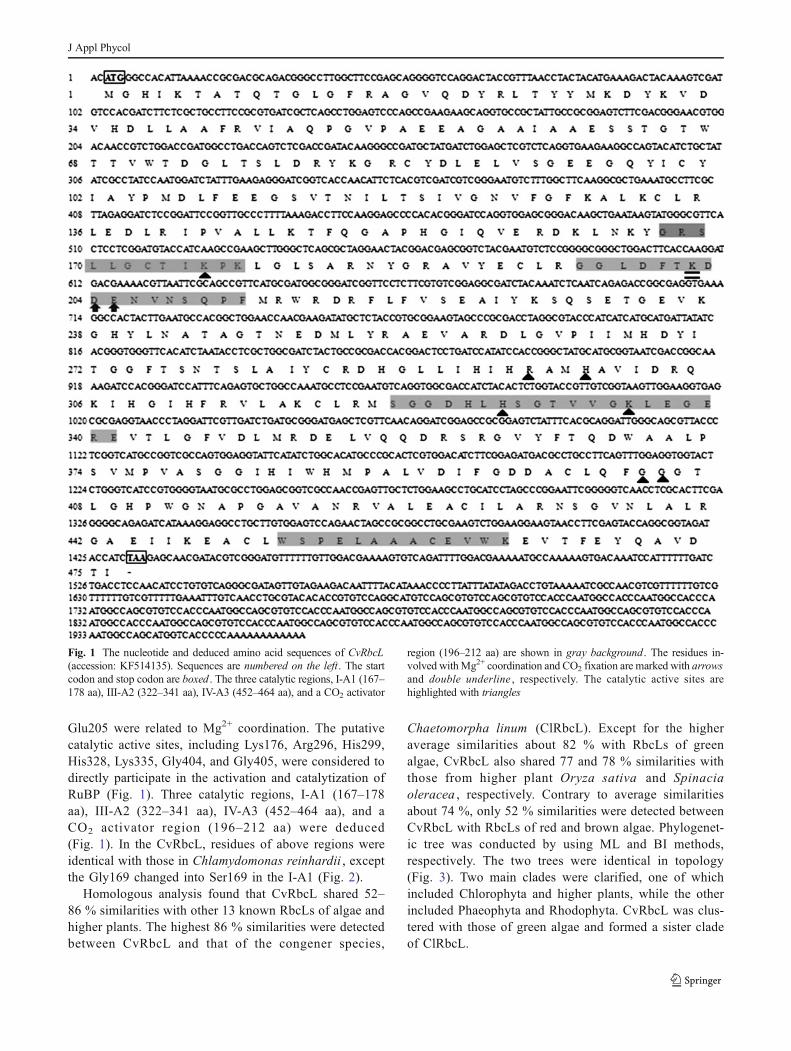
Glu205 were related to Mg2+ coordination. The putativecatalytic active sites, including Lys176, Arg296, His299,His328, Lys335, Gly404, and Gly405, were considered todirectly participate in the activation and catalytization ofRuBP (Fig. 1). Three catalytic regions, I-A1 (167–178aa), III-A2 (322–341 aa), IV-A3 (452–464 aa), and aCO2 activator region (196–212 aa) were deduced(Fig. 1). In the CvRbcL, residues of above regions wereidentical with those in Chlamydomonas reinhardii , exceptthe Gly169 changed into Ser169 in the I-A1 (Fig. 2).
Homologous analysis found that CvRbcL shared 52–86 % similarities with other 13 known RbcLs of algae andhigher plants. The highest 86 % similarities were detectedbetween CvRbcL and that of the congener species,
Chaetomorpha linum (ClRbcL). Except for the higheraverage similarities about 82 % with RbcLs of greenalgae, CvRbcL also shared 77 and 78 % similarities withthose from higher plant Oryza sativa and Spinaciaoleracea , respectively. Contrary to average similaritiesabout 74 %, only 52 % similarities were detected betweenCvRbcL with RbcLs of red and brown algae. Phylogenet-ic tree was conducted by using ML and BI methods,respectively. The two trees were identical in topology(Fig. 3). Two main clades were clarified, one of whichincluded Chlorophyta and higher plants, while the otherincluded Phaeophyta and Rhodophyta. CvRbcL was clus-tered with those of green algae and formed a sister cladeof ClRbcL.
Fig. 1 The nucleotide and deduced amino acid sequences of CvRbcL(accession: KF514135). Sequences are numbered on the left . The startcodon and stop codon are boxed . The three catalytic regions, I-A1 (167–178 aa), III-A2 (322–341 aa), IV-A3 (452–464 aa), and a CO2 activator
region (196–212 aa) are shown in gray background. The residues in-volved withMg2+ coordination and CO2 fixation are marked with arrowsand double underline , respectively. The catalytic active sites arehighlighted with triangles
J Appl Phycol

Transcriptional analysis of CvRbcL
For both CvRbcL and 28S rDNA genes, there was only onepeak at the corresponding melting temperature in the dissoci-ation curve analysis, which indicated that the q-PCR wasspecifically amplified. In the heat experiment, significant
difference in transcript level was detected under differenttemperature treatments (p <0.05). The transcriptions showeda general increasing trend with temperature increasing from 5to 20 °C and gradually decreased after that. The maximumoccurred at 20 °C, which was 1.6-fold and 1.8-fold comparedto that of 5 and 35 °C, respectively (p <0.05) (Fig. 4a). In the
Fig. 2 Comparison of amino acidresidues in the CO2 activatorregion (a) and the three catalyticregions, I-A1 (b), III-A2 (c), andIV-A3 (d). Conserved amino acidresidues are marked withasterisks on top. The variedresidues in I-A1 (b) are present ingray background. Abbreviationsare the same as in Table 1
Fig. 3 Phylogenetic tree inferredfrom RbcL amino acid sequencesalignment using the maximumlikelihood (ML) method. TheBayesian inference (BI) treeperformed using MrBayes isidentical with the ML tree intopology. Node support is asfollow:ML bootstrap/BI posteriorprobability. The new sequence ishighlighted in boldface
J Appl Phycol

light/dark test, CvRbcL were highly expressed under lightcondition for 12 h and markedly declined after 12-h darknessexposure (3.9-fold, 3.4-fold, and 3.1-fold, respectively) (p <0.01) (Fig. 4b).
Prokaryotic expression of recombinant CvRbcL protein
After induction with 0.5 mM IPTG, the recombinant proteinwere separated on the 12 % SDS–PAGE, and one distinctband about ∼55 kDa was identified (Fig. 5a).
No obvious expression of 55 kDa was observed in thecontrol test (IPTG induction for 0 h). Western blot analysis
proved that the ∼55 kDa band was positive to the anti-Hisantibody with high specificity (Fig. 5b).
Discussion
So far, only one record (accession: EU380531) has been anno-tated as partial RbcL sequence for Chaetomorpha taxa atGenBank. Limited information about RbcL is currently avail-able in these non-model and ecologically important species.Our study is the first to isolate full-length RbcL sequence fromChaetomorpha species. Structurally, RbcL does not containintrons and encodes ∼475 amino acids (Gutteridge andGatenby 1995). The new generated RbcL had no intron andencoded 476 amino acids polypeptide, which agrees with theprevious report. Homology alignments found CvRbcL sharedmuch higher similarities with RbcLs of green algae and higherplants. Phylogenetic analysis also revealed CvRbcL was closerwith green algae and higher plants RbcLs than those from redand brown algae (Fig. 3). These results were consistent with thephylogenetic affinity of these organisms.
In the CvRbcL, putative catalytic active sites (Lys176,Arg296, His299, His328, Lys335, Gly404, and Gly405) aswell as residues assumed for Mg2+ coordination (Asp204,Glu205) and CO2 fixation (Lys202) are in accordance withresults in previous reports (Lorimer and Miziorko 1980;Cleland et al. 1998), which implies the conservation ofRubisco function. In higher plants and algae, three catalyticregions and a CO2 activator region have been characterized inspinach (Zurawski et al. 1981), Chlamydomonas reinhardii(Dron et al. 1982), Dunaliella salina (Chai et al. 2007), andUlva linza (Ying et al. 2011). Here, parallel regions were alsodeduced in CvRbcL (Fig. 1). As shown in Fig. 2, sequenceswere highly conserved in these regions. For CvRbcL, residueswere identical with those in C. reinhardii , except the hydro-phobic Gly169 changed into neutral Ser169 in the I-A1(Fig. 2). It is suggested that the residue is probably associated
Fig. 4 Relative mRNA expression of CvRbcL gene under differenttemperatures (a) and exposed to the light (gray bar)/dark (black bar)cycle (b), respectively. Values are mean ± standard deviation
Fig. 5 SDS–PAGE (12 %) andwestern blot analysis of CvRbcLrecombinant protein. Note targetprotein at ∼55 kDa (arrows). aSDS–PAGE analysis of theinduced recombinant CvRbcL.Mprotein marker, lanes 1–7induced expression of CvRbcL by0.5 mM IPTG for 0–6 h,respectively. b Western blotanalysis of CvRbcL recombinantprotein.M protein marker, lane 1western blot result based on thesample of lane 4 in a
J Appl Phycol

with differences in enzyme activity between C. valida and C.reinhardii . It is interesting that same change was also detectedin the ClRbcL (Fig. 2), which infers that the unique residuemight be unified in the genus Chaetomorpha . Future work onRbcL sequences of otherChaetomorpha species is required toconfirm this hypothesis.
The expression level of CvRbcL with different tem-peratures and light/dark treatments were quantified byreal-time PCR. The light/dark test was expected to sim-ply replicate the natural light cycle in order to explorethe primary variation of CvRbcL expression. The tran-script level was dramatically upregulated in the light andstrongly suppressed in the dark (Fig. 5b), which impliesthat the CvRbcL was light-induced. This result corre-sponds with those of higher plants whose expression isstimulated by irradiance and constrained by darkness(Sugita and Gruissem 1987; Nishimura et al. 2008).Temperature also greatly affected the CvRbcL expressionin our study. Relatively high transcript level appearedbetween 15 and 25 °C, whereas both too high (35 °C)and too low temperatures (5 °C) reduced the mRNAaccumulations (Fig. 5a). Based on laboratory cultureresults, Deng et al. (2012) reported healthy growth andreproduction of C. valida in the range of 17–29 °C andan upper lethal temperature limit at 33 °C. Our resultshere are generally consistent with the ecological featuresof C. valida . Rubisco carboxylation reaction which re-sults in carbon fixation is considered to be the mainfactor limiting net photosynthesis (Yokota and Shigeoka2007). More efficient Rubisco could increase photosyn-thetic rate for faster growth rate and more biomass. Thissuggests that the highly expressed Rubisco was partlyresponsible for the faster growth of C. valida and facil-itates the rapid massive accumulation in favorable naturalenvironments.
In our study, the CvRbcL recombinant protein wasexpressed in E. coli and induced by 0.5 mM IPTG. Obviousband at ∼55 kDa appeared on the SDS–PAGE and westernblotting detection (Fig. 5), which consisted with the theoreti-cal molecular mass and demonstrated the successful heterol-ogous expression of CvRbcL . Further work is expected topurify the recombinant protein and evaluate biochemical pa-rameters to gain more insights into CvRbcL properties. How-ever, until now, no eukaryotic Rubisco has been assembledsuccessfully in vitro (Spreitzer and Salvucci 2002; Anderssonand Backlund 2008). More information about the mechanismsof Rubisco assembly remains to be uncovered to address thiselusive problem in future.
Acknowledgments This research was supported by Shandong Agri-culture Breeding Engineering Biological Resources Innovation of Re-search Project. The authors acknowledge the anonymous reviewers forthe critical comments and suggestions for the manuscript.
References
Altschul SF, Madden TL, Schaffer AA, Zhang JH, Zhang Z, Miller W,Lipman DJ (1997) Gapped BLAST and PSI-BLAST: a new gener-ation of protein database search programs. Nucleic Acids Res 25:3389–3402
Andersson I, Taylor TC (2003) Structural framework for catalysis andregulation in ribulose-1,5-bisphosphate carboxylase/oxygenase.Arch Biochem Biophys 414:130–140
Andersson I, Backlund A (2008) Structure and function of Rubisco. PlantPhysiol Biochem 46:275–291
Chai YR, Sun Y, Pan WD, Jia YL, Xue LX (2007) Isolation and charac-terization of rbcL gene from Dunaliella salina . Life Sci J 4:6–12
Chi YX, Wang LM, Luan RX, Wang HW (2009) Chaetomorpha valida ,a new recorded green alga species in genus Chaetomorpha Kützingin China. Fisheries Sci 28:162–163
Cleland WW, Andrews TJ, Gutteridge S, Hartman FC, Lorimer GH(1998) Mechanism of Rubisco: the carbamate as general base.Chem Rev 98:549–561
Dean C, Pichersky E, Dunsmuir P (1989) Structure, evolution, andregulation of RbcS genes in higher plants. Annu Rev Plant PhysiolPlant Mol Biol 40:415–439
Deng Y, Tang X, Huang B, Ding L (2011) Life history of Chaetomorphavalida (Cladophoraceae, Chlorophyta) in culture. BotMar 54:551–556
Deng Y, Tang X, Huang B, Ding L (2012) Effect of temperature andirradiance on the growth and reproduction of the green macroalga,Chaetomorpha valida (Cladophoraceae, Chlorophyta). J ApplPhycol 24:927–933
DronM, Rahire M, Rochaix JD (1982) Sequence of the chloroplast DNAregion of Chlamydomonas reinhardii containing the gene of thelarge subunit of ribulose bisphosphate carboxylase and parts of itsflanking genes. J Mol Biol 162:775–793
Edgar RC (2004) MUSCLE: multiple sequence alignment with highaccuracy and high throughput. Nucleic Acids Res 32:1792–1797
Fletcher RL (1996) The occurrence of “green tides”: a review. In:Schramm W, Nienhuis PH (eds) Marine benthic vegetation: recentchanges and the effects of eutrophication. Springer, Berlin, pp 7–43
Gasteiger E, Hoogland C, Gattiker A, Duvaud S,WilkinsMR,Appel RD,Bairoch A (2005) In: Walker JM (ed) The proteomics protocolshandbook. Humana Press, New Jersey
Geourjon C, Deleage G (1995) SOPMA: significant improvements inprotein secondary structure prediction by consensus prediction frommultiple alignments. Comput Appl Biosci 11:681–684
Ginrich JC, Hallick RB (1985) The Euglena gracilis chloroplast ribulose-1,5-bisphosphate carboxylase gene. II. The spliced mRNA and itsproduct. J Biol Chem 260:16162–16168
Gutteridge S, Gatenby AA (1995) Rubisco synthesis, assembly, mecha-nism, and regulation. Plant Cell 7:809–819
Houtz RL, Portis AR (2003) The life of ribulose 1,5-bisphosphatecarboxylase/oxygenase-posttranslational facts and mysteries. ArchBiochem Biophys 414:150–158
Huelsenbeck JP, Ronquist FR (2003) Mrbayes 3: Bayesian phylogeneticinference under mixed models. Bioinformatics 19:1572–1574
Jensen GR, Bahr TJ (1977) Ribulose 1,5-bisphosphate carboxylase-oxygenase. Annu Rev Plant Physiol 28:379–400
Krogh A, Larsson B, von Heijne B, Sonnhammer ELL (2001) Predictingtransmembrane protein topology with a hidden Markov model:application to complete genomes. J Mol Biol 305:567–580
Lorimer GH, Miziorko HM (1980) Carbamate formation on the epsilon-amino group of a lysyl residue as the basis for the activation ofribulosebisphosphate carboxylase by CO2 and Mg2+. Biochemistry19:5321–5328
Nishimura K, Ogawa T, Ashida H, Yokota A (2008) Molecular mecha-nisms of RuBisCO biosynthesis in higher plants. Plant Biotechnol25:285–290
J Appl Phycol

Petersen TN, Brunak S, von Heijne G, Nielsen H (2011) SignalP 4.0:discriminating signal peptides from transmembrane regions. NatMethods 8:785–786
Posada D, Crandall KA (1998) Modeltest: testing the model of DNAsubstitution. Bioinformatics 14:817–818
Raffaelli DG, Raven JA, Poole LJ (1998) Ecological impact of greenmacroalgal blooms. Oceanogr Mar Biol Ann Rev 36:97–125
Rombel IT, Sykes KF, RaynerS JSA (2002) ORF-Finder: a vector forhigh-throughput gene identification. Gene 282:33–41
Schmittgen TD, Zakrajsek BA, Mills AG, Gorn V, Singer MJ, Reed MW(2000) Quantitative reverse transcription-polymerase chain reactionto study mRNA decay: comparison of endpoint and real-timemethods. Anal Biochem 285:194–204
Schneider G, Lindqvist Y, Branden CI (1992) Rubisco: structure andmechanism. Annu Rev Biophys Biomol Struct 21:119–143
Spreitzer RJ, Salvucci ME (2002) Rubisco: structure, regulatory interac-tions, and possibilities for a better enzyme. Annu Rev Plant Biol 53:449–475
Stamatakis A, Hoover P, Rougemont J (2008) A fast bootstrappingalgorithm for the RAxMLWeb-Servers. Syst Biol 57:758–771
Sugita M, Gruissem W (1987) Developmental, organ-specific, and lightdependent expression of the tomato ribulose-1,5-bisphosphate car-boxylase small subunit gene family. Proc Natl Acad Sci U S A 84:7104–7108
Tabita FR, Satagopan S, Hanson TE, Kreel NE, Scott SS (2008) Distinctform I, II, III, and IV Rubisco proteins from the three kingdoms of
life provide clues about Rubisco evolution and structure/functionrelationships. J Exp Bot 59:1515–1524
Tamura K, Peterson D, Peterson N, Stecher G, Nei M, Kumar S (2011)MEGA5: molecular evolutionary genetics analysis using maximumlikelihood, evolutionary distance, and maximum parsimonymethods. Mol Biol Evol 28:2731–2739
Valiela I, Mccelland J, Hauxwell J, Behr PJ, Hersh D, Foreman K(1997) Macroalgal blooms in shallow estuaries: controls and eco-physiological and ecosystem consequences. Limnol Oceanogr 42:1105–1118
Yao JT, FuWD,Wang XL, Duan DL (2009) Improved RNA isolation forLaminaria japonica Aresch (Laminariaceae, Phaeophyta). J ApplPhycol 21:233–238
Ye N, Zhang X,Mao Y, Liang C, Xu D, Zou J, Zhuang Z,Wang Q (2011)“Green tides” are overwhelming the coastline of our blue planet:taking the world's largest example. Ecol Res 26:477–485
Ying CQ, Yin SJ, Shen Y, Lin SJ, He PM (2011) Cloning and analysis ofthe full-length Rubisco large subunit (rbcL) cDNA from Ulva linza(Chlorophyceae, Chlorophyta). Bot Mar 54:303–312
Yokota A, Shigeoka S (2007) Engineering photosynthetic pathways. In:Lewis NG, Bohnert HJ, Nguyen HT (eds) Advances in plant bio-chemistry and molecular biology, bioengineering and molecularbiology of plant pathways, volume 1. Elsevier, Netherlands
Zurawski G, Perrot B, Bottomley W (1981) The structure of the gene forthe large subunit of ribulose bisphosphate carboxylase from spinachchloroplast DNA. Nucleic Acids Res 14:3251–3270
J Appl Phycol


![WITPress BMTA fm · 2014. 5. 10. · Codium [11, 27], Caulerpa [11, 12] and Chaetomorpha [10] have been selected ... (temperature, light, oxygen and nutrient) [8]. Metal bioaccumulation](https://img.pdfslide.net/doc/110x75/614a65a812c9616cbc696380/witpress-bmta-fm-2014-5-10-codium-11-27-caulerpa-11-12-and-chaetomorpha.jpg)