Embed Size (px)
Citation preview
JOURNAL OF CLINICAL MICROBIOLOGY,0095-1137/01/$04.0010 DOI: 10.1128/JCM.39.4.1460–1466.2001
Apr. 2001, p. 1460–1466 Vol. 39, No. 4
Copyright © 2001, American Society for Microbiology. All Rights Reserved.
Molecular Epidemiology of Streptococcus uberis Isolates fromDairy Cows with Mastitis
PATCHARA PHUEKTES,1 PETER D. MANSELL,2 RODNEY S. DYSON,3 NARELLE D. HOOPER,3
JENNIFER S. DICK,3 AND GLENN F. BROWNING1*
Veterinary Preclinical Centre, Faculty of Veterinary Science, The University of Melbourne, Melbourne,1
Veterinary Clinical Centre, Faculty of Veterinary Science, The University of Melbourne,Werribee,2 and Kyabram Veterinary Clinic, Kyabram,3 Victoria, Australia
Received 21 June 2000/Returned for modification 11 October 2000/Accepted 29 January 2001
Pulsed-field gel electrophoresis and antimicrobial sensitivity testing were used as tools to investigate theepidemiology of Streptococcus uberis mastitis in dairy cows. A total of 62 different strains were found among 138isolates from the four herds investigated, and between 10 and 26 different strains were found in each herd.There was no strain common to all four herds. Identical strains of S. uberis were detected from differentquarters of individual cows and from cows within the same herd, suggesting that transmission from quarterto quarter and cow to cow had occurred. Despite the great variation in S. uberis strains, persistent infectionwith the same strain within a lactation was observed in most cows. Predominant strains were present in twoherds. Preliminary investigations could not clarify why these particular strains might predominate, but in oneherd there was a significant difference between the prevalence of clinical mastitis in quarters infected with thepredominant strain and that in quarters infected with other strains, suggesting the greater virulence of thepredominant strain. The wide variety of S. uberis strains found is consistent with an environmental source ofS. uberis. However, evidence of direct transmission, the persistence of infection, and the predominance ofparticular strains in some herds indicate that S. uberis infections are epidemiologically complex and that therelative importance of these factors in the occurrence of mastitis may differ between herds.
Implementation of mastitis control programs based on im-proved milking practices, postmilking teat disinfection, thera-peutic and prophylactic antimicrobial administration, and cull-ing of persistently infected animals has effectively controlledintramammary infections caused by contagious pathogens.However, these measures have been less effective against en-vironmental pathogens. Consequently, environmental mastitishas become a major problem, particularly in well-managedherds that have successfully controlled contagious pathogens.It has been hypothesized that the niche vacated by contagiousmastitis pathogens becomes occupied by environmental mas-titis pathogens, resulting in an increased prevalence of intra-mammary infection by environmental pathogens (16). Strepto-coccus uberis is one of the more important environmental patho-gens implicated in bovine mastitis, accounting for a significantproportion of subclinical and clinical intramammary infectionsin both lactating and nonlactating cows.
An increasing prevalence of S. uberis mastitis has been re-ported throughout the world. Approximately 14 to 26% ofclinical mastitis cases in Canada, the United States, The Neth-erlands, and the United Kingdom are caused by S. uberis (13).S. uberis is also one of the most significant causes of bovinemastitis in New Zealand and Australia, where the dairy indus-try is pasture based (21, 29). Despite its high prevalence, theepidemiology of S. uberis mastitis is incompletely understood.
S. uberis has been isolated from many sites on the cow includ-ing the skin surface, genital tract, intestinal tract, and tonsils (6,22, 23). It can also be isolated in large numbers from beddingmaterial (4), which is thought to be a major source for intra-mammary infections in housed cattle. Although many potentialreservoirs have been identified, their significance and theirassociation with the occurrence of mastitis in a herd are stillunclear. More information on reservoirs and modes of trans-mission is needed for development of better control programsfor this pathogen.
The ability to identify specific strains of a causative bacterialspecies is an essential tool for epidemiological investigations.A number of typing techniques for differentiation of S. uberisstrains have been investigated. The conventional typing meth-ods based on phage typing, serotyping, bacteriocin-like inhib-itory substance fingerprinting, and antibiograms have low type-ability for closely related strains of S. uberis (5, 11, 17, 24).DNA-based methods have shown potential for typing S. uberis(9, 12, 15, 17, 28). DNA macrorestriction analysis by pulsed-field gel electrophoresis (PFGE) appears to be a simple, reli-able, and highly discriminatory method. It produces distinctpatterns that are easy to interpret and is highly reproducible.The reproducibility of PFGE makes it useful for comparison ofstrains between laboratories, and it has been used successfullyto investigate genomic diversity among strains of S. uberis (2, 7,27).
The aim of this study was to explore the epidemiology ofmastitis caused by S. uberis in dairy cattle by using PFGE andantibiograms to investigate the persistence of strains and therelative importance of new infections from the environmentand from cow-to-cow spread.
* Corresponding author. Mailing address: Veterinary PreclinicalCentre, Faculty of Veterinary Science, The University of Melbourne,Melbourne, Victoria 3010, Australia. Phone: (613) 8344 7342. Fax:(613) 8344 7374. E-mail: [email protected].
1460
Dow
nloa
ded
from
http
s://j
ourn
als.
asm
.org
/jour
nal/j
cm o
n 30
Dec
embe
r 20
21 b
y 21
0.25
5.24
9.90
.
MATERIALS AND METHODS
Cattle herds and sample collection. Milk samples from 38 cows infected withS. uberis were obtained from four pasture-based dairy herds located in Kyabram,Victoria, Australia. Each herd had a history of a high prevalence of S. uberisinfection. Milk samples were collected aseptically from each quarter of most ofthese cows four times over two successive lactations, from 2 months prior todrying off to 2 months after the subsequent parturition. The samples werecollected at 4- to 6-week intervals in each lactation. The cows were dried off inJune and July and calved in August and September. Blanket dry-cow therapywith cefalonium dihydrate (Cepravin DC; Schering-Plough Animal Health,Baulkham Hills, New South Wales, Australia) was used by all four herds duringthe trial. Clinical mastitis and treatment data were available from herd 4.
Bacteriological methods. Milk samples were mixed thoroughly, and 10 ml wasplated onto Edward’s medium. S. uberis isolates were identified to the specieslevel by conventional tests and by PCR. Conventional tests included Gramstraining, catalase production, esculin hydrolysis, the CAMP test, and the sodiumhippurate test. PCR amplification of spacer regions between 16S and 23S rRNAof S. uberis was used to confirm identification (8).
Antimicrobial susceptibility testing. The calibrated dichotomous sensitivitytest of Bell (3) was used to assess the antimicrobial sensitivity of S. uberis isolates.Antimicrobial disks (Oxoid, Basingstoke, Hampshire, England) used includedpenicillin G (0.5 mg), cloxacillin (5 mg), erythromycin (5 mg), gentamicin (10 mg),trimethroprim (5 mg), sulfamethoxazole (300 mg), tetracycline (30 mg), vanco-mycin (5 mg), cephalexin (100 mg), neomycin (30 mg), novobiocin (30 mg), andkanamycin (50 mg). The diameter of the inhibition zone was measured after 18to 24 h of incubation at 37°C. A standard strain of Staphylococcus aureus wasused as a control strain.
S. uberis concentrations in milk from infected quarters. The numbers of S.uberis colonies isolated from 38 milk samples collected from herds 3 and 4 weredetermined. Each sample was mixed thoroughly, and 50 ml from each sample wasspread onto separate Edward’s medium agar plates. The inoculated plates werethen incubated at 37°C for 24 h. S. uberis colonies were observed using UV light(Wood’s lamp), the colonies were counted, and each sample was allocated to acategory depending on whether it yielded less than 50, 51 to 100, 101 to 200, 201to 300, 301 to 400, or more than 400 colonies per 50-ml sample. The distributionof samples containing strain C2 into these categories was compared to thedistribution of samples from herd 3 containing other strains, and the distributionof samples containing strain D2 was compared to the distribution of samplesfrom herd 4 containing other strains using chi-square tests.
Preparation of genomic DNA in agarose blocks. S. uberis isolates were grownin Todd-Hewitt broth (Oxoid) at 37°C overnight. Cells from 1 ml of culture werecollected by centrifugation at 13,000 3 g for 1 min, washed three times with 1 MNaCl–10 mM Tris-HCl (pH 7.6), and resuspended in 300 ml of the same solution.The cell suspension was mixed with an equal volume of molten 2% (wt/vol)low-melting-temperature agarose (SeaPlaque; FMC Bioproducts, Rockland,Maine) and dispensed into 100-ml molds. When solidified, blocks were incubatedin lysis buffer (6 mM Tris-HCl [pH 7.6], 1 M NaCl, 100 mM EDTA [pH 7.6], 1%Sarkosyl, 1 mg of lysozyme/ml) for 18 h at 37°C. The lysis buffer was thenreplaced with ESP buffer (0.5 M EDTA [pH 9.2], 1% Sarkosyl, 1 mg of protein-ase K/ml), and the blocks were incubated at 50°C for 72 h. Gel blocks were storedin 0.5 M EDTA (pH 8) until used.
Restriction endonuclease digestion of genomic DNA. Before digestion blockswere equilibrated with TE (10 mM Tris-HCl [pH 8], 1 mM EDTA [pH 8]). Slices1 to 2 mm thick were then cut from each block and incubated in 100 ml of 13restriction endonuclease buffer as supplied by the manufacturer. Digestions wereperformed with 10 U of SmaI (Boehringer GmbH, Mannheim, Germany) at25°C for 18 h or 20 U of ApaI (Boehringer GmbH) at 30°C for 18 h.
PFGE. DNA fragments were separated by clamped homogeneous electric field(CHEF) electrophoresis using a CHEF DRIII (Bio-Rad Laboratories, Rich-mond, Calif.). Electrophoresis of digested samples was performed through 1%(wt/vol) agarose gels in 0.53 TBE (13 TBE is 89 mM Tris-HCl, 89 mM boricacid, and 2 mM EDTA, pH 8.3) at 6 V/cm for 20 h at 14°C, with the pulse timeincreasing linearly from 1 to 20 s for SmaI-digested samples and from 1 to 17 sfor ApaI-digested samples. Gels were stained with 0.5 mg of ethidium bromide/liter, and DNA was visualized by UV transillumination. Lambda phage con-catamers (Bio-Rad) and HindIII digests of lambda phage DNA were used asmolecular size standards.
Strain classification. Strains were defined using the criteria proposed byTenover et al. (25). Isolates were designated different strains if the PFGEpatterns differed by more than three bands and different subtypes of the samestrain if patterns differed by one to three bands. Isolates that had similar PFGEpatterns were considered to be genetically related isolates and to be derived from
a common parent, as differences in two or three bands were most likely the resultof a single genetic change such as a point mutation or an insertion or deletion ofDNA. The discrimination of PFGE and antibiogram typing in this populationwere determined using Simpson’s index of diversity (14), assuming that all iso-lates obtained from the same quarter in the same lactation were not independentstrains if they belonged to the same subtype as determined by PFGE of SmaI-digested chromosomal DNA.
RESULTS
Discrimination and reproducibility of PFGE typing. PFGEof chromosomal DNA digested with SmaI yielded 8 to 14fragments in the 23- to 340-kb size range, and ApaI digestsyielded patterns of 10 to 16 fragments of 23 to 340 kb. Thestability of SmaI and ApaI PFGE patterns was examined byselecting one isolate (strain D2b) and looking at the PFGEpatterns before and after 10 passages in Todd-Hewitt broth.The reproducibility of the PFGE patterns was also examinedby repeated testing of the same isolate on separate occasionson different gels. In both cases identical patterns were ob-tained, indicating the stability and reproducibility of PFGE.
To increase the discriminatory power of PFGE and confirmthe results obtained from SmaI digestion, ApaI digestion wasused to examine isolates that had identical or similar SmaIrestriction patterns. Identical ApaI patterns were seen in allisolates that had identical SmaI patterns. However, ApaI di-gestion was found to be less discriminatory than SmaI diges-tion for closely related isolates. Isolates that belonged to sub-types C2a and C2c as determined by SmaI digestion werefound to have identical ApaI PFGE patterns. Isolates of sub-types D2a and D2b also had the same ApaI PFGE patterns.The SmaI and ApaI restriction patterns revealed a markedclonal diversity of S. uberis. A total of 62 different strains wereobtained from 138 S. uberis isolates from the four herds (Table1), indicating that the epidemiology of S. uberis infection couldbe very complex.
Discrimination of antibiogram patterns. Antimicrobial sus-ceptibility testing revealed that all isolates were susceptible tovancomycin and cephalexin and resistant to gentamicin, sulfa-methoxazole, neomycin, and kanamycin. A total of 11 differentantibiogram patterns were obtained (Table 2). The majority ofS. uberis isolates (91 of 138, 66%) were antibiogram type B orD (Tables 1 and 2). Although the discriminatory power of theantibiogram technique was relatively low, the results of anti-biograms mostly correlated with the results obtained by PFGE(Fig. 1). S. uberis isolates designated as the same strains orsubtypes of the same strains by PFGE had common antibio-gram patterns. However, some isolates from different herdsthat shared PFGE patterns were found to have different anti-biogram patterns. Two isolates (strain B12) from herd 2 andfour isolates (strain C19) from herd 3 had the same PFGEpattern but had antibiogram types F and K, respectively, andtwo isolates (strain A4) from herd 1 and five isolates (strainD2b) from herd 4 that had the same PFGE pattern had anti-biogram types B and D, respectively.
The index of discrimination for PFGE examination of SmaI-digested chromosomal DNA was 0.989, while that for antibio-gram typing was 0.731, indicating the much higher discrimina-tory power of PFGE typing.
Comparison of S. uberis isolates from the same quarters. Todetermine if multiple strains were present in one infected quar-
VOL. 39, 2001 EPIDEMIOLOGY OF S. UBERIS IN COWS WITH MASTITIS 1461
Dow
nloa
ded
from
http
s://j
ourn
als.
asm
.org
/jour
nal/j
cm o
n 30
Dec
embe
r 20
21 b
y 21
0.25
5.24
9.90
.
ter, milk samples from 12 S. uberis-infected quarters wereevaluated. Three S. uberis colonies isolated from each of 12milk samples were selected for PFGE. The 36 S. uberis isolatesincluded 11 strains, but all isolates from the same quarter werethe same strain, suggesting that infection with multiple strainsof S. uberis within the same quarter was not common.
Comparison of S. uberis isolates from different quarters ofindividual cows. Strains of S. uberis isolated from differentquarters of the same cow were analyzed to determine whetherdifferent quarters were infected with different strains. Of 11cows that had two quarters infected with S. uberis, 5 cows wereinfected with the same strain in both quarters. Two of the sixcows that had three quarters infected had different S. uberisstrains in each quarter, and the other four had the same strainin two of the three infected quarters. One cow was found to
have different strains of S. uberis in all four quarters, and twocows had the same strain in two of four infected quarters. In 5of 11 cows, the two quarters infected with the same strain werethe left forequarter and left hindquarter, and in 4 of 11 cowsthe left and right forequarters were infected with the samestrain. In no cow were diagonally opposite quarters infectedwith the same strain.
Comparison of S. uberis strains within and among herds. Agreat degree of genetic variation was found in S. uberis withinthe same herd and among herds. Between 10 and 26 differentS. uberis strains were present in each herd (Table 1). Withinherds 1 and 2, no common strains were found. However, pre-dominant strains were found in herds 3 and 4. Strain C2 wasthe most prevalent in herd 3 during the lactation prior todrying off, with 40% of isolates belonging to this strain. Strain
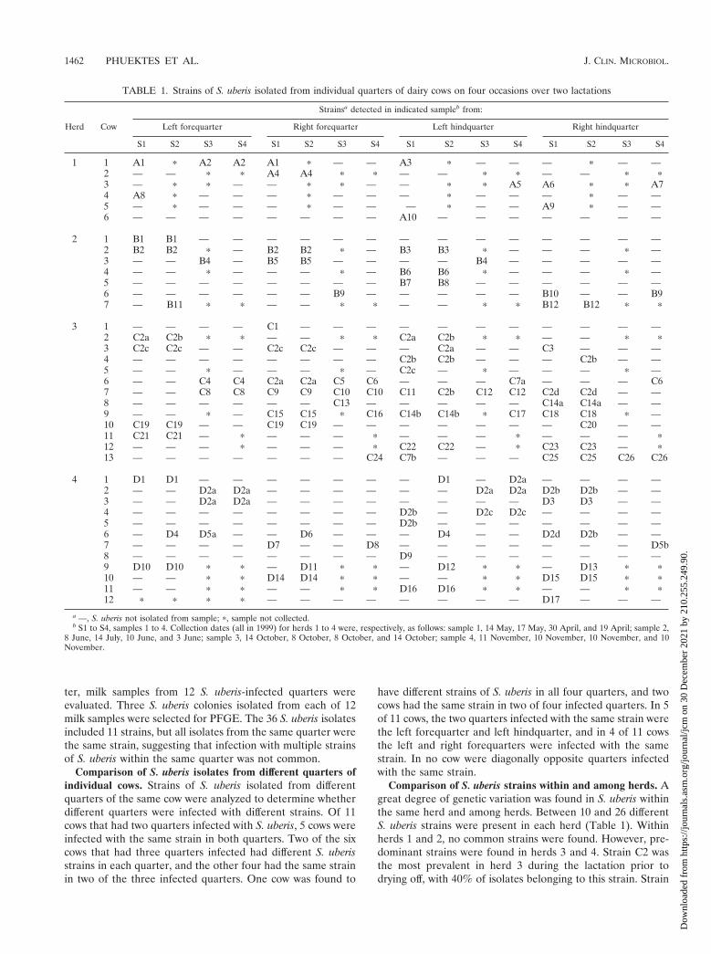
TABLE 1. Strains of S. uberis isolated from individual quarters of dairy cows on four occasions over two lactations
Herd Cow
Strainsa detected in indicated sampleb from:
Left forequarter Right forequarter Left hindquarter Right hindquarter
S1 S2 S3 S4 S1 S2 S3 S4 S1 S2 S3 S4 S1 S2 S3 S4
1 1 A1 p A2 A2 A1 p — — A3 p — — — p — —2 — — p p A4 A4 p p — — p p — — p p3 — p p — — p p — — p p A5 A6 p p A74 A8 p — — — p — — — p — — — p — —5 — p — — — p — — — p — — A9 p — —6 — — — — — — — — A10 — — — — — — —
2 1 B1 B1 — — — — — — — — — — — — — —2 B2 B2 p — B2 B2 p — B3 B3 p — — — p —3 — — B4 — B5 B5 — — — — B4 — — — — —4 — — p — — — p — B6 B6 p — — — p —5 — — — — — — — — B7 B8 — — — — — —6 — — — — — — B9 — — — — — B10 — — B97 — B11 p p — — p p — — p p B12 B12 p p
3 1 — — — — C1 — — — — — — — — — — —2 C2a C2b p p — — p p C2a C2b p p — — p p3 C2c C2c — — C2c C2c — — — C2a — — C3 — — —4 — — — — — — — — C2b C2b — — — C2b — —5 — — p — — — p — C2c — p — — — p —6 — — C4 C4 C2a C2a C5 C6 — — — C7a — — — C67 — — C8 C8 C9 C9 C10 C10 C11 C2b C12 C12 C2d C2d — —8 — — — — — — C13 — — — — — C14a C14a — —9 — — p — C15 C15 p C16 C14b C14b p C17 C18 C18 p —10 C19 C19 — — C19 C19 — — — — — — — C20 — —11 C21 C21 — p — — — p — — — p — — — p12 — — — p — — — p C22 C22 — p C23 C23 — p13 — — — — — — — C24 C7b — — — C25 C25 C26 C26
4 1 D1 D1 — — — — — — — D1 — D2a — — — —2 — — D2a D2a — — — — — — D2a D2a D2b D2b — —3 — — D2a D2a — — — — — — — — D3 D3 — —4 — — — — — — — — D2b — D2c D2c — — — —5 — — — — — — — — D2b — — — — — — —6 — D4 D5a — — D6 — — — D4 — — D2d D2b — —7 — — — — D7 — — D8 — — — — — — — D5b8 — — — — — — — — D9 — — — — — — —9 D10 D10 p p — D11 p p — D12 p p — D13 p p10 — — p p D14 D14 p p — — p p D15 D15 p p11 — — p p — — p p D16 D16 p p — — p p12 p p p p — — — — — — — — D17 — — —
a —, S. uberis not isolated from sample; p, sample not collected.b S1 to S4, samples 1 to 4. Collection dates (all in 1999) for herds 1 to 4 were, respectively, as follows: sample 1, 14 May, 17 May, 30 April, and 19 April; sample 2,
8 June, 14 July, 10 June, and 3 June; sample 3, 14 October, 8 October, 8 October, and 14 October; sample 4, 11 November, 10 November, 10 November, and 10November.
1462 PHUEKTES ET AL. J. CLIN. MICROBIOL.
Dow
nloa
ded
from
http
s://j
ourn
als.
asm
.org
/jour
nal/j
cm o
n 30
Dec
embe
r 20
21 b
y 21
0.25
5.24
9.90
.
D2 was the predominant strain found in herd 4 over bothlactations, accounting for 21.4 and 75% of S. uberis isolates inthe two successive lactations. Only two to six isolates in eachherd were found to be identical to isolates from other herds.Two isolates (strain B12) from herd 2 and four isolates (strainC19) from herd 3 appeared to be identical, two isolates (strainA4) from herd 1 had the same pattern as five isolates (strainD2b) from herd 4, and two isolates that belonged to strain D5aand D5b from herd 4 were identical to six isolates (strain C2b)and five isolates (strain C2a), respectively, from herd 3. Therewas no strain found in all four herds.
Comparison of concentrations of predominant strains andother strains in milk from infected quarters. The concentra-tions of S. uberis strains C2 and D2, which were the predom-inant strains in herds 3 and 4, respectively, and other strainsfrom herds 3 and 4 in milk from infected quarters were exam-ined by colony counting. There was no significant differencebetween the concentrations of strains C2 (P 5 0.29; x2 5 4.95)or D2 (P 5 0.609; x2 5 2.7) and the other strains in infectedquarters in herds 3 and 4, respectively.
Incidence of clinical mastitis caused by strain D2 and theother strains in herd 4. There was a significant differencebetween the prevalence of clinical mastitis in quarters infectedwith strain D2 and the prevalence of clinical mastitis in quar-ters infected with other strains (P 5 0.002). Only 2 of the 18quarters infected with the other strains developed clinical mas-titis during the trial. Of eight quarters infected with strain D2,six quarters developed clinical mastitis, and four out of the sixquarters were still infected with the same strain after treatmentwith a combination of oxytetracycline hydrochloride, oleando-mycin, and neomycin (Mastalone Blue; Pfizer Animal Health,Highett, Victoria, Australia).
New and persistent S. uberis infections during a lactationand over lactations. The 47 paired isolates of S. uberis recov-ered from the same quarters over a 4- to 6-week interval duringa single lactation were compared. In 41 pairs strains of S. uberiswere the same, and members of three pairs were differentsubtypes of the same strain. Different strains of S. uberis weredetected in only three paired samples. The persistence of S.
uberis in infected quarters over lactations was also investigatedby comparing strains isolated in one lactation with strainsfound in the next lactation. The prevalence of S. uberis infec-tion decreased significantly from one lactation to the next in allherds. The total number of infected quarters was reduced from64 in one lactation to 27 in the next lactation, with only 13quarters infected with S. uberis in both lactations. Of these 13quarters, different S. uberis strains were detected in each lac-tation in 12, suggesting that new infections had occurred, and1 quarter was found to have different subtypes of the samestrain in each lactation.
DISCUSSION
PFGE and antibiogram analysis were used to examinestrains of S. uberis to investigate the epidemiology of S. uberismastitis in dairy cattle. As shown in previous studies (2, 7, 27)PFGE was proven to be a highly discriminatory method. It wasable to resolve many isolates that were indistinguishable byantimicrobial susceptibility testing. Antibiograms alone werefound to be of limited value in differentiating closely relatedstrains, but agreement between antibiograms and PFGE wasobserved for most isolates. However, it was found that someisolates from different herds that were defined as the samestrain by PFGE had different antibiogram patterns. This couldbe due to differences in management and treatment practicesamong the herds, particularly differences in the type and fre-quency of antibiotic use, and thus of exposure to antibiotics,among herds.
A number of S. uberis strains were isolated from cows in thisstudy, demonstrating that a wide variety of strains are able tocause mastitis. This is consistent with the dogma that quartersare infected by this pathogen more frequently from their en-vironment than from other infected quarters. Strains of S.uberis recovered from clinically affected quarters were alsofound in subclinically infected quarters. This finding agreeswith the previous report of Jayarao et al. (18), who showed thatmost S. uberis strains isolated from clinical mastitis were alsoisolated from cows with subclinical mastitis.
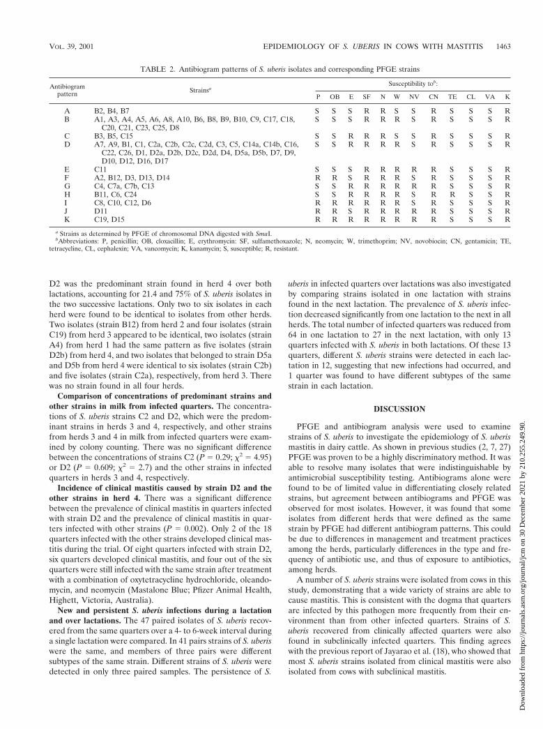
TABLE 2. Antibiogram patterns of S. uberis isolates and corresponding PFGE strains
Antibiogrampattern Strainsa
Susceptibility tob:
P OB E SF N W NV CN TE CL VA K
A B2, B4, B7 S S S R R S S R S S S RB A1, A3, A4, A5, A6, A8, A10, B6, B8, B9, B10, C9, C17, C18,
C20, C21, C23, C25, D8S S S R R R S R S S S R
C B3, B5, C15 S S R R R S S R S S S RD A7, A9, B1, C1, C2a, C2b, C2c, C2d, C3, C5, C14a, C14b, C16,
C22, C26, D1, D2a, D2b, D2c, D2d, D4, D5a, D5b, D7, D9,D10, D12, D16, D17
S S R R R R S R S S S R
E C11 S S S R R R R R S S S RF A2, B12, D3, D13, D14 R R S R R R S R S S S RG C4, C7a, C7b, C13 S S R R R R R R S S S RH B11, C6, C24 S S R R R R S R R S S RI C8, C10, C12, D6 R R R R R R S R S S S RJ D11 R R S R R R R R S S S RK C19, D15 R R R R R R R R S S S R
a Strains as determined by PFGE of chromosomal DNA digested with SmaI.bAbbreviations: P, penicillin; OB, cloxacillin; E, erythromycin: SF, sulfamethoxazole; N, neomycin; W, trimethoprim; NV, novobiocin; CN, gentamicin; TE,
tetracycline, CL, cephalexin; VA, vancomycin; K, kanamycin; S, susceptible; R, resistant.
VOL. 39, 2001 EPIDEMIOLOGY OF S. UBERIS IN COWS WITH MASTITIS 1463
Dow
nloa
ded
from
http
s://j
ourn
als.
asm
.org
/jour
nal/j
cm o
n 30
Dec
embe
r 20
21 b
y 21
0.25
5.24
9.90
.
The demonstration of identical strains of S. uberis in differ-ent quarters of individual cows and from some cows within thesame herd suggests that direct transmission from an infectedquarter to uninfected quarters and from cow to cow occurs,presumably during the milking process. Improper milking hy-giene and machine function could therefore contribute to theincidence of S. uberis infection. However, direct contact withthe same environmental source, although unlikely, cannot be
discounted entirely. As only a few strains were isolated frommore than one herd, it is unlikely that there is an environmen-tal source of S. uberis common to different farms.
Despite the large number of different strains found withineach herd, in two herds there was a predominant strain. Onlyone herd had the same strain predominating over two lacta-tions. It is possible that this strain was widespread in the en-vironment and relatively stable over time or, alternatively, that
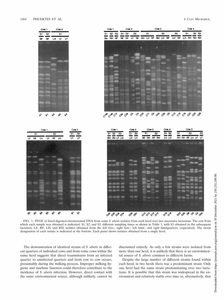
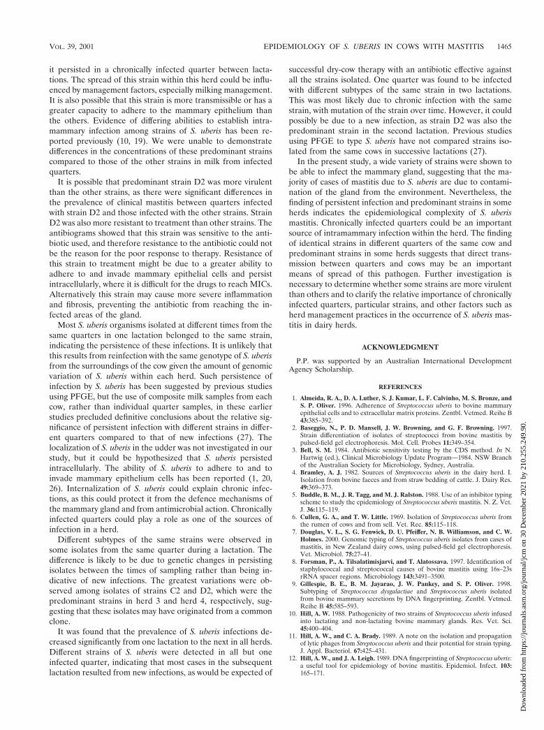
FIG. 1. PFGE of SmaI-digested chromosomal DNA from some S. uberis isolates from each herd over two successive lactations. The cow fromwhich each sample was obtained is indicated. S1, S2, and S3, different sampling times as shown in Table 1, with S3 obtained in the subsequentlactation; LF, RF, LH, and RH, isolates obtained from the left fore-, right fore-, left hind-, and right hindquarters, respectively. The straindesignation of each isolate is indicated at the bottom. Each panel shows isolates obtained from a single herd.
1464 PHUEKTES ET AL. J. CLIN. MICROBIOL.
Dow
nloa
ded
from
http
s://j
ourn
als.
asm
.org
/jour
nal/j
cm o
n 30
Dec
embe
r 20
21 b
y 21
0.25
5.24
9.90
.
it persisted in a chronically infected quarter between lacta-tions. The spread of this strain within this herd could be influ-enced by management factors, especially milking management.It is also possible that this strain is more transmissible or has agreater capacity to adhere to the mammary epithelium thanthe others. Evidence of differing abilities to establish intra-mammary infection among strains of S. uberis has been re-ported previously (10, 19). We were unable to demonstratedifferences in the concentrations of these predominant strainscompared to those of the other strains in milk from infectedquarters.
It is possible that predominant strain D2 was more virulentthan the other strains, as there were significant differences inthe prevalence of clinical mastitis between quarters infectedwith strain D2 and those infected with the other strains. StrainD2 was also more resistant to treatment than other strains. Theantibiograms showed that this strain was sensitive to the anti-biotic used, and therefore resistance to the antibiotic could notbe the reason for the poor response to therapy. Resistance ofthis strain to treatment might be due to a greater ability toadhere to and invade mammary epithelial cells and persistintracellularly, where it is difficult for the drugs to reach MICs.Alternatively this strain may cause more severe inflammationand fibrosis, preventing the antibiotic from reaching the in-fected areas of the gland.
Most S. uberis organisms isolated at different times from thesame quarters in one lactation belonged to the same strain,indicating the persistence of these infections. It is unlikely thatthis results from reinfection with the same genotype of S. uberisfrom the surroundings of the cow given the amount of genomicvariation of S. uberis within each herd. Such persistence ofinfection by S. uberis has been suggested by previous studiesusing PFGE, but the use of composite milk samples from eachcow, rather than individual quarter samples, in these earlierstudies precluded definitive conclusions about the relative sig-nificance of persistent infection with different strains in differ-ent quarters compared to that of new infections (27). Thelocalization of S. uberis in the udder was not investigated in ourstudy, but it could be hypothesized that S. uberis persistedintracellularly. The ability of S. uberis to adhere to and toinvade mammary epithelium cells has been reported (1, 20,26). Internalization of S. uberis could explain chronic infec-tions, as this could protect it from the defence mechanisms ofthe mammary gland and from antimicrobial action. Chronicallyinfected quarters could play a role as one of the sources ofinfection in a herd.
Different subtypes of the same strains were observed insome isolates from the same quarter during a lactation. Thedifference is likely to be due to genetic changes in persistingisolates between the times of sampling rather than being in-dicative of new infections. The greatest variations were ob-served among isolates of strains C2 and D2, which were thepredominant strains in herd 3 and herd 4, respectively, sug-gesting that these isolates may have originated from a commonclone.
It was found that the prevalence of S. uberis infections de-creased significantly from one lactation to the next in all herds.Different strains of S. uberis were detected in all but oneinfected quarter, indicating that most cases in the subsequentlactation resulted from new infections, as would be expected of
successful dry-cow therapy with an antibiotic effective againstall the strains isolated. One quarter was found to be infectedwith different subtypes of the same strain in two lactations.This was most likely due to chronic infection with the samestrain, with mutation of the strain over time. However, it couldpossibly be due to a new infection, as strain D2 was also thepredominant strain in the second lactation. Previous studiesusing PFGE to type S. uberis have not compared strains iso-lated from the same cows in successive lactations (27).
In the present study, a wide variety of strains were shown tobe able to infect the mammary gland, suggesting that the ma-jority of cases of mastitis due to S. uberis are due to contami-nation of the gland from the environment. Nevertheless, thefinding of persistent infection and predominant strains in someherds indicates the epidemiological complexity of S. uberismastitis. Chronically infected quarters could be an importantsource of intramammary infection within the herd. The findingof identical strains in different quarters of the same cow andpredominant strains in some herds suggests that direct trans-mission between quarters and cows may be an importantmeans of spread of this pathogen. Further investigation isnecessary to determine whether some strains are more virulentthan others and to clarify the relative importance of chronicallyinfected quarters, particular strains, and other factors such asherd management practices in the occurrence of S. uberis mas-titis in dairy herds.
ACKNOWLEDGMENT
P.P. was supported by an Australian International DevelopmentAgency Scholarship.
REFERENCES
1. Almeida, R. A., D. A. Luther, S. J. Kumar, L. F. Calvinho, M. S. Bronze, andS. P. Oliver. 1996. Adherence of Streptococcus uberis to bovine mammaryepithelial cells and to extracellular matrix proteins. Zentbl. Vetmed. Reihe B43:385–392.
2. Baseggio, N., P. D. Mansell, J. W. Browning, and G. F. Browning. 1997.Strain differentiation of isolates of streptococci from bovine mastitis bypulsed-field gel electrophoresis. Mol. Cell. Probes 11:349–354.
3. Bell, S. M. 1984. Antibiotic sensitivity testing by the CDS method. In N.Hartwig (ed.), Clinical Microbiology Update Program—1984. NSW Branchof the Australian Society for Microbiology, Sydney, Australia.
4. Bramley, A. J. 1982. Sources of Streptococcus uberis in the dairy herd. I.Isolation from bovine faeces and from straw bedding of cattle. J. Dairy Res.49:369–373.
5. Buddle, B. M., J. R. Tagg, and M. J. Ralston. 1988. Use of an inhibitor typingscheme to study the epidemiology of Streptococcus uberis mastitis. N. Z. Vet.J. 36:115–119.
6. Cullen, G. A., and T. W. Little. 1969. Isolation of Streptococcus uberis fromthe rumen of cows and from sell. Vet. Rec. 85:115–118.
7. Douglas, V. L., S. G. Fenwick, D. U. Pfeiffer, N. B. Williamson, and C. W.Holmes. 2000. Genomic typing of Streptococcus uberis isolates from cases ofmastitis, in New Zealand dairy cows, using pulsed-field gel electrophoresis.Vet. Microbiol. 75:27–41.
8. Forsman, P., A. Tilsalatimisjarvi, and T. Alatossava. 1997. Identification ofstaphylococcal and streptococcal causes of bovine mastitis using 16s–23srRNA spacer regions. Microbiology 143:3491–3500.
9. Gillespie, B. E., B. M. Jayarao, J. W. Pankey, and S. P. Oliver. 1998.Subtyping of Streptococcus dysgalactiae and Streptococcus uberis isolatedfrom bovine mammary secretions by DNA fingerprinting. Zentbl. Vetmed.Reihe B 45:585–593.
10. Hill, A. W. 1988. Pathogenicity of two strains of Streptococcus uberis infusedinto lactating and non-lactating bovine mammary glands. Res. Vet. Sci.45:400–404.
11. Hill, A. W., and C. A. Brady. 1989. A note on the isolation and propagationof lytic phages from Streptococcus uberis and their potential for strain typing.J. Appl. Bacteriol. 67:425–431.
12. Hill, A. W., and J. A. Leigh. 1989. DNA fingerprinting of Streptococcus uberis:a useful tool for epidemiology of bovine mastitis. Epidemiol. Infect. 103:165–171.
VOL. 39, 2001 EPIDEMIOLOGY OF S. UBERIS IN COWS WITH MASTITIS 1465
Dow
nloa
ded
from
http
s://j
ourn
als.
asm
.org
/jour
nal/j
cm o
n 30
Dec
embe
r 20
21 b
y 21
0.25
5.24
9.90
.
13. Hogan, J. S., and K. L. Smith. 1997. Occurrence of clinical and sub-clinicalenvironmental streptococcal mastitis, p. 59–75. In Proceedings of the Sym-posium on Udder Health Management for Environmental Streptococci—1997. National Mastitis Council Inc., Arlington, Tex.
14. Hunter, P. R., and M. A. Gaston. 1988. Numerical index of the discrimina-tory ability of typing systems: an application of Simpson’s index of diversity.J. Clin. Microbiol. 26:2465–2466.
15. Jayarao, B. M., B. J. Bassam, G. Caetano-Anolles, P. M. Gresshoff, and S. P.Oliver. 1992. Subtyping of Streptococcus uberis by DNA amplification finger-printing. J. Clin. Microbiol. 30:1347–1350.
16. Jayarao, B. M., B. E. Gillespie, M. J. Lewis, H. H. Dowlen, and S. P. Oliver.1999. Epidemiology of Streptococcus uberis intramammary infections in adairy herd. Zentbl. Vetmed. Reihe B 46:433–442.
17. Jayarao, B. M., S. P. Oliver, J. R. Tagg, and K. R. Matthews. 1991. Geno-typic and phenotypic analysis of Streptococcus uberis isolated from bovinemammary secretions. Epidemiol. Infect. 107:543–555.
18. Jayarao, B. M., E. E. Schilling, and S. P. Oliver. 1993. Genomic deoxyribo-nucleic acid restriction fragment length polymorphism of Streptococcusuberis: evidence of clonal diversity. J. Dairy Sci. 76:468–474.
19. Leigh, J. A., T. R. Field, and M. R. Williams. 1990. Two strains of Strepto-coccus uberis, of differing ability to cause clinical mastitis, differ in theirability to resist some host defence factors. Res. Vet. Sci. 49:85–87.
20. Matthews, K. R., R. A. Almeida, and S. P. Oliver. 1994. Bovine mammaryepithelial cell invasion by Streptococcus uberis. Infect. Immun. 62:5641–5646.
21. Pankey, J. W., P. B. Pankey, R. M. Barker, J. H. Williamson, and M. W.
Woolford. 1996. The prevalence of mastitis in primiparous heifers in elevenWaikato dairy herds. N. Z. Vet. J. 44:41–44.
22. Razavi-Rohani, M., and A. J. Bramley. 1981. A study of the frequency anddistribution of Streptococcus uberis contamination on the body of lactatingand non-lactating cows. Indian Vet. J. 58:804–811.
23. Sharma, R. M., and R. A. Packer. 1970. Occurrence and ecologic features ofStreptococcus uberis in the dairy cow. Am. J. Vet. Res. 31:1197–1202.
24. Tagg, J. R., and L. G. Vugler. 1986. An inhibitor typing scheme for Strepto-coccus uberis. J. Dairy Res. 53:451–456.
25. Tenover, F. C., R. D. Arbeit, R. V. Goering, P. A. Mickelsen, B. E. Murray,D. H. Persing, and B. Swaminathan. 1995. Interpreting chromosomal DNArestriction patterns produced by pulsed-field gel electrophoresis: criteria forbacterial strain typing. J. Clin. Microbiol. 33:2233–2239.
26. Thomas, L. H., W. Haider, A. W. Hill, and R. S. Cook. 1994. Pathologicfindings of experimentally induced Streptococcus uberis infection in the mam-mary gland of cows. Am. J. Vet. Res. 55:1723–1728.
27. Wang, S. M., M. A. Deighton, J. A. Capstick, and N. Gerraty. 1999. Epide-miological typing of bovine streptococci by pulsed-field gel electrophoresis.Epidemiol. Infect. 123:317–324.
28. Williams, A. M., and M. D. Collins. 1991. DNA fingerprinting of Streptococ-cus uberis based on polymorphism of DNA encoding rRNA. Lett. Appl.Microbiol. 12:23–28.
29. Williamson, J. H., M. W. Woolford, and A. M. Day. 1995. The prophylacticeffect of a dry-cow antibiotic against Streptococcus uberis. N. Z. Vet. J.43:228–234.
1466 PHUEKTES ET AL. J. CLIN. MICROBIOL.
Dow
nloa
ded
from
http
s://j
ourn
als.
asm
.org
/jour
nal/j
cm o
n 30
Dec
embe
r 20
21 b
y 21
0.25
5.24
9.90
.