Embed Size (px)
Citation preview
Molecular Genetics and Metabolism 105 (2012) 368–381
Contents lists available at SciVerse ScienceDirect
Molecular Genetics and Metabolism
j ourna l homepage: www.e lsev ie r .com/ locate /ymgme
Minireview
Treatable inborn errors of metabolism causing intellectual disability: A systematicliterature review
Clara D.M. van Karnebeek a,⁎, Sylvia Stockler b,⁎a Division of Biochemical Diseases, Department of Pediatrics, BC Children's Hospital, Room K3-201, 4480 Oak Street, Vancouver B.C. V6H 3V4, Vancouver, Canadab Division of Biochemical Diseases, Department of Pediatrics, BC Children's Hospital, Room K3-204, 4480 Oak Street, Vancouver B.C. V6H 3V4, Vancouver, Canada
Abbreviations: DD, global developmental delay; ID(s⁎ Correspondence to: C.D.M. van Karnebeek and S. St
E-mail addresses: [email protected] (C.D.M.
1096-7192/$ – see front matter © 2011 Elsevier Inc. Alldoi:10.1016/j.ymgme.2011.11.191
a b s t r a c t
a r t i c l e i n f oArticle history:
Received 18 October 2011Received in revised form 17 November 2011Accepted 17 November 2011Available online 30 November 2011Keywords:Inborn errors of metabolismIntellectual disabilityDevelopmental delayTherapyEvidenceSystematic review
Background: Intellectual disability (‘developmental delay’ at ageb5 years) affects 2.5% of population worldwide.Recommendations to investigate genetic causes of intellectual disability are based on frequencies of single condi-tions and on the yield of diagnosticmethods, rather than availability of causal therapy. Inborn errors of metabolismconstitute a subgroup of rare genetic conditions for which an increasing number of treatments has become avail-able. To identify all currently treatable inborn errors of metabolism presentingwith predominantly intellectual dis-ability, we performed a systematic literature review.Methods: We applied Cochrane Collaboration guidelines in formulation of PICO and definitions, and searched inPubmed (1960–2011) and relevant (online) textbooks to identify ‘all inborn errors of metabolism presentingwith intellectual disability as major feature’. We assessed levels of evidence of treatments and characterised theeffect of treatments on IQ/development and related outcomes.Results:We identified a total of 81 ‘treatable inborn errors of metabolism’ presenting with intellectual disability asa major feature, including disorders of amino acids (n=12), cholesterol and bile acid (n=2), creatine (n=3),
fatty aldehydes (n=1); glucose homeostasis and transport (n=2); hyperhomocysteinemia (n=7); lysosomes(n=12), metals (n=3), mitochondria (n=2), neurotransmission (n=7); organic acids (n=19), peroxisomes(n=1), pyrimidines (n=2), urea cycle (n=7), and vitamins/co-factors (n=8). 62% (n=50) of all disordersare identified by metabolic screening tests in blood (plasma amino acids, homocysteine) and urine (creatinemetabolites, glycosaminoglycans, oligosaccharides, organic acids, pyrimidines). For the remaining disor-ders (n=31) a ‘single test per single disease’ approach including primary molecular analysis is required.Therapeuticmodalities include: sick-daymanagement, diet, co-factor/vitamin supplements, substrate inhibition,stemcell transplant, gene therapy. Therapeutic effects include improvement and/or stabilisation of psychomotor/cognitive development, behaviour/psychiatric disturbances, seizures, neurologic and systemic manifestations.The levels of available evidence for the various treatments range from Level 1b,c (n=5); Level 2a,b,c (n=14);Level 4 (n=45), Level 4–5 (n=27). In clinical practice more than 60% of treatments with evidence level 4–5is internationally accepted as ‘standard of care’.Conclusion: This literature review generated the evidence to prioritise treatability in the diagnostic evaluation ofintellectual disability. Our results were translated into digital information tools for the clinician (www.treatable-id.org), which are part of a diagnostic protocol, currently implemented for evaluation of effectivenessin our institution. Treatments for these disorders are relatively accessible, affordable and with acceptable side-effects. Evidence for themajority of the therapies is limitedhowever; international collaborations, patient registries,and novel trial methodologies are key in turning the tide for rare diseases such as these.© 2011 Elsevier Inc. All rights reserved.
Contents
1. Introduction . . . . . . . . . . . . . . . . . . . . . . . . . . . . . . . . . . . . . . . . . . . . . . . . . . . . . . . . . . . . . 3692. Methods and results . . . . . . . . . . . . . . . . . . . . . . . . . . . . . . . . . . . . . . . . . . . . . . . . . . . . . . . . . . 369
2.1. Identification and characterisation of treatable IEMs causing ID . . . . . . . . . . . . . . . . . . . . . . . . . . . . . . . . . . 3692.1.1. Literature search . . . . . . . . . . . . . . . . . . . . . . . . . . . . . . . . . . . . . . . . . . . . . . . . . . . . 369
), intellectual disability (-ies); IEM(s), inborn errors of metabolism(s); MPS, Mucopolysaccharidosis.ockler, BC Children's Hospital, Room K3-204, 4480 Oak Street, Vancouver B.C. V6H 3V4 Canada. Fax: +1 604 875 2349.van Karnebeek), [email protected] (S. Stockler).
rights reserved.
369C.D.M. van Karnebeek, S. Stockler / Molecular Genetics and Metabolism 105 (2012) 368–381
2.1.2. Definition of outcomes . . . . . . . . . . . . . . . . . . . . . . . . . . . . . . . . . . . . . . . . . . . . . . . . . 3692.1.3. Inclusion/exclusion criteria . . . . . . . . . . . . . . . . . . . . . . . . . . . . . . . . . . . . . . . . . . . . . . . 3702.1.4. Treatable IDs . . . . . . . . . . . . . . . . . . . . . . . . . . . . . . . . . . . . . . . . . . . . . . . . . . . . . . 3712.1.5. Clinical features. . . . . . . . . . . . . . . . . . . . . . . . . . . . . . . . . . . . . . . . . . . . . . . . . . . . . 3712.1.6. Diagnostic tests . . . . . . . . . . . . . . . . . . . . . . . . . . . . . . . . . . . . . . . . . . . . . . . . . . . . . 371
2.2. Identification and characterisation of treatment modalities . . . . . . . . . . . . . . . . . . . . . . . . . . . . . . . . . . . . . 3712.2.1. Literature search . . . . . . . . . . . . . . . . . . . . . . . . . . . . . . . . . . . . . . . . . . . . . . . . . . . . 3712.2.2. Levels of evidence. . . . . . . . . . . . . . . . . . . . . . . . . . . . . . . . . . . . . . . . . . . . . . . . . . . . 3712.2.3. Effect(s) of treatments on outcome measures . . . . . . . . . . . . . . . . . . . . . . . . . . . . . . . . . . . . . . . 3712.2.4. Treatments and clinical practice . . . . . . . . . . . . . . . . . . . . . . . . . . . . . . . . . . . . . . . . . . . . . 3712.2.5. References and information sources . . . . . . . . . . . . . . . . . . . . . . . . . . . . . . . . . . . . . . . . . . . 373
3. Discussion . . . . . . . . . . . . . . . . . . . . . . . . . . . . . . . . . . . . . . . . . . . . . . . . . . . . . . . . . . . . . . . 373Role of funding source . . . . . . . . . . . . . . . . . . . . . . . . . . . . . . . . . . . . . . . . . . . . . . . . . . . . . . . . . . . 378Acknowledgments . . . . . . . . . . . . . . . . . . . . . . . . . . . . . . . . . . . . . . . . . . . . . . . . . . . . . . . . . . . . . 378
Appendix A. Suplementary data . . . . . . . . . . . . . . . . . . . . . . . . . . . . . . . . . . . . . . . . . . . . . . . . . . . . . . 378References . . . . . . . . . . . . . . . . . . . . . . . . . . . . . . . . . . . . . . . . . . . . . . . . . . . . . . . . . . . . . . . . . 378
1. Introduction
Intellectual disability (ID) is a life-long and debilitating conditionwith deficits in cognitive functioning (IQb70) and adaptive skills[1,2]. ID is often associated with behavioural problems (autism, hy-peractivity, aggressivity and self-injurious behaviour), epilepsy andother neurological disabilities, all resulting in psychological, socialand economic burdens [3,4]. In children b5years of age with deficitsin two or more developmental domains (e.g. fine/gross motor skills,speech, interaction, etc.), the term global developmental delay (DD)is applied [5]. Here we will use the term ID collectively for both IDand DD. ID is frequent, affecting 2–3% of children and adults world-wide. ID is the disease category with one of the largest health carecosts [6]. The etiology of ID is diverse, including infectious, traumaticand toxic causes. Genetic etiologies constitute the most frequentcause and are demonstrable in more than 50% of individuals with ID[7], ranging from numeric and structural chromosomal abnormalitiesand submicroscopic Copy Number Variants to methylation abnormal-ities, and to single gene defects [8].
Current guidelines aimed at structuring the evaluation of geneticcauses of ID, are based on frequencies of single conditions and yieldof diagnostic methods and procedures [9]. Therefore, karyotypingand array-comparative genomic hybridisation, which yield a causaldiagnosis in 20% of cases, is standard practice as part of the first-lineinvestigation [10]. Unfortunately these high diagnostic yields do nottranslate into therapeutic benefit, as at the present time causal therapyis not available for most conditions identified by these investigations.One category of genetic conditions is amenable to treatment however:inborn errors of metabolism (IEMs). This group of single gene disordersis not systematically screened for [11], despite increasing opportunitiesto causally treat and profoundly improve prognosis.
The yield of routine metabolic investigations of ID/DD patientsvaries from 0.8 to 2.5% [7,9,12], but detailed metabolic reassessmentyielded a previously unknown causative IEM in up to 14% of cases[13,14]. Based on these studies, concerns have been raised that treat-able diagnoses may be missed if we weigh too heavily on currentpractice parameters [15]. In addition, during the past decades thenumber of IEM which has become amenable to causal therapy hasconstantly increased. Although technologies for better recognitionhave been introduced into clinical practice, this has not translated intopractice guidelines for diagnostic evaluation children with ID such asthose of the American College of Medical Genetics (1997) [16], theAmerican Academy of Pediatrics (2006) [17], and the American Acade-my of Neurology (2003) [18]. To strengthen our level of understandingin an evidence-based manner, we performed a systematic literature
review to: 1) investigate the number of treatable IEM presenting withID; and 2) to characterise types of treatments and evidence for effect.In stark contrast to the general notion that only few IEMs are treatable,we identified as many as 81 IEMs with ID as a major clinical feature.
2. Methods and results
For the design of this systematic review we followed Cochrane Col-laboration methodology (http://www.cochrane.org/training/cochrane-handbook) as closely as possible. All steps were performed by twoindependent reviewers (CvK and SS) with regular consensus meet-ings. Themain goal of our reviewwas to identify all treatable IEMs pre-senting with ID as a major feature. We characterised the clinical anddiagnostic recognition patterns as well as treatment modalities perti-nent to the identified IEMs, and made an attempt to assess the level ofavailable evidence and effect of the various treatments on clinical out-come measures.
2.1. Identification and characterisation of treatable IEMs causing ID
2.1.1. Literature searchDefinitions of terms relevant for the search strategy and key
words for terms indicating developmental delay/intellectual disabili-ty, inborn error of metabolism, and treatment are shown in Table 1Aand B.
We searched Pubmed (http://www.ncbi.nlm.nih.gov/pubmed;1960–August 2011) using a combination of the keywords identified.We also reviewed all chapters of the textbook ‘Metabolic and MolecularBases of Inherited Disease’ [19] as well as the online version www.ommbid.com [20], with special attention to reports on treatment ofIEMs presenting with ID.
2.1.2. Definition of outcomesThe ideal outcome of therapy for an IEM is improvement of IQ and
related developmental scores. As improvement of co-morbid featuressuch as epilepsy, neurologic, behavioural or psychiatric problems isoften a prerequisite for improved cognitive outcomes these were in-cluded as ‘secondary outcomes’. An example of such developmentalimprovement is seen in patients with GLUT-1 deficiency in whomthe ketogenic diet is successful in controlling medicine refractory ep-ilepsy [21]. Beneficial changes in neuro-imaging and neurologic defi-cits were also designated secondary outcomes, as for some disordersthis is the most objective parameter of improvement, e.g. stemcelltransplant in X-linked Adrenoleukodystophy [22]. Improvements inbiochemical markers of disease indicating metabolic control were
Table 1Definitions and search terms.
A. Definitions used in systematic literature review.
Global developmental delay (DD): applied to ageb5 years; significant delay(=performance two standard deviations or more below the mean on age-appropriate, standardised norm-referenced testing) in two or more ofdevelopmental domains including gross/fine motor skills, speech/language,cognition, social/personal, activities of daily living [2].
Intellectual disability (ID): applied to age≥5 years and manifesting before age18 years, historically referred to as ‘mental retardation’; intellectual functioninglevel (IQ) less than 70 to 75 and significant limitations in two or more adaptiveskills [1,5].
Inborn error of metabolism (IEM): genetic disease involving a disorder of metabolismwith confirmation based on the internationally accepted diagnostic test(s) for thatIEM (gene mutations, enzyme deficiency, or specific biochemical marker). Thisterm excludes endocrine disorders such as hypothyroidism and hyperinsulinism.
Causal of ID/DD: sufficient evidence in literature from bench and/or clinical researchto make a pathophysiological relationship between IEM and ID/DD highly likely.
Treatable ID: if a particular therapeutic modality is capable of preventing orimproving ID/DD phenotype, or halting/slowing neurocognitive decline (withacceptable adverse effects) in the IEM, ie positively influencing the ‘outcomemeasures’ .
Therapeutic modalities: dietary restriction/supplement, co-factor/-enzyme, vitamin,substrate inhibition, (small molecule) substrate reduction, enzyme replacement,bone marrow and hematopoietic stem cell transplant, gene therapy.
Outcome measure/effect: primary= IQ, developmental testing score/performance,survival; secondary = epilepsy, behaviour, psychiatric, neurological deficit (e.g.movement disorder), neuro-imaging, systemic symptoms influencingdevelopmental/cognitive performance (e.g. ichtyosis, liver disease).
Levels of evidence: Level 1a=systematic reviewof RCT's, 1b= individual RCT, 1c= ‘Allor None’ [=(prolongation of) survival with therapy]; Level 2a= systematic review ofcohort studies, 2b = individual cohort study, 2c = ‘Outcomes Research’ [focussed onend results of therapy for chronic conditions, including functioning and quality of life(http://www.ahrq.gov/clinic.outfact.htm)]; Level 3 = systematic review of case–control studies; Level 4 = individual case–control study or case-series/report; Level5 = expert opinion without critical appraisal; based on physiology, bench researchor first principles.
Standard of care: a formal treatment process a physician will follow for a patientwith a specific illness, which experts generally accept as ‘best clinical practice’.
Individual patient basis: decision to start specific treatment depends on patientcharacteristics (ie disease stage), physician's opinion, availability of treatment,potential side-effects.
B. Terms used for search strategy in Pubmed (www.pubmed.org).
Developmental delay/intellectual disability: mental retardation, learning disorder(s),developmental disability/ disabilities, learning disability/disabilities, intellectualdisability/disabilities, developmental delay, intelligence/classification, mentallydisabled (persons), childhood/juvenile Alzheimer's, childhood/juvenile dementia,neurodegenerative disease].
Inborn error of metabolism: metabolic disease(s), inborn error(s) of metabolism,metabolic disorder(s), metabolic condition(s), inherited metabolic disease(s),inherited metabolic disorder(s), biochemical disease(s)].
Treatment: treatment, therapy, cure, trial, (dietary) supplement, (dietary) restriction,diet, substrate inhibition, small molecule substrate reduction, enzyme replacement,vitamin(s), co-factor(s), bone marrow transplant, hematopoietic stem celltransplant, umbilical cord blood transplant(−ation), gene therapy.
370 C.D.M. van Karnebeek, S. Stockler / Molecular Genetics and Metabolism 105 (2012) 368–381
also designated secondary outcomes, but only if these correlated closelywith neurodevelopmental outcome; e.g. Kuvan therapy which in addi-tion to dietary phenylalanine restriction can further improve blood phe-nylalanine levels, thereby prevent brain damage) [23]. Finally, as sometherapies make the difference between life and death (e.g. haemato-poietic stemcell transplant for Hurler syndrome) [24], ‘survival’ whichobviously allows for development was also included as an outcomemeasure.
2.1.3. Inclusion/exclusion criteriaIn general, we considered only IEMs for which a) a causal relation-
ship with ID is likely; b) articles which have been published in Englishlanguage and peer-reviewed journals, reporting one or more of thedefined treatment outcomes in human(s).
We included conditions irrespective of whether they are capturedin Newborn Screening panels.
We included IEMs presenting with severe co-morbid features suchas epilepsy (e.g. Pyridoxine Dependent Epilepsy due to ALDH7A1 de-ficiency) and or congenital malformations (Smith–Lemli–Opitz Syn-drome), because despite early presentation, the aetiology mayremain unclear until later in life thus presenting as unclarified com-plex ID.
IEMs for which treatment has only recently become available and/orreported to be effective, were included if the case report(s) provided asolid and detailed description of outcome: this applied in the followinginstances: cPMP (Cyclic Pyranopterin Monophosphate/Precursor Z)treatment resulted in seizure contol and improved psychomotor devel-opment and head growth in an infant with Molybdenum Co-factorDeficiency [25]; Creatine, glycine, and arginine therapy improvedepilepsy and behaviour in a female with creatine transporter deficiency[29]; Arginine therapy has proven effective in preventing metabolicstroke and thus slowing neurodegeneration both clinically and onneuro-imaging in patients with MELAS syndrome (13513G>A muta-tions) [26].
In case of contradictory literature reports on presence versus ab-sence of therapeutic effect in an IEM, the quality and level of evidencewere weighed in combination with the pathophysiologic rationaleand/or target of therapy. Effects of cholesterol supplements and sta-tins in Smith–Lemli–Opitz patients are contradictory in the literature,but included because of the qualitative strength of the study designs(including outcome measures) and reporting of the positive reportsand the rationale behind the treatment itself. [27,28]. This is truealso for creatine, arginine and glycine supplements in Creatine Trans-porter Deficiency. Mercimek-Mahmutoglu et al. (2010) [29] reportedpositive effects on behaviour and seizure control for single female pa-tient, whilst Valayannapoulos et al. [30] identified improvements inmuscular symptoms but not in cognitive or psychiatric manifestations.
We excluded IEMs for which ID is not a consitent finding and/orfor which treatment has not or inconsistently proven effect on intel-lectual or related outcomes:
• In Galactosemia treatment with a galactose free diet prevents life-threatening liver failure, but despite good diet control a majorityof patients develops speech delay, low IQ scores and ataxia [31];
• In Prolidase deficiency oral Ascorbate and Manganese (co-factor ofprolidase), consistently improves skin ulcers but neurological out-comes are only infrequently affected [32];
• In Hartnup disease and Tyrosinemia type 3, ID is not a consistentpart of the clinical picture [33,34] and treatment has only beenshown to be effective for skin lesions.
• Farber disease (a lysosomal storage disorder) causes somatic problemsdue to the granulomatous inflammation; but for mild cases – the onlyform amenable to treatment with haematopoietic stem cell transpla-nation – ID is not part of the clinical picture [35].
• In histidinemia, which was previously considered a treatable ID, nat-ural history studies of cases identified through newborn screeningsuggested that there is likely no causal relationship between the bio-chemical trait and ID [36].
Finally we excluded IEMs for which reports of therapeutic effect areonly available in conference abstract form. For example, Vockley et al.[37] presented two patients with SC4MOL deficiency (OMIM#607545),a defect in cholesterologenesis, with positive response to statins andcholesterol/bile acid supplements, at the American Medical Genetics2010meeting but the case has not been published yet. Another exampleis the presentation at the ‘Society for the Study of Inborn Errors of Me-tabolism’ Annual Symposium 2011 by Cario et al. [38] of DihydrofolateReductase Deficiency (OMIM#613839); folinic acid reportedly im-proves the features of this complex hematological and neurological dis-ease accompanied by cerebral folate deficiency. Also, case reports oftreatable IDs referred to only as ‘unpublished data’ in an articlewere ex-cluded from this review; e.g. S-adenosylmethionine supplementation in
371C.D.M. van Karnebeek, S. Stockler / Molecular Genetics and Metabolism 105 (2012) 368–381
PRPS1 spectrum diseases (phosphoribosylpyrophosphate synthetase)by de Brouwer et al. [39].
2.1.4. Treatable IDsThe literature search in Pubmed (1960–2011) yielded 2945 arti-
cles. Based on the defined inclusion/exclusion criteria we identified71 treatable IDs. The search in the textbook ‘Metabolic and MolecularBases of Inherited Disease’ [19] and its online version www.ommbid.org [20] yielded another 10 treatable ID. All 81 treatable IDs includ-ing MIM number, biochemical deficiency and correspondinggene(s), are listed in Table 2. In this table IEMs are grouped accordingto the biochemical phenotype as presented in standard textbooks,and alphabetically. This type of classification has proven valuablefor didactic purposes and systematic comprehension of IEMs.
2.1.5. Clinical featuresThe main clinical recognition patterns of each of the 81 treatable
IEM with ID as a predominant feature, are shown in Supplements Iand II in the online version of this journal. These supplementary ta-bles lists the main clinical presentation of each disease, i.e. the mostcharacteristic, specific and consistent signs and symptoms.
We subdivided the clinical features in neurological and non-neurological:
Neurologic features include ataxia, behavioural disturbance, de-mentia, dystonia, encephalopathic crisis, epilepsy, hearing loss,hypotonia/myopathy, neuro-imaging abnormalities (basal ganglia,cerebellum, cerebrum, cysts/dysgenesis, white matter, mixed), neu-ropathy, ocular movement abnormality, psychiatric disturbance,sensorineural hearing loss, spasticity, stroke, vision loss. All IEM ex-cept one (Tyrosinemia type II) are associated with at least one addi-tional prominent neurologic feature, of which the most frequent areepilepsy and various types and degrees of movement disorders (e.g.spasticity, dyskinesia, ataxia etc.). However, many of these condi-tions can present with ID as sole feature for a considerable timeprior tomanifestation of the full phenotype. Examples include disor-ders of creatine sythesis and transport, female OTC deficiency,unrecognised PKU, and mild Homocystinuria.The non-neurologic features affect the following anatomic/organ sys-tems: bones and joints, dermatology, endocrinology, eye, facial dys-morphism, growth and stature, heart, gastrointestinal, haematology,immunology, kidney, liver, odour. For 55 out of the 81 (69%) treatableIEM, a non-neurologic feature is a prominent part of the phenotype.
We emphasise that that absence or presence of specific signs and/or symptoms not fitting our list does not rule out the specific disorderin a patient. Also, these lists are subject to change as new diagnostictechniques provide novel insights into the spectrum of phenotypicpresentation and natural history of metabolic diseases. For the mostrecent and updated version of these lists, please visit our websitewww.treatable-id-org.
2.1.6. Diagnostic testsTo facilitate a practical guide for biochemical and genetic diagnosis,
we assessed which tests are necessary to diagnose each of the condi-tions. Accordingly we grouped the diseases into IEMs diagnosed via‘metabolic screening tests’ versus IEMs diagnosed via ‘single test persingle disease’ approach. As screening tests we defined those tests inblood and urine, which are readily available in biochemical laboratoriesin most developed countries, andwith a yield of at least 2 IEMs per test.Fig. 1 depicts the type and the yield of the specific metabolic screeningtests, demonstrating that urine organic acid profiling is a powerfulscreening test with the potential to identify 22 IEMs.
Overall, these screening tests reliably provide clues for diagnosisfor 62% (50/81) of all treatable IDs. For the remaining 31 treatable
IDs (38%), a specific ‘one test per one disease’ approach is required. Therespective conditions and the nature of the most specific diagnostictests are shown in Table 3. Treatable IDs, for which biochemical markersare difficult to interpret, and/or conventional diagnostic approach re-quires an invasive procedure or poorly accessible test (ie only performedin a very few centres worldwide) are shown in Table 4. Primary geneanalysis is likely the most effective diagnostic approach for the 20genes underlying these conditions.
2.2. Identification and characterisation of treatment modalities
2.2.1. Literature searchTo ensure comprehensiveness of treatment modalities, we identified
all relevant references reporting outcome/effect for each of the selectedtreatments and IEMs. We searched Pubmed (1960–2011) combining askeywords all known names for each IEM as well as gene and enzymewith the relevant therapeutic modalities. For all IEMs the pages on ‘ther-apy’ of each relevant chapter in the textbook ‘Metabolic and MolecularBases of Inherited Disease’ [19] as well as the online version www.ommbid.com [20] were searched as well the textbook ‘Inborn MetabolicDiseases: Diagnosis and Treatment’ [40]. TheCochraneDatabase of System-atic Reviews (www.cochrane.org/cochrane-reviews) and Cochrane Cen-tral Register of Controlled Trials (http://www.ovid.com/site/products/ovidguide/cctrdb.htm) were searched using as keywords the names foreach IEM.
A total of 91 causal therapies were identified, eachwith a proven ef-fect on primary and/or secondary outcomes as previously defined. For10 IEMs two distinct treatments are available. An overview of all thera-pies for each IEM is provided in Table 5, along with correspondinglevel(s) of evidence, therapeutic effect(s), current use in clinicalpractice.
2.2.2. Levels of evidenceWe assessed the quality of evidence for the beneficial effect of each
therapeutic modality, on primary and/or secondary outcome(s) mea-sure for each corresponding IEM by adopting the ‘Oxford Centre forEvidence Based Medicine Levels of Evidence 2009’ approach in ‘bestavailable’ fashion to the relevant peer-reviewed literature (http://www.cebm.net). Detailed critical appraisal of each literature reportfor the outcome of causal treatments in the 81 IEMs was outside ofthe scope of the study; instead we screened the studies for generalquality of study design (incl. outcome measures) and reporting. Asthe level of evidence of treatment may vary per literature report,the highest available level was awarded based on those studies withqualitatively strong study design and reporting. In summary, for 21%of causal therapies, the level of evidence is high (1 or 2), whilst forthe remainder (almost 80%) the evidence ranks at levels 4 to 5.
2.2.3. Effect(s) of treatments on outcome measuresWe defined and coded outcome measures as follows: treatment
improves psychomotor/cognitive development/IQ (A); treatment im-proves behaviour (B); treatment prevents acute metabolic decom-pensation (C); treatment prevents, halts, or slows deterioration (D);treatment improves neurological manifestations (E); treatment im-proves seizure/epilepsy control (F); treatment improves systemicmanifestations (G). Outcome measures of the various treatmentsare shown in Table 5. Most therapies sorted a positive effect on mul-tiple outcomes, varying from 1 to 5. Interestingly improvement ofcognitive/psychomotor development, ie the primary outcome, isonly achieved for 20% of IEM whilst for the majority of treatable IDsthe secondary outcomes are positively influenced by therapy.
2.2.4. Treatments and clinical practiceFor rare diseases, the level of evidence is usually not decisive in treat-
ment protocols; thereforewe also defined the clinical significance accord-ing to the current clinical practice in treating these IEMs, by specifying
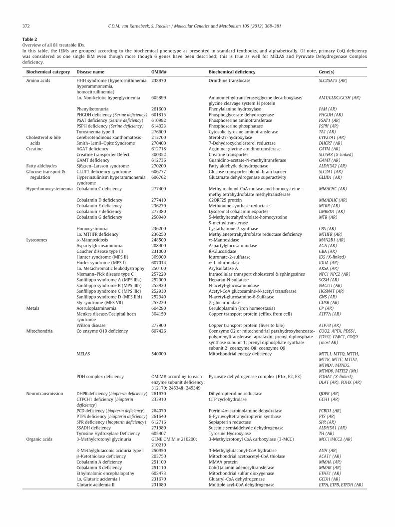
Table 2Overview of all 81 treatable IDs.In this table, the IEMs are grouped according to the biochemical phenotype as presented in standard textbooks, and alphabetically. Of note, primary CoQ deficiencywas considered as one single IEM even though more though 6 genes have been described; this is true as well for MELAS and Pyruvate Dehydrogenase Complexdeficiency.
Biochemical category Disease name OMIM# Biochemical deficiency Gene(s)
Amino acids HHH syndrome (hyperornithinemia,hyperammonemia,homocitrullinemia)
238970 Ornithine translocase SLC25A15 (AR)
l.o. Non-ketotic hyperglycinemia 605899 Aminomethyltransferase/glycine decarboxylase/glycine cleavage system H protein
AMT/GLDC/GCSH (AR)
Phenylketonuria 261600 Phenylalanine hydroxylase PAH (AR)PHGDH deficiency (Serine deficiency) 601815 Phosphoglycerate dehydrogenase PHGDH (AR)PSAT deficiency (Serine deficiency) 610992 Phosphoserine aminotransferase PSAT1 (AR)PSPH deficiency (Serine deficiency) 614023 Phosphoserine phosphatase PSPH (AR)Tyrosinemia type II 276600 Cytosolic tyrosine aminotransferase TAT (AR)
Cholesterol & bileacids
Cerebrotendinous xanthomatosis 213700 Sterol-27-hydroxylase CYP27A1 (AR)Smith–Lemli–Opitz Syndrome 270400 7-Dehydroxycholesterol reductase DHCR7 (AR)
Creatine AGAT deficiency 612718 Arginine: glycine amidinotransferase GATM (AR)Creatine transporter Defect 300352 Creatine transporter SLC6A8 (X-linked)GAMT deficiency 612736 Guanidino-acetate-N-methyltransferase GAMT (AR)
Fatty aldehydes Sjögren–Larsson syndrome 270200 Fatty aldehyde dehydrogenase ALDH3A2 (AR)Glucose transport ®ulation
GLUT1 deficiency syndrome 606777 Glucose transporter blood–brain barrier SLC2A1 (AR)Hyperinsulinism hyperammonemiasyndrome
606762 Glutamate dehydrogenase superactivity GLUD1 (AR)
Hyperhomocysteinemia Cobalamin C deficiency 277400 Methylmalonyl-CoA mutase and homocysteine :methyltetrahydrofolate methyltransferase
MMACHC (AR)
Cobalamin D deficiency 277410 C2ORF25 protein MMADHC (AR)Cobalamin E deficiency 236270 Methionine synthase reductase MTRR (AR)Cobalamin F deficiency 277380 Lysosomal cobalamin exporter LMBRD1 (AR)Cobalamin G deficiency 250940 5-Methyltetrahydrofolate-homocysteine
S-methyltransferaseMTR (AR)
Homocystinuria 236200 Cystathatione β-synthase CBS (AR)l.o. MTHFR deficiency 236250 Methylenetetrahydrofolate reductase deficiency MTHFR (AR)
Lysosomes α-Mannosidosis 248500 α-Mannosidase MAN2B1 (AR)Aspartylglucosaminuria 208400 Aspartylglucosaminidase AGA (AR)Gaucher disease type III 231000 ß-Glucosidase GBA (AR)Hunter syndrome (MPS II) 309900 Iduronate-2-sulfatase IDS (X-linked)Hurler syndrome (MPS I) 607014 α-L-iduronidase IDUA (AR)l.o. Metachromatic leukodystrophy 250100 Arylsulfatase A ARSA (AR)Niemann–Pick disease type C 257220 Intracellular transport cholesterol & sphingosines NPC1 NPC2 (AR)Sanfilippo syndrome A (MPS IIIa) 252900 Heparan-N-sulfatase SGSH (AR)Sanfilippo syndrome B (MPS IIIb) 252920 N-acetyl-glucosaminidase NAGLU (AR)Sanfilippo syndrome C (MPS IIIc) 252930 Acetyl-CoA glucosamine-N-acetyl transferase HGSNAT (AR)Sanfilippo syndrome D (MPS IIId) 252940 N-acetyl-glucosamine-6-Sulfatase GNS (AR)Sly syndrome (MPS VII) 253220 β-glucuronidase GUSB (AR)
Metals Aceruloplasminemia 604290 Ceruloplasmin (iron homeostasis) CP (AR)Menkes disease/Occipital hornsyndrome
304150 Copper transport protein (efflux from cell) ATP7A (AR)
Wilson disease 277900 Copper transport protein (liver to bile) ATP7B (AR)Mitochondria Co enzyme Q10 deficiency 607426 Coenzyme Q2 or mitochondrial parahydroxybenzoate-
polyprenyltransferase; aprataxin; prenyl diphosphatesynthase subunit 1; prenyl diphosphate synthasesubunit 2; coenzyme Q8; coenzyme Q9
COQ2, APTX, PDSS1,PDSS2, CABC1, COQ9(most AR)
MELAS 540000 Mitochondrial energy deficiency MTTL1, MTTQ, MTTH,MTTK, MTTC, MTTS1,MTND1, MTND5,MTND6, MTTS2 (Mt)
PDH complex deficiency OMIM# according to eachenzyme subunit deficiency:312170; 245348; 245349
Pyruvate dehydrogenase complex (E1α, E2, E3) PDHA1 (X-linked),DLAT (AR), PDHX (AR)
Neurotransmission DHPR deficiency (biopterin deficiency) 261630 Dihydropteridine reductase QDPR (AR)GTPCH1 deficiency (biopterindeficiency)
233910 GTP cyclohydrolase GCH1 (AR)
PCD deficiency (biopterin deficiency) 264070 Pterin-4α-carbinolamine dehydratase PCBD1 (AR)PTPS deficiency (biopterin deficiency) 261640 6-Pyruvoyltetrahydropterin synthase PTS (AR)SPR deficiency (biopterin deficiency) 612716 Sepiapterin reductase SPR (AR)SSADH deficiency 271980 Succinic semialdehyde dehydrogenase ALDH5A1 (AR)Tyrosine Hydroxylase Deficiency 605407 Tyrosine Hydroxylase TH (AR)
Organic acids 3-Methylcrotonyl glycinuria GENE OMIM # 210200;210210
3-Methylcrotonyl CoA carboxylase (3-MCC) MCC1/MCC2 (AR)
3-Methylglutaconic aciduria type I 250950 3-Methylglutaconyl-CoA hydratase AUH (AR)β-Ketothiolase deficiency 203750 Mitochondrial acetoacetyl-CoA thiolase ACAT1 (AR)Cobalamin A deficiency 251100 MMAA protein MMAA (AR)Cobalamin B deficiency 251110 Cob(I)alamin adenosyltransferase MMAB (AR)Ethylmalonic encephalopathy 602473 Mitochondrial sulfur dioxygenase ETHE1 (AR)l.o. Glutaric acidemia I 231670 Glutaryl-CoA dehydrogenase GCDH (AR)Glutaric acidemia II 231680 Multiple acyl-CoA dehydrogenase ETFA, ETFB, ETFDH (AR)
372 C.D.M. van Karnebeek, S. Stockler / Molecular Genetics and Metabolism 105 (2012) 368–381
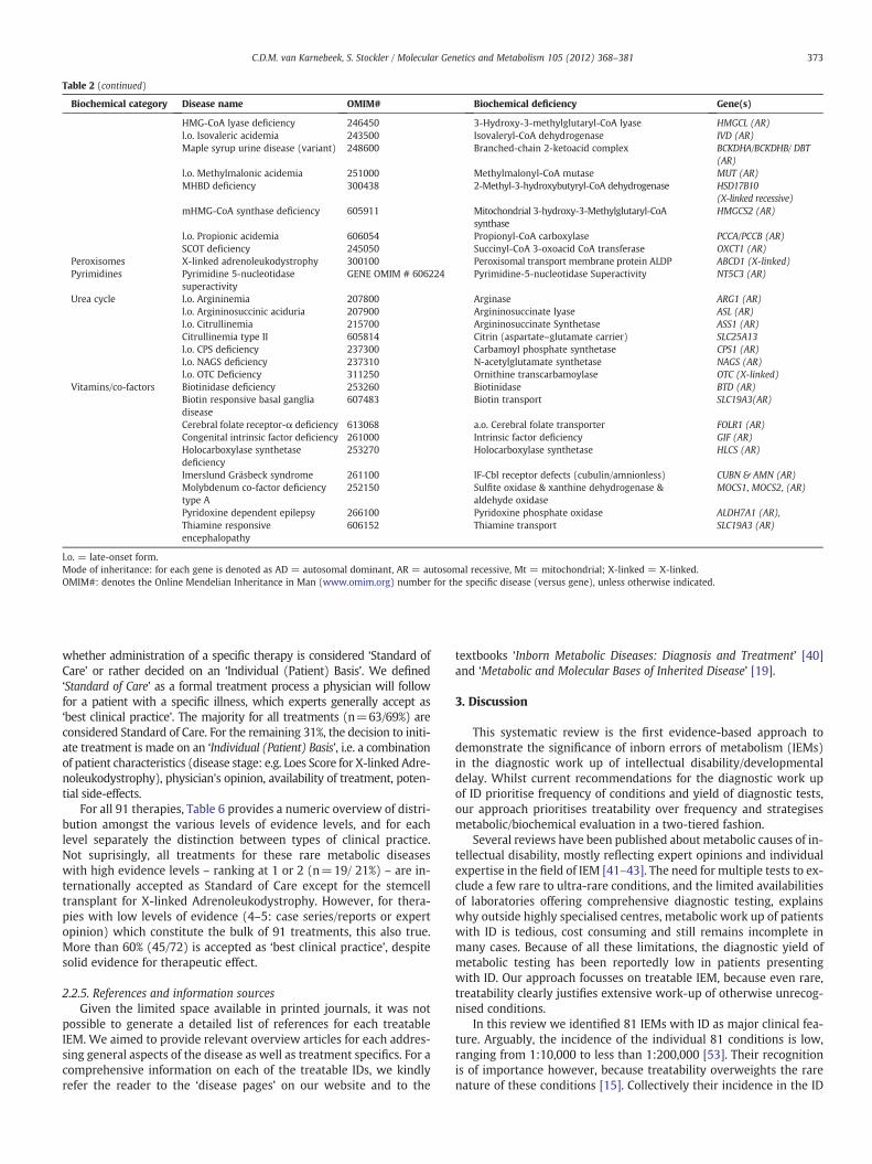
Table 2 (continued)
Biochemical category Disease name OMIM# Biochemical deficiency Gene(s)
HMG-CoA lyase deficiency 246450 3-Hydroxy-3-methylglutaryl-CoA lyase HMGCL (AR)l.o. Isovaleric acidemia 243500 Isovaleryl-CoA dehydrogenase IVD (AR)Maple syrup urine disease (variant) 248600 Branched-chain 2-ketoacid complex BCKDHA/BCKDHB/ DBT
(AR)l.o. Methylmalonic acidemia 251000 Methylmalonyl-CoA mutase MUT (AR)MHBD deficiency 300438 2-Methyl-3-hydroxybutyryl-CoA dehydrogenase HSD17B10
(X-linked recessive)mHMG-CoA synthase deficiency 605911 Mitochondrial 3-hydroxy-3-Methylglutaryl-CoA
synthaseHMGCS2 (AR)
l.o. Propionic acidemia 606054 Propionyl-CoA carboxylase PCCA/PCCB (AR)SCOT deficiency 245050 Succinyl-CoA 3-oxoacid CoA transferase OXCT1 (AR)
Peroxisomes X-linked adrenoleukodystrophy 300100 Peroxisomal transport membrane protein ALDP ABCD1 (X-linked)Pyrimidines Pyrimidine 5-nucleotidase
superactivityGENE OMIM # 606224 Pyrimidine-5-nucleotidase Superactivity NT5C3 (AR)
Urea cycle l.o. Argininemia 207800 Arginase ARG1 (AR)l.o. Argininosuccinic aciduria 207900 Argininosuccinate lyase ASL (AR)l.o. Citrullinemia 215700 Argininosuccinate Synthetase ASS1 (AR)Citrullinemia type II 605814 Citrin (aspartate–glutamate carrier) SLC25A13l.o. CPS deficiency 237300 Carbamoyl phosphate synthetase CPS1 (AR)l.o. NAGS deficiency 237310 N-acetylglutamate synthetase NAGS (AR)l.o. OTC Deficiency 311250 Ornithine transcarbamoylase OTC (X-linked)
Vitamins/co-factors Biotinidase deficiency 253260 Biotinidase BTD (AR)Biotin responsive basal gangliadisease
607483 Biotin transport SLC19A3(AR)
Cerebral folate receptor-α deficiency 613068 a.o. Cerebral folate transporter FOLR1 (AR)Congenital intrinsic factor deficiency 261000 Intrinsic factor deficiency GIF (AR)Holocarboxylase synthetasedeficiency
253270 Holocarboxylase synthetase HLCS (AR)
Imerslund Gräsbeck syndrome 261100 IF-Cbl receptor defects (cubulin/amnionless) CUBN & AMN (AR)Molybdenum co-factor deficiencytype A
252150 Sulfite oxidase & xanthine dehydrogenase &aldehyde oxidase
MOCS1, MOCS2, (AR)
Pyridoxine dependent epilepsy 266100 Pyridoxine phosphate oxidase ALDH7A1 (AR),Thiamine responsiveencephalopathy
606152 Thiamine transport SLC19A3 (AR)
l.o. = late-onset form.Mode of inheritance: for each gene is denoted as AD = autosomal dominant, AR = autosomal recessive, Mt = mitochondrial; X-linked = X-linked.OMIM#: denotes the Online Mendelian Inheritance in Man (www.omim.org) number for the specific disease (versus gene), unless otherwise indicated.
373C.D.M. van Karnebeek, S. Stockler / Molecular Genetics and Metabolism 105 (2012) 368–381
whether administration of a specific therapy is considered ‘Standard ofCare’ or rather decided on an ‘Individual (Patient) Basis’. We defined‘Standard of Care’ as a formal treatment process a physician will followfor a patient with a specific illness, which experts generally accept as‘best clinical practice’. The majority for all treatments (n=63/69%) areconsidered Standard of Care. For the remaining 31%, the decision to initi-ate treatment is made on an ‘Individual (Patient) Basis’, i.e. a combinationof patient characteristics (disease stage: e.g. Loes Score for X-linked Adre-noleukodystrophy), physician's opinion, availability of treatment, poten-tial side-effects.
For all 91 therapies, Table 6 provides a numeric overview of distri-bution amongst the various levels of evidence levels, and for eachlevel separately the distinction between types of clinical practice.Not suprisingly, all treatments for these rare metabolic diseaseswith high evidence levels – ranking at 1 or 2 (n=19/ 21%) – are in-ternationally accepted as Standard of Care except for the stemcelltransplant for X-linked Adrenoleukodystrophy. However, for thera-pies with low levels of evidence (4–5: case series/reports or expertopinion) which constitute the bulk of 91 treatments, this also true.More than 60% (45/72) is accepted as ‘best clinical practice’, despitesolid evidence for therapeutic effect.
2.2.5. References and information sourcesGiven the limited space available in printed journals, it was not
possible to generate a detailed list of references for each treatableIEM. We aimed to provide relevant overview articles for each addres-sing general aspects of the disease as well as treatment specifics. For acomprehensive information on each of the treatable IDs, we kindlyrefer the reader to the ‘disease pages’ on our website and to the
textbooks ‘Inborn Metabolic Diseases: Diagnosis and Treatment’ [40]and ‘Metabolic and Molecular Bases of Inherited Disease’ [19].
3. Discussion
This systematic review is the first evidence-based approach todemonstrate the significance of inborn errors of metabolism (IEMs)in the diagnostic work up of intellectual disability/developmentaldelay. Whilst current recommendations for the diagnostic work upof ID prioritise frequency of conditions and yield of diagnostic tests,our approach prioritises treatability over frequency and strategisesmetabolic/biochemical evaluation in a two-tiered fashion.
Several reviews have been published about metabolic causes of in-tellectual disability, mostly reflecting expert opinions and individualexpertise in the field of IEM [41–43]. The need for multiple tests to ex-clude a few rare to ultra-rare conditions, and the limited availabilitiesof laboratories offering comprehensive diagnostic testing, explainswhy outside highly specialised centres, metabolic work up of patientswith ID is tedious, cost consuming and still remains incomplete inmany cases. Because of all these limitations, the diagnostic yield ofmetabolic testing has been reportedly low in patients presentingwith ID. Our approach focusses on treatable IEM, because even rare,treatability clearly justifies extensive work-up of otherwise unrecog-nised conditions.
In this review we identified 81 IEMs with ID as major clinical fea-ture. Arguably, the incidence of the individual 81 conditions is low,ranging from 1:10,000 to less than 1:200,000 [53]. Their recognitionis of importance however, because treatability overweights the rarenature of these conditions [15]. Collectively their incidence in the ID
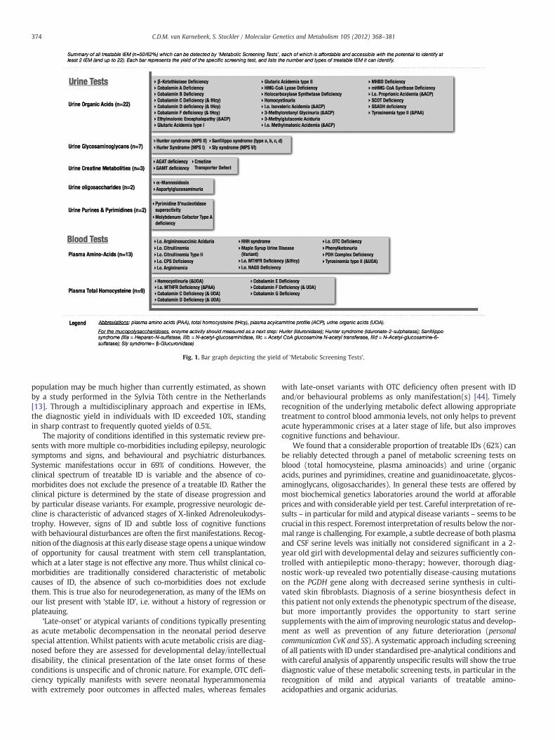
Fig. 1. Bar graph depicting the yield of ‘Metabolic Screening Tests’.
374 C.D.M. van Karnebeek, S. Stockler / Molecular Genetics and Metabolism 105 (2012) 368–381
population may be much higher than currently estimated, as shownby a study performed in the Sylvia Tòth centre in the Netherlands[13]. Through a multidisciplinary approach and expertise in IEMs,the diagnostic yield in individuals with ID exceeded 10%, standingin sharp contrast to frequently quoted yields of 0.5%.
The majority of conditions identified in this systematic review pre-sents with more multiple co-morbidities including epilepsy, neurologicsymptoms and signs, and behavioural and psychiatric disturbances.Systemic manifestations occur in 69% of conditions. However, theclinical spectrum of treatable ID is variable and the absence of co-morbidites does not exclude the presence of a treatable ID. Rather theclinical picture is determined by the state of disease progression andby particular disease variants. For example, progressive neurologic de-cline is characteristic of advanced stages of X-linked Adrenoleukodys-trophy. However, signs of ID and subtle loss of cognitive functionswith behavioural disturbances are often the first manifestations. Recog-nition of the diagnosis at this early disease stage opens a uniquewindowof opportunity for causal treatment with stem cell transplantation,which at a later stage is not effective any more. Thus whilst clinical co-morbidities are traditionally considered characteristic of metaboliccauses of ID, the absence of such co-morbidities does not excludethem. This is true also for neurodegeneration, as many of the IEMs onour list present with ‘stable ID’, i.e. without a history of regression orplateauing.
‘Late-onset’ or atypical variants of conditions typically presentingas acute metabolic decompensation in the neonatal period deservespecial attention. Whilst patients with acute metabolic crisis are diag-nosed before they are assessed for developmental delay/intellectualdisability, the clinical presentation of the late onset forms of theseconditions is unspecific and of chronic nature. For example, OTC defi-ciency typically manifests with severe neonatal hyperammonemiawith extremely poor outcomes in affected males, whereas females
with late-onset variants with OTC deficiency often present with IDand/or behavioural problems as only manifestation(s) [44]. Timelyrecognition of the underlying metabolic defect allowing appropriatetreatment to control blood ammonia levels, not only helps to preventacute hyperammonic crises at a later stage of life, but also improvescognitive functions and behaviour.
We found that a considerable proportion of treatable IDs (62%) canbe reliably detected through a panel of metabolic screening tests onblood (total homocysteine, plasma aminoacids) and urine (organicacids, purines and pyrimidines, creatine and guanidinoacetate, glycos-aminoglycans, oligosaccharides). In general these tests are offered bymost biochemical genetics laboratories around the world at afforableprices and with considerable yield per test. Careful interpretation of re-sults – in particular for mild and atypical disease variants – seems to becrucial in this respect. Foremost interpretation of results below the nor-mal range is challenging. For example, a subtle decrease of both plasmaand CSF serine levels was initially not considered significant in a 2-year old girl with developmental delay and seizures sufficiently con-trolled with antiepileptic mono-therapy; however, thorough diag-nostic work-up revealed two potentially disease-causing mutationson the PGDH gene along with decreased serine synthesis in culti-vated skin fibroblasts. Diagnosis of a serine biosynthesis defect inthis patient not only extends the phenotypic spectrum of the disease,but more importantly provides the opportunity to start serinesupplements with the aim of improving neurologic status and develop-ment as well as prevention of any future deterioration (personalcommunication CvK and SS). A systematic approach including screeningof all patients with ID under standardised pre-analytical conditions andwith careful analysis of apparently unspecific results will show the truediagnostic value of these metabolic screening tests, in particular in therecognition of mild and atypical variants of treatable amino-acidopathies and organic acidurias.
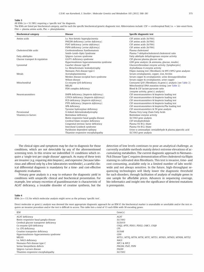
Table 3All IEMs (n=31/38%) requiring a ‘specific test’ for diagnosis.The IEMs are listed per biochemical category, and for each the specific biochemical/genetic diagnostic test. Abbreviations include: CSF = cerebrospinal fluid, l.o. = late-onset form,PAA = plasma amino acids, Phe = phenylalanine.
Biochemical category Disease Specific diagnostic test
Amino acids l.o. Non-ketotic hyperglycinemia CSF amino acids (& PAA)PHGDH deficiency (serine deficiency) CSF amino acids (& PAA)PSAT deficiency (serine deficiency) CSF amino acids (& PAA)PSPH deficiency (serine deficiency) CSF amino acids (& PAA)
Cholesterol/bile acids Cerebrotendinous Xanthomatosis Plasma cholestanolSmith–Lemli–Opitz Syndrome Plasma 7-dehydrocholesterol:cholesterol ratio
Fatty aldehydes Sjögren–Larsson syndrome Fatty aldehyde dehydrogenase enzyme activityGlucose transport & regulation GLUT1 deficiency syndrome CSF glucose:plasma glucose ratio
Hyperinsulinism hyperammonemia syndrome GDH gene analysis (& ammonia, glucose, insulin)Lysosomal Gaucher disease type III Glucocerebrosidase enzyme activity (lymphocytes)
l.o. Metachromatic leukodystrophy Arylsulfatase-A enzyme activityNiemann–Pick disease type C Filipin staining test (fibroblasts) & NPC1/NPC2 gene analyses
Metals Aceruloplasminemia Serum ceruloplasmin, copper, iron, ferritinMenkes disease-occipital horn syndrome Serum copper & ceruloplasmin; urine deoxypyridinolineWilson disease Serum copper & ceruloplasmin, urine copper
Mitochondrial Co enzyme Q10 deficiency Coenzyme Q10 (fibroblasts) & gene(s) analysis (see Table 2)MELAS Mitochondrial DNA mutation testing (see Table 2)PDH complex deficiency Blood & CSF lactate:pyruvate ratio
(enzyme activity, gene(s) analysis)Neurotransmitters DHPR deficiency (biopterin deficiency) CSF neurotransmitters & biopterin loading test
GTPCH deficiency (biopterin deficiency) CSF neurotransmitters & biopterin loading testPCD deficiency (biopterin deficiency) CSF neurotransmitters & biopterin loading testPTPS deficiency (biopterin deficiency) CSF neurotransmitters & biopterin loading testSPR deficiency CSF neurotransmitters & biopterin/Phe loading testTyrosine hydroxylase deficiency CSF neurotransmitters & TH gene analysis
Peroxisomal X-linked Adrenoleukodystrophy Plasma Very Long Chain Fatty AcidsVitamins/co-factors Biotinidase deficiency Biotinidase enzyme activity
Biotin responsive basal ganglia disease SLC19A3 gene analysisCerebral folate receptor deficiency CSF tetrahydrofolateCongenital intrinsic factor deficiency Plasma Vit B12, folateImerslund Gräsbeck syndrome Plasma Vit B12, folatePyridoxine dependent epilepsy Urine α-aminoadipic semialdehyde & plasma pipecolic acidThiamine-responsive encephalopathy SLC19A3 gene analysis
375C.D.M. van Karnebeek, S. Stockler / Molecular Genetics and Metabolism 105 (2012) 368–381
The clinical signs and symptoms may be clue to diagnosis for thoseconditions, which are not detectable by any of the aforementionedscreening tests. In this review we indentified 31 conditions which re-quire a ‘single test per single disease’ approach. As many of these testsare invasive (e.g. requiring skin biopsies), and expensive (because labo-rious and offered only by a few laboratories worldwide), a careful clin-ical differential diagnosis is mandatory for a time- and cost-effectivediagnostic evaluation.
Primary gene analysis is a way to enhance the diagnostic yield inconditions with unspecific clinical and biochemical presentation. Forexample, low urinary excretion of guanidinoacetate is characteristic ofAGAT deficiency, a treatable disorder of creatine synthesis, but the
Table 4IEMs (n=13) for which molecular analysis might serve as the primary ‘specific test’.
Direct molecular or gene(s) analysis was deemed the most appropriate diagnostic approacquires an invasive procedure and/or the test is difficult to access. This table lists a total of 1
IEM
AGAT deficiencyBiotin responsive basal ganglia diseaseCerebral glucose transporter deficiencyCo enzyme Q10 deficiencyl.o. CPS deficiencyCreatine transporter deficiencyHyperinsulinism–hyperammonia syndromeMELASl.o. NAGS deficiencyNiemann-Pick disease type CSerine biosynthesis defectsSjögren–Larssen diseaseThiamine-responsive encephalopathy
detection of low levels continues to pose an analytical challenge, ascurrently available methods mainly detect extreme elevations of ac-cumulating metabolites. The current diagnostic approach to Niemann–PickDisease Type C requires demonstration of free cholesterol viafilipinstaining in cultivated skin fibroblasts. This test is invasive, time- andcost-consuming, available only in a limited number of labs world-wide and not always sensitive. In the future, high-throughput se-quencing technologies will likely lower the diagnostic thresholdfor such disorders, through facilitation of analysis of multiple genes inone sample for afforfable prices. Advances in sequencing coverage,bio-informatics and insight into the significance of detected mutationsis prerequisite.
h for an IEM if: the biochemical marker is unavailable or unreliable and/or the test re-3 such IEMs with 30 encoding genes.
Gene(s)
AGATSLC19A3SLC6A19COQ2, APTX, PDSS1, PDSS2, CABC1, COQ9CPSSLC6A8GDHMTTL1, MTTQ, MTTH, MTTK, MTTC, MTTS1, MTND1, MTND5, MTND6, MTTS2NAGSNPC1 & NPC2PHGDH, PSAT, PSPHFALDHSLC19A3
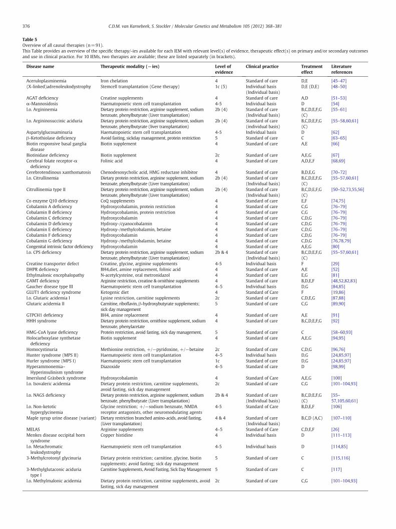
Table 5Overview of all causal therapies (n=91).This Table provides an overview of the specific therapy/-ies available for each IEM with relevant level(s) of evidence, therapeutic effect(s) on primary and/or secondary outcomesand use in clinical practice. For 10 IEMs, two therapies are available; these are listed separately (in brackets).
Disease name Therapeutic modality (− ies) Level ofevidence
Clinical practice Treatmenteffect
Literaturereferences
Aceruloplasminemia Iron chelation 4 Standard of care D,E [45–47](X-linked)adrenoleukodystrophy Stemcell transplantation (Gene therapy) 1c (5) Individual basis
(Individual basis)D,E (D,E) [48–50]
AGAT deficiency Creatine supplements 4 Standard of care A,D [51–53]α-Mannosidosis Haematopoietic stem cell transplantation 4-5 Individual basis D [54]l.o. Argininemia Dietary protein restriction, arginine supplement, sodium
benzoate, phenylbutyrate (Liver transplantation)2b (4) Standard of care
(Individual basis)B,C,D,E,F,G(C)
[55–61]
l.o. Argininosuccinic aciduria Dietary protein restriction, arginine supplement, sodiumbenzoate, phenylbutyrate (liver transplantation)
2b (4) Standard of care(individual basis)
B,C,D,E,F,G(C)
[55–58,60,61]
Aspartylglucosaminuria Haematopoietic stem cell transplantation 4-5 Individual basis D [62]β-Ketothiolase deficiency Avoid fasting, sickday management, protein restriction 5 Standard of care C [63–65]Biotin responsive basal gangliadisease
Biotin supplement 4 Standard of care A,E [66]
Biotinidase deficiency Biotin supplement 2c Standard of care A,E,G [67]Cerebral folate receptor-αdeficiency
Folinic acid 4 Standard of care A,D,E,F [68,69]
Cerebrotendinous xanthomatosis Chenodesoxycholic acid, HMG reductase inhibitor 4 Standard of care B,D,E,G [70–72]l.o. Citrullinemia Dietary protein restriction, arginine supplement, sodium
benzoate, phenylbutyrate (Liver transplantation)2b (4) Standard of care
(Individual basis)B,C,D,E,F,G(C)
[55–57,60,61]
Citrullinemia type II Dietary protein restriction, arginine supplement, sodiumbenzoate, phenylbutyrate (Liver transplantation)
2b (4) Standard of care(Individual basis)
B,C,D,E,F,G(C)
[50–52,73,55,56]
Co enzyme Q10 deficiency CoQ supplements 4 Standard of care E,F [74,75]Cobalamin A deficiency Hydroxycobalamin, protein restriction 4 Standard of care C,G [76–79]Cobalamin B deficiency Hydroxycobalamin, protein restriction 4 Standard of care C,G [76–79]Cobalamin C deficiency Hydroxycobalamin 4 Standard of care C,D,G [76–79]Cobalamin D deficiency Hydroxy-/cyanocobalamin 4 Standard of care C,D,G [76–79]Cobalamin E deficiency Hydroxy-/methylcobalamin, betaine 4 Standard of care C,D,G [76–79]Cobalamin F deficiency Hydroxycobalamin 4 Standard of care C,D,G [76–79]Cobalamin G deficiency Hydroxy-/methylcobalamin, betaine 4 Standard of care C,D,G [76,78,79]Congenital intrinsic factor deficiency Hydroxycobalamin 4 Standard of care A,E,G [80]l.o. CPS deficiency Dietary protein restriction, arginine supplement, sodium
benzoate, phenylbutyrate (Liver transplantation)2b & 4 Standard of care
(Individual basis)B,C,D,E,F,G(C)
[55–57,60,61]
Creatine transporter defect Creatine, glycine, arginine supplements 4-5 Individual basis F [29]DHPR deficiency BH4,diet, amine replacement, folinic acid 4 Standard of care A,E [52]Ethylmalonic encephalopathy N-acetylcysteine, oral metronidazol 4 Standard of care E,G [81]GAMT deficiency Arginine restriction, creatine & ornithine supplements 4 Standard of care B,D,E,F [48,52,82,83]Gaucher disease type III Haematopoietic stem cell transplantation 4–5 Individual basis D,G [84,85]GLUT1 deficiency syndrome Ketogenic diet 4 Standard of Care F [19,86]l.o. Glutaric acidemia I Lysine restriction, carnitine supplements 2c Standard of care C,D,E,G [87,88]Glutaric acidemia II Carnitine, riboflavin, β-hydroxybutyrate supplements;
sick day management5 Standard of care C,G [89,90]
GTPCH1 deficiency BH4, amine replacement 4 Standard of care A,E [91]HHH syndrome Dietary protein restriction, ornithine supplement, sodium
benzoate, phenylacetate4 Standard of care B,C,D,E,F,G [92]
HMG-CoA lyase deficiency Protein restriction, avoid fasting, sick day management, 5 Standard of care C [58–60,93]Holocarboxylase synthetasedeficiency
Biotin supplement 4 Standard of care A,E,G [94,95]
Homocystinuria Methionine restriction, +/−pyridoxine, +/−betaine 2c Standard of care C,D,G [96,76]Hunter syndrome (MPS II) Haematopoietic stem cell transplantation 4–5 Individual basis D,G [24,85,97]Hurler syndrome (MPS I) Haematopoietic stem cell transplantation 1c Standard of care D,G [24,85,97]Hyperammonemia–Hyperinsulinism syndrome
Diazoxide 4–5 Standard of care D [98,99]
Imerslund Gräsbeck syndrome Hydroxycobalamin 4 Standard of Care A,E,G [100]l.o. Isovaleric acidemia Dietary protein restriction, carnitine supplements,
avoid fasting, sick day management2c Standard of care C,G [101–104,93]
l.o. NAGS deficiency Dietary protein restriction, arginine supplement, sodiumbenzoate, phenylbutyrate (Liver transplantation)
2b & 4 Standard of care(Individual basis)
B,C,D,E,F,G(C)
[55–57,105,60,61]
l.o. Non-ketotichyperglycinemia
Glycine restriction; +/−sodium benzoate, NMDAreceptor antagonists, other neuromodulating agents
4-5 Standard of Care B,D,E,F [106]
Maple syrup urine disease (variant) Dietary restriction branched amino-acids, avoid fasting,(Liver transplantation)
4 & 4 Standard of care(Individual basis)
B,C,D (A,C) [107–110]
MELAS Arginine supplements 4–5 Standard of Care C,D,E,F [26]Menkes disease occipital hornsyndrome
Copper histidine 4 Individual basis D [111–113]
l.o. Metachromaticleukodystrophy
Haematopoietic stem cell transplantation 4-5 Individual basis D [114,85]
3-Methylcrotonyl glycinuria Dietary protein restriction; carnitine, glycine, biotinsupplements; avoid fasting; sick day management
5 Standard of care C [115,116]
3-Methylglutaconic aciduriatype I
Carnitine Supplements, Avoid Fasting, Sick DayManagement 5 Standard of care C [117]
l.o. Methylmalonic acidemia Dietary protein restriction, carnitine supplements, avoidfasting, sick day management
2c Standard of care C,G [101–104,93]
376 C.D.M. van Karnebeek, S. Stockler / Molecular Genetics and Metabolism 105 (2012) 368–381
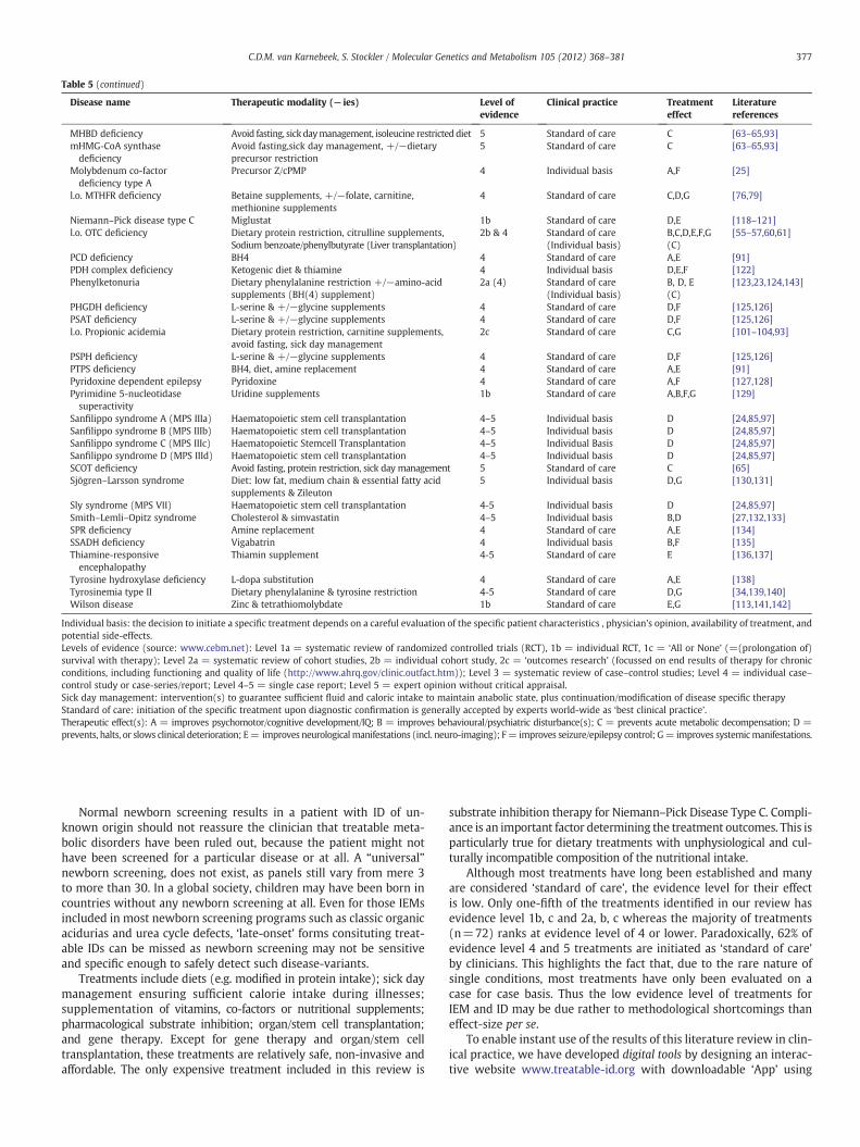
Table 5 (continued)
Disease name Therapeutic modality (− ies) Level ofevidence
Clinical practice Treatmenteffect
Literaturereferences
MHBD deficiency Avoid fasting, sick daymanagement, isoleucine restricted diet 5 Standard of care C [63–65,93]mHMG-CoA synthasedeficiency
Avoid fasting,sick day management, +/−dietaryprecursor restriction
5 Standard of care C [63–65,93]
Molybdenum co-factordeficiency type A
Precursor Z/cPMP 4 Individual basis A,F [25]
l.o. MTHFR deficiency Betaine supplements, +/−folate, carnitine,methionine supplements
4 Standard of care C,D,G [76,79]
Niemann–Pick disease type C Miglustat 1b Standard of care D,E [118–121]l.o. OTC deficiency Dietary protein restriction, citrulline supplements,
Sodium benzoate/phenylbutyrate (Liver transplantation)2b & 4 Standard of care
(Individual basis)B,C,D,E,F,G(C)
[55–57,60,61]
PCD deficiency BH4 4 Standard of care A,E [91]PDH complex deficiency Ketogenic diet & thiamine 4 Individual basis D,E,F [122]Phenylketonuria Dietary phenylalanine restriction +/−amino-acid
supplements (BH(4) supplement)2a (4) Standard of care
(Individual basis)B, D, E(C)
[123,23,124,143]
PHGDH deficiency L-serine & +/−glycine supplements 4 Standard of care D,F [125,126]PSAT deficiency L-serine & +/−glycine supplements 4 Standard of care D,F [125,126]l.o. Propionic acidemia Dietary protein restriction, carnitine supplements,
avoid fasting, sick day management2c Standard of care C,G [101–104,93]
PSPH deficiency L-serine & +/−glycine supplements 4 Standard of care D,F [125,126]PTPS deficiency BH4, diet, amine replacement 4 Standard of care A,E [91]Pyridoxine dependent epilepsy Pyridoxine 4 Standard of care A,F [127,128]Pyrimidine 5-nucleotidasesuperactivity
Uridine supplements 1b Standard of care A,B,F,G [129]
Sanfilippo syndrome A (MPS IIIa) Haematopoietic stem cell transplantation 4–5 Individual basis D [24,85,97]Sanfilippo syndrome B (MPS IIIb) Haematopoietic stem cell transplantation 4–5 Individual basis D [24,85,97]Sanfilippo syndrome C (MPS IIIc) Haematopoietic Stemcell Transplantation 4–5 Individual Basis D [24,85,97]Sanfilippo syndrome D (MPS IIId) Haematopoietic stem cell transplantation 4–5 Individual basis D [24,85,97]SCOT deficiency Avoid fasting, protein restriction, sick day management 5 Standard of care C [65]Sjögren–Larsson syndrome Diet: low fat, medium chain & essential fatty acid
supplements & Zileuton5 Individual basis D,G [130,131]
Sly syndrome (MPS VII) Haematopoietic stem cell transplantation 4-5 Individual basis D [24,85,97]Smith–Lemli–Opitz syndrome Cholesterol & simvastatin 4–5 Individual basis B,D [27,132,133]SPR deficiency Amine replacement 4 Standard of care A,E [134]SSADH deficiency Vigabatrin 4 Individual basis B,F [135]Thiamine-responsiveencephalopathy
Thiamin supplement 4-5 Standard of care E [136,137]
Tyrosine hydroxylase deficiency L-dopa substitution 4 Standard of care A,E [138]Tyrosinemia type II Dietary phenylalanine & tyrosine restriction 4-5 Standard of care D,G [34,139,140]Wilson disease Zinc & tetrathiomolybdate 1b Standard of care E,G [113,141,142]
Individual basis: the decision to initiate a specific treatment depends on a careful evaluation of the specific patient characteristics , physician's opinion, availability of treatment, andpotential side-effects.Levels of evidence (source: www.cebm.net): Level 1a = systematic review of randomized controlled trials (RCT), 1b = individual RCT, 1c = ‘All or None’ (=(prolongation of)survival with therapy); Level 2a = systematic review of cohort studies, 2b = individual cohort study, 2c = ‘outcomes research’ (focussed on end results of therapy for chronicconditions, including functioning and quality of life (http://www.ahrq.gov/clinic.outfact.htm)); Level 3 = systematic review of case–control studies; Level 4 = individual case–control study or case-series/report; Level 4–5 = single case report; Level 5 = expert opinion without critical appraisal.Sick day management: intervention(s) to guarantee sufficient fluid and caloric intake to maintain anabolic state, plus continuation/modification of disease specific therapyStandard of care: initiation of the specific treatment upon diagnostic confirmation is generally accepted by experts world-wide as ‘best clinical practice’.Therapeutic effect(s): A = improves psychomotor/cognitive development/IQ; B = improves behavioural/psychiatric disturbance(s); C = prevents acute metabolic decompensation; D =prevents, halts, or slows clinical deterioration; E= improves neurologicalmanifestations (incl. neuro-imaging); F= improves seizure/epilepsy control; G= improves systemicmanifestations.
377C.D.M. van Karnebeek, S. Stockler / Molecular Genetics and Metabolism 105 (2012) 368–381
Normal newborn screening results in a patient with ID of un-known origin should not reassure the clinician that treatable meta-bolic disorders have been ruled out, because the patient might nothave been screened for a particular disease or at all. A “universal”newborn screening, does not exist, as panels still vary from mere 3to more than 30. In a global society, children may have been born incountries without any newborn screening at all. Even for those IEMsincluded in most newborn screening programs such as classic organicacidurias and urea cycle defects, ‘late-onset’ forms consituting treat-able IDs can be missed as newborn screening may not be sensitiveand specific enough to safely detect such disease-variants.
Treatments include diets (e.g. modified in protein intake); sick daymanagement ensuring sufficient calorie intake during illnesses;supplementation of vitamins, co-factors or nutritional supplements;pharmacological substrate inhibition; organ/stem cell transplantation;and gene therapy. Except for gene therapy and organ/stem celltransplantation, these treatments are relatively safe, non-invasive andaffordable. The only expensive treatment included in this review is
substrate inhibition therapy for Niemann–Pick Disease Type C. Compli-ance is an important factor determining the treatment outcomes. This isparticularly true for dietary treatments with unphysiological and cul-turally incompatible composition of the nutritional intake.
Although most treatments have long been established and manyare considered ‘standard of care’, the evidence level for their effectis low. Only one-fifth of the treatments identified in our review hasevidence level 1b, c and 2a, b, c whereas the majority of treatments(n=72) ranks at evidence level of 4 or lower. Paradoxically, 62% ofevidence level 4 and 5 treatments are initiated as ‘standard of care’by clinicians. This highlights the fact that, due to the rare nature ofsingle conditions, most treatments have only been evaluated on acase for case basis. Thus the low evidence level of treatments forIEM and ID may be due rather to methodological shortcomings thaneffect-size per se.
To enable instant use of the results of this literature review in clin-ical practice, we have developed digital tools by designing an interac-tive website www.treatable-id.org with downloadable ‘App’ using
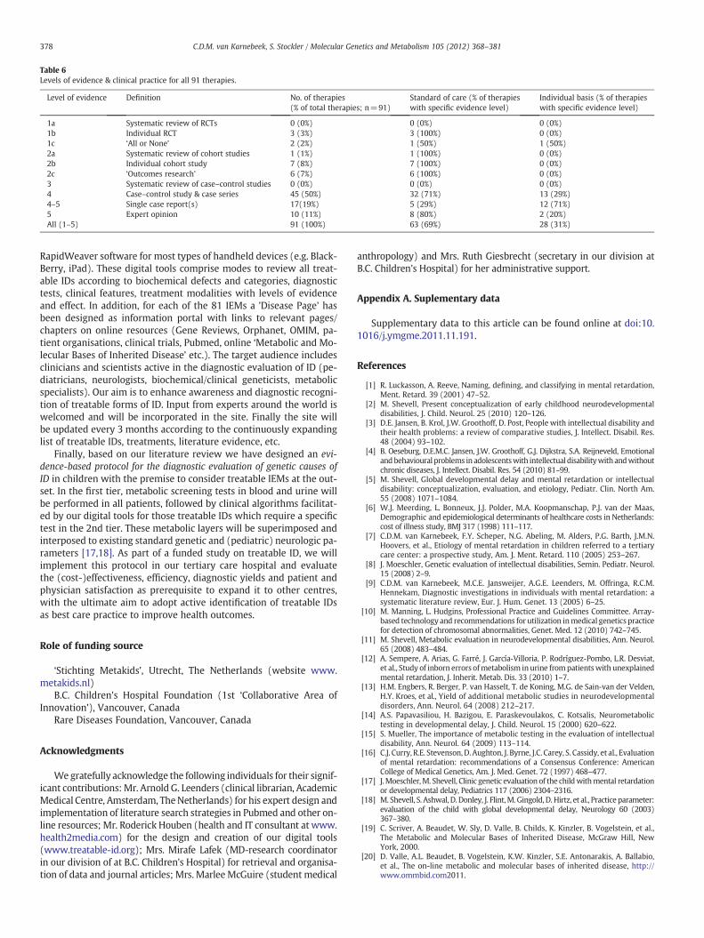
Table 6Levels of evidence & clinical practice for all 91 therapies.
Level of evidence Definition No. of therapies(% of total therapies; n=91)
Standard of care (% of therapieswith specific evidence level)
Individual basis (% of therapieswith specific evidence level)
1a Systematic review of RCTs 0 (0%) 0 (0%) 0 (0%)1b Individual RCT 3 (3%) 3 (100%) 0 (0%)1c ‘All or None’ 2 (2%) 1 (50%) 1 (50%)2a Systematic review of cohort studies 1 (1%) 1 (100%) 0 (0%)2b Individual cohort study 7 (8%) 7 (100%) 0 (0%)2c ‘Outcomes research’ 6 (7%) 6 (100%) 0 (0%)3 Systematic review of case–control studies 0 (0%) 0 (0%) 0 (0%)4 Case–control study & case series 45 (50%) 32 (71%) 13 (29%)4–5 Single case report(s) 17(19%) 5 (29%) 12 (71%)5 Expert opinion 10 (11%) 8 (80%) 2 (20%)All (1–5) 91 (100%) 63 (69%) 28 (31%)
378 C.D.M. van Karnebeek, S. Stockler / Molecular Genetics and Metabolism 105 (2012) 368–381
RapidWeaver software for most types of handheld devices (e.g. Black-Berry, iPad). These digital tools comprise modes to review all treat-able IDs according to biochemical defects and categories, diagnostictests, clinical features, treatment modalities with levels of evidenceand effect. In addition, for each of the 81 IEMs a 'Disease Page' hasbeen designed as information portal with links to relevant pages/chapters on online resources (Gene Reviews, Orphanet, OMIM, pa-tient organisations, clinical trials, Pubmed, online ‘Metabolic and Mo-lecular Bases of Inherited Disease’ etc.). The target audience includesclinicians and scientists active in the diagnostic evaluation of ID (pe-diatricians, neurologists, biochemical/clinical geneticists, metabolicspecialists). Our aim is to enhance awareness and diagnostic recogni-tion of treatable forms of ID. Input from experts around the world iswelcomed and will be incorporated in the site. Finally the site willbe updated every 3 months according to the continuously expandinglist of treatable IDs, treatments, literature evidence, etc.
Finally, based on our literature review we have designed an evi-dence-based protocol for the diagnostic evaluation of genetic causes ofID in children with the premise to consider treatable IEMs at the out-set. In the first tier, metabolic screening tests in blood and urine willbe performed in all patients, followed by clinical algorithms facilitat-ed by our digital tools for those treatable IDs which require a specifictest in the 2nd tier. These metabolic layers will be superimposed andinterposed to existing standard genetic and (pediatric) neurologic pa-rameters [17,18]. As part of a funded study on treatable ID, we willimplement this protocol in our tertiary care hospital and evaluatethe (cost-)effectiveness, efficiency, diagnostic yields and patient andphysician satisfaction as prerequisite to expand it to other centres,with the ultimate aim to adopt active identification of treatable IDsas best care practice to improve health outcomes.
Role of funding source
‘Stichting Metakids’, Utrecht, The Netherlands (website www.metakids.nl)
B.C. Children's Hospital Foundation (1st ‘Collaborative Area ofInnovation’), Vancouver, Canada
Rare Diseases Foundation, Vancouver, Canada
Acknowledgments
Wegratefully acknowledge the following individuals for their signif-icant contributions:Mr. Arnold G. Leenders (clinical librarian, AcademicMedical Centre, Amsterdam, TheNetherlands) for his expert design andimplementation of literature search strategies in Pubmed and other on-line resources; Mr. Roderick Houben (health and IT consultant at www.health2media.com) for the design and creation of our digital tools(www.treatable-id.org); Mrs. Mirafe Lafek (MD-research coordinatorin our division of at B.C. Children's Hospital) for retrieval and organisa-tion of data and journal articles; Mrs. MarleeMcGuire (student medical
anthropology) and Mrs. Ruth Giesbrecht (secretary in our division atB.C. Children's Hospital) for her administrative support.
Appendix A. Suplementary data
Supplementary data to this article can be found online at doi:10.1016/j.ymgme.2011.11.191.
References
[1] R. Luckasson, A. Reeve, Naming, defining, and classifying in mental retardation,Ment. Retard. 39 (2001) 47–52.
[2] M. Shevell, Present conceptualization of early childhood neurodevelopmentaldisabilities, J. Child. Neurol. 25 (2010) 120–126.
[3] D.E. Jansen, B. Krol, J.W. Groothoff, D. Post, People with intellectual disability andtheir health problems: a review of comparative studies, J. Intellect. Disabil. Res.48 (2004) 93–102.
[4] B. Oeseburg, D.E.M.C. Jansen, J.W. Groothoff, G.J. Dijkstra, S.A. Reijneveld, Emotionalandbehavioural problems in adolescentswith intellectual disabilitywith andwithoutchronic diseases, J. Intellect. Disabil. Res. 54 (2010) 81–99.
[5] M. Shevell, Global developmental delay and mental retardation or intellectualdisability: conceptualization, evaluation, and etiology, Pediatr. Clin. North Am.55 (2008) 1071–1084.
[6] W.J. Meerding, L. Bonneux, J.J. Polder, M.A. Koopmanschap, P.J. van der Maas,Demographic and epidemiological determinants of healthcare costs in Netherlands:cost of illness study, BMJ 317 (1998) 111–117.
[7] C.D.M. van Karnebeek, F.Y. Scheper, N.G. Abeling, M. Alders, P.G. Barth, J.M.N.Hoovers, et al., Etiology of mental retardation in children referred to a tertiarycare center: a prospective study, Am. J. Ment. Retard. 110 (2005) 253–267.
[8] J. Moeschler, Genetic evaluation of intellectual disabilities, Semin. Pediatr. Neurol.15 (2008) 2–9.
[9] C.D.M. van Karnebeek, M.C.E. Jansweijer, A.G.E. Leenders, M. Offringa, R.C.M.Hennekam, Diagnostic investigations in individuals with mental retardation: asystematic literature review, Eur. J. Hum. Genet. 13 (2005) 6–25.
[10] M. Manning, L. Hudgins, Professional Practice and Guidelines Committee. Array-based technology and recommendations for utilization inmedical genetics practicefor detection of chromosomal abnormalities, Genet. Med. 12 (2010) 742–745.
[11] M. Shevell, Metabolic evaluation in neurodevelopmental disabilities, Ann. Neurol.65 (2008) 483–484.
[12] A. Sempere, A. Arias, G. Farré, J. García-Villoria, P. Rodríguez-Pombo, L.R. Desviat,et al., Study of inborn errors ofmetabolism in urine frompatientswith unexplainedmental retardation, J. Inherit. Metab. Dis. 33 (2010) 1–7.
[13] H.M. Engbers, R. Berger, P. van Hasselt, T. de Koning, M.G. de Sain-van der Velden,H.Y. Kroes, et al., Yield of additional metabolic studies in neurodevelopmentaldisorders, Ann. Neurol. 64 (2008) 212–217.
[14] A.S. Papavasiliou, H. Bazigou, E. Paraskevoulakos, C. Kotsalis, Neurometabolictesting in developmental delay, J. Child. Neurol. 15 (2000) 620–622.
[15] S. Mueller, The importance of metabolic testing in the evaluation of intellectualdisability, Ann. Neurol. 64 (2009) 113–114.
[16] C.J. Curry, R.E. Stevenson,D.Aughton, J. Byrne, J.C. Carey, S. Cassidy, et al., Evaluationof mental retardation: recommendations of a Consensus Conference: AmericanCollege of Medical Genetics, Am. J. Med. Genet. 72 (1997) 468–477.
[17] J.Moeschler,M. Shevell, Clinic genetic evaluation of the childwithmental retardationor developmental delay, Pediatrics 117 (2006) 2304–2316.
[18] M. Shevell, S. Ashwal, D. Donley, J. Flint,M. Gingold, D. Hirtz, et al., Practice parameter:evaluation of the child with global developmental delay, Neurology 60 (2003)367–380.
[19] C. Scriver, A. Beaudet, W. Sly, D. Valle, B. Childs, K. Kinzler, B. Vogelstein, et al.,The Metabolic and Molecular Bases of Inherited Disease, McGraw Hill, NewYork, 2000.
[20] D. Valle, A.L. Beaudet, B. Vogelstein, K.W. Kinzler, S.E. Antonarakis, A. Ballabio,et al., The on-line metabolic and molecular bases of inherited disease, http://www.ommbid.com2011.

379C.D.M. van Karnebeek, S. Stockler / Molecular Genetics and Metabolism 105 (2012) 368–381
[21] W.G. Leen, J. Klepper, M.M. Verbeek, M. Leferink, T. Hofste, B.G. van Engelen,et al., Glucose transporter-1 deficiency syndrome: the expanding clinical andgenetic spectrum of a treatable disorder, Brain 133 (2010) 655–670.
[22] N. Cartier, P. Aubourg, Hematopoietic stem cell transplantation and hematopoieticstem cell gene therapy in X-linked adrenoleukodystrophy, Brain Pathol. 20 (2010)857–862.
[23] F.J. van Spronsen, G.M. Enns, Future treatment strategies in phenylketonuria,Mol. Genet. Metab. 99 (2010) S90–S95.
[24] V.K. Prasad, J. Kurtzberg, Transplant outcomes in mucopolysaccharidoses,Semin. Hematol. 47 (2010) 59–69.
[25] A. Veldman, J.A. Santamaria-Araujo, S. Sollazzo, J. Pitt, R. Gianello, J. Yaplito-Lee,et al., Successful treatment of molybdenum cofactor deficiency type A withcPMP, Pediatrics 125 (2010) e1249–e1254.
[26] R. Shigemi, M. Fukuda, Y. Suzuki, T. Morimoto, E. Ishii, L-arginine is effective instroke-like episodes of MELAS associated with the G13513A mutation, BrainDev. 33 (2011) 518–520.
[27] D. Haas, S.F. Garbade, C. Vohwinkel, N. Muschol, F.K. Trefz, J.M. Penzien, et al.,Effects of cholesterol and simvastatin treatment in patients with Smith–Lemli–Opitz syndrome (SLOS), J. Inherit. Metab. Dis. 30 (2007) 375–387.
[28] N.A. Nwokoro, J.J. Mulvihill, Cholesterol and bile acid replacement therapy inchildren and adults with Smith–Lemli–Opitz (SLO/RSH) syndrome, Am. J. Med.Genet. 31 (68) (1997) 315–321.
[29] S. Mercimek-Mahmutoglu, M.B. Connolly, K.J. Poskitt, G.A. Horvath, N. Lowry,G.S. Salomons, et al., Treatment of intractable epilepsy in a female withSLC6A8 deficiency, Mol. Genet. Metab. 101 (2010) 409–412.
[30] V. Valayannopoulos, N. Boddaert, A. Chabli, V. Barbier, I. Desguerre, A. Philippe,et al., Treatment by oral creatine, L-arginine and L-glycine in six severely affectedpatients with creatine transporter defect, J. Inherit. Metab. Dis. (Jun 10 2011)(Epub ahead of print), doi:10.1007/s10545-011-9358-9.
[31] A.M. Bosch, H.D. Bakker, A.H. van Gennip, J.V. van Kempen, R.J. Wanders, F.A.Wijburg, Clinical features of galactokinase deficiency: a review of the literature,J. Inherit. Metab. Dis. 25 (2002) 629–634.
[32] M. Di Rocco, A.R. Fantasia, M. Taro, A. Loy, A. Forlino, A. Martini, Systemic lupuserythematosus-like disease in a 6-year-old boywith prolidase deficiency, J. Inherit.Metab. Dis. 30 (2007) 814.
[33] C.K. Cheon, B.H. Lee, J.M. Ko, H.J. Kim, H.W. Yoo, Novel mutation in SLC6A19 causinglate-onset seizures in Hartnup disorder, Pediatr. Neurol. 42 (2010) 369–371.
[34] C.R. Scott, The genetic tyrosinemias, Am. J. Med. Genet. C. Semin. Med. Genet.142C (2006) 121–126.
[35] K. Ehlert, M. Frosch, N. Fehse, A. Zander, J. Roth, J. Vormoor, Farber disease: clinicalpresentation, pathogenesis and a new approach to treatment, Pediatr. Rheumatol.Online J. 5 (2007) 15.
[36] J.P. Brosco, L.M. Sanders, R. Dharia, G. Guez, C. Feudtner, The lure of treatment:expanded newborn screening and the curious case of histidinemia, Pediatrics125 (2010) 417–419.
[37] J. Vockley, L. Smith, R. Chang, J. Michel, L. Kratz, R.I. Kelley, A. Vallejo, et al.,Clinical evaluation and treatment of two patients with SC4MOL deficiency, anew disorder in cholesteroligenesis, ACMG Annual Clinical Genetics Meeting,Abstract, 7, 2011, p. 148.
[38] H. Cario, D.E.C. Smith, H. Blom, N. Blau, H. Bode, K. Holzmann, et al., Dihydrofolatereductase deficiency due to a homozygous DHFR mutation leads to congenitalmegaloblastic anemia and cerebral folate deficiency, J. Inherit. Metab. Dis. 34 (3)(2011) S118.
[39] A.P.M. de Brouwer, H. van Bokhoven, S.B. Nabuurs, W.F. Arts, J. Christodoulou, J.Duley, PRPS1 mutations: four distinct syndromes and potential treatment[Review], Am. J. Hum. Genet. 86 (2010) 506–518.
[40] J. Fernandes, J.M. Saudubray, G. van den Berghe, J.H. Walter, Inborn MetabolicDiseases: Diagnosis and Treatment, fourth ed. Springer, 2006.
[41] S.G. Kahler, M.C. Fahey, Metabolic disorders and mental retardation, Am. J. Med.Genet. C. Semin. Med. Genet. 117C (2003) 31–41.
[42] M.A. Kayser, Inherited metabolic diseases in neurodevelopmental andneurobehavioral disorders, Semin. Pediatr. Neurol. 15 (2008) 127–131.
[43] A. Garcia-Cazorla, N.I. Wolf, M. Serrano, U. Moog, B. Pérez-Dueñas, P. Póo, et al.,Mental retardation and inborn errors of metabolism, J. Inherit. Metab. Dis. 32(2009) 597–608.
[44] M.J. Ahrens, S.A. Berry, C.B. Whitley, D.J. Markowitz, R.J. Plante, M. Tuchman,Clinical and biochemical heterogeneity in females of a large pedigreewith ornithinetranscarbamylase deficiency due to the R141Q mutation, Am. J. Med. Genet. 66(1996) 311–315.
[45] H. Miyajima, Y. Takahashi, T. Kamata, H. Shimizu, N. Sakai, J.D. Gitlin, Use ofdesferrioxamine in the treatment of aceruloplasminemia, Ann. Neurol. 41(1997) 404–407.
[46] A.McNeill,M. Pandolfo, J. Kuhn,H. Shang,H.Miyajima, Theneurological presentationof ceruloplasmin gene mutations, Eur. Neurol. 60 (2008) 200–205.
[47] P.L. Pan, H.H. Tang, Q. Chen, W. Song, H.F. Shang, Desferrioxamine treatment ofaceruloplasminemia: long-term follow-up, Mov. Disord. 26 (2011) 2142–2144.
[48] C. Peters, L.R. Charnas, Y. Tan, R.S. Ziegler, E.G. Shapiro, T. DeFor, et al., CerebralX-linked adrenoleukodystrophy: the international hematopoietic cell transplanta-tion experience from 1982 to 1999, Blood 104 (2004) 881–888.
[49] W.P. Miller, S.M. Rothman, D. Nascene, T. Kivisto, T.E. Defor, R.S. Ziegler, et al.,Outcomes following allogeneic hematopoietic cell transplantation for childhoodcerebral adrenoleukodystrophy: the largest single-institution cohort report,Blood 118 (2011) 1971–1978.
[50] N. Cartier, S. Hacein-Bey-Abina, C.C. Bartholomae, G. Veres, M. Schmidt, I.Kutschera, et al., Hematopoietic stem cell gene therapy with a lentiviral vectorin X-linked adrenoleukodystrophy, Science 326 (2009) 818–823.
[51] S. Edvardson, S.H. Korman, A. Livne, A. Shaag, A. Saada, R. Nalbandian, et al.,L-arginine:glycine amidinotransferase (AGAT) deficiency: clinical presenta-tion and response to treatment in two patients with a novel mutation, Mol.Genet. Metab. 101 (2010) 228–232.
[52] N. Longo, O. Ardon, R. Vanzo, E. Schwartz, M. Pasquali, Disorders of creatine transportand metabolism, Am. J. Med. Genet. C. Semin. Med. Genet. 157 (2011) 72–78.
[53] S. Stockler, P.W. Schutz, G.S. Salomons, Cerebral creatine deficiency syndromes:clinical aspects, treatment and pathophysiology, Subcell. Biochem. 46 (2007)149–166.
[54] M.Mynarek, J. Tolar,M.H. Albert,M.L. Escolar, J.J. Boelens,M.J. Cowan, et al., Allogeneichematopoietic SCT for alpha-mannosidosis: an analysis of 17 patients, Bone MarrowTransplant. (May 9 2011) (Epub ahead of print), doi:10.1038/bmt.2011.99.
[55] M. Serrano, C. Martins, B. Perez-Duenas, L. Gomez-Lopez, E. Murgui, C. Fons,et al., Neuropsychiatric manifestations in late-onset urea cycle disorder patients,J. Child. Neurol. 25 (2010) 352–358.
[56] M.L. Summar, D. Dobbelaere, S. Brusilow, B. Lee, Diagnosis, symptoms, frequencyand mortality of 260 patientswith urea cycle disorders froma 21-year,multicentrestudy of acute hyperammonaemic episodes, Acta Paediatr. 97 (2008) 1420–1425.
[57] M. Tuchman, B. Lee, U. Lichter-Konecki, M.L. Summar, M. Yudkoff, S.D. Cederbaum,et al., Cross-sectional multicenter study of patients with urea cycle disorders in theUnited States, Mol. Genet. Metab. 94 (2008) 397–402.
[58] A. Erez, S.C. Nagamani, B. Lee, Argininosuccinate lyase deficiency-argininosuccinicaciduria and beyond, Am. J. Med. Genet. C. Semin. Med. Genet. 157 (2011) 45–53.
[59] F. Scaglia, B. Lee, Clinical, biochemical, and molecular spectrum of hyperargini-nemia due to arginase I deficiency, Am. J. Med. Genet. C. Semin. Med. Genet.142C (2) (2006) 113–120.
[60] G.M. Enns, M.T. Millan, Cell-based therapies for metabolic liver disease, Mol.Genet. Metab. 95 (2008) 3–10.
[61] J. Meyburg, G.F. Hoffmann, Liver, liver cell and stem cell transplantation for thetreatment of urea cycle defects, Mol. Genet. Metab. 100 (1) (2010) S77–S83.
[62] O. Ringden, M. Remberger, B.M. Svahn, L. Barkholt, J. Mattsson, J. Aschan, et al.,Allogeneic hematopoietic stem cell transplantation for inherited disorders:experience in a single center, Transplantation 81 (2006) 718–725.
[63] S.H. Korman, Inborn errors of isoleucine degradation: a review, Mol. Genet.Metab. 89 (2006) 289–299.
[64] M.A. Kayer, Disorders of ketone production and utilization, Mol. Genet. Metab.87 (2006) 281–283.
[65] P.T. Ozand, M. Rashed, G.G. Gascon, A. al Odaib, A. Shums, M. Nester, et al., 3-Ketothiolase deficiency: a review and four newpatients with neurologic symptoms,Brain Dev. 16 (1994) 38–45.
[66] T.I. El-Hajj, P.E. Karam, M.A. Mikati, Biotin-responsive basal ganglia disease: casereport and review of the literature, Neuropediatrics 39 (2008) 268–271.
[67] B. Wolf, The neurology of biotinidase deficiency, Mol. Genet. Metab. 104 (2011)27–39.
[68] K. Hyland, J. Shoffner, S.J. Heales, Cerebral folate deficiency, J. Inherit. Metab. Dis.33 (2010) 563–570.
[69] R. Steinfeld, M. Grapp, R. Kraetzner, S. Dreha-Kulaczewski, G. Helms, P. Dechent,R. Wevers, S. Grosso, J. Gärtner, Folate receptor alpha defect causes cerebral folatetransport deficiency: a treatable neurodegenerative disorder associated withdisturbed myelin metabolism, Am. J. Hum. Genet. 85 (3) (2009) 354–363.
[70] A. Federico, M.T. Dotti, Cerebrotendinous xanthomatosis: clinical manifestations,diagnostic criteria, pathogenesis, and therapy, J. Child. Neurol. 18 (2003) 633–638.
[71] V.M. Berginer, B. Gross, K.Morad, N. Kfir, S.Morkos, S. Aaref, et al., Chronic diarrheaand juvenile cataracts: think cerebrotendinous xanthomatosis and treat, Pediatrics123 (2009) 143–147.
[72] O. Bonnot, M.J. Fraidakis, R. Lucanto, D. Chauvin, N. Kelley, M. Plaza, O. Dubourg,O. Lyon-Caen, F. Sedel, D. Cohen, Cerebrotendinous xanthomatosis presentingwith severe externalized disorder: improvement after one year of treatmentwith chenodeoxycholic acid, CNS Spectr. 15 (2010) 231–236.
[73] T. Saheki, K. Inoue, A. Tushima, K.Mutoh, K. Kobayashi, Citrin deficiency and currenttreatment concepts, Mol. Genet. Metab. 100 (1) (2010) S59–S64.
[74] L. Salviati, S. Sacconi, L. Murer, G. Zacchello, L. Franceschini, A.M. Laverda, et al.,Infantile encephalomyopathy and nephropathy with CoQ10 deficiency: aCoQ10-responsive condition, Neurology 23 (65) (2005) 606–608.
[75] C.M. Quinzii, M. Hirano, Coenzyme Q and mitochondrial disease, Dev. Disabil.Res. Rev. 16 (2010) 183–188.
[76] B.H. Ogier de Baulny, M. Gerard, J.M. Saudubray, J. Zittoun, Remethylation defects:guidelines for clinical diagnosis and treatment, Eur. J. Pediatr. 157 (2) (1998) S77–S83.
[77] B. Fowler, J.V. Leonard, M.R. Baumgartner, Causes of and diagnostic approach tomethylmalonic acidurias, J. Inherit. Metab. Dis. 31 (2008) 350–360.
[78] D.Watkins, D.S. Rosenblatt, Inborn errors of cobalamin absorption andmetabolism,Am. J. Med. Genet. C. Semin. Med. Genet. 157 (2011) 33–44.
[79] M. Schiff, J.F. Benoist, B. Tilea, N. Royer, S. Giraudier, H. Ogier de Baulny, Isolatedremethylation disorders: do our treatments benefit patients? J. Inherit. Metab.Dis. 34 (2011) 137–145.
[80] S.M. Tanner, Z. Li, J.D. Perko, C. Oner, M. Cetin, C. Altay, et al., Hereditary juvenilecobalamin deficiency caused by mutations in the intrinsic factor gene, Proc. Natl.Acad. Sci. U. S. A. 102 (2005) 4130–4133.
[81] C. Viscomi, A.B. Burlina, I. Dweikat, M. Savoiardo, C. Lamperti, T. Hildebrandt,et al., Combined treatment with oral metronidazole and N-acetylcysteine is effectivein ethylmalonic encephalopathy, Nat. Med. 16 (2010) 869–871.
[82] N. Gordon, Guanidinoacetate methyltransferase deficiency (GAMT), Brain Dev.32 (2010) 79–81.
[83] S. Mercimek-Mahmutoglu, S. Stoeckler-Ipsiroglu, A. Adami, R. Appleton, H.C.Araujo, M. Duran, et al., GAMT deficiency. Features, treatment and outcome inan inborn error of creatine synthesis, Neurology 67 (2006) 480–484.
380 C.D.M. van Karnebeek, S. Stockler / Molecular Genetics and Metabolism 105 (2012) 368–381
[84] U.R. Somaraju, K. Tadepalli, Hematopoietic stem cell transplantation for Gaucherdisease, Cochrane Database Syst. Rev. (4) (Oct 8 2008) CD006974.
[85] A. Gassas, J. Raiman, L. White, T. Schechter, J. Clarke, J. Doyle, Long-term adap-tive functioning outcomes of children with inherited metabolic and geneticdiseases treated with hematopoietic stem cell transplantation in a singlelarge pediatric center: parents' perspective, J. Pediatr. Hematol. Oncol. 33(2011) 216–220.
[86] J. Klepper, GLUT1 deficiency syndrome in clinical practice, Epilepsy Res. (2011),doi:10.1016/j.eplepsyres.2011.02.007.
[87] D.I. Zafeiriou, J. Zschocke, P. Augoustidou-Savvopoulou, I. Mauromatis, A.Sewell, E. Kontopoulos, G. Katzos, G.F. Hoffmann, Atypical and variableclinical presentation of glutaric aciduria type I, Neuropediatrics 31 (2000)303–306.
[88] J. Heringer, S.P. Boy, R. Ensenauer, B. Assmann, J. Zschocke, I. Harting, T. Lücke,E.M. Maier, C. Mühlhausen, G. Haege, G.F. Hoffmann, P. Burgard, S. Kölker, Useof guidelines improves the neurological outcome in glutaric aciduria type I,Ann. Neurol. 68 (5) (2010) 743–752.
[89] M. Nasser, H. Javaheri, Z. Fedorowicz, Z. Noorani, Carnitine supplementationfor inborn errors of metabolism, Cochrane Database Syst. Rev. 2 (2009)CD006659.
[90] N. Gordon, Glutaric aciduria types I and II, Brain Dev. 28 (2006) 136–140.[91] N. Longo, Disorders of biopterin metabolism, J. Inherit. Metab. Dis. 32 (2009)
333–342.[92] F.G.Debray,M. Lambert, B. Lemieux, J.F. Soucy, R. Drouin,D. Fenyves, et al., Phenotypic
variability among patients with hyperornithinaemia–hyperammonaemia–homocitrullinuria syndrome homozygous for the delF188 mutation inSLC25A15, J. Med. Genet. 45 (2008) 759–764.
[93] I. Knerr, N. Weinhold, J. Vockley, K.M. Gibson, Advances and challenges in thetreatment of branched-chain amino/keto acid metabolic defects, J. Inherit.Metab. Dis., doi:10.1007/s10545-010-9269-1.
[94] J.L. Van Hove, S. Josefsberg, C. Freehauf, J.A. Thomas, I.P. Thuy, B.A. Barshop, et al.,Management of a patient with holocarboxylase synthetase deficiency, Mol.Genet. Metab. 95 (2008) 201–205.
[95] R. Tammachote, S. Janklat, S. Tongkobpetch, K. Suphapeetiporn, V. Shotelersuk,Holocarboxylase synthetase deficiency: novel clinical and molecular findings,Clin. Genet. 78 (2010) 88–93.
[96] S. Yap, E. Naughten, Homocystinuria due to cystathionine beta-synthase deficiencyin Ireland: 25 years' experience of a newborn screened and treated populationwithreference to clinical outcome and biochemical control, J. Inherit. Metab. Dis. 21(1998) 738–747.
[97] J.J. Boelens, V.K. Prasad, J. Tolar, R.F.Wynn, C. Peters, Current international perspectiveson hematopoietic stem cell transplantation for inheritedmetabolic disorders, Pediatr.Clin. North Am. 57 (2010) 123–145.
[98] N. Bahi-Buisson, E. Roze, C. Dionisi, F. Escande, V. Valayannopoulos, F. Feillet,et al., Neurological aspects of hyperinsulinism–hyperammonaemia syndrome,Dev. Med. Child. Neurol. 50 (2008) 945–949.
[99] A. Palladino, C. Stanley, The hyperinsulinism/hyperammonemia syndrome, Rev.Endocr. Metab. Disord. 11 (2010) 171–178.
[100] R. Gräsbeck, Imerslund-Gräsbeck syndrome (selective vitaminB(12)malabsorptionwith proteinuria) [Review], Orphanet. J. Rare Dis. 19 (1) (2006) 17.
[101] E.Martin-Hernandez, P.J. Lee, A.Micciche, S. Grunewald, R.H. Lachmann, Long-termneeds of adult patients with organic acidaemias: outcome and prognostic factors, J.Inherit. Metab. Dis. 32 (2009) 523–533.
[102] H.O. de Baulny, J.F. Benoist, O. Rigal, G. Touati, D. Rabier, J.M. Saudubray,Methylmalonic and propionic acidaemias: management and outcome, J. Inherit.Metab. Dis. 28 (2005) 415–423.
[103] C. Dionisi-Vici, F. Deodato, W. Roschinger, W. Rhead, B. Wilcken, ‘Classical’ organicacidurias, propionic aciduria, methylmalonic aciduria and isovaleric aciduria:long-term outcome and effects of expanded newborn screening using tandemmass spectrometry, J. Inherit. Metab. Dis. 29 (2006) 383–389.
[104] F. Deodato, S. Boenzi, F.M. Santorelli, C. Dionisi-Vici, Methylmalonic andpropionic aciduria [Review], Am. J. Med. Genet. C. Semin. Med. Genet.142C (2006) 104–112.
[105] P. Gessler, P. Buchal, H.U. Schwenk, B. Wermuth, Favourable long-term outcomeafter immediate treatment of neonatal hyperammonemia due to N-acetylglutamate synthase deficiency, Eur. J. Pediatr. 169 (2010) 197–199.
[106] A. Dinopoulos, Y. Matsubara, S. Kure, Atypical variants of nonketotic hyperglyci-nemia, Mol. Genet. Metab. 86 (2005) 61–69.
[107] E. Simon, N. Flaschker, P. Schadewaldt, U. Langenbeck, U. Wendel, Variant maplesyrup urine disease (MSUD)—the entire spectrum, J. Inherit. Metab. Dis. 29(2006) 716–724.
[108] U. Wendel, J.M. Saudubray, A. Bodner, P. Schadewaldt, Liver transplantation inmaple syrup urine disease, Eur. J. Pediatr. 158 (2) (1999) S60–S64.
[109] G.V. Mazariegos, D.H. Morton, R. Sindhi, K. Soltys, N. Nayyar, G. Bond, D.Shellmer, B. Shneider, J. Vockley, K.A. Strauss, Liver transplantation for clas-sical maple syrup urine disease: long-term follow-up in 37 patients andcomparative united network for organ sharing experience, J. Pediatr. 160(2012) 116–121.
[110] D.A. Shellmer, A. DeVito Dabbs, M.A. Dew, R.B. Noll, H. Feldman, K.A. Strauss,D.H. Morton, J. Vockley, G.V. Mazariegos, Cognitive and adaptive functioningafter liver transplantation formaple syrupurinedisease: a case series, Pediatr. Trans-plant. 15 (2011) 58–64.
[111] J. Christodoulou, D.M. Danks, B. Sarkar, K.E. Baerlocher, R. Casey, N. Horn,et al., Early treatment of Menkes disease with parenteral copper-histidine:long-term follow-up of four treated patients, Am. J. Med. Genet. 76 (1998)154–164.
[112] J. Tang, A. Donsante, V. Desai, N. Patronas, S.G. Kaler, Clinical outcomes inMenkes disease patients with a copper-responsive ATP7A mutation, G727R,Mol. Genet. Metab. 95 (2008) 174–181.
[113] H. Kodama, C. Fujisawa,W. Bhadhprasit, Pathology, clinical features and treatmentsof congenital copper metabolic disorders—focus on neurologic aspects, Brain Dev.33 (2011) 243–251.
[114] T.M. Pierson, C.G. Bonnemann, R.S. Finkel, N. Bunin, G. Tennekoon, Umbilicalcord blood transplantation for juvenile metachromatic leukodystrophy I, Ann.Neurol. 64 (2008) 583–587.
[115] G. Visser, T. Suormala, G.P. Smit, D.J. Reijngoud, M.T. Bink-Boelkens, K.E. Niezen-Koning, et al., 3-methylcrotonyl-CoA carboxylase deficiency in an infant withcardiomyopathy, in her brother with developmental delay and in their asymp-tomatic father, Eur. J. Pediatr. 159 (2000) 901–904.
[116] D. Friebel, M. von der Hagen, E.R. Baumgartner, B. Fowler, G. Hahn, P. Feyh, et al.,The first case of 3-methylcrotonyl-CoA carboxylase (MCC) deficiency responsiveto biotin, Neuropediatrics 37 (2006) 72–78.
[117] G. Di Rosa, F. Deodato, F.J. Loupatty, C. Rizzo, R. Carrozzo, F.M. Santorelli,et al., Hypertrophic cardiomyopathy, cataract, developmental delay, lacticacidosis: a novel subtype of 3-methylglutaconic aciduria, J. Inherit. Metab.Dis. 29 (2006) 546–550.
[118] M.C. Patterson, D. Vecchio, H. Prady, L. Abel, J.E. Wraith, Miglustat for treatmentof Niemann–Pick C disease: a randomised controlled study, Lancet Neurol. 6(2007) 765–772.
[119] M.C. Patterson, D. Vecchio, E. Jacklin, L. Abel, H. Chadha-Boreham, C. Luzy, et al.,Long-term miglustat therapy in children with Niemann–Pick disease type C, J.Child. Neurol. 25 (2010) 300–305.
[120] M. Pineda, M.S. Perez-Poyato, M. O'Callaghan, M.A. Vilaseca, M. Pocovi, R.Domingo, et al., Clinical experience with miglustat therapy in pediatric patientswith Niemann–Pick disease type C: a case series, Mol. Genet. Metab. 99 (2010)358–366.
[121] J.E. Wraith, D. Vecchio, E. Jacklin, L. Abel, H. Chadha-Boreham, C. Luzy, et al.,Miglustat in adult and juvenile patients with Niemann–Pick disease type C:long-term data from a clinical trial, Mol. Genet. Metab. 99 (2010) 351–357.
[122] C. Barnerias, J.M. Saudubray, G. Touati, P. De Lonlay, O. Dulac, G. Ponsot, et al.,Pyruvate dehydrogenase complex deficiency: four neurological phenotypeswith differing pathogenesis, Dev. Med. Child. Neurol. 52 (2010) e1–e9.
[123] V.J. Poustie, J. Wildgoose, Dietary interventions for phenylketonuria, CochraneDatabase Syst. Rev. 1 (2010) CD001304.
[124] A. Burlina, N. Blau, Effect of BH(4) supplementation on phenylalanine tolerance,J. Inherit. Metab. Dis. 32 (2009) 40–45.
[125] T.J. de Koning, Treatment with amino acids in serine deficiency disorders, J. Inherit.Metab. Dis. 29 (2006) 347–351.
[126] L. Tabatabaie, L.W. Klomp, R. Berger, T.J. de Koning, L-serine synthesis in the centralnervous system: a review on serine deficiency disorders, Mol. Genet. Metab. 99(2010) 256–262.
[127] P.B. Mills, E.J. Footitt, K.A. Mills, K. Tuschl, S. Aylett, S. Varadkar, et al., Genotypicand phenotypic spectrumof pyridoxine-dependent epilepsy (ALDH7A1deficiency),Brain 133 (2010) 2148–2159.
[128] S. Stockler, B. Plecko, S.M.Gospe Jr.,M. Coulter-Mackie,M. Connolly, C. vanKarnebeek,et al., Pyridoxine dependent epilepsy and antiquitin deficiency clinical andmolecularcharacteristics and recommendations for diagnosis, treatment and follow-up, Mol.Genet. Metab. 104 (2011) 48–60.
[129] T. Page, A. Yu, J. Fontanesi, W.L. Nyhan, Developmental disorder associated withincreased cellular nucleotidase activity, Proc. Natl. Acad. Sci. U. S. A. 94 (1997)11601–11606.
[130] B. Taube, C. Billeaud, C. Labrèze, B. Entressangles, D. Fontan,A. Taïeb, Sjögren–Larssonsyndrome: early diagnosis, dietary management and biochemical studies in twocases, Dermatology 198 (1999) 340–345.
[131] M.A. Willemsen, M.A. Lutt, P.M. Steijlen, J.R. Cruysberg, M. Van Der Graaf, M.W.Nijhuis-van der Sanden, et al., Clinical and biochemical effects of zileuton inpatients with the Sjogren–Larsson syndrome, Eur. J. Pediatr. 160 (2001)711–717.
[132] Y.M. Chan, L.S. Merkens, W.E. Connor, J.B. Roullet, J.A. Penfield, J.M. Jordan, et al.,Effects of dietary cholesterol and simvastatin on cholesterol synthesis in Smith–Lemli–Opitz syndrome, Pediatr. Res. 65 (2009) 681–685.
[133] G.P. Szabo, A.V. Olah, L. Kozak, E. Balogh, A. Nagy, I. Blahakova, et al., A patientwith Smith–Lemli–Opitz syndrome: novel mutation of the DHCR7 gene and effectsof therapywith simvastatin and cholesterol supplement, Eur. J. Pediatr. 169 (2010)121–123.
[134] K. Kusmierska, E.E. Jansen, C. Jakobs, K. Szymanska, E. Malunowicz, D. Meilei, B.Thony, N. Blau, J. Tryfon, D. Rokicki, E. Pronicka, J. Sykut-Cegielska, Sepiapterinreductase deficiency in a 2-year-old girl with incomplete response to treatment dur-ing short-term follow-up, J. Inherit. Metab. Dis. (2009), doi:10.1007/s10545-008-1009-4 (Short Report #137).
[135] N. Gordon, Succinic semialdehyde dehydrogenase deficiency (SSADH) (4-hydroxybutyric aciduria, gamma-hydroxybutyric aciduria), Eur. J. Paediatr.Neurol. 8 (2004) 261–265.
[136] S. Kono, H. Miyajima, K. Yoshida, A. Togawa, K. Shirakawa, H. Suzuki, Mutationsin a thiamine-transporter gene and Wernicke's-like encephalopathy, N. Engl. J.Med. 360 (2009) 1792–1794.
[137] K. Yamada, K. Miura, K. Hara, M. Suzuki, K. Nakanishi, T. Kumagai, et al., A widespectrum of clinical and brain MRI findings in patients with SLC19A3 mutations,BMC Med. Genet. 11 (2010) 171.
[138] M.A. Willemsen, M.M. Verbeek, E.J. Kamsteeg, J.F. de Rijk-van Andel, A. Aeby, N.Blau, et al., Tyrosine hydroxylase deficiency: a treatable disorder of braincatecholamine biosynthesis, Brain 133 (2010) 1810–1822.

381C.D.M. van Karnebeek, S. Stockler / Molecular Genetics and Metabolism 105 (2012) 368–381
[139] D.G. Barr, J.M. Kirk, S.C. Laing, Outcome in tyrosinaemia type II, Arch. Dis. Child.66 (1991) 1249–1250.
[140] M.S. Macsai, T.L. Schwartz, D. Hinkle, M.B. Hummel, M.G. Mulhern, D. Rootman,Tyrosinemia type II: nine cases of ocular signs and symptoms, Am. J. Ophthalmol.132 (2001) 522–527.
[141] G.J. Brewer, F. Askari, M.T. Lorincz, M. Carlson, M. Schilsky, K.J. Kluin, et al.,Treatment of Wilson disease with ammonium tetrathiomolybdate: IV. Com-parison of tetrathiomolybdate and trientine in a double-blind study of
treatment of the neurologic presentation of Wilson disease, Arch. Neurol. 63(2006) 521–527.
[142] A.B. Taly, S. Meenakshi-Sundaram, S. Sinha, H.S. Swamy, G.R. Arunodaya, Wilsondisease: description of 282patients evaluated over 3 decades,Medicine (Baltimore)86 (2007) 112–121.
[143] N. Blau, A. Belanger-Quintana, M. Demirkol, F. Feillet, M. Giovannini, A. Macdonald,et al., Optimizing the use of sapropterin (BH(4)) in the management ofphenylketonuria, Mol. Genet. Metab. 96 (2009) 158–163.