Embed Size (px)
Citation preview
Molecular Genetics
Chapter 17
Biology 3201
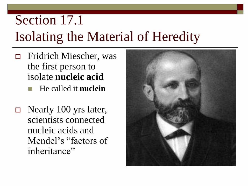
Section 17.1
Isolating the Material of Heredity
Fridrich Miescher, was the first person to isolate nucleic acid
He called it nuclein
Nearly 100 yrs later, scientists connected nucleic acids and Mendel’s “factors of inheritance”
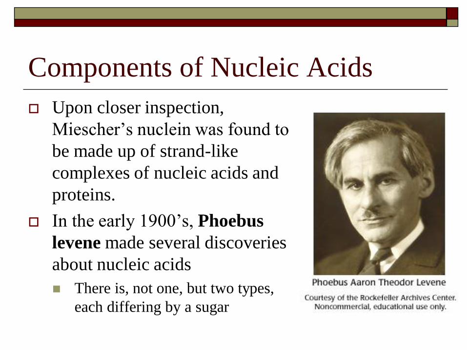
Components of Nucleic Acids
Upon closer inspection,
Miescher’s nuclein was found to
be made up of strand-like
complexes of nucleic acids and
proteins.
In the early 1900’s, Phoebus
levene made several discoveries
about nucleic acids
There is, not one, but two types,
each differing by a sugar
Two Types of Nucleic Acid
1. Ribonucleic Acid Contained a 5-carbon sugar called ribose
Also called RNA
2. Deoxyribonucleic Acid Contained a different 5-carbon sugar called deoxyribose
Also called DNA
Levene determined that these nucleic acids were composed of long chains of individual units called Nucleotides
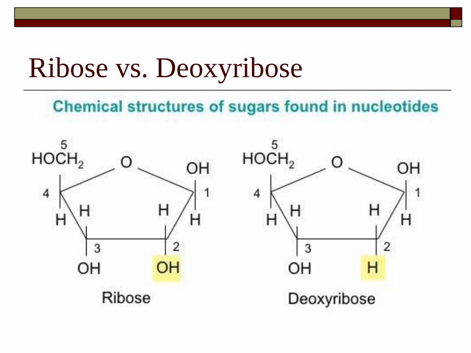
Ribose vs. Deoxyribose
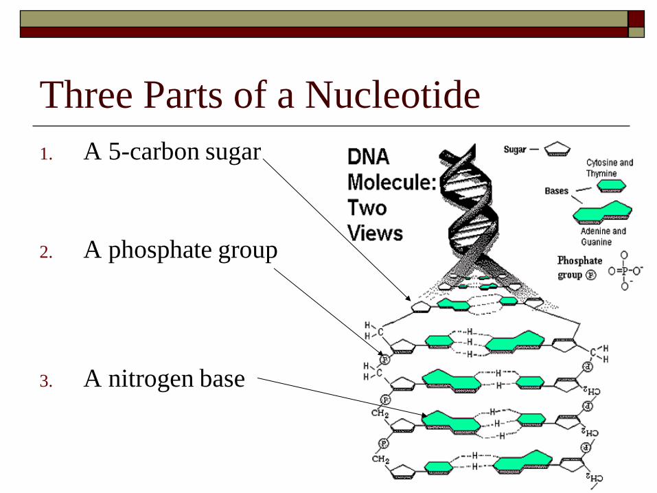
Three Parts of a Nucleotide
1. A 5-carbon sugar
2. A phosphate group
3. A nitrogen base

The Nitrogen Bases
DNA Bases
Thymine (T)
Cytosine (C)
Guanine (G)
Adenine (A)
RNA Bases
Uracil (U)
Replaces thymine
Cytosine
Guanine
Adenine
Sugar – phosphate bonds allow long nucleic acid chains to be formed
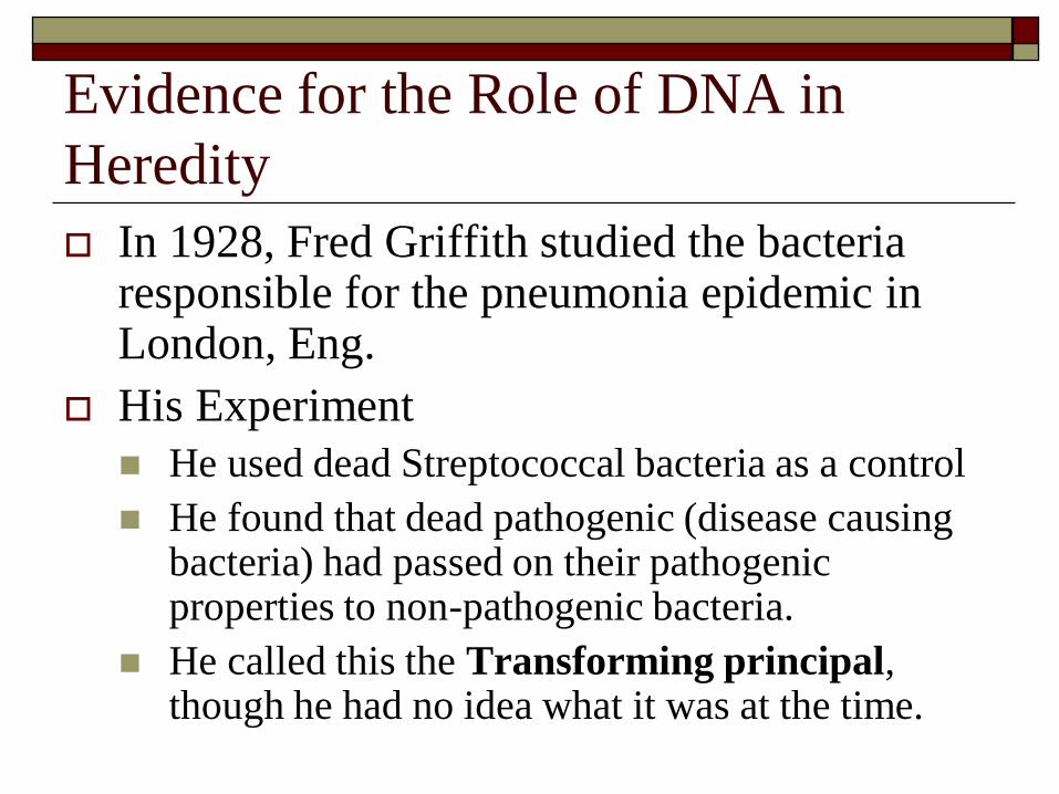
Evidence for the Role of DNA in
Heredity
In 1928, Fred Griffith studied the bacteria responsible for the pneumonia epidemic in London, Eng.
His Experiment
He used dead Streptococcal bacteria as a control
He found that dead pathogenic (disease causing bacteria) had passed on their pathogenic properties to non-pathogenic bacteria.
He called this the Transforming principal, though he had no idea what it was at the time.
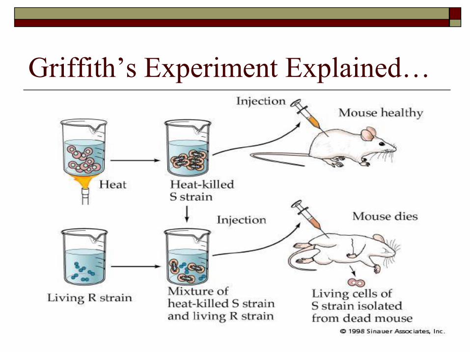
Griffith’s Experiment Explained…
Avery, MacLeod and McCarty
1944 - Took up the challenge to figure out the
transformation principal after Griffith’s death
Their results on pathogenic bacteria:
When treated with a protein-destroying enzyme
transformation still took place
When treated with DNA-destroying enzyme
transformation did NOT occur
When treated with RNA-destroying enzyme
transformation took place
The conclusion: DNA caused the transformation!!
Erwin Chargaff
Late 1940’s – Studied DNA and made the
following discoveries
The 4 nucleotides in DNA are NOT present in
equal amounts, as once thought
Nucleotide composition varies from species to
species
Composition within a species, however, is constant
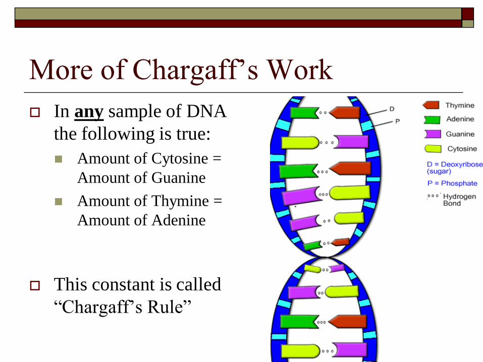
More of Chargaff’s Work
In any sample of DNA
the following is true:
Amount of Cytosine =
Amount of Guanine
Amount of Thymine =
Amount of Adenine
This constant is called
“Chargaff’s Rule”

Hershey and Chase
1952 – Did an experiment using T4 bacteriophage viruses and radioactive labeling techniques
They performed two “Blender” experiments
1. Viruses with radioactively labeled DNA and a normal protein coat
2. Viruses without radioactive DNA, but had a radioactive protein coat
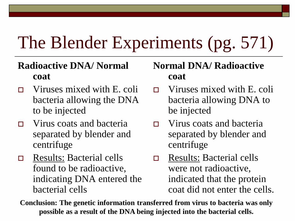
The Blender Experiments (pg. 571)
Radioactive DNA/ Normal coat
Viruses mixed with E. coli bacteria allowing the DNA to be injected
Virus coats and bacteria separated by blender and centrifuge
Results: Bacterial cells found to be radioactive, indicating DNA entered the bacterial cells
Normal DNA/ Radioactive coat
Viruses mixed with E. coli bacteria allowing DNA to be injected
Virus coats and bacteria separated by blender and centrifuge
Results: Bacterial cells were not radioactive, indicated that the protein coat did not enter the cells.
Conclusion: The genetic information transferred from virus to bacteria was only
possible as a result of the DNA being injected into the bacterial cells.
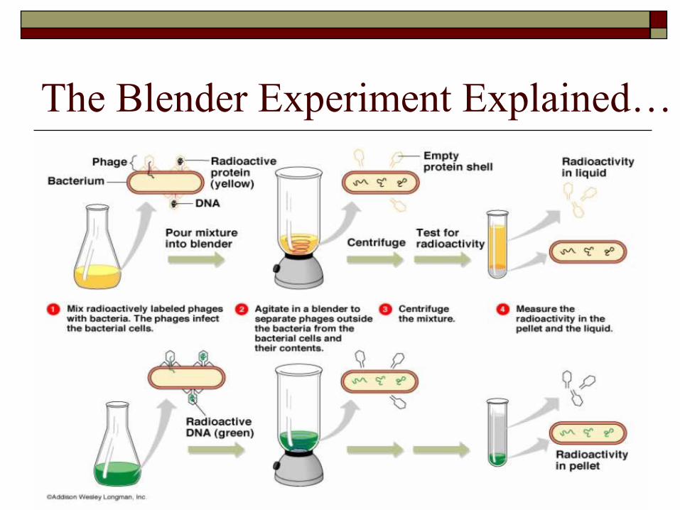
The Blender Experiment Explained…

Suggested Section Review
Read pages 566 – 572 in the textbook
Questions page 572
1, 2, 3, 4, 5,
Be able to label the diagram in question 6
Explain the work of the scientists in question 8
You do not need to hand in these questions, but
they are good review for the exam
Section 17.2
The Structure of Nucleic Acids
By the late 1940’s scientists knew that DNA
was made up of:
A sugar
Phosphate group
Nitrogenous base
What they did not know was how the DNA
strand was arranged
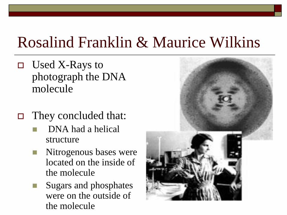
Rosalind Franklin & Maurice Wilkins
Used X-Rays to photograph the DNA molecule
They concluded that:
DNA had a helical structure
Nitrogenous bases were located on the inside of the molecule
Sugars and phosphates were on the outside of the molecule
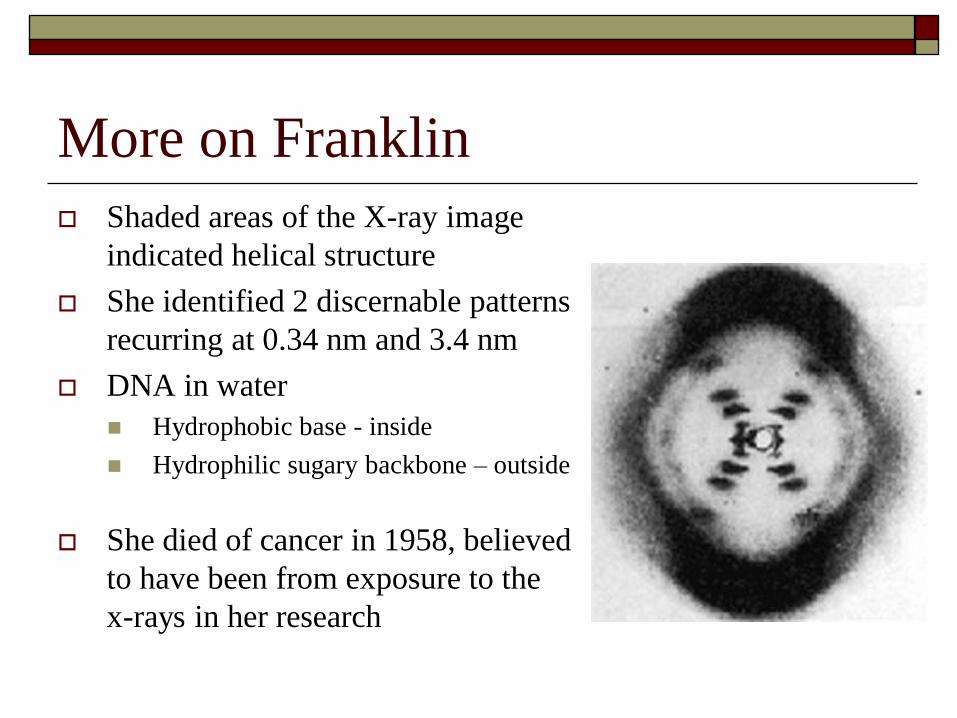
More on Franklin
Shaded areas of the X-ray image
indicated helical structure
She identified 2 discernable patterns
recurring at 0.34 nm and 3.4 nm
DNA in water
Hydrophobic base - inside
Hydrophilic sugary backbone – outside
She died of cancer in 1958, believed
to have been from exposure to the
x-rays in her research
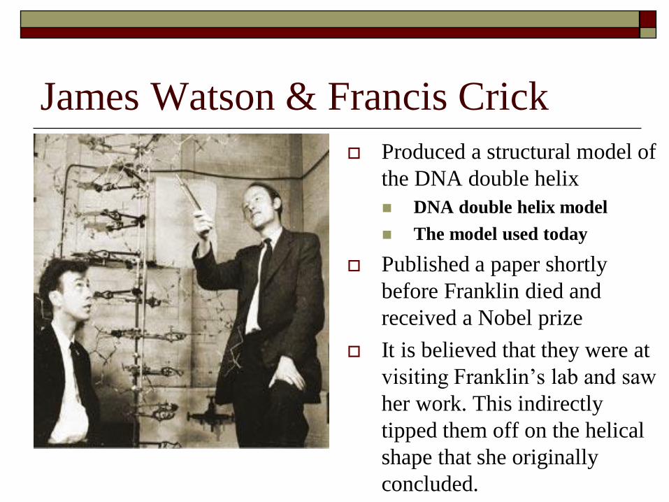
James Watson & Francis Crick
Produced a structural model of
the DNA double helix
DNA double helix model
The model used today
Published a paper shortly
before Franklin died and
received a Nobel prize
It is believed that they were at
visiting Franklin’s lab and saw
her work. This indirectly
tipped them off on the helical
shape that she originally
concluded.
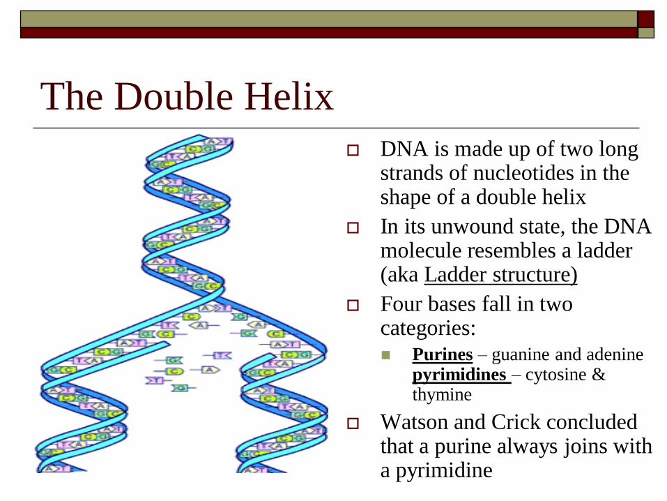
The Double Helix
DNA is made up of two long strands of nucleotides in the shape of a double helix
In its unwound state, the DNA molecule resembles a ladder (aka Ladder structure)
Four bases fall in two categories: Purines – guanine and adenine
pyrimidines – cytosine & thymine
Watson and Crick concluded that a purine always joins with a pyrimidine
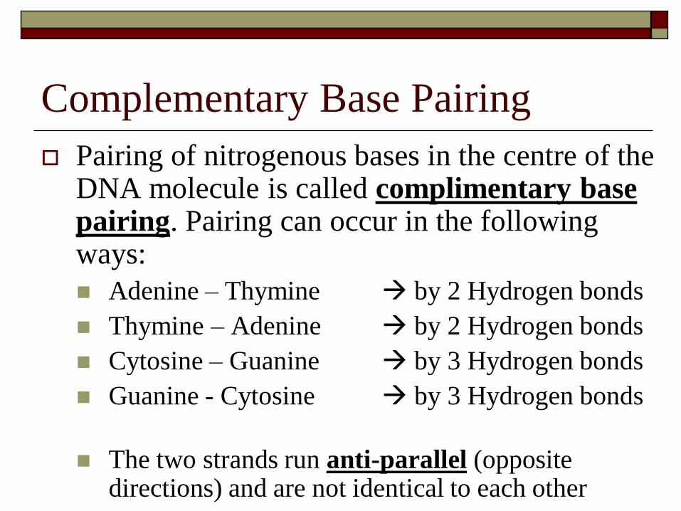
Complementary Base Pairing
Pairing of nitrogenous bases in the centre of the DNA molecule is called complimentary base pairing. Pairing can occur in the following ways:
Adenine – Thymine by 2 Hydrogen bonds
Thymine – Adenine by 2 Hydrogen bonds
Cytosine – Guanine by 3 Hydrogen bonds
Guanine - Cytosine by 3 Hydrogen bonds
The two strands run anti-parallel (opposite directions) and are not identical to each other

Anti-parallelism

RNA
Three differnces from DNA
1. Sugar is a ribose, while DNA has a deoxyribose
2. RNA has uracil instead of thymine as in DNA
3. RNA is only a single strand
Organization of Genetic Material
Scientists examine cells to determine how DNA is organized within a cell
There are two main types of cells:
Prokaryotes (bacteria)
Eukaryotes (everything else)
Structure of the DNA varies in each type of cell
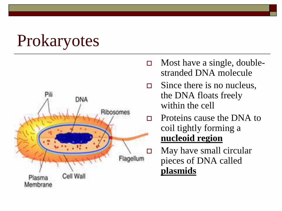
Prokaryotes
Most have a single, double-stranded DNA molecule
Since there is no nucleus, the DNA floats freely within the cell
Proteins cause the DNA to coil tightly forming a nucleoid region
May have small circular pieces of DNA called plasmids
Eukaryotes
All cells have double-stranded DNA
DNA is arranged into chromosomes within the nucleus
Each chromosome contains a double stranded DNA molecule and a protein called a histone
A typical chromosome contains:
60% Protein
35% DNA
5% RNA
Chromosomes are joined together to form a long, fibrous material called Chromatin
Genes and the Genome
Studies have shown that there
are patterns in how heredity
information is organized at the
molecular level that are shared
by different organisms. They
are:
How individual genes are
organized
How the individual’s genome is
organized
Genes
A gene is a subunit of DNA
Chromosomes in a cell carry genes
Different species have their own unique
arrangement of genes
Though many genes are common between species
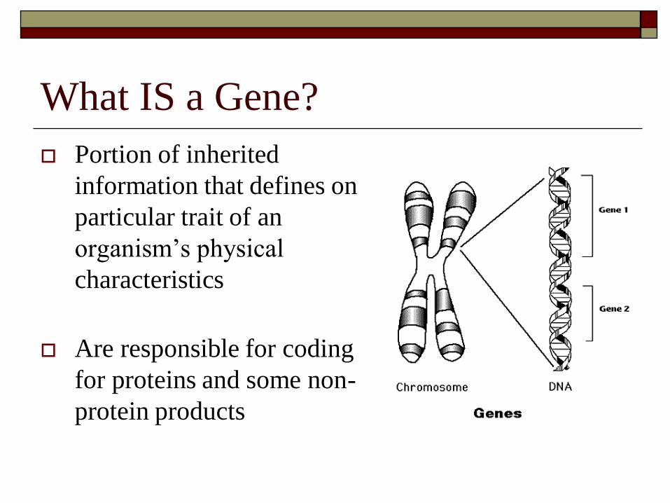
What IS a Gene?
Portion of inherited
information that defines one
particular trait of an
organism’s physical
characteristics
Are responsible for coding
for proteins and some non-
protein products
DNA Humour
Arrangement of the Genome
Each chromosome has its own unique
arrangement of genes
Gene density varies among chromosomes
Ex. Ch. # 4 has about 200 genes, while Ch. # 14 has
about 1450 genes
Different organisms have different numbers of
genes
An ameoba has about 7000 genes while humans
have about 35,000 genes
Eukaryote Genes
Each genes if made up of two different regions
Exons Coding or expressed regions of a gene
Introns Non-coding nucleotide sequences
Can make up over 50% of the length of a gene
More complex organisms tend to have more introns, while simple organisms like bacteria or yeasts have none or few introns
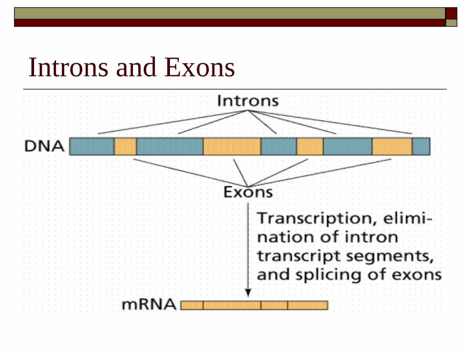
Introns and Exons
Suggested Section Review
Read Pages 573 – 581 in textbook
Review DNA extraction Lab
We will do this before the week is over
Questions on Page 581
1, 2, 3, 4, 5, 9, 10, 11, 14
These questions are not currently due, but are recommended in your exam review
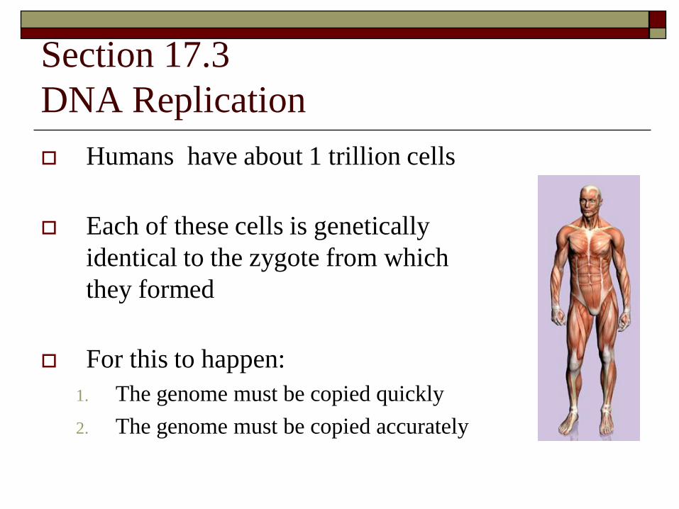
Section 17.3
DNA Replication
Humans have about 1 trillion cells
Each of these cells is genetically
identical to the zygote from which
they formed
For this to happen:
1. The genome must be copied quickly
2. The genome must be copied accurately

The Replication Process
DNA replication is a process from which two
molecules of DNA are made from one
Called a semi-conservative model
Meaning each of the two new DNA molecules
contains one original (parent) strand and one new
strand
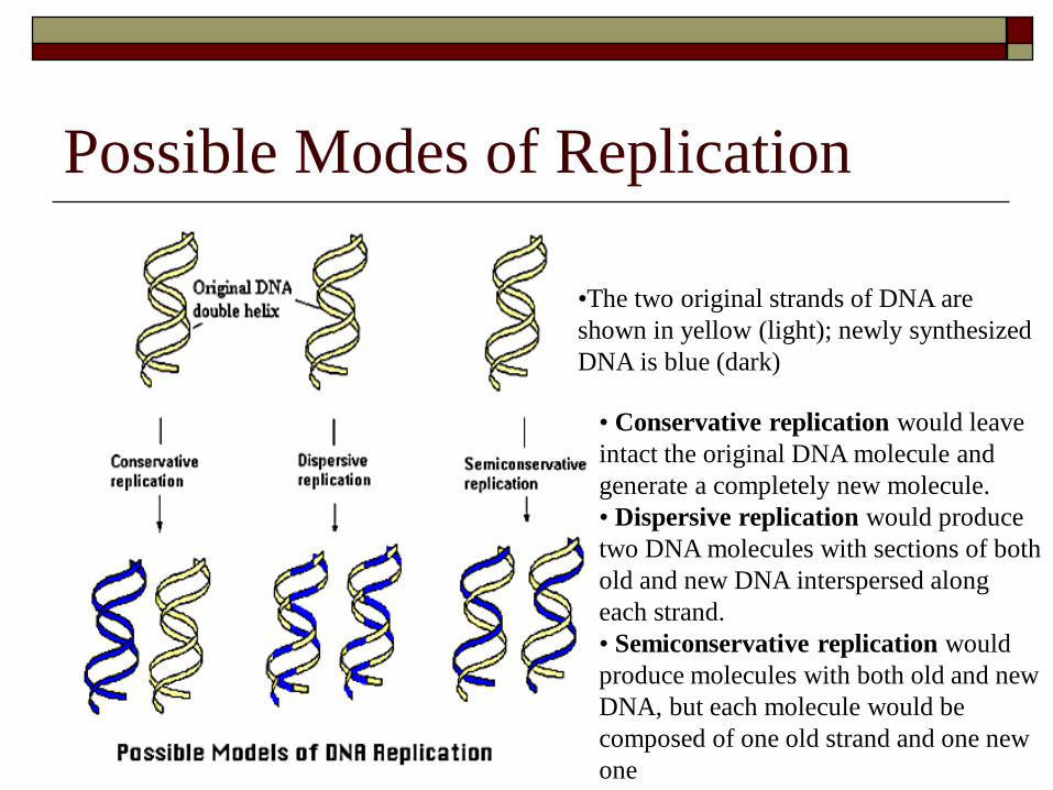
Possible Modes of Replication
•The two original strands of DNA are
shown in yellow (light); newly synthesized
DNA is blue (dark)
• Conservative replication would leave
intact the original DNA molecule and
generate a completely new molecule.
• Dispersive replication would produce
two DNA molecules with sections of both
old and new DNA interspersed along
each strand.
• Semiconservative replication would
produce molecules with both old and new
DNA, but each molecule would be
composed of one old strand and one new
one

Three Stages of the Replication
Process
1. Initiation
2. Elongation
3. Termination
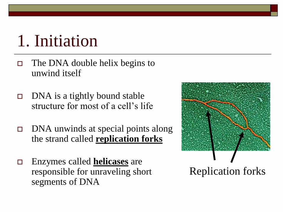
1. Initiation
The DNA double helix begins to unwind itself
DNA is a tightly bound stable structure for most of a cell’s life
DNA unwinds at special points along the strand called replication forks
Enzymes called helicases are responsible for unraveling short segments of DNA
Replication forks
2. Elongation
Assembly of two new DNA strands begins
An enzyme called DNA polymerase helps to
attach new nucleotides to the DNA strand
Newly replicated DNA can be found in short
segments called Okazaki fragments ranging
from 1 to 2 thousand nucleotides in lenth
Still Elongating
Replication occurs in the 5’ to 3’ direction of one DNA strand while it occurs in the 3’ to 5’ direction on the other strand. The enzyme DNA primase begins this process
Leading strand - The strand replicating in the 5’ to 3’ direction
Laggin strand – The strand replicating in the 3’ to 5’ direction
Okazaki fragments are joined together by an enzyme called DNA ligase
3. Termination
The stage when the new DNA molecules
reform into helices or double helices
Daughter DNA strands rewind forming their
stable helical structure
Each new daughter DNA molecule is slightly
shorter than its parent
Chromosomes lose about 100 base pairs with
each replication
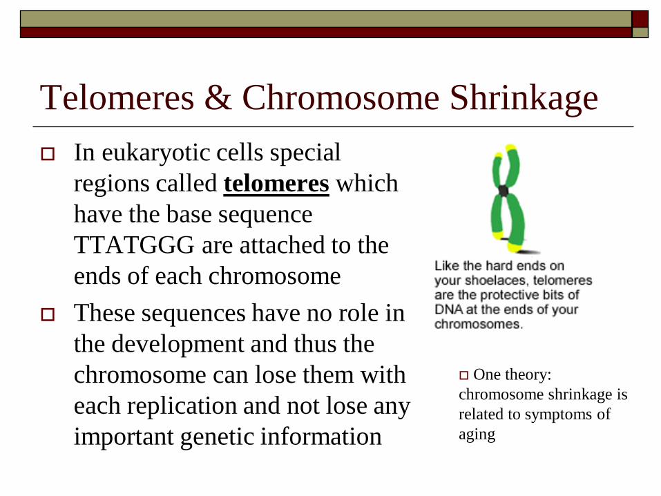
Telomeres & Chromosome Shrinkage
In eukaryotic cells special
regions called telomeres which
have the base sequence
TTATGGG are attached to the
ends of each chromosome
These sequences have no role in
the development and thus the
chromosome can lose them with
each replication and not lose any
important genetic information
One theory:
chromosome shrinkage is
related to symptoms of
aging

Err is to human… and DNA replication
Though we would like to believe that DNA
replication is an orderly step by step process,
this is usually not the case. Just as we make
mistakes, so can the replication process
Wrong bases may be inserted into the new DNA
Nucleotide bases may be damaged (ie. By
radiation)
When this happens, mutations or other serious
problems can occur in the DNA molecule
Proofreading and Correction
To prevent errors from occurring, the enzyme DNA
polymerase is able to check to see whether bases are
actually bonding together by hydrogen bonding
No H-bonding means there is a base mismatch
The incorrect base is replace with the correct one
DNA replication involves dozens different enzymes
and other proteins working together as a replication
machine to get the job done correctly and virtually
error-free
DNA Repair Animation
Suggested Section Review
Read Pages 582 – 588
You MUST know the enzymes involved and their
functions
Page 587 – Table 17.1
You must be able to explain the replication
process and draw basic diagrams on a test
Questions on page 588
1, 2, 3, 4, 9,
Section 17.4
Protein Synthesis & Gene Expression
DNA stores information in the form of a code
that we call the genetic code
Genetic code is based on the order of the base
pairs that make up the DNA molecule
The sequence of nucleotide determines the
sequence of amino acids within a protein
Genetic Code
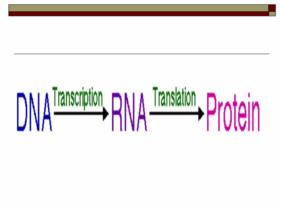
Transfer of genetic information from DNA to
protein is called genetic expression which
occurs in two stages:
1. Transcription
Information is copied from DNA onto an RNA
molecule (inside the nucleus of the cell)
2. Translation
RNA moves from the nucleus to the cytoplasm where
it helps to make a polypeptide (protein)
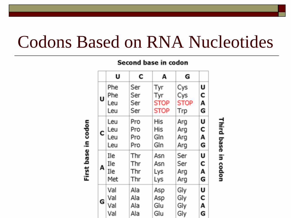
Codons Based on RNA Nucleotides

The Genetic Code
Using combinations of three nucleotides, the DNA
molecule creates code words that represent the 20
amino acids (Pg. 590 table 17.2)
Each set of three bases is called a codon
Some amino acids (AA) are coded for by more than one
codon, while others, only by one
Each set of 3 amino acids is called a reading frame
Codons are represented by the RNA base sequences
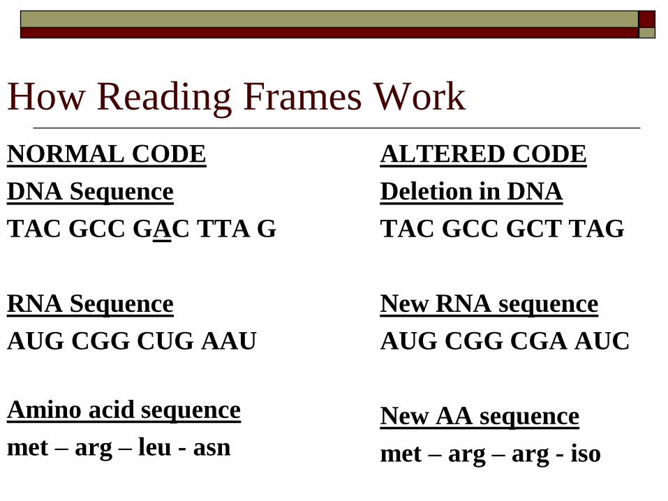
How Reading Frames Work
NORMAL CODE
DNA Sequence
TAC GCC GAC TTA G
RNA Sequence
AUG CGG CUG AAU
Amino acid sequence
met – arg – leu - asn
ALTERED CODE
Deletion in DNA
TAC GCC GCT TAG
New RNA sequence
AUG CGG CGA AUC
New AA sequence
met – arg – arg - iso
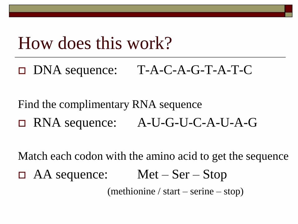
How does this work?
DNA sequence: T-A-C-A-G-T-A-T-C
Find the complimentary RNA sequence
RNA sequence: A-U-G-U-C-A-U-A-G
Match each codon with the amino acid to get the sequence
AA sequence: Met – Ser – Stop
(methionine / start – serine – stop)

3 Characteristics of the Code
1. Redundancy
More than one codon can code for the same amino acid – lots of repetition
2. Continuous
Code reads as a series of 3-letter codons without spaces, punctuation or overlap
3. Universal
Code is virtually the same in all organisms making is possible to transfer information
Transcription I
Process by which a small portion of the DNA is copied onto a special type of RNA called messenger RNA or mRNA
mRNA carries information from the nucleus of a cell to the cytoplasm to become a protein
RNA polymerase is the catalyst for the production of the RNA molecule
Transcription II
DNA has two strands
Sense strand and Anti-sense strand
ONLY the sense strand is transcribed into RNA
RNA polymerase opens up the DNA double helix allowing the mRNA to be formed from exposed nucleotide bases
Transcription continues along the DNA until a stop codon is reached. The RNA and polymerase separate and a special nucleotide sequence is added to the 3’ and 5’ ends

Transcription Illustrated
Sense strand
Anti-sense strand
Transcription Animation
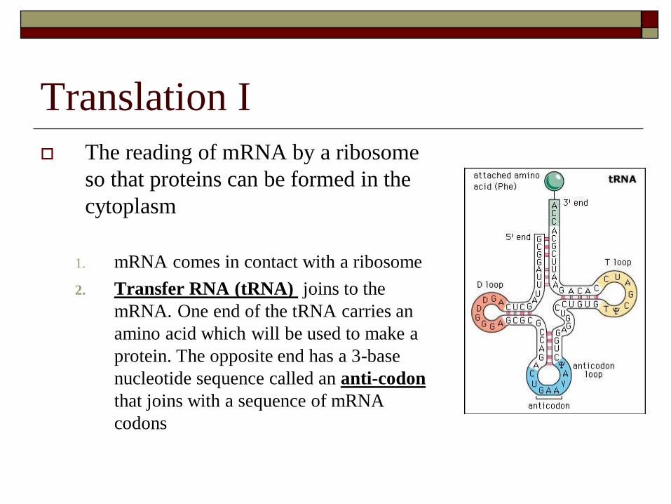
Translation I
The reading of mRNA by a ribosome
so that proteins can be formed in the
cytoplasm
1. mRNA comes in contact with a ribosome
2. Transfer RNA (tRNA) joins to the
mRNA. One end of the tRNA carries an
amino acid which will be used to make a
protein. The opposite end has a 3-base
nucleotide sequence called an anti-codon
that joins with a sequence of mRNA
codons
Translation II - Animation
After the first tRNA binds to the mRNA a second will join next to it, adding its amino acid to the chain. When the third tRNA binds the first tRNA molecule is “bumped” out of the ribosome. With each new tRNA a new amino acid is added to the polypeptide chain.
The cycle of amino acids linking together is repeated until a “stop” codon (UAA, UAG or UGA) is reached. Once this tRNA is read, the amino acid is released from the ribosome and the protein is formed
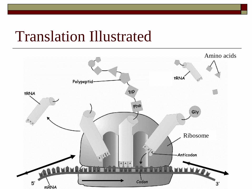
Translation Illustrated Amino acids
Ribosome

Regulating Gene Expression
Every living cell has the ability to respond to
it environment by changing the kinds and
amounts of polypeptide (proteins) it produces
By controlling this process, the cell can regulate
gene expression
There are a number of factors that control the
rate of transcription and translation
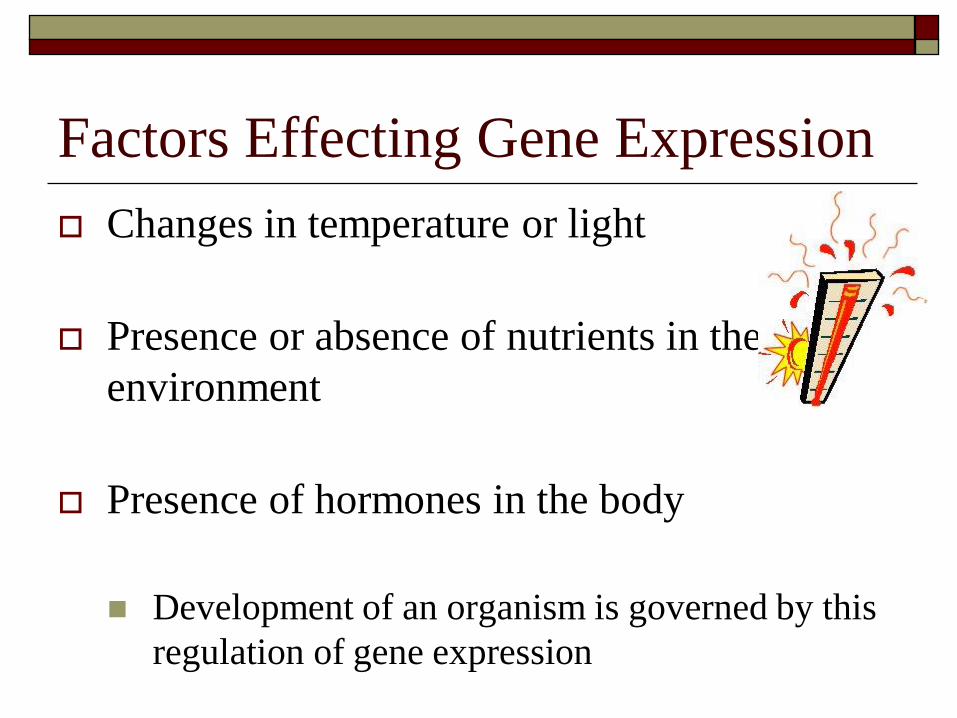
Factors Effecting Gene Expression
Changes in temperature or light
Presence or absence of nutrients in the
environment
Presence of hormones in the body
Development of an organism is governed by this
regulation of gene expression
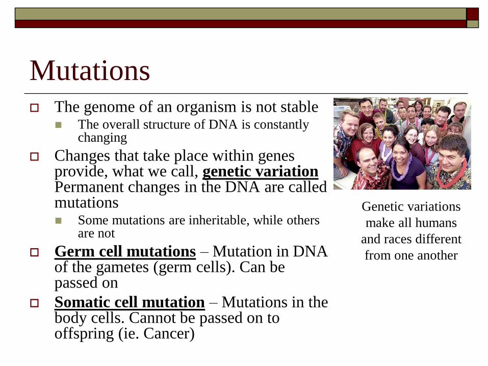
Mutations The genome of an organism is not stable
The overall structure of DNA is constantly changing
Changes that take place within genes provide, what we call, genetic variation Permanent changes in the DNA are called mutations Some mutations are inheritable, while others
are not
Germ cell mutations – Mutation in DNA of the gametes (germ cells). Can be passed on
Somatic cell mutation – Mutations in the body cells. Cannot be passed on to offspring (ie. Cancer)
Genetic variations
make all humans
and races different
from one another
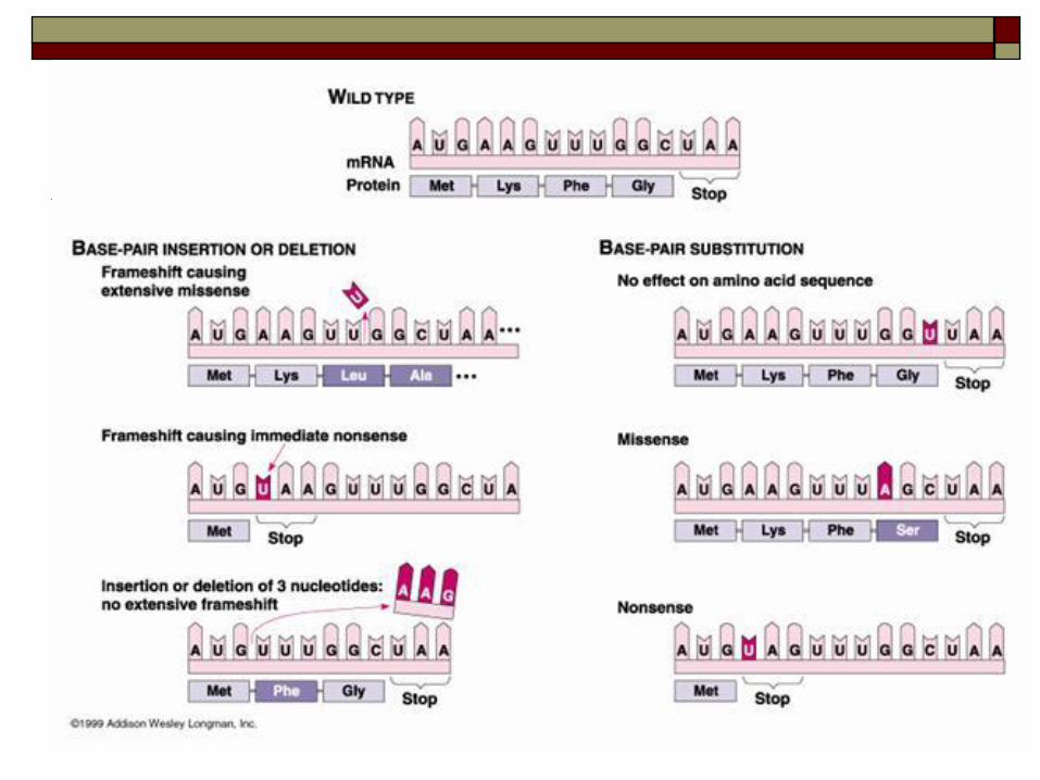
Types of Mutations
Point mutations – small changes in the nucleotide sequence of genes. Maybe be one nucleotide replacing another, deletion or insertion
Silent mutations – Has no negative effect on the cells in which they occur. May be in exons or simply in “unused” DNA
Mis-sense mutations – Cause slight alteration of a protein. May be beneficial or harmful depending on the protein(s) affected
Nonsense mutations – Make a gene unable to code for a functional protein. Usually caused by changes to the start/ stop codons
Nucleotide Insertions/ Deletions
One or two nucleotides in a sequence of
codons can produce a frameshift mutation
This is when a nucleotide insertion or deletion
causes and entire frame of a gene to be altered
See page 597 – fig. 17.33 for an example

Chromosomal Mutations
Involve the rearrangement of genetic material which affects genes
May involve:
Exchange of portions of chromosomes between sister chromatids or chromosomes
Loss of chromosome pieces
Duplication of chromosome segments
Barbara McClintock found jumping genes called transposons that are short strands of DNA capable of moving from one location to another. (pg 597-598)
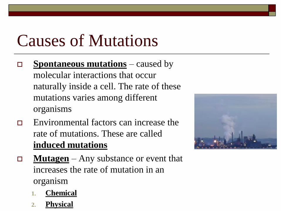
Causes of Mutations
Spontaneous mutations – caused by
molecular interactions that occur
naturally inside a cell. The rate of these
mutations varies among different
organisms
Environmental factors can increase the
rate of mutations. These are called
induced mutations
Mutagen – Any substance or event that
increases the rate of mutation in an
organism
1. Chemical
2. Physical
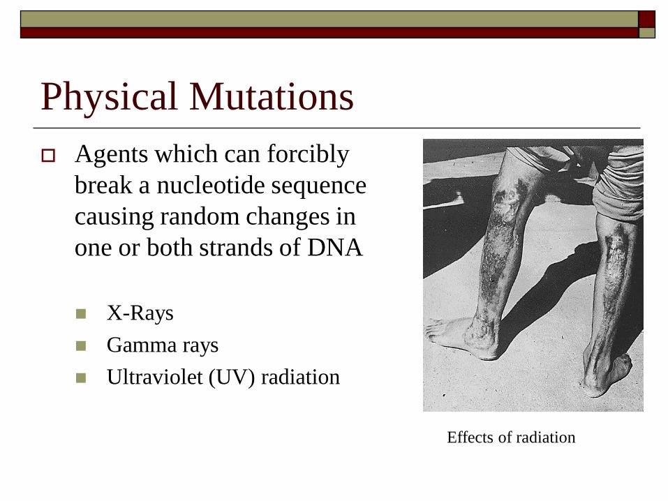
Physical Mutations
Agents which can forcibly
break a nucleotide sequence
causing random changes in
one or both strands of DNA
X-Rays
Gamma rays
Ultraviolet (UV) radiation
Effects of radiation
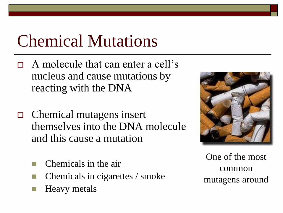
Chemical Mutations
A molecule that can enter a cell’s nucleus and cause mutations by reacting with the DNA
Chemical mutagens insert themselves into the DNA molecule and this cause a mutation
Chemicals in the air
Chemicals in cigarettes / smoke
Heavy metals
One of the most
common
mutagens around
Mutations: General Information
Each organisms genes undergoes 1000’s of mutations during a lifetime
Most mutations are repaired by the cell’s own enzymes
Some mutations cannot be repaired, and these build up over the lifetime of the cell leading to cellular damage
Cancer is an example of a disorder caused by accumulated mutations – cells begin to divide uncontrollably
Any mutagen which can cause cancer is called a carcinogen
Suggested Section Review
Read Pages 589 – 600
Be able to draw basic diagrams showing
transcription & translation
Know the types of mutations and examples
Be able to transcribe DNA/RNA/Amino Acid
sequence
Questions page
1, 2, 3, 4, 5,
Chapter Overview Assignment
Questions Page 601 – 602
1 – 9, 11, 13, 15, 16, 17, 18, 20, 21 Due Tuesday April 3, 2007
Test scheduled for:
THURSDAY APRIL 5, 2007
All Animations will be available on the Bio 3201 website for download