Embed Size (px)
Citation preview
Molecular Interactions of Type III Secretion System Transcriptional
Regulators in Pseudomonas aeruginosa: ExsA and ExsD
Robert Cory Bernhards
Dissertation submitted to the faculty of the Virginia Polytechnic Institute
and State University in partial fulfillment of the requirements for the degree
of
Doctor of Philosophy
In
Biological Sciences
Florian D. Schubot, Chair
Marcy Hernick
Stephen B. Melville
Ann M. Stevens
May 2, 2013
Blacksburg, Virginia
Keywords: Pseudomonas aeruginosa, ExsA, ExsD, type III secretion system,
transcriptional regulation
Molecular Interactions of Type III Secretion System Transcriptional
Regulators in Pseudomonas aeruginosa: ExsA and ExsD
Robert Cory Bernhards
ABSTRACT
The opportunistic pathogen Pseudomonas aeruginosa ranks among the leading
causes of nosocomial infections. The type III secretion system (T3SS) aids acute P.
aeruginosa infections by injecting potent cytotoxins (effectors) into host cells to suppress
the host's innate immune response. Expression of all T3SS-related genes is strictly
dependent upon the transcription factor ExsA. Consequently, ExsA and the biological
processes that regulate ExsA function are of great biomedical interest. The ExsA-ExsC-
ExsD-ExsE signaling cascade ties host cell contact to the up-regulation of T3SS gene
expression. Prior to T3SS induction, the antiactivator protein ExsD binds to ExsA and
blocks ExsA-dependent transcription by interfering with ExsA dimerization and promoter
interactions. Upon host cell contact, ExsD is sequestered by the T3SS chaperone ExsC,
resulting in the release of ExsA and an up-regulation of the T3SS.
ExsA is an AraC/XylS-type transcriptional regulator and belongs to a subfamily
of activators that regulate the T3SS in a variety of Gram-negative pathogens. These
regulators are characteristically difficult to purify due to the low solubility of their C-
terminal DNA binding domains. A new method for purifying ExsA was developed and
produced ExsA with improved solubility. The interaction of ExsA and its PexsD promoter
was examined using fluorescence anisotropy. An in vitro transcription assay was
developed and it was determined that ExsA is sufficient to activate T3SS transcription.
Next, the ExsD–ExsA inhibitory mechanism was examined. It was demonstrated
for the first time that ExsD alone is sufficient to inhibit ExsA-dependent transcription in
iii
vitro without the aid of any other cellular factors. More significantly and contrary to
previously published results, it was discovered that independently folded ExsD and ExsA
are capable of interacting, but only at 37 C and not at 30 C. Guided by the crystal
structure of ExsD, a monomeric variant of the protein was designed to demonstrate that
ExsD trimerization prevents ExsD from inhibiting ExsA-dependent transcription at 30 C.
To further elucidate the ExsD-ExsA inhibitory mechanism, the ExsD-ExsA
interface was examined. ExsD variants were generated and used to determine which
region of ExsD interacts with ExsA. Interestingly, ExsD was also found to bind DNA,
although it is unclear whether or not this plays a role in ExsA inhibition. Fully
understanding the mechanism by which ExsD inhibits ExsA may enable the development
of drugs that target ExsA in order to shut down the T3SS, thereby eliminating P.
aeruginosa infection.

iv
ACKNOWLEDGEMENTS
First of all, I would like thank my advisor Dr. Florian Schubot for giving me the
opportunity to be a part of his lab and for all of his guidance throughout the years. I
would also like to thank my committee members, Dr. Ann Stevens, Dr. Stephen Melville,
and Dr. Marcy Hernick for all of their advice and support during my graduate studies. Dr.
David Popham and Dr. Birgit Scharf have also been very helpful during my time at
Virginia Tech.
I would like to thank all of my fellow lab members, Dr. Nancy Vogelaar, Jing
Xing, Xiao Yi, Manisha Shrestha, Jordan Mancl, Shannon Esher, Doug Ryan, Sarah
Williams, Amber Sholl, Lisa Swallow, Peter Grinc, and Rika Judd for all of their help. I
wish you all the best in your future careers. I would particularly like to recognize David
Mishkel, who was the first undergraduate that I mentored as well as my friend. He was a
brilliant young scientist and his memory will never be forgotten.
Thank you to all of the people who I’ve met during grad school for your advice
and friendship, especially Dr. Trevor Glaros, Dr. Wes Black, Dr. Jared Heffron, Katie
Rodgers, Jared Geissinger, Jake Kocher, Sean Mury, Yan Chen, Hualan Liu, Regina
Wallace, Revathy Ramachandran, Alison Kernell, Hardik Zatakia, and Ben Webb. A
special thanks to all of those who played softball with me. I had a blast playing with you
guys.
To my family, you’ve been an incredible support system for me throughout my
entire life, and I am forever grateful for that. Mom and Dad, you’ve given me the
opportunity to pursue my dreams. To my sister Kirsten, you have always been my best
friend and your level of kindness is truly unique. Most importantly, to my wife Casey, I

v
couldn’t have succeeded without your support. You bring out the best in me and always
push me to improve. It is such a blessing to have someone who I can talk to at any time
about my research. I look forward to seeing where our careers take us.
vi
ATTRIBUTIONS
Several colleagues aided in the research in this dissertation. A brief description of their
contributions is included here.
Chapter Two: Characterization of ExsA, the main transcriptional regulator of the type
III secretion system in Pseudomonas aeruginosa
Nancy Vogelaar, Ph.D. (Department of Biological Sciences) is a former post doctorate in
the Schubot Lab at Virginia Tech who helped develop the ExsA purification protocol as
well as the in vitro transcription assay.
Dr. Marcy Hernick, Ph.D. (Department of Biochemistry) is an assistant professor at
Virginia Tech. Dr. Hernick advised on the fluorescence anisotropy experiments.
Chapter Three: Self-trimerization of ExsD limits inhibition of the Pseudomonas
aeruginosa transcriptional activator ExsA in vitro
Appendix: Extension of Chapter Three
Chapter Three was published in FEBS Journal in 2013.
Anne Marsden (Department of Microbiology) is a graduate student in the lab of Dr.
Timothy Yahr at the University of Iowa. Ms. Marsden was co-author on this paper and
performed the bacterial monohybrid experiment.
Shannon Esher (Department of Biological Sciences) is a former undergraduate student in
the Schubot Lab. Ms. Esher was co-author on this paper and constructed the ExsDM59R
variant expression plasmid.
Timothy Yahr, Ph.D. (Department of Microbiology) is a currently an associate professor
at the University of Iowa. Dr. Yahr was co-author on this paper, principal investigator
for one of the grants supporting the research, and contributed editorial comments.
Chapter Four: Mapping the ExsD–ExsA interface and further elucidation of the type III
secretion system inhibitory mechanism in Pseudomonas aeruginosa
Yi Xiao (Department of Biological Sciences) is a former graduate student in the Schubot
Lab. Yi constructed, expressed, and purified the ExsDΔC-C variant.
Shannon Esher performed the differential scanning fluorimetry experiments.
Anne Marsden performed the electophoretic mobility shift assay.
vii
TABLE OF CONTENTS
ABSTRACT……………………………………………………………………………...ii
ACKNOWLEDGEMENTS ……………………………………………………………iv
ATTRIBUTIONS…………………………………………………………………….....vi
LIST OF FIGURES……………………………………………………………………..x
LIST OF TABLES………………………………………………………………………xi
CHAPTER ONE: Literature Review…………………………………………………...1
Pseudomonas aeruginosa…………………………………………………………2
P. aeruginosa infection…………………………………………………………...2
Chronic infection………………………………………………………………….3
Acute infection……………………………………………………………………3
Antibiotic resistance………………………………………………………………4
Treatment………………………………………………………………………….5
Type III secretion system (T3SS)…………………………………………………5
T3SS effector proteins…………………………………………………………….5
ExoS………………………………………………………………………………6
ExoT………………………………………………………………………………7
ExoU……………………………………………………………………………...7
ExoY……………………………………………………………………………...8
T3SS chaperones………………………………………………………………….8
T3SS needle complex……………………………………………………………..8
T3SS translocation apparatus……………………………………………………..9
T3SS regulation…………………………………………………………………...9
ExsACDE regulatory cascade…………………………………………………....10
ExsA……………………………………………………………………………...11
ExsA homologs…………………………………………………………………..12
ExsA promoters………………………………………………………………….12
ExsA–RNAP interactions………………………………………………………..14
ExsD……………………………………………………………………………..15
ExsC……………………………………………………………………………..15
ExsE……………………………………………………………………………..16
Medical importance……………………………………………………………...17
Research objectives……………………………………………………………...17
CHAPTER TWO: Characterization of ExsA, the main transcriptional regulator
of the type III secretion system in Pseudomonas aeruginosa…..25
Abstract………………………………………………………………………….26
Introduction……………………………………………………………………...27
Results …………………………………………………………………………...28
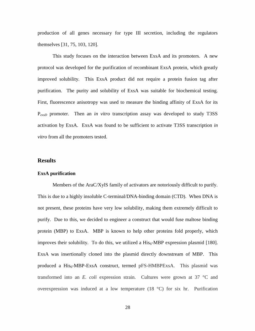
ExsA purification………………………………………………………...28
viii
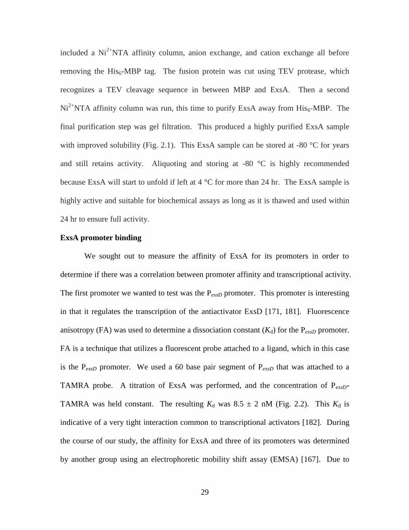
ExsA promoter binding…………………………………………………..29
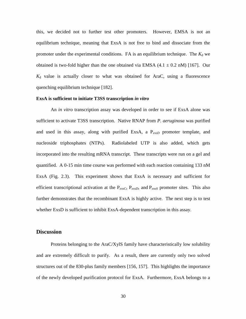
ExsA is sufficient to initiate T3SS transcription in vitro………………...30
Discussion………………………………………………………………………..30
Materials and Methods…………………………………………………………..32
ExsA expression and purification………………………………………..32
Fluorescence anisotropy…………………………………………………34
RNA polymerase purification and specific activity determination……...35
In vitro transcription assay………………………………………………36
Acknowledgements……………………………………………………………...38
CHAPTER THREE: Self-trimerization of ExsD limits inhibition of the
Pseudomonas aeruginosa transcriptional activator ExsA
in vitro…………………………………………………………..42
Abstract………………………………………………………………………….43
Introduction……………………………………………………………………...44
Results …………………………………………………………………………...46
ExsA-mediated in vitro transcription is not inhibited by wild-type
ExsD at 30 C, but is strongly inhibited at 37 C………………………..46
A single mutation generating the ExsDM59R
variant disrupts ExsD
trimer formation………………………………………………………….48
Monomeric ExsDM59R
efficiently inhibits ExsA-dependent
transcription in vitro at 30 C……………………………………………50
ExsDM59R
disrupts ExsA dimerization, but does not interfere with
ExsA promoter binding………………………………………………….50
Discussion……………………………………………………………………….51
Materials and Methods…………………………………………………………..54
Recombinant protein expression and purification……………………….54
RNA polymerase purification and specific activity determination……...57
Site-directed mutagenesis………………………………………………..58
ExsA-dependent in vitro transcription assays……………………………58
Analytical size exclusion chromatography………………………………60
Circular dichroism (CD) measurements…………………………………60
Differential scanning fluorimetry (DSF)………………………………...61
LexA monohybrid assay………………………………………………....61
Acknowledgements……………………………………………………………...62
CHAPTER FOUR: Mapping the ExsD–ExsA interface and further elucidation
of the type III secretion system inhibitory mechanism in
Pseudomonas aeruginosa………………………………………...72
Abstract…………………………………………………………………………..73
Introduction……………………………………………………………………...74
Results …………………………………………………………………………...75
ExsD is a DNA-binding protein………………………………………….75
The coiled-coil region of ExsD is not important for ExsA binding……...77
The amino terminus of ExsD is important for ExsA binding……………78
Discussion………………………………………………………………………..79

ix
Materials and Methods…………………………………………………………..81
Recombinant protein expression and purification……………………….81
RNA polymerase purification and specific activity determination……...84
Site-directed mutagenesis………………………………………………..85
Differential scanning fluorimetry (DSF)………………………………...86
Limited proteolysis to obtain ExsDΔ20…………………………………86
In vitro transcription assay………………………………………………87
Acknowledgements……………………………………………………………...88
CHAPTER FIVE: Overall Conclusions………………………………………………99
APPENDIX: Extension of Chapter Three: Self-trimerization of ExsD limits
inhibition of the Pseudomonas aeruginosa transcriptional
activator ExsA in vitro………………………………………………105
Abstract………………………………………………………………………...106
Introduction…………………………………………………………………….107
Results………………………………………………………………………….108
Inhibition by ExsD is based on a temperature gradient………………...108
Effect of ExsDM59R
on ExsA-dependent transcription at 37 °C………...108
Effect of ExsDM59R
in vivo……………………………………………...109
Discussion………………………………………………………………………109
Materials and Methods………………………………………………………....111
In vitro transcription assay……………………………………………...111
β-galactosidase assay…………………………………………………...112
REFERENCES……………………………………………………………………….117

x
LIST OF FIGURES
CHAPTER ONE
Figure 1.1. Structural proteins of the T3SS needle apparatus…………………………..20
Figure 1.2. T3SS regulatory network…………………………………………………...21
Figure 1.3. Regulatory cascade of ExsA-dependent activation of the T3SS…………....22
Figure 1.4. Regulatory and secretory portion of T3SS regulon…………………………23
Figure 1.5. ExsD structure………………………………………………………………24
CHAPTER TWO Figure 2.1. ExsA purification…………………………………………………………....39
Figure 2.2. ExsA promoter affinity……………………………………………………...40
Figure 2.3. T3SS transcriptional activation by ExsA…………………………………...41
CHAPTER THREE Figure 3.1. Temperature-dependent regulation of ExsA by ExsD……………………...64
Figure 3.2. Characterization of the ExsDM59R
variant…………………………………..65
Figure 3.3. Effect of ExsDM59R
on ExsA-dependent transcription………………….......66
Figure 3.4. ExsDM59R
disrupts ExsA dimerization………………………………………67
Figure S3.1. SDS-PAGE images for the purified proteins used in the different
assays……………………………………………………………………...68
Figure S3.2. In vitro transcription assay time-course at 37 °C…………………………69
Figure S3.3. Temperature dependence of ExsA-dependent transcription, both in the
absence and presence of 50 μM ExsD, at 30 °C versus 37 °C……………70
Figure S3.4. Neither wild-type ExsD nor ExsDM59R
affects ExsA-independent
transcription……………………………………………………………….71
CHAPTER FOUR Figure 4.1. ExsD binds to DNA as a monomer…………………………………………89
Figure 4.2. Does ExsDM59R
disrupt ExsA-binding activity?……………………………90
Figure 4.3. ExsD variant purification…………………………………………………...91
Figure 4.4. Effect of ExsDΔC-C on ExsA-dependent transcription at 30 and
37 °C………………………………………………………………………...92
Figure 4.5. Effect of ExsDΔ20 on ExsA dependent transcription at 37 °C……………..93
Figure 4.6. ExsDΔ20 has no effect on ExsA-independent transcription………………..94
Figure 4.7. Effect of ExsD1-46 on ExsA dependent transcription at 37 °C…………….95
Figure 4.8. ExsD1-46 has no effect on ExsA-independent transcription……………….96
Figure 4.9. Effect of ExsD variants on ExsA dependent transcription at 37 °C………..97
APPENDIX
Figure A.1. Effect of ExsD on ExsA-dependent transcription at 33 °C……………….114
Figure A.2. Effect of ExsDM59R
on ExsA-dependent transcription at 37 °C…………..115
Figure A.3. T3SS transcription activity of PA103 WT vs. PA103 exsDM59R
……..…...116

xi
LIST OF TABLES
CHAPTER FOUR
Table 4.1. IC50 values of ExsD variants at 30 and 37 °C……………………………......98

1
CHAPTER ONE
LITERATURE REVIEW
2
Pseudomonas aeruginosa
Pseudomonas aeruginosa is a Gram-negative, rod-shaped bacterium that is
ubiquitous in nature; occurring in soil, water, and on surfaces of plants and the skin of
humans and animals. It is free-living, aerobic, and versatile in that it uses a wide range of
organic materials for food and can inhabit many natural and artificial environments. P.
aeruginosa is motile by a single polar flagellum [1] and is distinguishable by its
production of pyocyanin, a blue-green pigment, as well as pyoverdin, a yellow
fluorescent pigment [2]. It is capable of causing disease not only in humans and animals,
but in plants as well [3].
P. aeruginosa infection
P. aeruginosa causes two distinct types of infections in humans: chronic and
acute. It causes chronic infections in the lungs of cystic fibrosis (CF) patients by
producing a biofilm. As a result, P. aeruginosa has been established as a model organism
for the study of biofilms. In acute infections, P. aeruginosa utilizes a type III secretion
system (T3SS) as the main virulence mechanism to infect its host. P. aeruginosa is the
premier example of an opportunistic pathogen as it exploits a breach in host defenses in
order to start an infection. P. aeruginosa almost never infects uncompromised tissues.
However, it can infect almost any tissue that becomes compromised, causing a wide
variety of infections including bone and joint infections, dermatitis, gastrointestinal
infections, respiratory system infections, soft tissue infections, and urinary tract
infections [4-9]. Infection can progress to endocarditis, peritonitis, meningitis,
bacteremia, septicemia, and death [10-13]. Patients susceptible to systemic infections
include burn victims, cancer patients, and AIDS patients [8, 14-16].
3
P. aeruginosa expresses a number of virulence factors including adhesins,
invasins, and toxins. The adhesins include type IV pili [17], a polysaccharide capsule or
glycocalyx [18], and alginate slime which helps produce the biofilm [19]. Invasins
include alkaline protease [20], elastase [21], hemolysins phospholipase C [22] and
lecithinase [23], leukocidin [24], pyocyanin [25], and siderophores [26]. Toxins include
lipopolysaccharide (LPS) [27], exotoxin A [28], and exoenzymes S, T, U and Y [29-32].
The capsule, slime layers, biofilm, and LPS also contribute to phagocyte resistance [33,
34].
Chronic infection
Infection by P. aeruginosa is the leading cause of death in CF patients where a
biofilm develops in the lungs [35]. P. aeruginosa utilizes the T3SS to initiate these
infections, but quickly switches to a biofilm-producing state [36]. CF patients usually
become colonized with P. aeruginosa during childhood [37]. These chronic infections
are minimally invasive and rarely progress to septicemia [35]. Rather, the inflammation
and slow deterioration of pulmonary function is the main cause of death in these patients
[35].
Acute infection
Acute infection is typically composed of three distinct stages: (1) bacterial
attachment and colonization, (2) local invasion and (3) disseminated systemic disease.
However, disease progression may stop at any stage. P. aeruginosa is a leading cause of
nosocomial infections, especially in immunocompromised and critically ill patients [38].
Patient-to-patient transmission and hospital water sources are known to be responsible for
spreading infection [39]. The risk factors in a hospital setting include use of steroids or
4
chemotherapy, and the use of medical devices/procedures such as vascular and urinary
catheters, ventilators, drainage tubes, and endotracheal intubation [40-42]. Previous
exposure to antimicrobial agents also increases the risk of P. aeruginosa infection, due to
the development of antibiotic resistance [43]. Underlying diseases/conditions of
hospitalized patients are risk factors as well. Cancer, diabetes, renal failure, chronic
obstructive pulmonary disease (COPD), solid organ transplantation, and AIDS all
increase the risk of infection [14, 15, 44]. According to the Centers for Disease Control
and Prevention, P. aeruginosa is the leading Gram-negative organism causing pneumonia
in intensive care units in the United States. Furthermore, P. aeruginosa is the third most
frequently isolated Gram-negative pathogen from blood [45]. A prolonged hospital stay
increases the risk of infection due to an elevated likelihood of P. aeruginosa developing
resistance to antibiotics [46].
Antibiotic resistance
Multi-drug resistance is an important clinical feature of P. aeruginosa [47]. High
resistance has been seen with all major classes of antibiotics especially β-lactams [48],
quinolones [49], and aminoglycosides [50]. Resistance is due in part to very low
permeability of the P. aeruginosa outer membrane to small, hydrophobic molecules [51,
52]. In addition, P. aeruginosa is particularly adept at acquiring antibiotic-resistance
genes through genetic mutation or horizontal gene transfer [53]. Mechanisms of
resistance include production of enzymes such as β-lactamase [48], alterations in target
sites [54], loss of outer membrane proteins or porins [55], and production of efflux pumps
[56].

5
Treatment
Appropriate antimicrobial therapy is critical for treating acute P. aeruginosa
infections, but finding the proper regimen can be problematic due to the wide range of
resistance. Typically, a drug cocktail is administered to the patient. For instance, the
combination of gentamicin and carbenicillin is frequently used to treat severe P.
aeruginosa infections [57, 58]. However, if an effective drug cocktail is not given
swiftly, high mortality rates ensue. Choosing a proper combination of drugs can be
challenging because different strains are resistant to different antibiotics and strains can
quickly acquire new antibiotic resistance. There is no current vaccine for P. aeruginosa
infection, although several types are currently being tested.
Type III secretion system (T3SS)
The main virulence mechanism for P. aeruginosa acute infections is the T3SS
[59]. The T3SS works as a molecular syringe to inject effector molecules (cytotoxins)
directly into the cytoplasm of the host eukaryotic cell [60]. Many different types of
Gram-negative pathogens utilize a T3SS including Salmonella, Shigella, Vibrio, and
Yersinia spp [61, 62]. The components of the T3SS can be divided into five categories:
effector proteins, chaperones, needle complex proteins, translocation apparatus proteins
and regulatory proteins.
T3SS effector proteins
Only four effector proteins have been identified in P. aeruginosa which is less
than any other well-characterized T3SS. They are called exoenzymes S, T, U and Y.
These effectors have a variety of functions, including promotion of tissue destruction,
evasion of phagocytosis, and dissemination from initial sites of colonization [30, 59, 63-

6
74]. Each effector contains an amino-terminal signal sequence that is presumably used
for targeting to the type III secretion apparatus [75]. Different strains of P. aeruginosa
secrete different combinations of these effectors with ExoS and ExoU being mutually
exclusive [76, 77]. Strains that express ExoS cause delayed cell death via apoptosis,
while strains possessing ExoU cause quick and robust cell lysis [77, 78].
ExoS
The best characterized effector is exoenzyme S (ExoS). ExoS has GTPase
activating protein (GAP) activity as well as ADP ribosyl transferase (ADPRT) activity
[29, 79]. The GAP domain of ExoS targets small GTPases that maintain organization of
the host cell actin cytoskeleton, specifically Rac, Rho, and cell division cycle 42
(CDC42) [79, 80]. Normally, these regulatory GTPases switch between an inactive
GDP-bound form and an active GTP-bound form, but ExoS GAP domain changes the
equilibrium to favor the inactive GDP-bound form [79, 80]. This leads to disruption of
the host cell actin cytoskeleton [79, 80], which is associated with cell rounding and
decreased internalization of P. aeruginosa by certain types of cells. This suggests that
ExoS GAP activity has a role in preventing phagocytosis [30, 59, 63]. The ADPRT
domain of ExoS binds a eukaryotic 14-3-3 protein to activate ADPRT activity [81-84].
The known ExoS ADPRT host cell targets are apolipoprotein A1, CDC42, cyclophilin A,
ezrin, IgG3, moesin, radixin, RAB1, 3, 5, 7, 8 and 11, RAC1, RALA, RAP1, RAP2,
RAS, and vimentin [85]. Affects of the ExoS ADPRT activity include actin cytoskeleton
disruption associated with cell rounding, cell death, and inhibition of DNA synthesis,
vesicular trafficking and endocytosis [64-66, 86]. Cytoskeleton disruption may lead to

7
loss in cell-to-cell adherence, which may facilitate P. aeruginosa penetration through
epithelial barriers [64-66].
ExoT
The amino acid sequence of ExoT is very similar to that of ExoS, and as a result,
ExoT has GAP and ADPRT activity as well [87-89]. In addition to having the same GAP
activity as ExoS, the GAP domain of ExoT also contributes to the inhibition of
cytokinesis by inactivating Rho [90, 91]. The ADPRT domain of ExoT is also activated
by a 14-3-3 protein, but its targets are unique, yet limited. The known host cell target
proteins are CRKI, CRKII, and phosphoglycerate kinase. Collectively, the ExoT ADPRT
domain acts to alter the host cell actin cytoskeleton and to inhibit cell adhesion, migration
and proliferation. These activities help P. aeruginosa to disseminate and avoid
phagocytosis [63, 67-69]. The enzymatic activities of ExoT have also been correlated
with delays in wound healing [92, 93]. ExoT causes apoptotic-like cell death, but it’s
delayed compared to ExoS cell-mediated killing [94]. The role of ExoT in pathogenesis
is modest compared to that of ExoS and ExoU [63, 67-69].
ExoU
ExoU is a potent phospholipase capable of causing rapid eukaryotic cell death
[31]. The patatin-like domain of ExoU gives it phospholipase A2 (PLA2) activity [95, 96].
ExoU has a wide range of substrates including phospholipids, lysophospholipids, and
neutral lipids [95-97]. Similar to other P. aeruginosa effector proteins, ExoU requires a
eukaryotic factor for activation [95, 96]. In this case, the factor is Cu2+
, Zn2+
-superoxide
dismutase (SOD1) [98]. The C-terminal domain of ExoU is used for targeting to the
plasma membrane [99, 100]. ExoU intoxication causes rapid loss of cell membrane
8
integrity, resulting in cell death consistent with necrosis [31, 101]. ExoU killing is
directed against phagocytes and epithelial barriers to prevent phagocytosis and enhance
dissemination [67, 69-72].
ExoY
ExoY is an adenylyl cyclase and works by binding ATP [32]. This results in an
increase in intracellular cAMP, causing disruption of the actin cytoskeleton [32, 102],
inhibition of phagocytosis [73], and increased endothelial permeability [74]. ExoY also
requires a host factor for activity, however, the identity of this factor is unknown [32].
T3SS chaperones
In P. aeruginosa, some but not all of the T3SS effectors have chaperones to which
they bind before secretion. These chaperones allow for proper storage of the effectors in
the bacterial cytosol and delivery of the effectors to the secretion apparatus. SpcS serves
as the chaperone for both ExoS and ExoT [103, 104], SpcU is the chaperone for ExoU
[105], and no chaperone has been identified for ExoY to date. Chaperones also bind
secreted proteins that aren’t effectors. Translocation proteins PopB and PopD are bound
by the chaperone PcrH [106]. PscF, a structural subunit of the needle complex, has two
chaperones: PscE and PscG [107]. ExsC is also a chaperone for the secreted regulatory
protein ExsE [108, 109].
T3SS needle complex
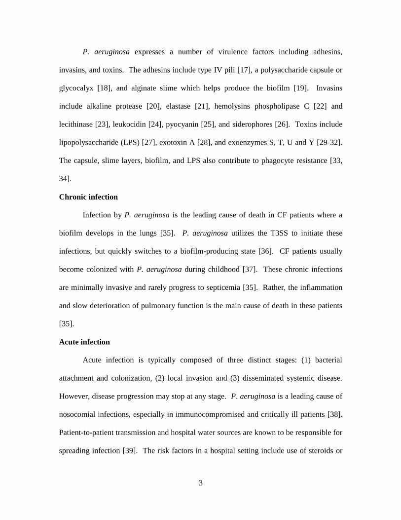
The structural proteins of the T3SS that make up the needle apparatus are
encodedby genes pscUTSRQPON, pcr1234DR, and pscBCDEFGHIJKL. Much of what
is known about the structural proteins is from studies in Yersinia enterocolitica, which
has a needle complex that is homologous to that in P. aeruginosa (Fig. 1.1). PscN
9
encodes an ATPase that uses energy created from the proton motive force to unfold
effector proteins and push them through the needle apparatus [110, 111]. PscN is
regulated by PscL [111]. PscC is a secretin-like protein that is thought to oligomerize
with the help of the lipoprotein PscW in order to form a channel through the bacterial
outer membrane [112, 113]. PscP is thought to serve as a molecular ruler to regulate the
length of the needle [114]. PscJ is hypothesized to be a lipoprotein component of the
basal structure of the needle complex [115]. Further work needs to be conducted to
determine the properties and function of all the components of the needle complex and
how they work together for secretion.
T3SS translocation apparatus
The translocation apparatus consists of a pore that delivers the effector molecules
through the host cell plasma membrane. It is encoded by pcrGVH and popBD. PopB and
PopD are actually secreted themselves and interact with the host cell membrane to form
the pore [106]; however, this mechanism is poorly understood. PcrV is also secreted and
is thought to form a multimeric scaffold at the tip of the needle to facilitate the assembly
of the PopB–PopD translocation pore [116]. Alternatively, PcrV could serve to connect
the needle to preformed PopB–PopD pores [117]. PcrV is a potential vaccine candidate,
given that it is required for the translocation of effector proteins [116] and has been
shown to elicit protection in animal infection models [118, 119].
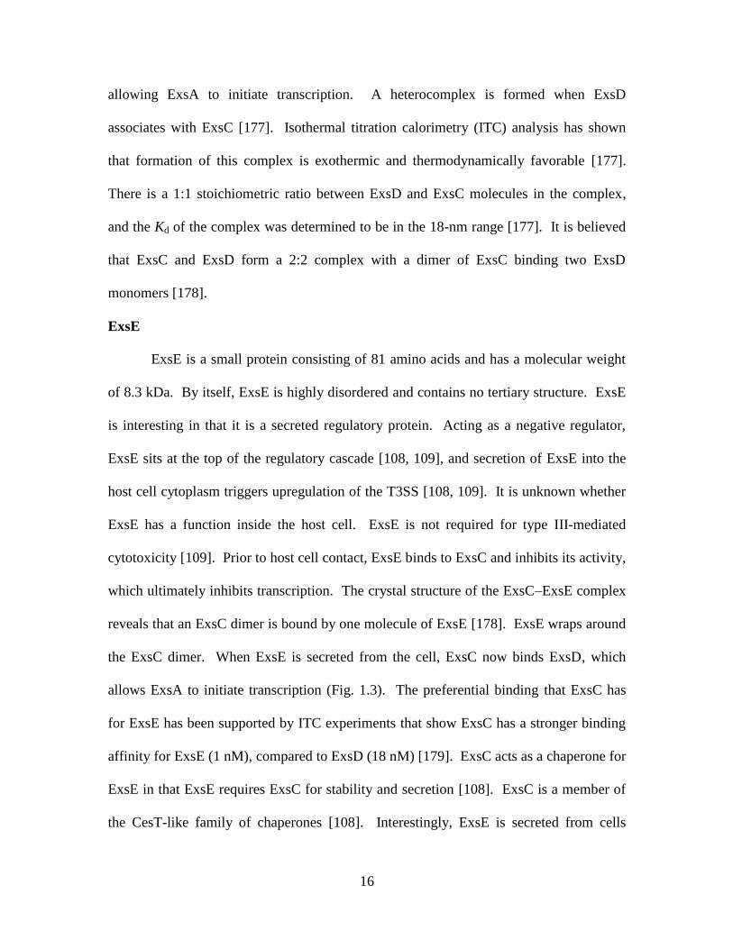
T3SS regulation
Effector secretion as well as the expression of all T3SS components is tightly
controlled by a regulatory mechanism consisting of four interacting proteins (ExsA, ExsC,
ExsD and ExsE), with ExsA being the main transcription factor [120]. In addition to the
10
ExsACDE regulatory cascade, the T3SS is regulated by a multitude of other proteins (Fig.
1.2). These proteins include CpdA, a cAMP phosphodiesterase [121]; Crc, a catabolite
repression control protein involved in carbon regulation [122]; adenylate cyclases CyaA
and CyaB [123]; FimL, a type IV pili biogenesis protein [124]; FlhA, a transmembrane
protein [125]; KynA, a tryptophan dioxygenase [126]; MgtE, a magnesium transporter
[127]; MucA/AlgU/AlgR, alginate biosynthesis proteins [128]; NirS, a nitrite reductase
[129]; PrpC, a methylcitrate synthase [122]; PsrA, a transcriptional activator [130]; small
proteins PtrB and PtrC [131, 132]; RpoS, the stationary-phase sigma factor [133]; RsmA,
an RNA-binding protein [134, 135]; two-component response regulatory systems,
LadS/RetS, GacA/GacS and GtrS/GltR [36, 136-138]; TrpA, a tryptophan synthase
[126]; and Vfr, a cyclic AMP (cAMP)-binding protein [139]. There are also small RNAs,
RsmZ and RsmY, which contribute to T3SS regulation [36, 140]. In addition, the T3SS
is regulated by a number of small molecules including calcium [141], acetyl-CoA [142],
indole acetic acid (IAA), histidine [143], spermidine [144], and the quorum-sensing
signals homoserine lactone (HSL) [145] and Pseudomonas quinolone signal (PQS) [146].
However, for the most part, it is unclear how these regulatory proteins/molecules exert
their influence on the T3SS.
ExsACDE regulatory cascade
The ExsACDE cascade constitutes perhaps the most direct link between effector
secretion and upregulation of T3SS genes [109] (Fig. 1.3). ExsA, the main
transcriptional regulator, is inhibited by ExsD when ExsD binds to ExsA in a 1:1
complex [147]. Meanwhile, ExsE sequesters ExsC [108, 109]. Upon host cell contact,
ExsE is translocated into the host cell cytoplasm [108, 109]. Now ExsC sequesters ExsD
11
[148], which frees ExsA to bind to the T3SS promoters and recruit RNA polymerase
(RNAP), thus initiating T3SS transcription [149].
ExsA
ExsA is the main transcriptional activator of the T3SS and a member of the
AraC/XylS family of transcriptional regulators [120]. These regulators are found in
bacteria and fungi and are involved in the regulation of virtually all types of cellular
processes [150]. AraC/XylS-type transcription factors are characterized by structurally
conserved AraC domains that interact with specific DNA sequences near the -35
promoter region [150]. This facilitates the recruitment of RNAP to the transcription
initiation site [150]. On the other hand, the amino-terminal domains of the AraC/XylS-
type proteins are highly variable and thought to mediate diverse regulatory signals [150-
154]. Typical of an AraC/XylS-like activator, ExsA is composed of two functional
domains: a carboxy-terminal DNA binding domain (CTD) and an amino-terminal
regulatory domain (NTD) [155]. ExsA is 278 amino acids in length and has a molecular
weight of 31.6 kDa.
It is estimated that there are at least 830 members in the AraC/XylS family of
proteins [150]. However, out of these, only two complete structures have been published,
and they are both from E. coli: MarA and Rob [156, 157]. The crystal structures were
only obtained when these proteins were in complex with DNA to help stabilize their
carboxy-terminal domains. Both MarA and Rob activate genes that are involved in
antibiotic resistance.
12
ExsA homologs
There are close homologs to ExsA in a variety of Gram-negative pathogens
including InvF from Salmonella enterica serovar Typhimurium, LcrF in Yersinia spp.,
Rns from enterotoxigenic E. coli (ETEC), ToxT from Vibrio cholerae, and ExsA from
Vibrio parahaemolyticus. These proteins make up a subfamily of activators that regulate
virulence factor expression. InvF from S. enterica regulates the T3SS found on the SPI–1
pathogenicity island [158]. LcrF activates the T3SS in the three pathogenic Yersinia
species: Y. pestis, Y. enterocolitica, and Y. pseudotuberculosis [159, 160]; but it is
unknown whether a regulatory protein binds to LcrF. It is known, however, that LcrF is
thermoregulated through post-translational mechanisms [161]. Rns regulates fimbriae
expression in ETEC, which is required for pathogenesis [162]. ToxT regulates
expression of cholera toxin and toxin-coregulated pilus in V. cholerae [163]. The most
closely related regulatory mechanism to the T3SS in P. aeruginosa is found in T3SS1 of
V. parahaemolyticus. This system is regulated by a similar ExsACDE regulatory cascade
[164-166]; however, the main difference in this system is that ExsA does not regulate its
own expression [164].
ExsA promoters
ExsA is the transcriptional activator of the T3SS regulon (Fig. 1.4) [120] and is
known to bind to all 10 promoters involved in expression of T3SS genes: PexsC, PexsD, PexoS,
PexoT, PexoU, PexoY, Porf1, PpcrG, PpcrN and PpopN [31, 75, 103, 120]. ExsA is monomeric in
solution, but it forms a dimer when bound to one of its promoters [167]. This is in
contrast to MarA, which lacks a dimerization domain and binds to DNA as a monomer
[156]. It is thought that one molecule of ExsA initially binds to the promoter at binding
13
site 1 and recruits the second ExsA molecule to binding site 2 [167]. Binding site 1, or
the promoter-proximal site, contains highly conserved GnC and TGnnA sequences
separated by ~10 base pairs [167]. The ExsA-CTD contains two helix-turn-helix (HTH)
motifs, and the first HTH interacts with the GnC sequence while the second HTH
interacts with the TGnnA sequence [168]. On the other hand, binding site 2, or the
promoter-distal site, contains no obvious sequence similarity [167]. Given that ExsA
always binds to binding site 1 in the same manner, binding site 2 is responsible for
promoter-specific properties [169]. Promoter strength, ExsA-binding affinity, and the
degree of promoter bending are properties that are primarily determined by binding site 2
[169]. Cooperative binding has been demonstrated with the PexsC promoter [167], and the
way in which ExsA binds to PexsC is distinct from the interactions that occur at the other
promoters [169]. For PexsC, there is a GnC located in binding site 2 that interacts with
HTH1 of ExsA, and is functionally equivalent to the GnC sequence in binding site 1
[169]. The PexsD, PexoT, and PpcrG promoters also contain GnC sequences, but these
sequences are not required for ExsA binding or transcriptional activation [169]. It has
been shown that ExsA binds to the PexoT promoter in an ordered fashion in that
occupation of binding site 1 is required for efficient occupation of site 2 [167]. This
sequential binding mechanism is dependent upon interactions mediated through the
amino-terminal/dimerization domain of the ExsA monomers [155]. Recent studies show
that ExsA binds to the PexsC, PexsD, PexoT, and PpcrG promoters in a head-to-tail orientation
[168]. It has been demonstrated that ExsA induces DNA-bending upon binding [167].
ExsA shows modest DNA-bending with the PexsD and PexoT promoters, whereas the DNA-
bending at the PexsC promoter is more pronounced [167]. An electrophoretic mobility

14
shift assay (EMSA) was used to determine equilibrium constants (Keq) for the
interactions between ExsA and three of its promoters [167]. The apparent equilibrium
constants were 1.1 ± 0.2 nM, 4.1 ± 0.2 nM and 5.4 ± 0.6 nM for PexsC, PexsD and PexoT,
respectively [167]. These high affinities are characteristic of a transcriptional activator.
Once ExsA forms the dimer on the promoter, it is able to recruit RNAP so that
transcription can be initiated. DNase I footprinting experiments were used to map the
regions of the PexsC, PexsD, and PexoT promoters to which ExsA binds [167]. For each
promoter, the ExsA binding site overlaps the -35 RNAP binding site and extends
upstream for approximately 34 base pairs [167]. The -35 and -10 sites for typical σ70
-
dependent promoters are separated by ~17 nucleotides. However, for ExsA-dependent
promoters, this gap is 21-22 bases [167]. These features suggest that the -35 site is not
involved in RNAP binding.
ExsA–RNAP interactions
Recent work suggests that the -35 RNAP binding site of the PexsC promoter does
not contribute to RNAP binding [170]. Instead, ExsA recruits σ70
-RNAP to an extended
-10 promoter [149, 170]. It has been shown that the carboxy-terminal domain of the α
subunit of RNAP is not needed for ExsA-dependent transcriptional activation [170]. This
is unusual for transcriptional activators. Instead, studies indicate that ExsA interacts with
region 4.2 of σ70
because this region is required for ExsA-dependent transcription [170].
It is proposed that this is how ExsA makes up for the lack of a -35 RNAP binding site
since region 4.2 typically interacts with the -35 site for most bacterial promoters [170].

15
ExsD
When the T3SS is turned off, ExsA is inhibited by an antiactivator protein ExsD
[171]. An antiactivator is distinguished from a repressor in that it does not have a known
DNA recognition site (operator). An antiactivator can bind directly to the activator or
bind non-specifically to DNA. ExsD–ExsA interactions have been demonstrated in
bacterial two-hybrid studies [171]. It has been shown that ExsD binds to ExsA to form a
1:1 complex [147]. In the current model, ExsD inhibits ExsA activity by disrupting the
ExsA dimer [172] and preventing ExsA from binding to the promoters [147, 172].
ExsD (Fig. 1.5) is 276 amino acids in length and has a molecular weight of 31.4
kDa. ExsD self-associates into a trimer when not bound to a different molecule [173].
The crystal structure of ExsD [173] reveals surprising structural homology between ExsD
and a DNA binding protein in E. coli called KorB [174]. KorB is found on a broad-host-
range plasmid called RP4 [174]. It is a repressor protein that binds to DNA operator
sequences located as far as 500 base pairs away from the regulated promoter and is
thought to mediate transcriptional repression through DNA looping [175].
ExsD also shows structural homology to GreB; a RNAP binding protein from E.
coli [176]. RNAP occasionally loses its grip on the growing mRNA end during
elongation and backtracks on the DNA template [176]. GreB binds to RNAP and cleaves
the disengaged 3´ RNA segment so that RNAP can proceed with transcription [176].
ExsC
ExsC is the anti-antiactivator composed of 141 amino acids and has a molecular
weight of 16.2 kDa. ExsC acts as a positive regulator of the T3SS and is required for
T3SS expression [148]. When the T3SS is turned on, ExsC binds to ExsD [148],
16
allowing ExsA to initiate transcription. A heterocomplex is formed when ExsD
associates with ExsC [177]. Isothermal titration calorimetry (ITC) analysis has shown
that formation of this complex is exothermic and thermodynamically favorable [177].
There is a 1:1 stoichiometric ratio between ExsD and ExsC molecules in the complex,
and the Kd of the complex was determined to be in the 18-nm range [177]. It is believed
that ExsC and ExsD form a 2:2 complex with a dimer of ExsC binding two ExsD
monomers [178].
ExsE
ExsE is a small protein consisting of 81 amino acids and has a molecular weight
of 8.3 kDa. By itself, ExsE is highly disordered and contains no tertiary structure. ExsE
is interesting in that it is a secreted regulatory protein. Acting as a negative regulator,
ExsE sits at the top of the regulatory cascade [108, 109], and secretion of ExsE into the
host cell cytoplasm triggers upregulation of the T3SS [108, 109]. It is unknown whether
ExsE has a function inside the host cell. ExsE is not required for type III-mediated
cytotoxicity [109]. Prior to host cell contact, ExsE binds to ExsC and inhibits its activity,
which ultimately inhibits transcription. The crystal structure of the ExsC–ExsE complex
reveals that an ExsC dimer is bound by one molecule of ExsE [178]. ExsE wraps around
the ExsC dimer. When ExsE is secreted from the cell, ExsC now binds ExsD, which
allows ExsA to initiate transcription (Fig. 1.3). The preferential binding that ExsC has
for ExsE has been supported by ITC experiments that show ExsC has a stronger binding
affinity for ExsE (1 nM), compared to ExsD (18 nM) [179]. ExsC acts as a chaperone for
ExsE in that ExsE requires ExsC for stability and secretion [108]. ExsC is a member of
the CesT-like family of chaperones [108]. Interestingly, ExsE is secreted from cells

17
under low calcium conditions. The link between low calcium and T3SS initiation is
currently unknown, but it allows for studies of type III secretion in the laboratory setting.
Medical importance
ExsA is a potential drug target due to the fact that it is absolutely required for type
III secretion. An ExsA mutant is unable to secrete type III effectors and unable to initiate
an acute infection. Therefore, a drug that inhibits ExsA would be extremely valuable due
to the high antibiotic resistance of P. aeruginosa. This type of drug would not kill P.
aeruginosa, but just render it harmless. Due to this, there would be less evolutionary
pressure for the organism to develop resistance. Currently, there are no drugs that act on
P. aeruginosa in this way. An inhibitor of ExsA should also have no harmful effects on
human cells because AraC/XylS family members are only found in bacteria [150]. There
is much that needs to be discovered about the interaction between ExsD and ExsA and
how ExsD inhibits ExsA-dependent transcription. Elucidation of the ExsD–ExsA
inhibitory mechanism could result in the development of drugs that mimic ExsD function
and inhibit ExsA to shut down the T3SS, thereby eliminating acute infection.
Research objectives
The purpose of the following studies is to characterize ExsA and ExsD, the main
transcriptional regulators of the T3SS in P. aeruginosa, through a variety of in vitro
biochemical techniques to learn the properties of ExsA and ExsD individually and also
how they interact. If enough is known about these proteins and the way they regulate
transcription, then drugs can be designed to shut down the T3SS. In Chapter Two, the
goal was to purify a more soluble recombinant ExsA protein and examine its ability to
interact with its promoters and initiate transcription. The affinity of ExsA for its PexsD
18
promoter was obtained using fluorescence anisotropy. An in vitro transcription assay
was developed to examine the ability of ExsA to initiate transcription from three of its
promoters.
Chapter Three examines the interaction between ExsD and ExsA. Previous
studies have shown that the ExsD–ExsA interactions are not freely reversible. Because
independently folded ExsD and ExsA were not found to interact, it was hypothesized that
folding intermediates of the two proteins form the complex. This study demonstrates for
the first time that ExsD alone is sufficient to inhibit ExsA-dependent transcription in vitro
and that no other cellular factors are required. More significantly, it was discovered that
independently folded ExsD and ExsA are capable of interacting, but only at 37 C and
not at 30 C. ExsD is known to form a trimer in solution when not bound to a different
protein [173], but the role of ExsD self-trimerization was previously unknown. Guided
by the crystal structure of ExsD, a monomeric variant of the protein was designed to
demonstrate that ExsD trimerization prevents ExsD from inhibiting ExsA-dependent
transcription at 30 C.
In Chapter Four, the main goal was to map the ExsD–ExsA interface. ExsD
variants lacking certain regions of the protein were used in the in vitro transcription assay
to determine which regions of ExsD are important for ExsA binding and inhibition. The
ExsD–ExsA inhibitory mechanism was also further examined. Contrary to the current
model, ExsD was also found to bind to DNA. However, it is unclear whether this plays a
role in the inhibition of ExsA. Preliminary results show that ExsD also binds to RNAP,
but how this fits into the mechanism remains unclear. Overall, this study contributes to
the understanding of how ExsD and ExsA regulate the T3SS and adds to the current
19
model. These studies will pave the way for future work that will lead to a complete
understanding of how ExsD and ExsA regulate the T3SS in P. aeruginosa. This will not
only help in the development of drugs to treat P. aeruginosa infection, but other types of
infections caused by the many Gram-negative pathogens that depend on the T3SS to
infect humans.

20
Fig. 1.1. Structural proteins of the T3SS needle apparatus. Figure adapted by author
with permission from Izoré et al. Structure (2011).
21
Fig. 1.2. T3SS regulatory network. Model of the known factors that regulate the T3SS.
22
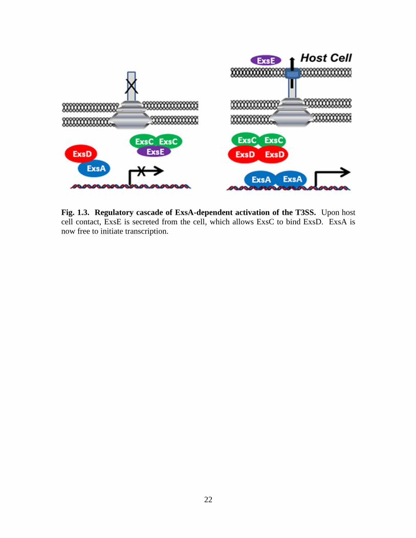
Fig. 1.3. Regulatory cascade of ExsA-dependent activation of the T3SS. Upon host
cell contact, ExsE is secreted from the cell, which allows ExsC to bind ExsD. ExsA is
now free to initiate transcription.
23
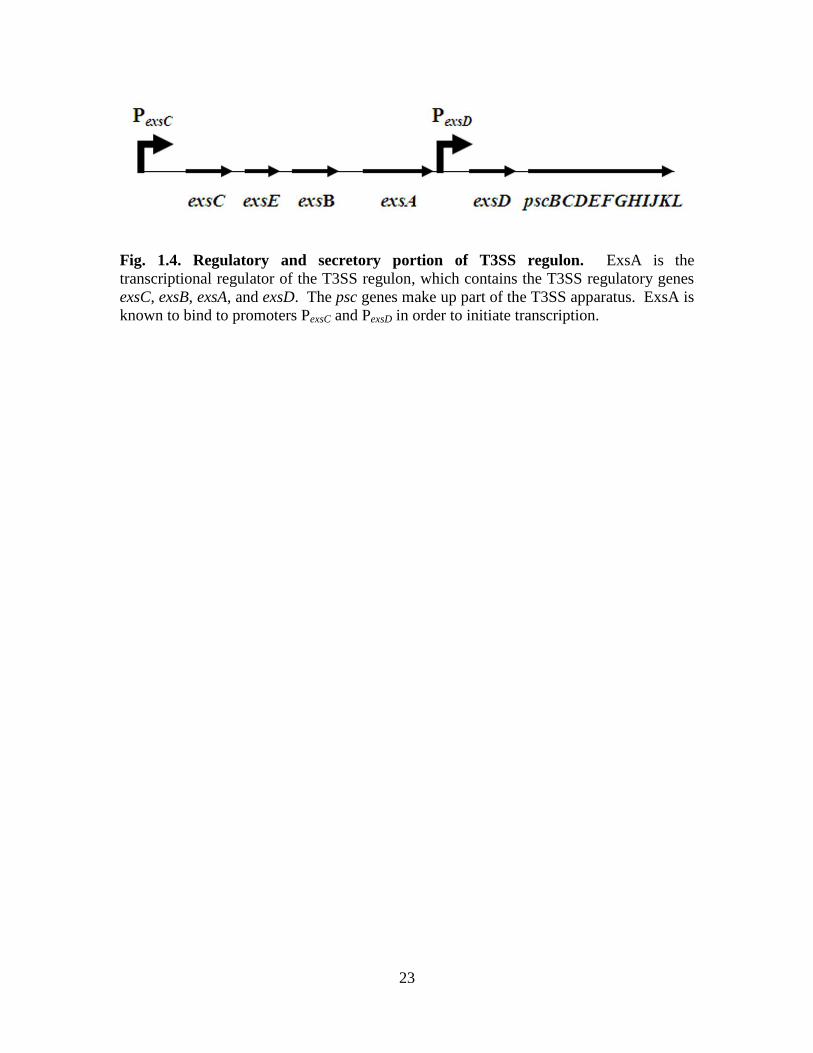
Fig. 1.4. Regulatory and secretory portion of T3SS regulon. ExsA is the
transcriptional regulator of the T3SS regulon, which contains the T3SS regulatory genes
exsC, exsB, exsA, and exsD. The psc genes make up part of the T3SS apparatus. ExsA is
known to bind to promoters PexsC and PexsD in order to initiate transcription.

24
Fig. 1.5. ExsD structure. Two views of the structure of ExsD as a monomer.
Bernhards, et al. Protein Science (2009).
Used with permission of Protein Science and John Wiley & Sons.
Published by Wiley-Blackwell. © 2009 The Protein Society.
25
CHAPTER TWO
Characterization of ExsA, the main transcriptional regulator of the type
III secretion system in Pseudomonas aeruginosa
Robert C. Bernhards1, Nancy J. Vogelaar
1, Marcy Hernick
2, Florian D. Schubot
1*
1Virginia Polytechnic Institute and State University
Department of Biological Sciences
125 Life Sciences 1 Building
Washington Street
Blacksburg, VA 24061
2Virginia Polytechnic Institute and State University
Department of Biochemistry
305 Engel Hall
West Campus Drive
Blacksburg, VA 24061
*Corresponding author: Phone: (540) 231-2393
Fax: (540) 231-7126
Email: [email protected]
Keywords: ExsA, AraC, Pseudomonas aeruginosa, thermoregulation, type III secretion.
Abbreviations: EMSA = electrophoretic mobility shift assay; ExsACDE = ExsA-ExsC-
ExsD-ExsE; FA = fluorescence anisotropy; IPTG = isopropyl-β-D-thiogalacto-
pyranoside; MBP = maltose binding protein; TCEP = Tris(2-carboxyethyl)phosphine;
TEV = tobacco etch virus; T3SS = type III secretion system.
26
Abstract
ExsA is the main transcription factor or activator of the T3SS system in
Pseudomonas aeruginosa. ExsA is a member of the AraC/XylS family of transcriptional
activators. These proteins are composed of two domains: a C-terminal activation domain
and an N-terminal regulatory domain. The activation domain of AraC/XylS-type
transcription factors binds to the promoter to initiate transcription. The regulatory
domain is the ligand binding domain. In most AraC/XylS-type regulators, small
molecules bind to either activate or inhibit the protein. In the case of ExsA, the
antiactivator protein ExsD is thought to bind to the regulatory domain and inhibit ExsA
function. A variety of ExsA homologs exist in other Gram-negative pathogens of
medical relevance, so the study of ExsA could help shed light on the regulatory
mechanisms of other T3SS’s. Due to this, ExsA is a potential drug target. The activation
domain of ExsA homologs is conserved, so initial drug development has targeted the
DNA binding domain in hope of finding an inhibitor to DNA binding. Potentially, a drug
that effectively inhibits the DNA-binding activity of the ExsA activation domain could be
used to treat other types of infections. AraC-type activators have been difficult to purify
and crystallize due to their poor solubility. In this study, a new ExsA purification
protocol was developed, which led to improved solubility. Fluorescence anisotropy was
used to measure the DNA binding activity of ExsA. A dissociation constant (Kd) was
determined for ExsA and its PexsD promoter. An in vitro transcription assay was also
developed to observe ExsA activation with its T3SS promoters. This assay could be used
in the future to test drug candidates for their ability to inhibit ExsA-dependent
transcription.

27
Introduction
The type III secretion system (T3SS) is the main virulence mechanism for
Pseudomonas aeruginosa acute infections. It works as a molecule syringe to inject
effector proteins (cytotoxins) directly into the host cell cytoplasm [60]. These effectors
aid in infection by preventing phagocytosis by cells of the immune system and allowing
for the dissemination of the bacteria throughout the body [30, 59, 63, 74]. P. aeruginosa
is unable to infect humans without a properly functioning T3SS. ExsA is the main
activator of the T3SS in P. aeruginosa and is essential for expression of all the genes
necessary for type III secretion [31, 75, 103, 120].
ExsA is a member of the AraC/XylS family of transcriptional activators [120].
This family is characterized by the presence of two distinct domains: a C-terminal
activation/DNA binding domain and N-terminal regulatory/ligand binding domain. Most
AraC/XylS proteins are regulated by the binding of a small molecule. ExsA is different
in that its ligand is a protein called ExsD [171]. ExsA sits at the bottom of a regulatory
cascade consisting of four interacting proteins: ExsA, ExsD, ExsC, and ExsE [108, 109,
120, 148, 171]. When there is no host cell contact and the T3SS is turned off, ExsA is
inhibited by the antiactivator ExsD [171]. Meanwhile, the anti-antiactivator ExsC
sequesters ExsE [109, 178]. When host cell contact occurs, ExsE is secreted through the
T3SS needle into the host cell [108, 109]. This frees ExsC to now bind to ExsD [148],
which allows ExsA to become free and bind to the T3SS promoters as a dimer [167].
ExsA serves to recruit RNA polymerase (RNAP) in order to initiate transcription [149].
ExsA activates transcription from all 10 T3SS promoters, so it is required for the
28
production of all genes necessary for type III secretion, including the regulators
themselves [31, 75, 103, 120].
This study focuses on the interaction between ExsA and its promoters. A new
protocol was developed for the purification of recombinant ExsA protein, which greatly
improved solubility. This ExsA product did not require a protein fusion tag after
purification. The purity and solubility of ExsA was suitable for biochemical testing.
First, fluorescence anisotropy was used to measure the binding affinity of ExsA for its
PexsD promoter. Then an in vitro transcription assay was developed to study T3SS
activation by ExsA. ExsA was found to be sufficient to activate T3SS transcription in
vitro from all the promoters tested.
Results
ExsA purification
Members of the AraC/XylS family of activators are notoriously difficult to purify.
This is due to a highly insoluble C-terminal/DNA-binding domain (CTD). When DNA is
not present, these proteins have very low solubility, making them extremely difficult to
purify. Due to this, we decided to engineer a construct that would fuse maltose binding
protein (MBP) to ExsA. MBP is known to help other proteins fold properly, which
improves their solubility. To do this, we utilized a His6-MBP expression plasmid [180].
ExsA was insertionally cloned into the plasmid directly downstream of MBP. This
produced a His6-MBP-ExsA construct, termed pFS-HMBPExsA. This plasmid was
transformed into an E. coli expression strain. Cultures were grown at 37 °C and
overexpression was induced at a low temperature (18 °C) for six hr. Purification
29
included a Ni2+
NTA affinity column, anion exchange, and cation exchange all before
removing the His6-MBP tag. The fusion protein was cut using TEV protease, which
recognizes a TEV cleavage sequence in between MBP and ExsA. Then a second
Ni2+
NTA affinity column was run, this time to purify ExsA away from His6-MBP. The
final purification step was gel filtration. This produced a highly purified ExsA sample
with improved solubility (Fig. 2.1). This ExsA sample can be stored at -80 °C for years
and still retains activity. Aliquoting and storing at -80 °C is highly recommended
because ExsA will start to unfold if left at 4 °C for more than 24 hr. The ExsA sample is
highly active and suitable for biochemical assays as long as it is thawed and used within
24 hr to ensure full activity.
ExsA promoter binding
We sought out to measure the affinity of ExsA for its promoters in order to
determine if there was a correlation between promoter affinity and transcriptional activity.
The first promoter we wanted to test was the PexsD promoter. This promoter is interesting
in that it regulates the transcription of the antiactivator ExsD [171, 181]. Fluorescence
anisotropy (FA) was used to determine a dissociation constant (Kd) for the PexsD promoter.
FA is a technique that utilizes a fluorescent probe attached to a ligand, which in this case
is the PexsD promoter. We used a 60 base pair segment of PexsD that was attached to a
TAMRA probe. A titration of ExsA was performed, and the concentration of PexsD-
TAMRA was held constant. The resulting Kd was 8.5 ± 2 nM (Fig. 2.2). This Kd is
indicative of a very tight interaction common to transcriptional activators [182]. During
the course of our study, the affinity for ExsA and three of its promoters was determined
by another group using an electrophoretic mobility shift assay (EMSA) [167]. Due to
30
this, we decided not to further test other promoters. However, EMSA is not an
equilibrium technique, meaning that ExsA is not free to bind and dissociate from the
promoter under the experimental conditions. FA is an equilibrium technique. The Kd we
obtained is two-fold higher than the one obtained via EMSA (4.1 ± 0.2 nM) [167]. Our
Kd value is actually closer to what was obtained for AraC, using a fluorescence
quenching equilibrium technique [182].
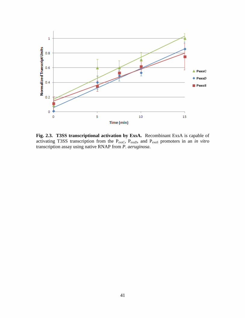
ExsA is sufficient to initiate T3SS transcription in vitro
An in vitro transcription assay was developed in order to see if ExsA alone was
sufficient to activate T3SS transcription. Native RNAP from P. aeruginosa was purified
and used in this assay, along with purified ExsA, a PexsD promoter template, and
nucleoside triphosphates (NTPs). Radiolabeled UTP is also added, which gets
incorporated into the resulting mRNA transcript. These transcripts were run on a gel and
quantified. A 0-15 min time course was performed with each reaction containing 133 nM
ExsA (Fig. 2.3). This experiment shows that ExsA is necessary and sufficient for
efficient transcriptional activation at the PexsC, PexsD, and PexoS promoter sites. This also
further demonstrates that the recombinant ExsA is highly active. The next step is to test
whether ExsD is sufficient to inhibit ExsA-dependent transcription in this assay.
Discussion
Proteins belonging to the AraC/XylS family have characteristically low solubility
and are extremely difficult to purify. As a result, there are currently only two solved
structures out of the 830-plus family members [156, 157]. This highlights the importance
of the newly developed purification protocol for ExsA. Furthermore, ExsA belongs to a
31
medically important subfamily of transcriptional activators that regulate virulence factors
in a variety of Gram-negative pathogens. These include ExsA from Vibrio
parahaemolyticus, InvF from Salmonella enterica serovar Typhimurium, LcrF in
Yersinia spp., Rns from enterotoxigenic E. coli (ETEC), ToxT from V. cholerae. T3SS1
of V. parahaemolyticus is regulated by a similar ExsACDE regulatory cascade [166].
InvF from S. enterica regulates the T3SS located on the SPI–1 pathogenicity island [158].
LcrF activates the T3SS in all three pathogenic Yersinia species: Y. pestis, Y.
enterocolitica, and Y. pseudotuberculosis [159, 160]; although it is unknown whether a
regulatory protein binds to LcrF. However, it is know that LcrF is thermoregulated
through post-translational mechanisms [161]. Rns regulates fimbriae expression in
ETEC [162], and ToxT controls expression of cholera toxin in V. cholerae [163]. As one
can anticipate, what is learned about ExsA can be translated to these other important
Gram-negative pathogens. Given that ExsA can be purified with relatively good
solubility, it can serve as a model protein for this subfamily. Recently, using the
expression and purification protocols outlined in this study, the structure of the amino
terminal domain of ExsA (ExsA-NTD) was solved, and current work is being done to
crystallize full-length ExsA in complex with DNA. This study demonstrates that
recombinant ExsA is properly folded and highly active. It was shown to bind to the PexsD
promoter with high affinity. ExsA was also shown to be sufficient to activate P.
aeruginosa-specific T3SS transcription in an in vitro transcription assay. This assay
serves as a reliable tool and was used in the following chapters to study the ExsD–ExsA
inhibitory mechanism.
32
With a better understanding of how ExsA interacts with its promoters and
activates transcription, drugs can be developed to target ExsA and prevent it from binding
to the T3SS promoters. These drugs can potentially be used to treat infections from all of
the aforementioned bacterial pathogens.
Materials and Methods
ExsA expression and purification
ExsA was overexpressed in E. coli from a vector constructed by Gateway
recombinational cloning (Invitrogen, Carlsbad, CA, USA). A tobacco etch virus (TEV)
protease recognition site and the appropriate att recombination sites (attB1 and attB2)
were added to the exsA gene during PCR, and the amplicons were subsequently
recombined into pDONR201 (Invitrogen). The nucleotide sequences of the ORFs were
verified, then recombined into the destination vector pDEST-HisMBP [180] to create the
expression vector pFS-HMBPExsA. This vector was designed to produce ExsA as a
fusion to the C-terminus of an N-terminally His6-tagged E. coli maltose-binding protein
(MBP).
A single colony of E. coli BL21(DE3) CodonPlus RIL cells (Stratagene, La Jolla,
CA, USA) containing the pFS-HMBPExsA expression plasmid was used to inoculate 125
mL of Luria broth (LB) supplemented with 2 g/L dextrose, 100 µg/mL ampicillin, and 30
µg/mL chloramphenicol. The cultures were grown with shaking (225 rpm) to saturation
overnight at 37 °C and then diluted 66-fold into 6 L of fresh medium. They were grown
to an OD600 of 1.0 then induced with IPTG at a final concentration of 1 mM. The
induction temperature was 18 °C, and the cultures were shaken for six hr. Cells were
harvested by centrifugation at 5,000 x g for 15 min. The cell pastes were resuspended in
33
200 mL of 500 mM NaCl, 25 mM imidazole, 50 mM Tris-HCl (pH 7.4), 2 mM DTT
(buffer A), along with three tablets of Complete, EDTA-free Protease Inhibitor Cocktail
(Roche Applied Science, Indianapolis, IN, USA). The cells were lysed via sonication
and centrifuged at 40,000 x g for 25 min. The supernatants were filtered through 0.45-
μm polyethersulfone membranes and applied to a 30 mL Ni-NTA Superflow affinity
column (Qiagen, Valencia, CA, USA) equilibrated with buffer A. During the run, the
column was washed with five column volumes of buffer A, and proteins were eluted with
a linear gradient from 25 to 250 mM imidazole (pH 7.4). The His6-MBP-ExsA fusion
protein sample was then dialyzed against a buffer of 50 mM NaCl, 25 mM Tris-HCl (pH
7.4), and 2 mM DTT and loaded onto a HiTrap Q HP column (GE Healthcare) that had
been equilibrated with the same buffer. The His6-MBP-ExsA fusion protein was eluted
using a linear NaCl gradient from 0.05 M to 1 M. The sample was dialyzed against 2 L
of 45 mM NaCl, 25 mM Tris-HCl (pH 7.15), and 2 mM DTT (buffer B) overnight. The
sample was then loaded onto a HiTrap Heparin HP column (GE Healthcare) equilibrated
in buffer B and eluted with a 0.05 M to 1 M gradient of NaCl. The NaCl concentration in
the His6-MBP-ExsA sample was adjusted to 0.5 M, and the fusion protein was digested
with 3 mg of His-tagged TEV(S219V) protease [183] at 4 C overnight. The TEV
protease cleaves at the TEV recognition site located between MBP and ExsA. After
digestion, ExsA was run through a second Ni-NTA Superflow affinity column to remove
both the His6-MBP tag and the protease, using the same buffers as the first Ni-NTA
column. The ExsA sample was collected in the flow through. Finally, gel filtration using
a HighLoad 26/60 Superdex 200 prep grade column (GE Healthcare) was performed with
the ExsA sample using 500 mM NaCl, 25 mM Tris-HCl (pH 7.4), and 2 mM TCEP
34
(ExsA storage buffer). The sample was concentrated to 1 mg/mL, flash-frozen using
liquid nitrogen, and stored at -80 °C.
Fluorescence anisotropy
A 60 base single strand of the PexsD promoter fragment attached to a 5´–TAMRA
probe was purchased from Integrated DNA Technologies, Coralville, IA, USA: 5´–
TAMRA–
GGAAGGACGAATGCCGGGCTAAAAATAACTGACGTTTTTTGAAAGCCCGGTA
GCGGCTGC–3´. This region contains both ExsA binding sites. The complementary
strand (5´–
GCAGCCGCTACCGGGCTTTCAAAAAACGTCAGTTATTTTTAGCCCGGCATTCG
TCCTTCC–3´) was annealed by incubation at 75 °C for 10 min and cooling at room
temperature for 5 min. The experiment consisted of individual samples of a titration of
ExsA with a final concentration ranging from 0 to 50.7 nM. Each sample also contained
a final concentration of 150 mM NaCl, 25 mM Tris-HCl pH 7.4, 0.6 mM TCEP pH 8.0,
and 10 nM PexsD-TAMRA. The final volume of each sample was 30 μL. Each sample
was pipetted into separate wells in a 384 well, flat bottom, black polystyrene assay plate
(Corning, Corning, NY, USA). Measurements at Ex. 559 nm and Em. 600 nm were
taken using SPECTRAmax M5e ROM v2.00b255 with SoftMax Pro 5.2 software. The
Kd was determined by data analysis using XLfit (IDBS, Bridgewater, NJ, USA). The
data points were fitted using the Boltzmann constant. The experimental samples were in
triplicate.
35
RNA polymerase purification and specific activity determination
RNA polymerase (RNAP) was purified from P. aeruginosa PAO1 cells following
the original procedure of Allan and Kropinski [184]. However, changes were made to the
later chromatographic steps. All purification steps were performed at 4 °C. P.
aeruginosa PAO1 cultures were grown in LB broth to an OD600 of 0.8, harvested by
centrifugation at 6,000 x g, then lysed by sonication. The cell debris was removed by
centrifugation at 35,000 x g for 30 min, and 25% polyethyleneimine (pH 7.5) was added
to the supernatant to a final concentration of 0.5% in order to precipitate the RNAP. The
supernatant was centrifuged at 35,000 x g for 30 min. The polyethyleneimine precipitate
was washed with 10 mM Tris-HCl (pH 8.0), 250 mM NaCl, 5% glycerol, 0.05 mM
EDTA, 1 mM DTT, and 0.1 mM PMSF (wash buffer) and centrifuged at 35,000 x g for
30 min. RNAP was released by resuspending the pellet in 10 mM Tris-HCl (pH 8.0), 800
mM NaCl, 5% glycerol, 0.05 mM EDTA, 1 mM DTT, and 0.1 mM PMSF (release
buffer) and centrifuged at 25,000 x g for 30 min. Ammonium sulfate was added to the
supernatant to a final concentration of 30%, followed by gentle stirring for one hr, and
centrifugation at 35,000 x g for 30 min. Additional ammonium sulfate was then added to
bring the supernatant to 60% saturation. After a second centrifugation at 35,000 x g for
30 min, the pellet was resuspended in 1 mL wash buffer per liter of original culture. The
suspension was dialyzed versus 2 L wash buffer overnight. The dialyzed RNAP sample
was centrifuged, and the supernatant was filtered in preparation for gel filtration. The
sample was run through a Sephacryl S-300 HR column (GE Healthcare) using wash
buffer. The fractions were analyzed by SDS-PAGE and collected to run on a Hi-Trap
Heparin HP column (GE Healthcare) using a loading buffer composed of 10 mM Tris-
36
HCl (pH 8.0), 250 mM NaCl, 5% glycerol, 0.05 mM EDTA, and 1 mM TCEP. RNAP
was eluted using a linear gradient of 0.25 M to 1 M NaCl. Fractions were analyzed via
SDS-PAGE, pooled, and concentrated to 1 mg/mL of total protein. Glycerol was added
to a final concentration of 50%. RNAP was aliquoted and stored at -20 °C.
The specific activity of the purified P. aeruginosa RNAP was determined by
comparing its activity to a standard curve generated with different amounts of E. coli
RNA polymerase holoenzyme (Epicentre Biotechnologies, Madison, WI, USA) using an
ExsA-independent RNA-1 promoter which produces a 108 base transcript [185].
In vitro transcription assay
The PexsC promoter template encompassed positions -268 to 123 of the PexsC
promoter, relative to the transcription start site; and from this template, RNA polymerase
synthesizes a 154 base mRNA transcript. The PexsC template was produced by PCR using
forward primer 5´-CGGGAAGGAGAGGTCAACGC-3´ and reverse primer 5´-
CAGGAGGCTCGCCATGC-3´. The PexsD promoter template encompassed positions
-207 to 94 of the PexsD promoter and it codes for an 82 base mRNA transcript. The PexsD
template was produced by PCR using forward primer 5´-
CATCAGTTGCTGCTCAACAGCG-3´ and reverse primer 5´-
CACCGCTTCTCGGGAGTACTGC-3´. The PexoS promoter template encompassed
positions -126 to 112 of the PexoS promoter and coded for a 137 base mRNA transcript.
The PexoS template was produced by PCR using forward primer 5´-
CCTCAATCTGTCCCAAACGCCC-3´ and reverse primer 5´-
GCAGTGCCAGCCCGGAGAGAC-3´. All three PCR products were run on 2% agarose
gels and purified using the Wizard SV Gel and PCR Clean-up System (Promega,
37
Madison, WI, USA). Each 30 µL transcription assay reaction contained 4.4 fM of
promoter template, 3.3 U purified RNA polymerase from P. aeruginosa (see above), 1 U
RiboGuard RNase Inhibitor (Epicentre Biotechnologies), 15 ng/µL poly(deoxyinosinic-
deoxycytidylic) acid (to prevent non-specific transcription initiation), 133 mM NaCl, 32
mM Tris-HCl (pH 7.4), 10 mM MgCl2, 25 µM EDTA, 0.9 mM TCEP, 0.2 mM DTT, and
15.5% glycerol. Each experiment contained 133 nM ExsA. Samples were mixed and
allowed to equilibrate at room temperature for five min. To start the reaction, 3 µL NTPs
(stock concentrations of 200 µM ATP, CTP, GTP and 40 µM UTP) mixed with 0.2 µL
(0.2 µCi) of 3.3 mM P32
-alpha UTP was added to each sample, and samples were
incubated at 30 °C. The reactions were stopped by adding 12 µL 1X stop solution (3M
ammonium acetate, 50 mM EDTA, 0.11 mg/mL glycogen). Then 170 µL 100% cold
ethanol was added, and the samples were incubated at -20 °C for one hr. Following
centrifugation at 12,000 x g for 15 min, the supernatant was discarded and pellets were
resuspended in 12 μL 1X TBE (Tris/Borate/EDTA)-urea sample buffer and heated at
70 °C for five min. After a brief centrifugation, the samples were loaded onto a 10%
TBE-urea gel and run at 200 mV for 60 min. Gels were exposed to a storage phosphor
screen (GE Healthcare) for 16 hr. The phosphor screen was scanned using a Typhoon
Trio Variable Mode Imager (GE Healthcare), and gel bands were quantified using Image
Quant TL v2005 (Amersham Biosciences, Piscataway, NJ, USA). Each experiment was
performed in triplicate.
38
Acknowledgements
This study was supported by the American Heart Association (09SDG2260401 to
FDS).

39
Fig. 2.1. ExsA purification. Each gel strip represents the protein sample after the
indicated purification steps.

40
Fig. 2.2. ExsA promoter affinity. Fluorescence anisotropy was used to measure the
affinity of ExsA for its PexsD promoter. The resulting Kd was 8.5 ± 2 nM. This
experiment was performed in triplicate.
41
Fig. 2.3. T3SS transcriptional activation by ExsA. Recombinant ExsA is capable of
activating T3SS transcription from the PexsC, PexsD, and PexoS promoters in an in vitro
transcription assay using native RNAP from P. aeruginosa.
42
CHAPTER THREE
Self-trimerization of ExsD limits inhibition of the Pseudomonas
aeruginosa transcriptional activator ExsA in vitro
Robert C. Bernhards1, Anne E. Marsden
2, Shannon K. Esher
1, Timothy L. Yahr
2, and
Florian D. Schubot1*
FEBS Journal 280 (2013) 1084–1094
1Virginia Polytechnic Institute and State University
Department of Biological Sciences
125 Life Sciences 1 Building
Washington Street
Blacksburg, VA 24061
2University of Iowa
Department of Microbiology
540B Eckstein Medical Research Building
Iowa City, IA 52242
* Corresponding author: Phone: (540) 231-2393
Fax: (540) 231-7126
Email: [email protected]
Keywords: ExsA, ExsD, Pseudomonas aeruginosa, thermoregulation, type III secretion.
Abbreviations: CD = circular dichroism; DSF = differential scanning fluorimetry; ITC =
isothermal titration calorimetry; DTT = dithiothreitol; EMSA = electrophoretic mobility
shift assay; ExsACDE = ExsA-ExsC-ExsD-ExsE; IPTG = isopropyl-β-D-thiogalacto-
pyranoside; MBP = maltose binding protein; TCEP = Tris(2-carboxyethyl)phosphine;
TEV = tobacco etch virus; T3SS = type III secretion system.
Used with permission of FEBS Journal and John Wiley & Sons.
Copyright © 1999–2013 John Wiley & Sons, Inc. All Rights Reserved.

43
Abstract
The opportunistic pathogen Pseudomonas aeruginosa ranks among leading causes
of nosocomial infections. The type III secretion system (T3SS) aids acute P. aeruginosa
infections by injecting potent cytotoxins into host cells to suppress the host's innate
immune response. Expression of all T3SS-related genes is strictly dependent upon the
transcription factor ExsA. Consequently, ExsA and the biological processes that regulate
ExsA function are of great biomedical interest. The presented work focuses on the ExsA-
ExsC-ExsD-ExsE signaling cascade that ties host cell contact to the up-regulation of
T3SS gene expression. Prior to T3SS induction, the antiactivator protein ExsD binds to
ExsA and blocks ExsA-dependent transcription by interfering with ExsA dimerization
and promoter interactions. Upon host cell contact, ExsD is sequestered by the T3SS
chaperone ExsC resulting in the release of ExsA and an up-regulation of the T3SS.
Previous studies have shown that the ExsD-ExsA interactions are not freely reversible.
Because independently folded ExsD and ExsA were not found to interact, it has been
hypothesized that folding intermediates of the two proteins form the complex. Here we
demonstrate for the first time that ExsD alone is sufficient to inhibit ExsA-dependent
transcription in vitro and that no other cellular factors are required. More significantly,
we show that independently folded ExsD and ExsA are capable of interacting, but only at
37 C and not at 30 C. Guided by the crystal structure of ExsD, we designed a
monomeric variant of the protein and demonstrate that ExsD trimerization prevents ExsD
from inhibiting ExsA-dependent transcription at 30 C. We propose that this unique
mechanism plays an important role in T3SS regulation.
44
Introduction
The opportunistic human pathogen Pseudomonas aeruginosa poses a significant
medical threat due to its high levels of natural and acquired antibiotic resistance [8, 42,
186-190]. P. aeruginosa utilizes a broad array of virulence mechanisms to establish and
sustain infections. The type III secretion system (T3SS) is a hallmark of acute infections
and aids infection by translocating at least four distinct effector proteins into the
eukaryotic host cell [191-193]. Inside the host, these effectors act to subvert the host-
immune response by interfering with critical signal transduction pathways [31, 194-200].
The needle complex that constitutes the secretion apparatus is assembled from multiple
copies of 27 distinct proteins. Protein translocation through the T3SS is powered by the
proton motive force, while a cytoplasmic ATPase (PscN in P. aeruginosa) is thought to
mediate targeting and unfolding of the transported effectors at the base of the needle
complex [201, 202]. Because expression, assembly, and operation of the T3SS are
energy-intensive, T3SS-related gene expression is tightly regulated via a number of
regulatory pathways and closely tied to host infection [108, 109, 130]. The ExsA-ExsC-
ExsD-ExsE (ExsACDE) signaling cascade constitutes perhaps the most direct link
between opening of the T3SS channel and activation of T3SS-gene expression [108].
The AraC-type transcriptional activator, ExsA, facilitates the recruitment of RNA
polymerase to the transcription initiation site and is required for transcription from all 10
T3SS-related promoters including its own expression as well as genes of the other
members of the signaling cascade: exsC, exsE, and exsD [120]. While unusual, the
underlying regulatory mechanism appears to be relatively straightforward: Prior to the
host cell contact-induced opening of the secretion channel, ExsC and ExsE form a tight

45
2:1 complex, while the antiactivator ExsD sequesters ExsA to prevent transcription
activation [171]. Host cell contact triggers the opening of the T3SS channel. Now, ExsE
is secreted thereby releasing ExsC, which in turn binds to ExsD to activate ExsA-
mediated transcription [108, 109].
Recent work has focused on the question of how ExsD inhibits ExsA function.
Thibault et al. determined that ExsD and ExsA form a 1:1 complex which fails to bind to
ExsA-dependent promoters in vitro, suggesting that ExsD interferes with ExsA-promoter
interactions [147]. Brutinel et al. subsequently discovered that ExsD also interferes with
ExsA self-association using a monohybrid study [172]. While both phenomena have not
been connected experimentally, the enhancement of DNA binding affinity through self-
association is a widespread feature of DNA binding protein factors. In both studies,
researchers reported an unusual feature of the signaling mechanism. ExsD could only
bind to ExsA when both proteins were synthesized at the same time. Thibault et al.
unsuccessfully added ExsD to see if it would interfere with ExsA-DNA interactions in
EMSA studies [147]. When attempting to reconstitute the entire signaling cascade in
vitro, Brutinel et al. observed that ExsC was indeed capable of dissociating the ExsD-
ExsA complex [172]. Perhaps mirroring a scenario where the bacterial cell loses host
cell contact, the addition of ExsE to the sample readily brought about the formation of an
ExsC-ExsE complex. However, the released ExsD protein was not able to rebind to
ExsA, suggesting that the signaling process is not freely reversible. To explain this
phenomenon, it was proposed that concurrent expression of both ExsD and ExsA might
be required, because folding intermediates of either ExsD, ExsA, or both proteins might
actually associate to form this complex [172]. Under this scenario, dissociation of the
46
ExsD-ExsA complex would allow for complete folding of the subunit(s) and create a
formidable kinetic barrier preventing reassociation of the complex. In the present study,
we demonstrate that, rather than the folding of either protein, it is ExsD self-trimerization
that accounts for the observed irreversible dissociation of the ExsD-ExsA complex. We
also demonstrate that this barrier may be overcome by shifting the temperature from 30 to
37 C.
Results
ExsA-mediated in vitro transcription is not inhibited by wild-type ExsD at 30 C,
but is strongly inhibited at 37 C
The above cited work demonstrated that ExsD interferes with ExsA dimerization
and ExsA-promoter interactions [147]. In vitro transcription studies subsequently
confirmed that ExsA is necessary and sufficient for activation of T3SS promoters [149].
Yet, what has not been explicitly shown is that ExsD alone can block ExsA-mediated
transcription in vitro. We believe that this distinction is important, because the
interaction of ExsA with RNA polymerase could significantly impact the interactions
between ExsD and ExsA; for example, by stabilizing the ExsA dimer or binding of ExsA
to the promoter region. We designed and optimized an efficient in vitro transcription
assay using purified ExsA, ExsD, and P. aeruginosa RNA polymerase. In the process,
we also developed a new expression and purification protocol for the transcriptional
regulator ExsA, which produces a highly homogeneous sample suitable for structural
studies. Unlike previous preparations, ExsA purified according to the described protocol
does not require detergent and may be concentrated up to 50 μM. Figure S3.1 in the
47
supplemental material contains the SDS-polyacrylamide gel lanes of all three purified
samples. Our initial in vitro transcription experiments closely mirrored the protocol used
in previously published experiments [149]. As anticipated, activation of the PexsD
promoter requires the presence of ExsA, and it was established that transcript production
proceeded in a linear fashion up to at least 20 min under the given experimental
conditions (Supplementary Fig. S3.2). Also, confirming results of previous studies, even
the addition of vast excess of ExsD (up to 50 μM) had no significant effect on the rate of
transcription (Fig. 3.1a). We now applied this assay to examine previously unexplored
parameters of ExsA-dependent transcription. Although temperature is likely important in
the context of infection, to this point all studies of the ExsACDE cascade, including our
initial assays, had been performed at or below 30 C. Because the body temperature of a
human host is approximately 37 C, we conducted a second set of in vitro transcription
assays at this temperature. At 37 C, the overall rate of ExsA-dependent transcription
increased by about 13% compared to the 30 C assay experiment (Supplementary Fig.
S3.3). When ExsD was included in the assay at the higher temperature, ExsD now
strongly inhibited ExsA-dependent transcription, suggesting that the elevated temperature
had alleviated the kinetic barrier that had previously prevented ExsD-ExsA interactions
(Fig. 3.1a). Dose-response studies yielded a half maximal inhibitory concentration (IC50)
value of 7.7 µM for ExsD under the given experimental conditions, which is indicative of
a relatively weak inhibitor (Fig. 3.1b). To determine if the observed effect of ExsD is
specific to ExsA-dependent transcription, we repeated the in vitro transcription reactions,
this time using a template containing the constitutively expressed RNA-1 promoter [185].
ExsD had no effect on transcript levels from this promoter at 37 C (Supplementary Fig.
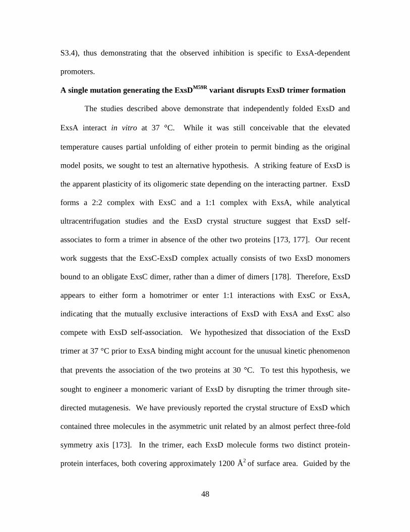
48
S3.4), thus demonstrating that the observed inhibition is specific to ExsA-dependent
promoters.
A single mutation generating the ExsDM59R
variant disrupts ExsD trimer formation
The studies described above demonstrate that independently folded ExsD and
ExsA interact in vitro at 37 C. While it was still conceivable that the elevated
temperature causes partial unfolding of either protein to permit binding as the original
model posits, we sought to test an alternative hypothesis. A striking feature of ExsD is
the apparent plasticity of its oligomeric state depending on the interacting partner. ExsD
forms a 2:2 complex with ExsC and a 1:1 complex with ExsA, while analytical
ultracentrifugation studies and the ExsD crystal structure suggest that ExsD self-
associates to form a trimer in absence of the other two proteins [173, 177]. Our recent
work suggests that the ExsC-ExsD complex actually consists of two ExsD monomers
bound to an obligate ExsC dimer, rather than a dimer of dimers [178]. Therefore, ExsD
appears to either form a homotrimer or enter 1:1 interactions with ExsC or ExsA,
indicating that the mutually exclusive interactions of ExsD with ExsA and ExsC also
compete with ExsD self-association. We hypothesized that dissociation of the ExsD
trimer at 37 °C prior to ExsA binding might account for the unusual kinetic phenomenon
that prevents the association of the two proteins at 30 C. To test this hypothesis, we
sought to engineer a monomeric variant of ExsD by disrupting the trimer through site-
directed mutagenesis. We have previously reported the crystal structure of ExsD which
contained three molecules in the asymmetric unit related by an almost perfect three-fold
symmetry axis [173]. In the trimer, each ExsD molecule forms two distinct protein-
protein interfaces, both covering approximately 1200 Å2
of surface area. Guided by the
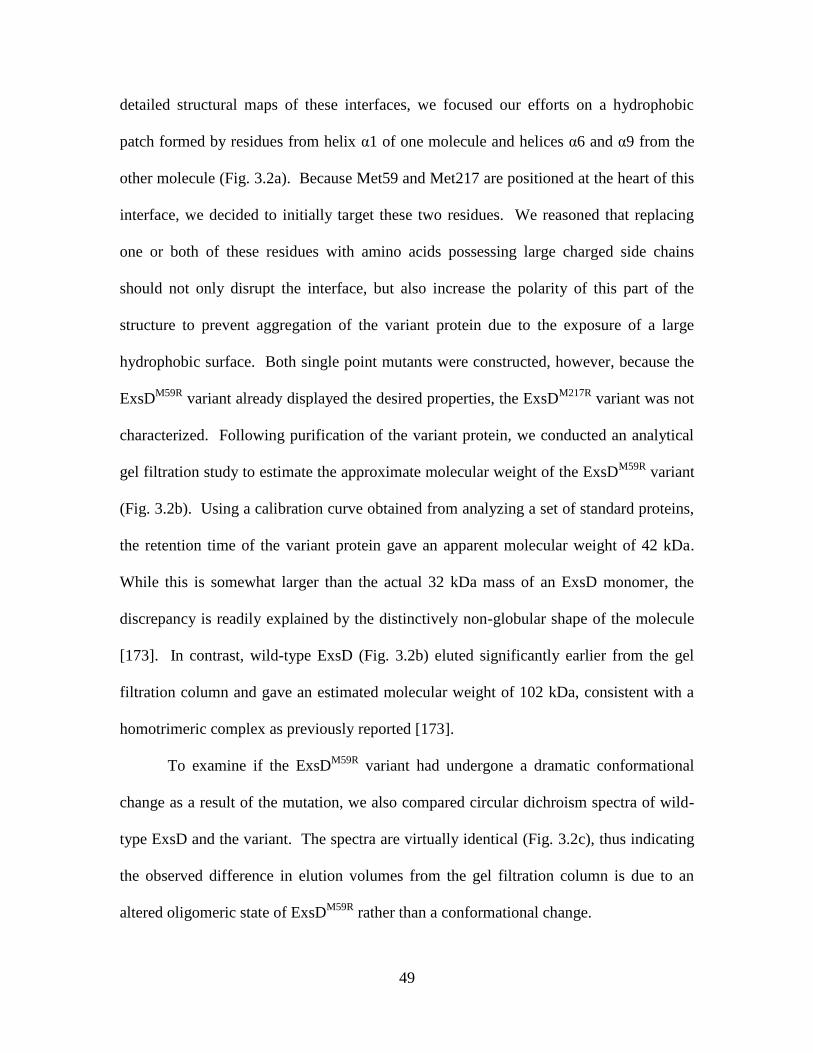
49
detailed structural maps of these interfaces, we focused our efforts on a hydrophobic
patch formed by residues from helix α1 of one molecule and helices α6 and α9 from the
other molecule (Fig. 3.2a). Because Met59 and Met217 are positioned at the heart of this
interface, we decided to initially target these two residues. We reasoned that replacing
one or both of these residues with amino acids possessing large charged side chains
should not only disrupt the interface, but also increase the polarity of this part of the
structure to prevent aggregation of the variant protein due to the exposure of a large
hydrophobic surface. Both single point mutants were constructed, however, because the
ExsDM59R
variant already displayed the desired properties, the ExsDM217R
variant was not
characterized. Following purification of the variant protein, we conducted an analytical
gel filtration study to estimate the approximate molecular weight of the ExsDM59R
variant
(Fig. 3.2b). Using a calibration curve obtained from analyzing a set of standard proteins,
the retention time of the variant protein gave an apparent molecular weight of 42 kDa.
While this is somewhat larger than the actual 32 kDa mass of an ExsD monomer, the
discrepancy is readily explained by the distinctively non-globular shape of the molecule
[173]. In contrast, wild-type ExsD (Fig. 3.2b) eluted significantly earlier from the gel
filtration column and gave an estimated molecular weight of 102 kDa, consistent with a
homotrimeric complex as previously reported [173].
To examine if the ExsDM59R
variant had undergone a dramatic conformational
change as a result of the mutation, we also compared circular dichroism spectra of wild-
type ExsD and the variant. The spectra are virtually identical (Fig. 3.2c), thus indicating
the observed difference in elution volumes from the gel filtration column is due to an
altered oligomeric state of ExsDM59R
rather than a conformational change.
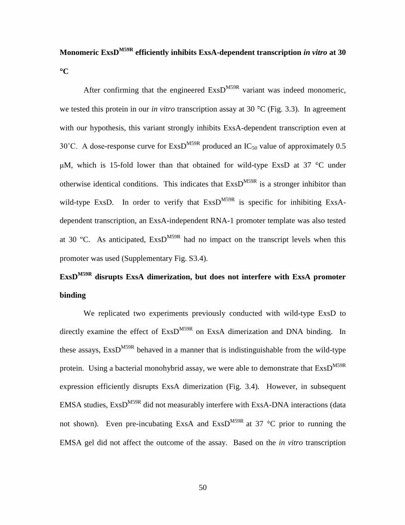
50
Monomeric ExsDM59R
efficiently inhibits ExsA-dependent transcription in vitro at 30
C
After confirming that the engineered ExsDM59R
variant was indeed monomeric,
we tested this protein in our in vitro transcription assay at 30 C (Fig. 3.3). In agreement
with our hypothesis, this variant strongly inhibits ExsA-dependent transcription even at
30˚C. A dose-response curve for ExsDM59R
produced an IC50 value of approximately 0.5
μM, which is 15-fold lower than that obtained for wild-type ExsD at 37 C under
otherwise identical conditions. This indicates that ExsDM59R
is a stronger inhibitor than
wild-type ExsD. In order to verify that ExsDM59R
is specific for inhibiting ExsA-
dependent transcription, an ExsA-independent RNA-1 promoter template was also tested
at 30 C. As anticipated, ExsDM59R
had no impact on the transcript levels when this
promoter was used (Supplementary Fig. S3.4).
ExsDM59R
disrupts ExsA dimerization, but does not interfere with ExsA promoter
binding
We replicated two experiments previously conducted with wild-type ExsD to
directly examine the effect of ExsDM59R
on ExsA dimerization and DNA binding. In
these assays, ExsDM59R
behaved in a manner that is indistinguishable from the wild-type
protein. Using a bacterial monohybrid assay, we were able to demonstrate that ExsDM59R
expression efficiently disrupts ExsA dimerization (Fig. 3.4). However, in subsequent
EMSA studies, ExsDM59R
did not measurably interfere with ExsA-DNA interactions (data
not shown). Even pre-incubating ExsA and ExsDM59R
at 37 C prior to running the
EMSA gel did not affect the outcome of the assay. Based on the in vitro transcription
51
results, we had anticipated that the ExsD variant would also interfere with ExsA-DNA
binding interactions despite not being co-expressed with ExsA.
Discussion
The ExsACDE signaling cascade displays a number of remarkable features, the
most striking perhaps being that signal transduction does not involve the type of
phosphate transfer events that mediate most of the signaling between extracellular milieu
and bacterial gene expression. Instead, signal transduction in the ExsACDE pathway was
shown to be based entirely on the competitive association and dissociation of various
bimolecular complexes formed by the four involved proteins [109, 148, 171]. The newly
discovered role of ExsD trimer formation in the regulation of T3SS gene expression adds
an intriguing new wrinkle to the mechanism. Interestingly, as ITC studies have shown,
ExsD self-association does not interfere with heterocomplex formation between ExsC
and ExsD, suggesting that the affinity of the ExsC-ExsD complex is sufficiently high to
overcome this obstacle [177]. The actual Kd for the ExsD-ExsA interactions is not
known, however, consistent with its position at the bottom of the regulatory cascade, the
complex is readily disrupted by ExsC [172]. Our data suggest that the affinity of the
ExsD-ExsA complex falls perhaps in the hundreds of nanomolar range. Therefore, one
would anticipate that the requirement for dissociation of the ExsD trimer prior to binding
poses a more significant barrier for the ExsD-ExsA complex.
The thermoregulatory effect observed in the in vitro transcription assay appears to
be produced by the self-association of ExsD into trimers. While the ExsDM59R
variant
has lost the ability to trimerize, we do not believe that the temperature increase from 30
52
to 37 C causes a dramatic shift in the monomer-trimer equilibrium of wild-type ExsD.
Rather, we propose that the temperature increase weakens this homotrimer sufficiently to
permit the thermodynamically favored association of ExsD and ExsA. Two pieces of
experimental data support the idea that ExsD is still primarily trimeric at 37 C. The IC50
value for the inhibition of ExsA-dependent transcription by wild-type ExsD is
significantly larger than that observed for the ExsDM59R
variant, suggesting that the wild-
type protein still has a poorer affinity for ExsA. Second, our differential scanning
fluorimetry experiments reveal that the ExsDM59R
variant itself is significantly more
temperature sensitive than the wild-type protein, presumably because the additional
surface areas buried at the trimer interfaces stabilize the wild-type protein (Fig. 3.2d).
Even though the melting temperature (Tm) of a protein depends on many factors, such as
buffer conditions and the presence of ligands, we observed a striking drop in Tm for the
variant, which would indicate that monomeric ExsD is not stable at 37 C unless it is
associated with a different protein, such as ExsA or ExsC.
The observation that ExsDM59R
did not affect ExsA-DNA interactions in the
EMSA experiments is intriguing. In conjunction with the results of our in vitro
transcription experiments, which indirectly demonstrate that ExsDM59R
does interact with
ExsA at 30 C, these findings appear to suggest that the presence of RNA polymerase is
required for the association of ExsD and ExsA. Perhaps the binding of ExsA to RNA
polymerase triggers a conformational change in the transcription factor that reveals the
otherwise obscured ExsD binding site.
Even though ExsD self-association has been documented in two independent
studies, its biological role is not clear [173, 177]. We carried out an initial comparison of
53
wild-type P. aeruginosa and a mutant strain carrying an M59R mutation in the
chromosomal exsD gene using a PexsD-lacZ transcriptional reporter [171]. In this study, we
found no difference in reporter activity under non-inducing or inducing conditions at 30
C (data not shown). These results were perhaps not completely unexpected and
highlight the fundamental question of when the cellular concentration of free ExsD will
most likely reach significant levels. Because ExsD is not a secreted protein and
expression is positively regulated by ExsA, the protein may accumulate inside the cell
during a prolonged period of T3SS induction. However, the pool of free ExsD is
diminished by its associations with ExsA and ExsC. Increases in the cellular levels of
ExsE, on the other hand, should cause the concentration of free ExsD to rise. In the
context of an infection, this could reflect a scenario where a bacterial cell releases from
the host cell at a later stage, causing the closure the T3SS channel, and thereby, an
intracellular accumulation of the ExsE-ExsC complex. While the temperature increase
causes an overall rise in the expression levels of T3SS genes [203, 204], the ability of
ExsD to re-associate with ExsA at 37 C may serve to dampen this effect at this stage,
and thus, fine-tune virulence gene expression. The ExsDM59R
variant provides a useful
system for our molecular studies, however, due to its temperature sensitivity, it is not
well suited for testing this model in vivo. Perhaps, a mutation that attenuates, but does
not fully disrupt ExsD self-association, would be a better choice for these studies. In vivo,
the temperature change from 30 C to 37 C is likely to affect the expression of multiple
P. aeruginosa genes, and it might be instructive to examine the role of ExsD self-
association in the larger context of these changes through computational modeling studies.

54
In summary, we have shown in vitro that independently expressed ExsD does not
inhibit ExsA-dependent transcription at 30 C, but inhibits efficiently at 37 C. A
monomeric ExsD variant strongly inhibits ExsA-dependent transcription at 30 C,
suggesting that the temperature effect is caused by ExsD self-association. ExsD self-
association appears to have no impact on basal T3SS expression levels at 30 or 37 C.
Instead, we propose that trimerization limits the effective concentration of ExsD and
stabilizes the protein. Re-association of the accumulated ExsD protein with ExsA may
assist in fine-tuning T3SS gene expression at a later stage of an infection.
Materials and Methods
Recombinant protein expression and purification
ExsA and ExsD were overexpressed in E. coli from a vector constructed by
Gateway recombinational cloning (Invitrogen, Carlsbad, CA, USA). A tobacco etch
virus (TEV) protease recognition site and the appropriate att recombination sites (attB1
and attB2) were added to the exsA and exsD genes during PCR, and the amplicons were
subsequently recombined into pDONR201 (Invitrogen). The nucleotide sequences of the
ORFs were verified, then recombined into the destination vector pDEST-HisMBP [180]
to create the expression vectors pFS-HMBPExsD and pFS-HMBPExsA. These vectors
were designed to produce either ExsA or ExsD as a fusion to the C-terminus of an N-
terminally His6-tagged E. coli maltose-binding protein (MBP).
Single colonies of E. coli BL21(DE3) CodonPlus RIL cells (Stratagene, La Jolla,
CA, USA) containing either expression plasmid were used to inoculate 125 mL of Luria
broth (LB) supplemented with 2 g/L dextrose, 100 µg/mL ampicillin, and 30 µg/mL
55
chloramphenicol. The cultures were grown with shaking (225 rpm) to saturation
overnight at 37 °C and then diluted 66-fold into 6 L of fresh medium. ExsA cultures
were grown to an OD600 of 1.0, ExsD cultures were grown to an OD600 of 0.5, and
ExsDM59R
cultures were grown to an OD600 of 0.8. All three cultures were induced with
IPTG at a final concentration of 1 mM. The induction temperature for the ExsA cultures
was 18 °C, and they were shaken for six hr. ExsD cultures were induced at 28 °C for
four hr, and ExsDM59R
cultures were induced at 17 °C overnight. Cells were harvested by
centrifugation at 5,000 x g for 15 min. The cell pastes were resuspended in 200 mL of
500 mM NaCl, 25 mM imidazole, 50 mM Tris-HCl (pH 7.4), 2 mM DTT (buffer A),
along with three tablets of Complete, EDTA-free Protease Inhibitor Cocktail (Roche
Applied Science, Indianapolis, IN, USA). The cells were lysed via sonication and
centrifuged at 40,000 x g for 25 min. The supernatants were filtered through 0.45-μm
polyethersulfone membranes and applied to a 30 mL Ni-NTA Superflow affinity column
(Qiagen, Valencia, CA, USA) equilibrated with buffer A. For each run, the column was
washed with five column volumes of buffer A, and proteins were eluted with a linear
gradient from 25 to 250 mM imidazole (pH 7.4).
The His6-MBP-ExsD protein was digested with 5 mg His-tagged TEV (S219V)
protease [183] while being dialyzed overnight in 150 mM NaCl, 50 mM Tris-HCl (pH
7.4), 11.6 mM imidazole (pH 7.4), and 1 mM DTT. The sample was then passed through
a second Ni-NTA column to remove both the His6-MBP tag and the protease, using the
same buffers as the first Ni-NTA column. The protein sample was collected in the flow
through. The sample was diluted with 50 mM Tris-HCl (pH 7.4) and 2 mM DTT in order
to lower the NaCl concentration to 50 mM. The ExsD sample was loaded onto a HiTrap

56
Q HP column (GE Healthcare, Waukesha, WI, USA) that had been equilibrated with 50
mM NaCl, 25 mM Tris-HCl (pH 7.4), and 2 mM DTT, and elution was achieved by
applying a linear gradient of NaCl from 50 mM to 1 M. Finally, gel filtration was
performed using 150 mM NaCl, 25 mM Tris-HCl (pH 7.4), and 2 mM TCEP (ExsD
storage buffer). ExsDM59R
was purified in the same manner as wild-type ExsD, except
that no anion exchange was performed. All purification steps were performed at 4 °C.
After each purification step, fractions were analyzed via SDS-PAGE and pooled
accordingly. ExsD and ExsDM59R
were concentrated to 4.5 mg/mL and 6.8 mg/mL,
respectively. Protein samples were flash-frozen using liquid nitrogen and stored at
-80 °C.
The His6-MBP-ExsA fusion protein was treated differently. Following the initial
Ni-NTA affinity purification step, the fusion protein was dialyzed against a buffer of 50
mM NaCl, 25 mM Tris-HCl (pH 7.4), and 2 mM DTT and loaded onto a HiTrap Q HP
column (GE Healthcare) that had been equilibrated with the same buffer. The His6-MBP-
ExsA fusion protein was eluted using a linear NaCl gradient from 0.05 M to 1 M. The
sample was dialyzed against 2 L of 45 mM NaCl, 25 mM Tris-HCl (pH 7.15), and 2 mM
DTT (buffer B) overnight. The sample was then loaded onto a HiTrap Heparin HP
column (GE Healthcare) equilibrated in buffer B and eluted with a 0.05 M to 1 M
gradient of NaCl. The NaCl concentration in the His-MBP-ExsA sample was adjusted to
0.5 M, and the fusion protein was digested with 3 mg of His-tagged TEV(S219V)
protease at 4 C overnight. Next, ExsA was run through a second Ni-NTA Superflow
affinity column, this time collecting ExsA in the flow through. Finally, gel filtration
using a HighLoad 26/60 Superdex 200 prep grade column (GE Healthcare) was
57
performed with the ExsA sample using 500 mM NaCl, 25 mM Tris-HCl (pH 7.4), and 2
mM TCEP (ExsA storage buffer). The sample was concentrated to 1 mg/mL, flash-
frozen using liquid nitrogen, and stored at -80 °C.
RNA polymerase purification and specific activity determination
RNA polymerase (RNAP) was purified from P. aeruginosa PAO1 cells following
the original procedure of Allan and Kropinski [184]. However, changes were made to the
later chromatographic steps. All purification steps were performed at 4 °C. P.
aeruginosa PAO1 cultures were grown in LB broth to an OD600 of 0.8, harvested by
centrifugation at 6,000 x g, then lysed by sonication. The cell debris was removed by
centrifugation at 35,000 x g for 30 min, and 25% polyethyleneimine (pH 7.5) was added
to the supernatant to a final concentration of 0.5% in order to precipitate the RNAP. The
supernatant was centrifuged at 35,000 x g for 30 min. The polyethyleneimine precipitate
was washed with 10 mM Tris-HCl (pH 8.0), 250 mM NaCl, 5% glycerol, 0.05 mM
EDTA, 1 mM DTT, and 0.1 mM PMSF (wash buffer) and centrifuged at 35,000 x g for
30 min. RNAP was released by resuspending the pellet in 10 mM Tris-HCl (pH 8.0), 800
mM NaCl, 5% glycerol, 0.05 mM EDTA, 1 mM DTT, and 0.1 mM PMSF (release
buffer) and centrifuged at 25,000 x g for 30 min. Ammonium sulfate was added to the
supernatant to a final concentration of 30%, followed by gentle stirring for one hr, and
centrifugation at 35,000 x g for 30 min. Additional ammonium sulfate was then added to
bring the supernatant to 60% saturation. After a second centrifugation at 35,000 x g for
30 min, the pellet was resuspended in 1 mL wash buffer per liter of original culture. The
suspension was dialyzed versus 2 L wash buffer overnight. The dialyzed RNAP sample
was centrifuged, and the supernatant was filtered in preparation for gel filtration. The
58
sample was run through a Sephacryl S-300 HR column (GE Healthcare) using wash
buffer. The fractions were analyzed by SDS-PAGE and collected to run on a Hi-Trap
Heparin HP column (GE Healthcare) using a loading buffer composed of 10 mM Tris-
HCl (pH 8.0), 250 mM NaCl, 5% glycerol, 0.05 mM EDTA, and 1 mM TCEP. RNAP
was eluted using a linear gradient of 0.25 M to 1 M NaCl. Fractions were analyzed via
SDS-PAGE, pooled, and concentrated to 1 mg/mL of total protein. Glycerol was added
to a final concentration of 50%. RNAP was aliquoted and stored at -20 °C.
The specific activity of the purified P. aeruginosa RNAP was determined by
comparing its activity to a standard curve generated with different amounts of E. coli
RNA polymerase holoenzyme (Epicentre Biotechnologies, Madison, WI, USA) using an
ExsA-independent RNA-1 promoter which produces a 108 base transcript [185].
Site-directed mutagenesis
The ExsDM59R
variant was generated by site-directed mutagenesis using Quik-
Change (Stratagene) and the manufacturer’s suggested protocol. The following primers
were used:
5´–CTGCAGCGGCGGCTGCCGCGCCTGCGGCTGGAGC–3´
5´–GGCGCGGCAGCCGCCGCTGCAGCAACGCCAG–3´.
ExsA-dependent in vitro transcription assays
The linear DNA template used in each assay encompassed positions -207 to 94 of
the PexsD promoter, relative to the transcription start site; and from this template, RNA
polymerase synthesizes an 82 base mRNA transcript. The template was produced by
PCR using forward primer 5´-CATCAGTTGCTGCTCAACAGCG-3´ and reverse primer
5´-CACCGCTTCTCGGGAGTACTGC-3´. The PCR product was run on a 2% agarose
59
gel and purified using the Wizard SV Gel and PCR Clean-up System (Promega, Madison,
WI, USA). Each 30 µL transcription assay reaction contained 4.4 fM of promoter
template, 50.4 µM bovine serum albumin (to eliminate non-specific protein-protein
interactions), 10 U purified RNA polymerase from P. aeruginosa (see above), 1 U
RiboGuard RNase Inhibitor (Epicentre Biotechnologies), 15 ng/µL poly(deoxyinosinic-
deoxycytidylic) acid (to prevent non-specific transcription initiation), 133 mM NaCl, 32
mM Tris-HCl (pH 7.4), 10 mM MgCl2, 25 µM EDTA, 0.9 mM TCEP, 0.2 mM DTT, and
15.5% glycerol. The time-course experiments contained 64 nM ExsA and either no ExsD
or 50 µM ExsD (no ExsA was added for the RNA-1 control experiments). Samples were
mixed and allowed to equilibrate at room temperature for five min. Samples were then
pre-incubated for 10 min at either 30 or 37 °C, depending on the experiment. Next, 3 µL
NTPs (stock concentrations of 200 µM ATP, CTP, GTP and 40 µM UTP) mixed with 0.2
µL (0.2 µCi) of 3.3 mM P32
-alpha UTP was added to each sample to start the reaction,
and samples were incubated at either 30 or 37 °C, depending on the experiment. After
the reactions were stopped by adding 12 µL 1X stop solution (3M ammonium acetate, 50
mM EDTA, 0.11 mg/mL glycogen), 170 µL 100% cold ethanol was added, and the
samples were incubated at -20 °C for one hr. Following centrifugation at 12,000 x g for
15 min, the supernatant was discarded and pellets were resuspended in 12 μL 1X TBE
(Tris/Borate/EDTA)-urea sample buffer and heated at 70 °C for five min. After a brief
centrifugation, the samples were loaded onto a 10% TBE-urea gel and run at 200 mV for
60 min. Gels were exposed to a storage phosphor screen (GE Healthcare) for 16 hr. The
phosphor screen was scanned using a Typhoon Trio Variable Mode Imager (GE
Healthcare), and gel bands were quantified using Image Quant TL v2005 (Amersham

60
Biosciences, Piscataway, NJ, USA). Each experiment was performed in triplicate, and
curve fits were analyzed with XLfit (IDBS, Bridgewater, NJ, USA).
Analytical size exclusion chromatography
One hundred µL samples of 2.1 µM purified wild-type ExsD and ExsDM59R
, each
containing 150 mM NaCl, 25 mM Tris-HCl (pH 7.4), and 2 mM TCEP, were separately
loaded onto a Superdex 200 10/300 GL column (GE Healthcare). The proteins were
eluted with 150 mM NaCl, 25 mM Tris-HCl (pH 7.4), and 2 mM TCEP. The absorbance
at UV280 was plotted on the y-axis and Ve/Vo was plotted on the x-axis, where Ve is the
elution volume and Vo is the void volume (7.93 mL). Cytochrome C, carbonic anhydrase,
bovine serum albumin, and β-amylase standards were individually run using the same
elution buffer. These standards were plotted using the log of their known molecular
weights on the y-axis and Ve/Vo on the x-axis; from this, a best fit line was determined.
The molecular weights of wild-type ExsD and ExsDM59R
were subsequently estimated
using the fitted linear equation.
Circular dichroism (CD) measurements
Circular dichroism measurements using far-UV (200-240 nm) of wild-type ExsD
and ExsDM59R
, each at a concentration of 5.3 µM, were separately measured at 4 °C using
a JASCO J-815 Circular Dichroism Spectrometer (JASCO, Easton, MD, USA) with a 1
mm pathlength cuvette. Each protein sample contained 150 mM NaCl, 25 mM Tris-HCl
(pH 7.4), and 2 mM TCEP. The spectra obtained represent an average of three scans that
were corrected for the buffer baseline.
61
Differential scanning fluorimetry (DSF)
The DSF experiments were performed utilizing an ABI 7300 Real-Time PCR
System (Applied Biosystems, Foster City, CA, USA). Each 30 μL reaction contained 5x
Sypro Orange (Ex. 490 nm, Em. 530 nm) (Invitrogen), 10 μM wild-type ExsD or
ExsDM59R
, 150 mM NaCl, 8.3 mM Tris-HCl (pH 7.4), and 0.67 mM TCEP. Starting at
10 °C, the temperature was incrementally increased to 68 °C at a rate of 1 °C per min.
Data analysis was conducted using XLfit (IDBS).
LexA monohybrid assay
Quik-Change (Stratagene) mutagenesis was used to modify the previously
constructed arabinose-inducible vector pJNexsD [172] by introducing an M59R
substitution in exsD using primer ExsDM59R
(5´-
CGTTGCTGCAGCGGCGCCTGCCGCGCCTGC-3´). The resulting plasmid was
designated pAM102. E. coli strain SU101, which carries a LexA-repressible PsulA-lacZ
reporter, was transformed with pAM102 and the IPTG-inducible pSR658-exsA vector
[172] and selected on LB agar with gentamicin (15 µg/mL) and tetracycline (12 µg/mL).
Self-association of ExsA in the presence of ExsD or ExsDM59R
was measured as
previously described [172, 205]. Briefly, strains containing the appropriate expression
vectors were grown overnight at 30 °C with shaking in 5 mL LB with 50 µM IPTG, 0.5%
arabinose, and appropriate antibiotics. The following day, cultures were diluted to OD600
= 0.1 in 5 mL trypticase soy broth supplemented with 100 mM monosodium glutamate
and 1% glycerol, as well as 50 μM IPTG, 0.5% arabinose, and antibiotics and grown at
30 °C with shaking to OD600 = 1.0. β-galactosidase levels were measured as previously
described [172]. Miller units were reported as the average of three replicates with error

62
bars representing the standard error of the mean (SEM). Immunoblots were performed
with -ExsD and -LexA1-87 to confirm the stability of ExsD and ExsA-LexA1-87,
respectively.
Acknowledgements
This study was supported by the National Institutes of Health (1R21AI101774-01
to FDS and RO1- AI055042 to TLY) and the American Heart Association
(09SDG2260401 to FDS).
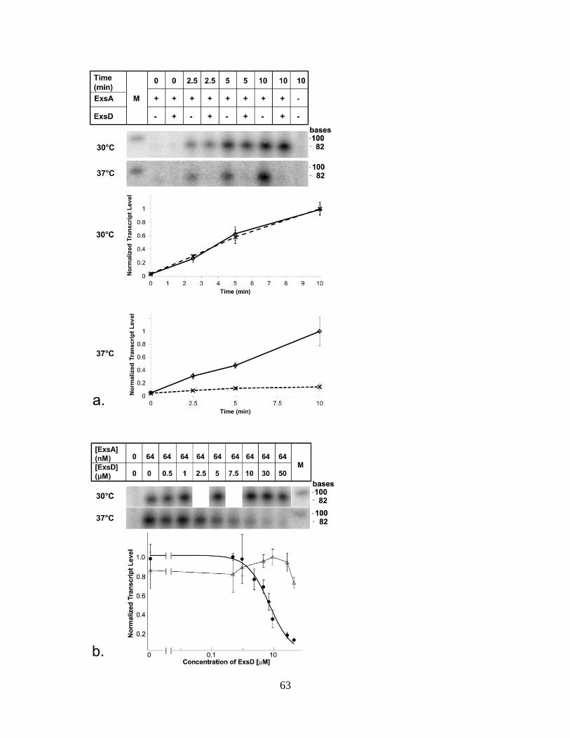
63
64
Fig. 3.1. Temperature-dependent regulation of ExsA by ExsD. a. Autoradiograms
and graphical representations for the in vitro transcription of an 86 nucleotide transcript
from an ExsA-dependent PexsD promoter template. Assays were performed at 30 C and
37 °C and in absence and presence of 50 μM ExsD. In both graphs, the ExsD-free data
are represented by solid trend lines and empty diamonds, whereas the data obtained in the
presence of ExsD are represented by dashed trend lines and crosses. b. Dose response
data and fit generated by measuring in vitro transcription levels at the 10-min time point
at 37 C in the presence of increasing amounts of ExsD protein (filled circles). Data
(empty triangles) and dashed trend line obtained when the same experiment was
conducted at 30 C. The 30 C gel strip has been digitally manipulated to align the fewer
data points with those generated in the experiments performed at 37 C. In order to
obtain a reliable dose-response curve, two additional concentrations were included in the
37 C experiment.
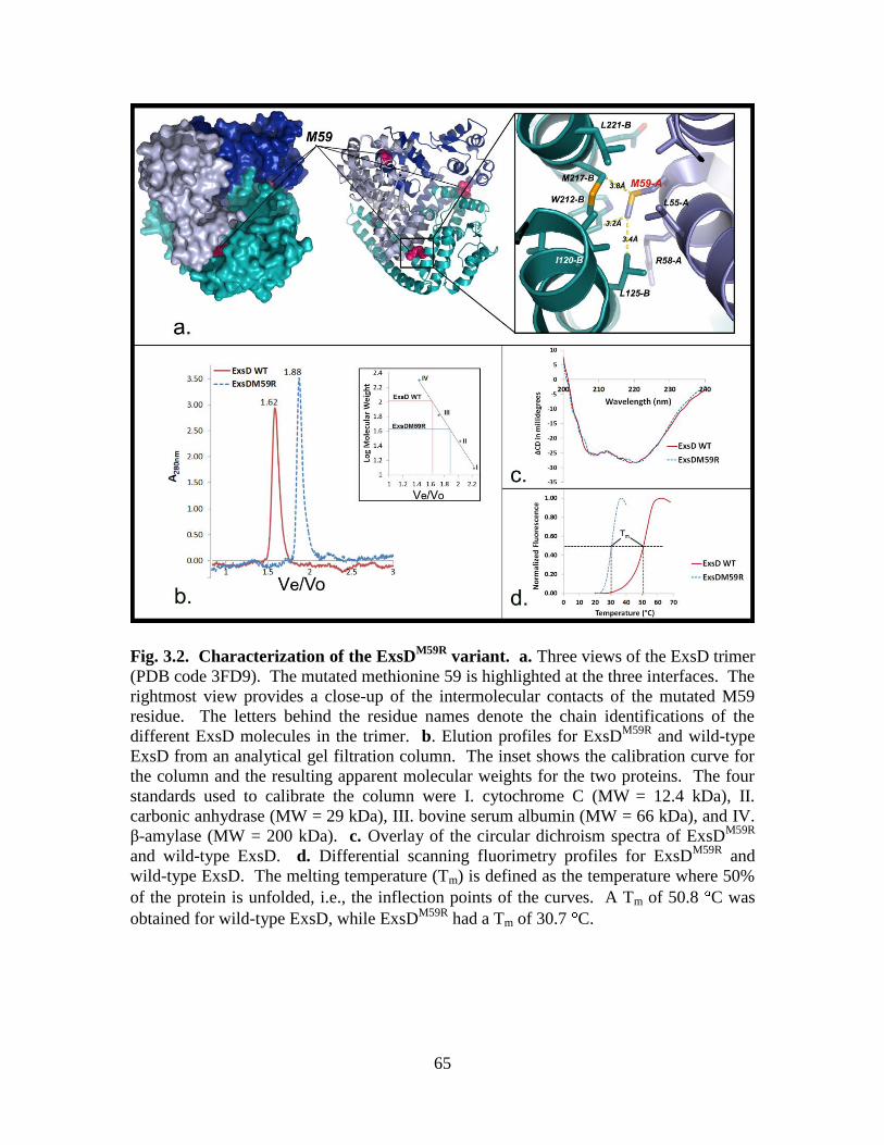
65
Fig. 3.2. Characterization of the ExsDM59R
variant. a. Three views of the ExsD trimer
(PDB code 3FD9). The mutated methionine 59 is highlighted at the three interfaces. The
rightmost view provides a close-up of the intermolecular contacts of the mutated M59
residue. The letters behind the residue names denote the chain identifications of the
different ExsD molecules in the trimer. b. Elution profiles for ExsDM59R
and wild-type
ExsD from an analytical gel filtration column. The inset shows the calibration curve for
the column and the resulting apparent molecular weights for the two proteins. The four
standards used to calibrate the column were I. cytochrome C (MW = 12.4 kDa), II.
carbonic anhydrase (MW = 29 kDa), III. bovine serum albumin (MW = 66 kDa), and IV.
β-amylase (MW = 200 kDa). c. Overlay of the circular dichroism spectra of ExsDM59R
and wild-type ExsD. d. Differential scanning fluorimetry profiles for ExsDM59R
and
wild-type ExsD. The melting temperature (Tm) is defined as the temperature where 50%
of the protein is unfolded, i.e., the inflection points of the curves. A Tm of 50.8 C was
obtained for wild-type ExsD, while ExsDM59R
had a Tm of 30.7 C.

66
Fig. 3.3. Effect of ExsDM59R
on ExsA-dependent transcription. Dose response data
and curve fit generated by measuring in vitro transcription levels in the presence of
increasing amounts of ExsDM59R
protein at the 10-min time point and at 30 C (clear
diamonds). To highlight the differences, data shown in figure 3.1b are reproduced. Clear
triangles show the results for the titration experiment with wild-type ExsD at 30 C,
while filled circles show data obtained at 37 C.
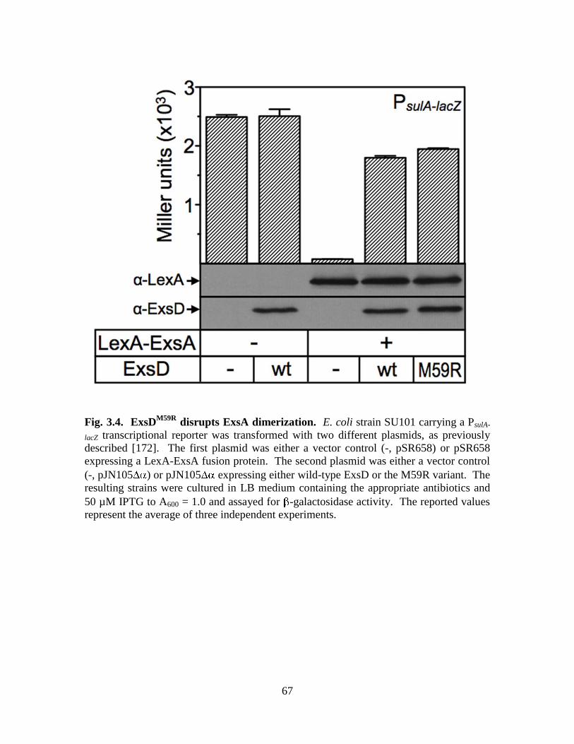
67
Fig. 3.4. ExsDM59R
disrupts ExsA dimerization. E. coli strain SU101 carrying a PsulA-
lacZ transcriptional reporter was transformed with two different plasmids, as previously
described [172]. The first plasmid was either a vector control (-, pSR658) or pSR658
expressing a LexA-ExsA fusion protein. The second plasmid was either a vector control
(-, pJN105 ) or pJN105 expressing either wild-type ExsD or the M59R variant. The
resulting strains were cultured in LB medium containing the appropriate antibiotics and
50 µM IPTG to A600 = 1.0 and assayed for -galactosidase activity. The reported values
represent the average of three independent experiments.
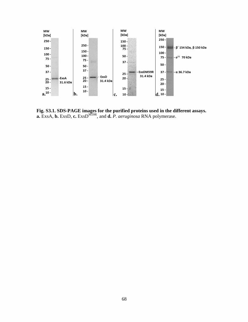
68
Fig. S3.1. SDS-PAGE images for the purified proteins used in the different assays. a. ExsA, b. ExsD, c. ExsD
M59R, and d. P. aeruginosa RNA polymerase.

69
Fig. S3.2. In vitro transcription assay time-course at 37 °C. Initial time-course study
performed to determine a suitable time-point for the dose response experiments. Under
the described experimental conditions, the rate of transcription remains linear for at least
20 min.
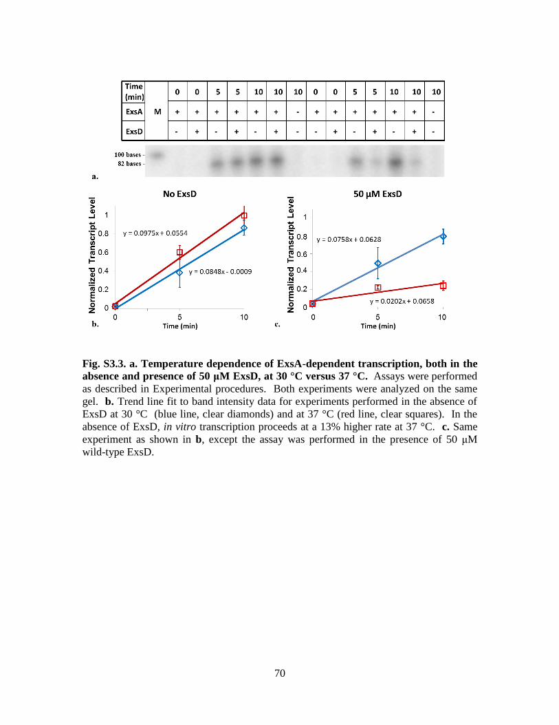
70
Fig. S3.3. a. Temperature dependence of ExsA-dependent transcription, both in the
absence and presence of 50 μM ExsD, at 30 °C versus 37 °C. Assays were performed
as described in Experimental procedures. Both experiments were analyzed on the same
gel. b. Trend line fit to band intensity data for experiments performed in the absence of
ExsD at 30 °C (blue line, clear diamonds) and at 37 °C (red line, clear squares). In the
absence of ExsD, in vitro transcription proceeds at a 13% higher rate at 37 °C. c. Same
experiment as shown in b, except the assay was performed in the presence of 50 μM
wild-type ExsD.
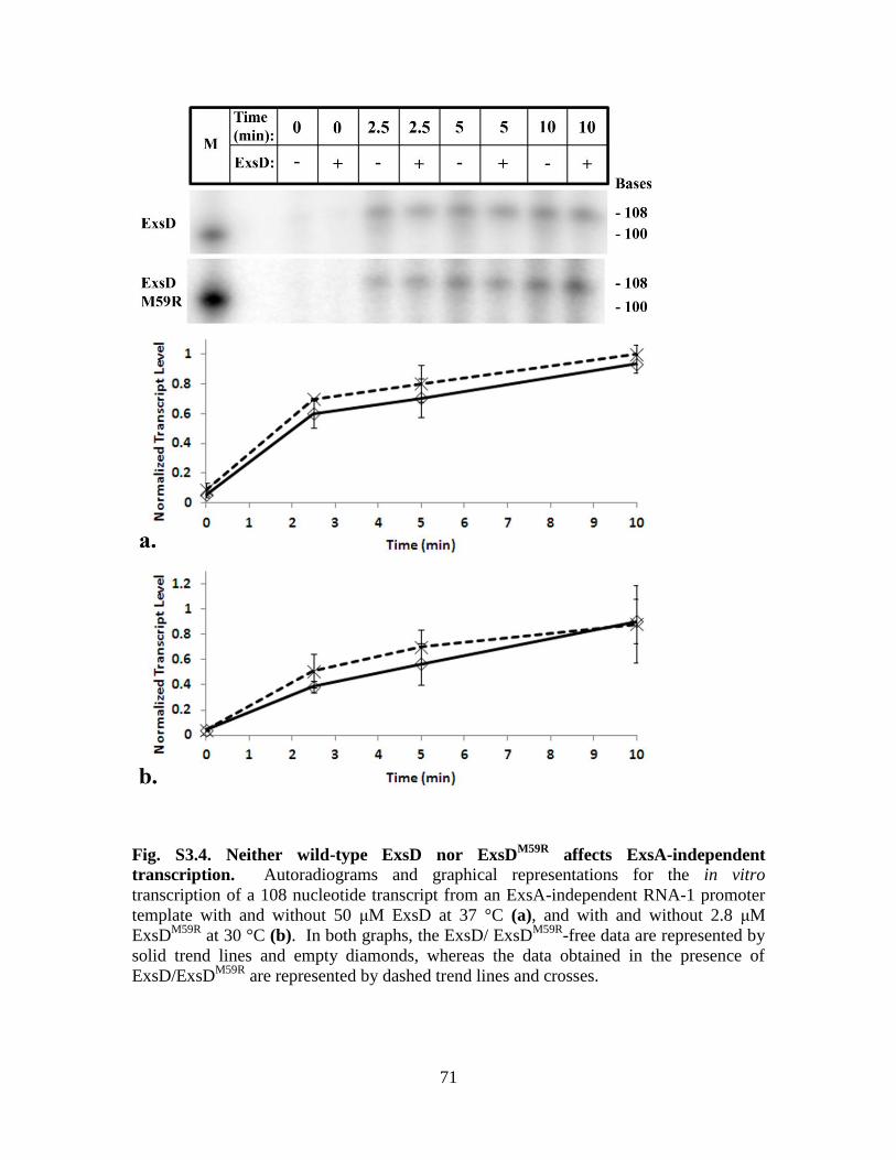
71
Fig. S3.4. Neither wild-type ExsD nor ExsDM59R
affects ExsA-independent
transcription. Autoradiograms and graphical representations for the in vitro
transcription of a 108 nucleotide transcript from an ExsA-independent RNA-1 promoter
template with and without 50 μM ExsD at 37 °C (a), and with and without 2.8 μM
ExsDM59R
at 30 °C (b). In both graphs, the ExsD/ ExsDM59R
-free data are represented by
solid trend lines and empty diamonds, whereas the data obtained in the presence of
ExsD/ExsDM59R
are represented by dashed trend lines and crosses.
72
CHAPTER FOUR
Mapping the ExsD–ExsA interface and further elucidation of the type
III secretion system inhibitory mechanism in Pseudomonas aeruginosa
Robert C. Bernhards1, Yi Xiao
1, Anne E. Marsden
2, Shannon K. Esher
1, Timothy L.
Yahr2, and Florian D. Schubot
1*
Manuscript in Preparation
1Virginia Polytechnic Institute and State University
Department of Biological Sciences
125 Life Sciences 1 Building
Washington Street
Blacksburg, VA 24061
2University of Iowa
Department of Microbiology
540B Eckstein Medical Research Building
Iowa City, IA 52242
*Corresponding author: Phone: (540) 231-2393
Fax: (540) 231-7126
Email: [email protected]
Keywords: ExsA, ExsD, Pseudomonas aeruginosa, type III secretion.
Abbreviations: DSF = differential scanning fluorimetry; EMSA = electrophoretic
mobility shift assay; FA = fluorescence anisotropy; IPTG = isopropyl-β-D-thiogalacto-
pyranoside; MBP = maltose binding protein; TCEP = Tris(2-carboxyethyl)phosphine;
TEV = tobacco etch virus; T3SS = type III secretion system.
73
Abstract
Pseudomonas aeruginosa is an emerging opportunistic human pathogen that is
problematic due to its resistance to a multitude of common antibiotics. P. aeruginosa
utilizes a type III secretion system (T3SS) to cause acute infections. The T3SS is
regulated by a cascade of four interacting proteins: ExsA, ExsD, ExsC and ExsE. ExsA
is the main transcriptional activator and regulates transcription of all the genes necessary
for the T3SS. ExsD is the antiactivator and inhibits ExsA when there is no contact of the
T3SS needle and the host cell. Meanwhile, ExsC sequesters ExsE. Upon host cell
contact, ExsE is secreted into the host cell cytoplasm, which frees ExsC to bind to ExsD.
ExsA is now free to activate transcription. The way in which ExsD inhibits ExsA is
largely unknown. The current model posits that ExsD inhibits ExsA by binding in a 1:1
complex, which interrupts ExsA dimerization and prevents ExsA from binding to the
T3SS promoters. This study reveals that there could be more to this inhibitory
mechanism. We show conclusive evidence that ExsD is a DNA-binding protein through
differential scanning fluorimetry (DSF) and electrophoretic mobility shift assays
(EMSAs). We also begin to map the ExsD–ExsA binding interface by testing an
assortment of ExsD variants in an in vitro transcription assay to determine which regions
of ExsD are important for ExsA binding and inhibition. A complete understanding of
how ExsD inhibits ExsA would allow for the development of drugs that mimic ExsD
function and target ExsA to shut down the T3SS.

74
Introduction
Pseudomonas aeruginosa utilizes a type III secretion system (T3SS) to infect its
host and cause acute infections [191, 193]. The T3SS is regulated by the ExsA-ExsC-
ExsD-ExsE cascade, which works by direct protein-protein interactions [179]. In this
cascade, ExsA is main transcriptional activator and controls expression of all the genes
necessary for type III secretion [120]. ExsA is a member of the AraC/XylS family of
transcriptional activators [120]. When the T3SS is turned off, ExsA is inhibited by the
antiactivator protein ExsD [171]. ExsD binds to ExsA in a 1:1 complex [147].
Meanwhile, ExsC sequesters ExsE [108, 109]. Upon host cell contact, ExsE is secreted
through the T3SS needle apparatus into the host cell cytoplasm [108, 109]. This now
allows for ExsC to bind to ExsD [148, 177], which frees ExsA to bind to the T3SS
promoters as a dimer and recruit RNA polymerase (RNAP) to activate T3SS transcription
[149].
ExsD is a dynamic protein in that it interacts with different protein binding
partners in different ways. ExsD exists in a 2:2 complex with ExsC [177], where a dimer
of ExsC is bound to two ExsD monomers [178]. In addition to ExsD binding to ExsA in
a 1:1 complex [147], ExsD also forms a self-trimer when not bound to a different protein
[173]. Furthermore, structural evidence as well as preliminary experimental evidence
suggests that ExsD is a DNA-binding protein and binds to DNA as a monomer [173].
The plasticity of ExsD gives it the ability to function in many different ways. ExsD is of
significant medical importance given that it is capable of inhibiting the T3SS in P.
aeruginosa [171]. Much can be gained by studying ExsD and how it is able to inhibit
ExsA to shut down the T3SS.
75
This study examines the interaction between ExsD and ExsA. The current model
proposes that ExsD disrupts ExsA dimerization and prevents ExsA from binding to the
T3SS promoters [172]. ExsD does not interact with DNA in this model. However, based
on the crystal structure of ExsD, which was previously solved by our lab, ExsD
resembled a DNA-binding protein [173]. Here we further examine the ability of ExsD to
bind DNA. Wild type ExsD as well as an ExsDM59R
monomeric variant were examined
using differential scanning fluorimetry (DSF) to demonstrate that ExsD is a DNA-
binding protein. ExsD DNA-binding was confirmed using ExsDM59R
in an
electrophoretic mobility shift assay (EMSA).
The main focus of this study was to map the ExsD–ExsA binding interface. A
group of ExsD variants lacking certain regions of the protein were examined in an in
vitro transcription assay to determine which regions of ExsD are important for ExsA
binding and inhibition. It was determined that the coiled-coil region of ExsD is not
important for ExsA binding. On the other hand, we determined that the amino terminus
of ExsD is important, but not sufficient to inhibit ExsA-dependent transcription.
Results
ExsD is a DNA-binding protein
The crystal structure of ExsD shows that it resembles a DNA binding protein
from E. coli called KorB [173, 174]. Initial examination by DSF provided preliminary
evidence that ExsD binds nonspecifically to DNA [173]. DSF measures the melting
temperature of a protein. If a protein binds to DNA, the protein typically becomes more
stable, which results in an increase in melting temperature. However, when DNA is
76
added to ExsD, it results in a decrease in melting temperature [173]. The ExsD structure
suggests that ExsD binds to DNA as a monomer [173], and this could explain the
decrease in melting temperature. The ExsD trimer without DNA could be more stable
than an individual monomer bound to DNA. To further test this hypothesis, a monomeric
ExsD variant called ExsDM59R
[206] was examined using DSF for its ability to bind DNA
(Fig. 4.1). A portion of the PexsD promoter was used as the dsDNA. Without DNA, the
melting temperature of ExsDM59R
was 27.64 ± 0.06 °C, and when DNA was added the
ExsDM59R
melting temperature increased to 31.57 ± 0.22 °C. Along with the wild type
ExsD data, this supports the hypothesis that ExsD binds to DNA as a monomer.
The lab of Dr. Timothy Yahr at the University of Iowa used EMSA to determine
whether ExsD and ExsDM59R
disrupt ExsA promoter binding (Fig. 4.2). In this assay,
ExsA forms two distinct shift products when it binds to one of its promoters. The ExsA-
dependent PexsC promoter was used in this experiment. The lower shift product
corresponds to one ExsA molecule bound to the promoter, and the higher and more
pronounced shift product occurs when two ExsA molecules bind to the promoter [167].
If ExsD inhibits ExsA DNA binding then they expected to see a lower amount of shifted
DNA when ExsD is added. Although they were not able to see an effect at these
particular protein concentrations and temperature (30 °C), they were able to examine the
ability of ExsD and ExsDM59R
to bind to DNA. At 30 °C, ExsD wild type was unable to
bind to PexsC. This is most likely due to the fact that the ExsD trimer is very stable at this
temperature and does not dissociate to bind to DNA. This could also explain why there
was no disruption of ExsA DNA binding by wild type ExsD. However, ExsDM59R
shifted
the PexsC as well as the nonspecific DNA control (pscF) at 30 °C (Fig. 4.2). This
77
confirms that ExsD is a DNA binding protein. However, since ExsDM59R
nonspecifically
binds to DNA, it is not known whether or not ExsA-dependent promoters are specific
targets for ExsD.
The coiled-coil region of ExsD is not important for ExsA binding
In order to map the ExsD–ExsA interface, we designed a variety of ExsD variants.
The first variant was ExsD lacking the coiled-coil region (ExsDΔC-C). The ExsDΔC-C
variant was constructed using site-directed mutagenesis and lacked residues 138-202.
The two separated regions were fused by a linker consisting of four glycines. Following
overexpression and purification, the ExsDΔC-C variant (Fig. 4.3) was examined using an
in vitro transcription assay. We hypothesized that if the coiled-coil region was important
for ExsA binding, then we would see less ExsA-dependent inhibition in the assay. A
titration of ExsDΔC-C was performed at 37 °C (Fig. 4.4). To our surprise, ExsDΔC-C
turned out to be a more potent inhibitor than wild type ExsD (IC50 = 0.97 ± 0.4 μM). We
reasoned that deleting the coiled-coil region made the protein primarily monomeric. We
previously demonstrated that the monomeric ExsD variant (ExsDM59R
) is a more potent
inhibitor of ExsA dependent transcription [206]. At 37 °C, ExsDΔC-C had an IC50 value
of 0.97 ± 0.4 μM, which closely resembles the IC50 value of ExsDM59R
(1.3 ± 0.1 μM) at
37 °C. The ExsDΔC-C titration was also performed at 30 °C, and it produced an IC50
value of 4.3 ± 0.4 μM (Fig. 4.4). We suspect that at the lower temperature, ExsDΔC-C is
still capable of forming trimers, so a certain percentage is trimeric. This would explain
the increase in IC50 value. Nevertheless, we concluded that the coiled-coil region of
ExsD is not important for ExsA binding or inhibition.
78
The amino terminus of ExsD is important for ExsA binding
The next region of ExsD we wanted to examine was the N-terminus, since it was
structurally located on the opposite side of the protein from the coiled-coil region. We
also suspected that the ExsD N-terminus of ExsD could be important for ExsA binding,
because the N-terminus of ExsD is predicted to be important for binding to ExsC [178].
It would be reasonable to assume that the binding of ExsD to either ExsC or ExsA
involves a competitive ExsD binding region. In order to test this hypothesis, a variant of
ExsD lacking the first 20 amino acids on the N-terminus was made by limited proteolysis.
A simple thermolysin digest results in a highly stable ExsDΔ20 product [173]. ExsDΔ20
was tested in the in vitro transcription assay at 37 °C using the same titration as for
ExsDΔC-C. The results show a decrease in ExsA-inhibition, indicating that the ExsD N-
terminus is important for ExsA binding and inhibition (Fig 4.5). To confirm that the
observed effect of ExsDΔ20 is specific to inhibit ExsA-dependent transcription, we
performed an in vitro transcription assay time course using a DNA template containing
the ExsA-independent RNA-1 promoter [185] (Fig 4.6). ExsDΔ20 showed no significant
inhibition, indicating that ExsD1-46 is specific to inhibit ExsA.
To confirm that the ExsD N-terminus is important for ExsA binding, we ordered a
synthetically made polypeptide of the first 46 amino acids of the ExsD N-terminus
(ExsD1-46). A titration in the in vitro transcription assay at 37 °C shows ExsD1-46 does
inhibit ExsA-dependent transcription, albeit weakly (Fig. 4.7). Since the inhibition was
weak, we wanted to confirm that the observed effect of ExsD1-46 is specific to ExsA-
dependent transcription, so we performed an in vitro transcription assay time course
using a DNA template containing the ExsA-independent RNA-1 promoter (Fig. 4.8).
79
ExsD1-46 showed no significant inhibition, indicating that ExsD1-46 is specific to inhibit
ExsA. Overall, these results suggest that the amino terminus of ExsD is important for
ExsA binding, but is not sufficient to inhibit ExsA-dependent transcription.
Discussion
In P. aeruginosa, ExsA-dependent T3SS transcription is inhibited by the
antiactivator ExsD [171]. ExsD is a dynamic protein with many different binding
partners and most likely multiple functions [147, 148, 173, 178]. This study confirms
that ExsD is a DNA binding protein. However, the role of ExsD DNA binding is unclear.
Even though ExsD exists as a trimer, we discovered that ExsD can only bind to DNA as a
monomer. Thus, one of the functions of the ExsD trimer could be to prevent ExsD from
binding to DNA non-specifically. The ExsD trimer also most likely serves to stabilize
the protein, given that the ExsD monomer is significantly less stable. It is unclear
whether ExsD has a specific DNA binding site. A ChIP-sequencing study is currently
underway to investigate this question.
The main goal of this study was to begin to map the ExsD–ExsA interface, as no
information on the way ExsD and ExsA interact currently exists. ExsD variants were
constructed and purified, and an in vitro transcription assay was utilized to determine
which region of ExsD is important for ExsA-binding. The results are shown together in
Fig. 4.9 for comparison. It was determined that the ExsD coiled-coil region is not
important for ExsA binding. However, the ExsD amino terminus is important for ExsA
binding, but not sufficient to inhibit ExsA-dependent transcription. We suspect that
another portion of ExsD somewhere between the amino terminus and the coiled-coil
80
region is also important for ExsA-binding. Future studies will further map the ExsD–
ExsA interface by testing amino acid substitution mutants in the in vitro transcription
assay to determine which amino acids are critical for ExsA-binding. We are also
attempting to crystallize the ExsD–ExsA complex.
The way in which ExsD inhibits ExsA remains a mystery. We propose three
possible scenarios. The first is the current model where ExsD disrupts ExsA dimerization
and promoter interactions by binding to ExsA in a 1:1 complex [147, 172]. The second
scenario is ExsD binds to ExsA while ExsA is bound to the promoter and disrupts ExsA
dimer formation. ExsD may or may not be binding to DNA under this scenario. The
third possibility is that ExsD binds to ExsA when the dimer is formed on the promoter
and disrupts ExsA–RNAP interactions. ExsD shows structural similarity to GreB, which
is a RNAP binding protein in E. coli [176]. During transcription, RNAP occasionally
loses its grip on the growing mRNA end during elongation and backtracks on the DNA
template [176]. GreB binds to RNAP and cleaves the disengaged 3´ RNA segment, so
that RNAP can proceed with transcription [176]. The coiled-coil region of GreB inserts
into RNAP [176], and given that the coiled-coil regions of ExsD and GreB are very
similar structurally, one could imagine that ExsD interacts with RNAP in the same
manner. Given that the coiled-coil region of ExsD does not interact with ExsA, ExsD
could be binding to both ExsA and RNAP at the same time. ExsD could function to stall
RNAP, thereby inhibiting T3SS transcription.
Insights into the way ExsD inhibits ExsA could lead to the discovery of drugs that
mimic ExsD function and target ExsA to shut down the T3SS in P. aeruginosa. Drugs
that shut down the T3SS would eliminate acute P. aeruginosa infection. These drugs
81
would also induce less selective pressure than current antibiotics, because P. aeruginosa
can survive without a T3SS outside the host. Drugs that do not induce selective pressure
are of particular interest for bacteria such as P. aeruginosa, which have the ability to
develop quick and wide-ranging resistance to first generation antibiotics. These drugs
could also potentially be used to treat human infection from other organisms that contain
close homologs to ExsA, such as Vibrio parahaemolyticus and Yersinia pestis.
Materials and Methods
Recombinant protein expression and purification
ExsA and ExsD were overexpressed in E. coli from a vector constructed by
Gateway recombinational cloning (Invitrogen, Carlsbad, CA, USA). A tobacco etch
virus (TEV) protease recognition site and the appropriate att recombination sites (attB1
and attB2) were added to the exsA and exsD genes during PCR, and the amplicons were
subsequently recombined into pDONR201 (Invitrogen). The nucleotide sequences of the
ORFs were verified, then recombined into the destination vector pDEST-HisMBP [180]
to create the expression vectors pFS-HMBPExsD and pFS-HMBPExsA. These vectors
were designed to produce either ExsA or ExsD as a fusion to the C-terminus of an N-
terminally His6-tagged E. coli maltose-binding protein (MBP).
Single colonies of E. coli BL21(DE3) CodonPlus RIL cells (Stratagene, La Jolla,
CA, USA) containing either expression plasmid were used to inoculate 125 mL of Luria
broth (LB) supplemented with 2 g/L dextrose, 100 µg/mL ampicillin, and 30 µg/mL
chloramphenicol. The cultures were grown with shaking (225 rpm) to saturation
overnight at 37 °C and then diluted 66-fold into 6 L of fresh medium. ExsA cultures
82
were grown to an OD600 of 1.0, ExsD cultures were grown to an OD600 of 0.5, and
ExsDM59R
cultures were grown to an OD600 of 0.8. All three cultures were induced with
IPTG at a final concentration of 1 mM. The induction temperature for the ExsA cultures
was 18 °C, and they were shaken for six hr. ExsD cultures were induced at 28 °C for
four hr, and ExsDM59R
cultures were induced at 17 °C overnight. Cells were harvested by
centrifugation at 5,000 x g for 15 min. The cell pastes were resuspended in 200 mL of
500 mM NaCl, 25 mM imidazole, 50 mM Tris-HCl (pH 7.4), 2 mM DTT (buffer A),
along with three tablets of Complete, EDTA-free Protease Inhibitor Cocktail (Roche
Applied Science, Indianapolis, IN, USA). The cells were lysed via sonication and
centrifuged at 40,000 x g for 25 min. The supernatants were filtered through 0.45-μm
polyethersulfone membranes and applied to a 30 mL Ni-NTA Superflow affinity column
(Qiagen, Valencia, CA, USA) equilibrated with buffer A. For each run, the column was
washed with five column volumes of buffer A, and proteins were eluted with a linear
gradient from 25 to 250 mM imidazole (pH 7.4).
The His6-MBP-ExsD protein was digested with 5 mg His-tagged TEV (S219V)
protease [183], while being dialyzed overnight in 150 mM NaCl, 50 mM Tris-HCl (pH
7.4), 11.6 mM imidazole (pH 7.4), and 1 mM DTT. The sample was then passed through
a second Ni-NTA column to remove both the His6-MBP tag and the protease, using the
same buffers as the first Ni-NTA column. The protein sample was collected in the flow
through. The sample was diluted with 50 mM Tris-HCl (pH 7.4) and 2 mM DTT in order
to lower the NaCl concentration to 50 mM. The ExsD sample was loaded onto a HiTrap
Q HP column (GE Healthcare, Waukesha, WI, USA) that had been equilibrated with 50
mM NaCl, 25 mM Tris-HCl (pH 7.4), and 2 mM DTT, and elution was achieved by

83
applying a linear gradient of NaCl from 50 mM to 1 M. Finally, gel filtration was
performed using 150 mM NaCl, 25 mM Tris-HCl (pH 7.4), and 2 mM TCEP (ExsD
storage buffer). ExsDM59R
was purified in the same manner as wild-type ExsD, except
that no anion exchange was performed. All purification steps were performed at 4 °C.
After each purification step, fractions were analyzed via SDS-PAGE and pooled
accordingly. ExsD and ExsDM59R
were concentrated to 4.5 mg/mL and 6.8 mg/mL,
respectively. Protein samples were flash-frozen using liquid nitrogen and stored at
-80 °C.
The His6-MBP-ExsA fusion protein was treated differently. Following the initial
Ni-NTA affinity purification step, the fusion protein was dialyzed against a buffer of 50
mM NaCl, 25 mM Tris-HCl (pH 7.4), and 2 mM DTT and loaded onto a HiTrap Q HP
column (GE Healthcare) that had been equilibrated with the same buffer. The His6-MBP-
ExsA fusion protein was eluted using a linear NaCl gradient from 0.05 M to 1 M. The
sample was dialyzed against 2 L of 45 mM NaCl, 25 mM Tris-HCl (pH 7.15), and 2 mM
DTT (buffer B) overnight. The sample was then loaded onto a HiTrap Heparin HP
column (GE Healthcare) equilibrated in buffer B and eluted with a 0.05 M to 1 M
gradient of NaCl. The NaCl concentration in the His-MBP-ExsA sample was adjusted to
0.5 M, and the fusion protein was digested with 3 mg of His-tagged TEV(S219V)
protease at 4 C overnight. Next, ExsA was run through a second Ni-NTA Superflow
affinity column, this time collecting ExsA in the flow through. Finally, gel filtration
using a HighLoad 26/60 Superdex 200 prep grade column (GE Healthcare) was
performed with the ExsA sample using 500 mM NaCl, 25 mM Tris-HCl (pH 7.4), and 2
84
mM TCEP (ExsA storage buffer). The sample was concentrated to 1 mg/mL, flash-
frozen using liquid nitrogen, and stored at -80 °C.
RNA polymerase purification and specific activity determination
RNA polymerase (RNAP) was purified from P. aeruginosa PAO1 cells following
the original procedure of Allan and Kropinski [184]. However, changes were made to the
later chromatographic steps. All purification steps were performed at 4 °C. P.
aeruginosa PAO1 cultures were grown in LB broth to an OD600 of 0.8, harvested by
centrifugation at 6,000 x g, then lysed by sonication. The cell debris was removed by
centrifugation at 35,000 x g for 30 min, and 25% polyethyleneimine (pH 7.5) was added
to the supernatant to a final concentration of 0.5% in order to precipitate the RNAP. The
supernatant was centrifuged at 35,000 x g for 30 min. The polyethyleneimine precipitate
was washed with 10 mM Tris-HCl (pH 8.0), 250 mM NaCl, 5% glycerol, 0.05 mM
EDTA, 1 mM DTT, and 0.1 mM PMSF (wash buffer) and centrifuged at 35,000 x g for
30 min. RNAP was released by resuspending the pellet in 10 mM Tris-HCl (pH 8.0), 800
mM NaCl, 5% glycerol, 0.05 mM EDTA, 1 mM DTT, and 0.1 mM PMSF (release
buffer) and centrifuged at 25,000 x g for 30 min. Ammonium sulfate was added to the
supernatant to a final concentration of 30%, followed by gentle stirring for one hr, and
centrifugation at 35,000 x g for 30 min. Additional ammonium sulfate was then added to
bring the supernatant to 60% saturation. After a second centrifugation at 35,000 x g for
30 min, the pellet was resuspended in 1 mL wash buffer per liter of original culture. The
suspension was dialyzed versus 2 L wash buffer overnight. The dialyzed RNAP sample
was centrifuged, and the supernatant was filtered in preparation for gel filtration. The
sample was run through a Sephacryl S-300 HR column (GE Healthcare) using wash
85
buffer. The fractions were analyzed by SDS-PAGE and collected to run on a Hi-Trap
Heparin HP column (GE Healthcare) using a loading buffer composed of 10 mM Tris-
HCl (pH 8.0), 250 mM NaCl, 5% glycerol, 0.05 mM EDTA, and 1 mM TCEP. RNAP
was eluted using a linear gradient of 0.25 M to 1 M NaCl. Fractions were analyzed via
SDS-PAGE, pooled, and concentrated to 1 mg/mL of total protein. Glycerol was added
to a final concentration of 50%. RNAP was aliquoted and stored at -20 °C.
The specific activity of the purified P. aeruginosa RNAP was determined by
comparing its activity to a standard curve generated with different amounts of E. coli
RNA polymerase holoenzyme (Epicentre Biotechnologies, Madison, WI, USA) using an
ExsA-independent RNA-1 promoter which produces a 108 base transcript [185].
Site-directed mutagenesis
The ExsDM59R
variant was generated by site-directed mutagenesis using Quik-
Change (Stratagene) and the manufacturer’s suggested protocol. The following primers
were used:
5´–CTGCAGCGGCGGCTGCCGCGCCTGCGGCTGGAGC–3´
5´–GGCGCGGCAGCCGCCGCTGCAGCAACGCCAG–3´. The ExsDΔC-C variant
was created by two sequential rounds of PCR. The first set of primers was 5´–
GTGGAGAACCTGTACTTCCAGGGTATGGAGCAGGAAGAC–3´ and 5´–
GTGGAGAACCTGTACTTCCAGGGTGCGATCCCCGGCTGG–3´. The second set
was 5´–CGGGTCAACCTCGGAGGAGGAGGATCGGCACTGGCG–3´ 5´–
GGGGACAACTTTGTACAAGAAAGTTGCTCATACTGGCAGAGCTGA–3´
86
Differential scanning fluorimetry (DSF)
The DSF experiments were performed utilizing a C1000TM
Thermal Cycler with a
CFX96TM
Real-Time System (Bio-Rad, Hercules, CA, USA). The DNA used was a 24
base pair segment of the PexsD promoter made by annealing the following primers:
5´–CGAATGCCGGGCTAAAAATAACTG–3´ and 5´–
CAGTTATTTTTAGCCCGGCATTCG–3´. The primers were annealed by incubating at
67 °C for 3 min and cooling at room temperature for 5 min. Each 30 μL reaction
contained 5x Sypro Orange (Ex. 490 nm, Em. 530 nm) (Invitrogen), 10 μM wild-type
ExsD or ExsDM59R
, 50 μM dsDNA (PexsD), 150 mM NaCl, 8.3 mM Tris-HCl (pH 7.4),
and 0.67 mM TCEP. Starting at 10 °C, the temperature was incrementally increased to
68 °C at a rate of 1 °C per min, and readings were taken every 30 sec. Data analysis was
conducted using XLfit (IDBS, Bridgewater, NJ, USA).
Limited proteolysis to obtain ExsDΔ20
ExsDΔ20 was made by performing limited proteolysis on wild type ExsD using
thermolysin. The digest contained 500 μL of 2.5 mg/mL ExsD in 150 mM NaCl, 25 mM
Tris-HCl pH 7.4 and 2 mM TCEP as well as 500 μL 2× thermolysin buffer (20 mM Tris-
HCl pH 8.0, 4 mM CaCl2, 400 mM NaCl, and 10% glycerol). To start the reaction, 10
μL of 0.25 mg/mL thermolysin in 1 × thermolysin buffer was added and the sample was
incubated at 37 °C for 1 hr. The reaction was stopped by adding 10 μL of 0.5 M EDTA
pH 8.0. In order to remove precipitated protein, the sample was centrifuged at 10,500 x g
at 4 °C for 10 min then spin filtered at 10,000 x g at 4 °C for 3 min. The sample was then
loaded onto a HighLoad 26/60 Superdex 200 prep grade column (GE Healthcare). The
column was washed with 150 mM NaCl, 25 mM Tris-HCl pH 7.4, and 2 mM TCEP pH

87
8.0. Sample fractions were run on SDS-PAGE. Fractions containing ExsDΔ20 were
pooled then the protein sample was aliquoted, flash-frozen, and stored at -80 °C.
In vitro transcription assay
The linear DNA template used in each assay encompassed positions -207 to 94 of
the PexsD promoter, relative to the transcription start site; and from this template, RNA
polymerase synthesizes an 82 base mRNA transcript. The template was produced by
PCR using forward primer 5´-CATCAGTTGCTGCTCAACAGCG-3´ and reverse primer
5´-CACCGCTTCTCGGGAGTACTGC-3´. The PCR product was run on a 2% agarose
gel and purified using the Wizard SV Gel and PCR Clean-up System (Promega, Madison,
WI, USA). Each 30 µL transcription assay reaction contained 4.4 fM of promoter
template, 50.4 µM bovine serum albumin (to eliminate non-specific protein-protein
interactions), 10 U purified RNA polymerase from P. aeruginosa (see above), 1 U
RiboGuard RNase Inhibitor (Epicentre Biotechnologies), 15 ng/µL poly(deoxyinosinic-
deoxycytidylic) acid (to prevent non-specific transcription initiation), 133 mM NaCl, 32
mM Tris-HCl (pH 7.4), 10 mM MgCl2, 25 µM EDTA, 0.9 mM TCEP, 0.2 mM DTT,
15.5% glycerol, 64 nM ExsA, and the specified concentration of ExsD variant. Samples
were mixed and allowed to equilibrate at room temperature for five min. Samples were
then pre-incubated for five min at either 30 or 37 °C, depending on the experiment. Next,
3 µL NTPs (stock concentrations of 200 µM ATP, CTP, GTP and 40 µM UTP) mixed
with 0.2 µL (0.2 µCi) of 3.3 mM P32
-alpha UTP was added to each sample to start the
reaction, and samples were incubated for 10 min at either 30 or 37 °C, depending on the
experiment. The reactions were stopped by adding 12 µL 1X stop solution (3M
ammonium acetate, 50 mM EDTA, 0.11 mg/mL glycogen). Then 170 µL 100% cold
88
ethanol was added, and the samples were incubated at -20 °C for one hr. Following
centrifugation at 12,000 x g for 15 min, the supernatant was discarded and pellets were
resuspended in 12 μL 1X TBE (Tris/Borate/EDTA)-urea sample buffer and heated at
70 °C for five min. After a brief centrifugation, the samples were loaded onto a 10%
TBE-urea gel and run at 200 mV for 60 min. Gels were exposed to a storage phosphor
screen (GE Healthcare) for 16 hr. The phosphor screen was scanned using a Typhoon
Trio Variable Mode Imager (GE Healthcare), and gel bands were quantified using Image
Quant TL v2005 (Amersham Biosciences, Piscataway, NJ, USA). Each experiment was
performed in triplicate, and curve fits were analyzed with XLfit (IDBS, Bridgewater, NJ,
USA).
Acknowledgements
This study was supported by the National Institutes of Health (1R21AI101774-01
to FDS and RO1- AI055042 to TLY) and the American Heart Association
(09SDG2260401 to FDS).
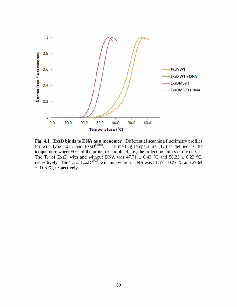
89
Fig. 4.1. ExsD binds to DNA as a monomer. Differential scanning fluorimetry profiles
for wild type ExsD and ExsDM59R
. The melting temperature (Tm) is defined as the
temperature where 50% of the protein is unfolded, i.e., the inflection points of the curves.
The Tm of ExsD with and without DNA was 47.71 ± 0.43 °C and 50.22 ± 0.23 °C,
respectively. The Tm of ExsDM59R
with and without DNA was 31.57 ± 0.22 °C and 27.64
± 0.06 °C, respectively.
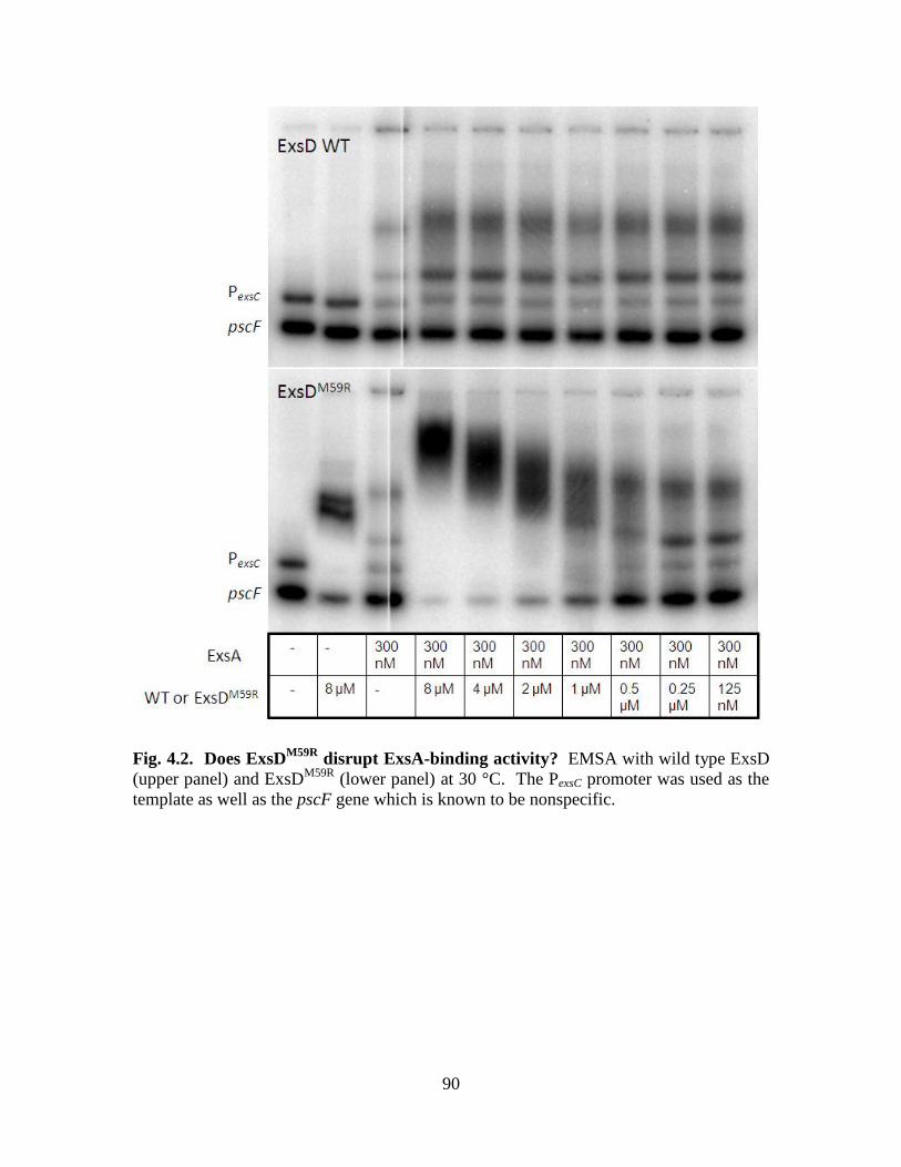
90
Fig. 4.2. Does ExsDM59R
disrupt ExsA-binding activity? EMSA with wild type ExsD
(upper panel) and ExsDM59R
(lower panel) at 30 °C. The PexsC promoter was used as the
template as well as the pscF gene which is known to be nonspecific.

91
Fig. 4.3. ExsD variant purification. Purified wild type ExsD (lane 1), ExsDΔ20 (lane
2), ExsDΔC-C (lane 3), and ExsD1-46 (lane 4).
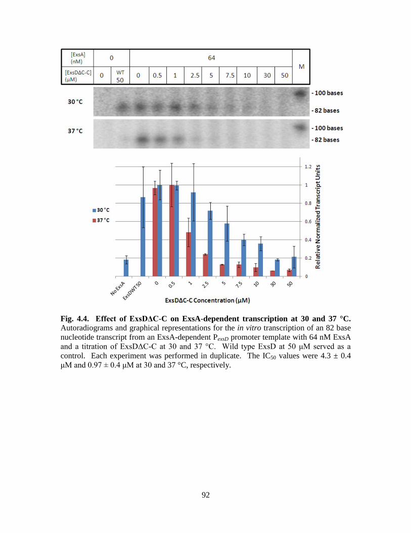
92
Fig. 4.4. Effect of ExsDΔC-C on ExsA-dependent transcription at 30 and 37 °C. Autoradiograms and graphical representations for the in vitro transcription of an 82 base
nucleotide transcript from an ExsA-dependent PexsD promoter template with 64 nM ExsA
and a titration of ExsDΔC-C at 30 and 37 °C. Wild type ExsD at 50 μM served as a
control. Each experiment was performed in duplicate. The IC50 values were 4.3 ± 0.4
μM and 0.97 ± 0.4 μM at 30 and 37 °C, respectively.
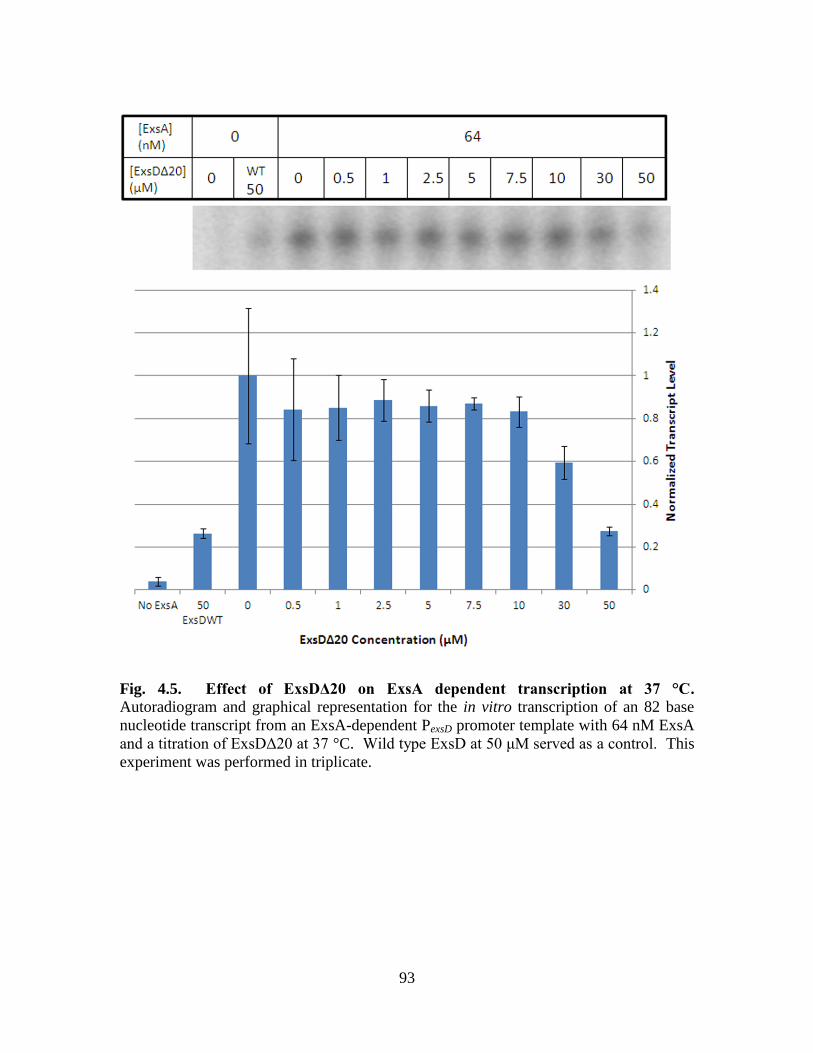
93
Fig. 4.5. Effect of ExsDΔ20 on ExsA dependent transcription at 37 °C. Autoradiogram and graphical representation for the in vitro transcription of an 82 base
nucleotide transcript from an ExsA-dependent PexsD promoter template with 64 nM ExsA
and a titration of ExsDΔ20 at 37 °C. Wild type ExsD at 50 μM served as a control. This
experiment was performed in triplicate.

94
Fig. 4.6. ExsDΔ20 has no effect on ExsA-independent transcription. Autoradiogram
and graphical representation for the in vitro transcription of a 108 nucleotide transcript
from an ExsA-independent RNA-1 promoter template with and without 50 μM ExsDΔ20
at 37 °C. This experiment was performed in duplicate.
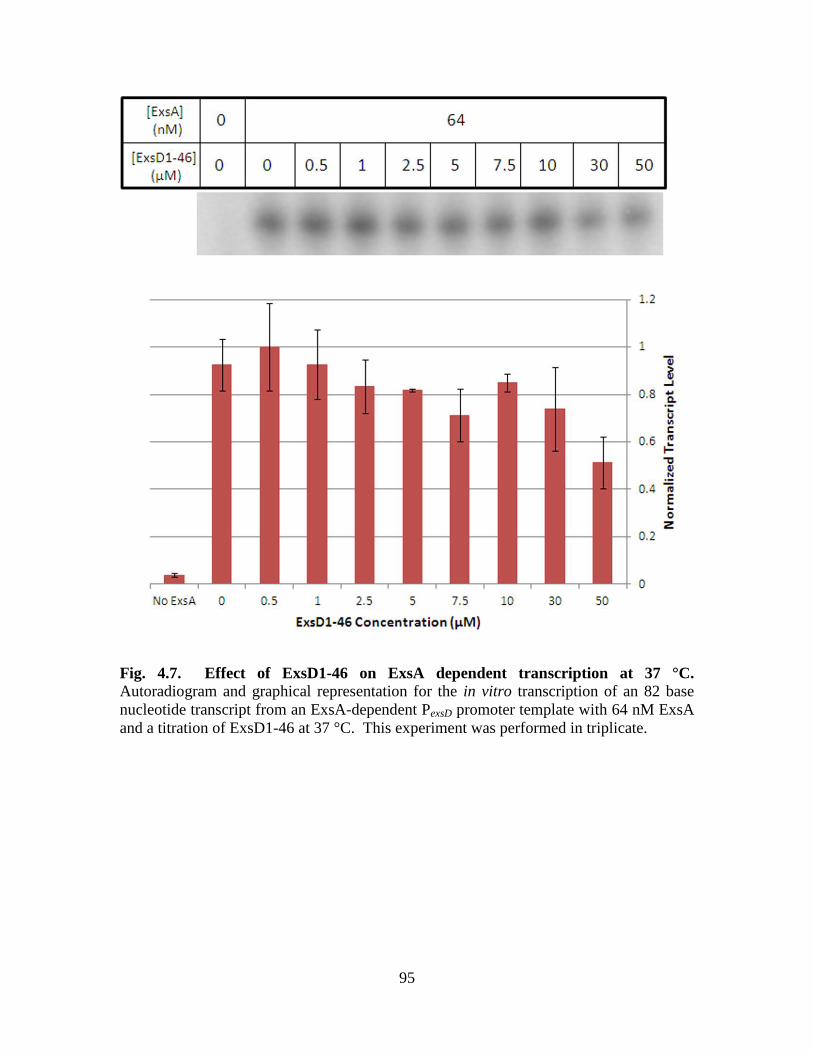
95
Fig. 4.7. Effect of ExsD1-46 on ExsA dependent transcription at 37 °C. Autoradiogram and graphical representation for the in vitro transcription of an 82 base
nucleotide transcript from an ExsA-dependent PexsD promoter template with 64 nM ExsA
and a titration of ExsD1-46 at 37 °C. This experiment was performed in triplicate.
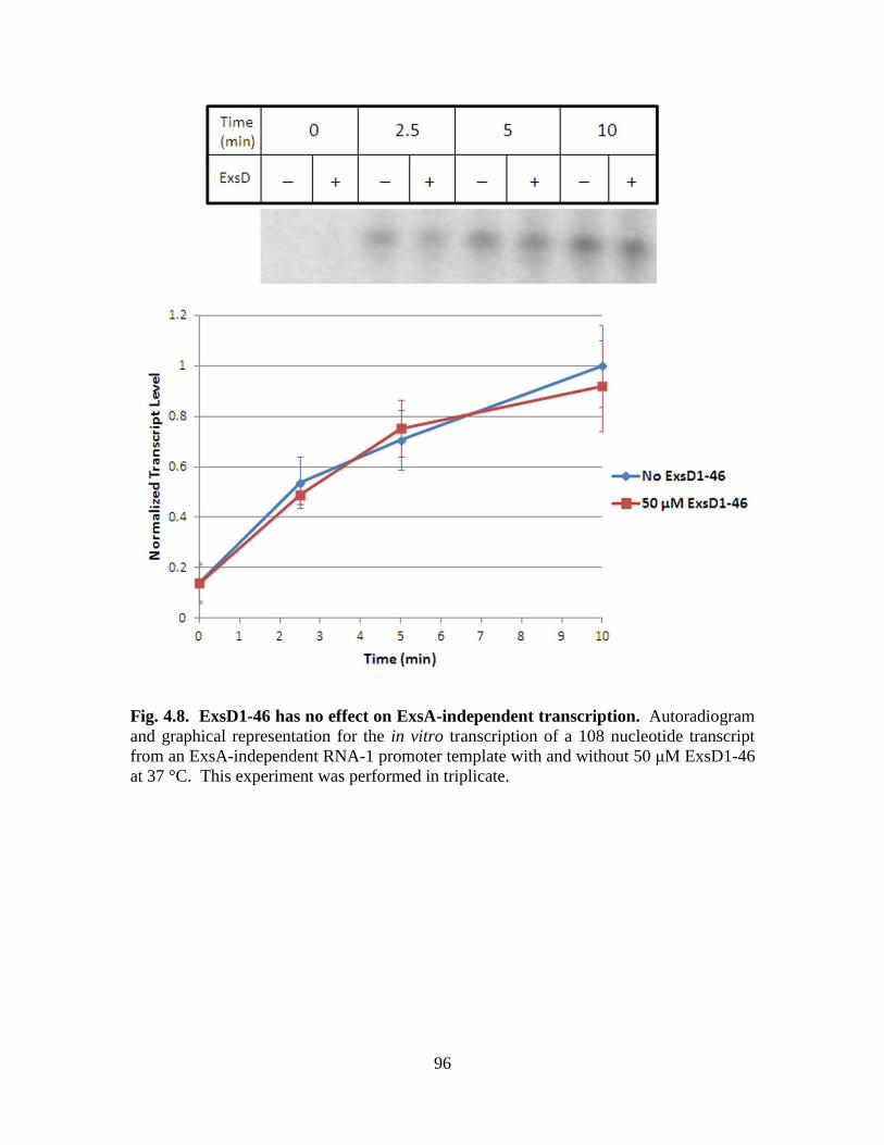
96
Fig. 4.8. ExsD1-46 has no effect on ExsA-independent transcription. Autoradiogram
and graphical representation for the in vitro transcription of a 108 nucleotide transcript
from an ExsA-independent RNA-1 promoter template with and without 50 μM ExsD1-46
at 37 °C. This experiment was performed in triplicate.
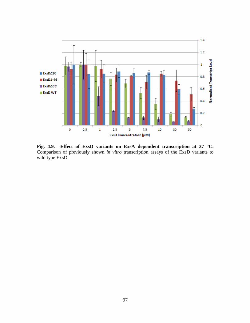
97
Fig. 4.9. Effect of ExsD variants on ExsA dependent transcription at 37 °C. Comparison of previously shown in vitro transcription assays of the ExsD variants to
wild type ExsD.
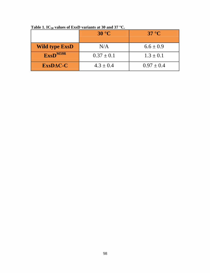
98
Table 1. IC50 values of ExsD variants at 30 and 37 °C.
30 °C 37 °C
Wild type ExsD N/A 6.6 ± 0.9
ExsDM59R
0.37 ± 0.1 1.3 ± 0.1
ExsDΔC-C 4.3 ± 0.4 0.97 ± 0.4

99
CHAPTER FIVE
Overall Conclusions
100
Pseudomonas aeruginosa is a major public health concern due to the increasing
number of immunocompromised patients in the United States and around the world as
well as its growing resistance to common antibiotics. The T3SS is the main virulence
mechanism of P. aeruginosa acute infection; therefore it is of great medical importance.
The ExsA-ExsC-ExsD-ExsE regulatory cascade in P. aeruginosa is highly unusual in that
it functions solely on protein–protein interactions to regulate the T3SS. Understanding
the way in which these regulatory proteins interact is essential to developing drugs to shut
down the T3SS. The ExsD–ExsA interaction is of critical importance, because it sits at
the bottom of the regulatory cascade. ExsA is the main transcriptional activator and is
absolutely essential for every aspect of the T3SS. ExsD directly binds to ExsA and
inhibits the T3SS. Without ExsD, the T3SS is constitutively expressed. Understanding
how ExsA and ExsD function will lay the groundwork for the development of novel
therapeutics to combat P. aeruginosa infection.
Chapter Two examines the T3SS activator ExsA. We detail a procedure for
greatly improving the solubility of recombinant ExsA utilizing a His-MBP tag and TEV
protease during the purification process. The end result is a highly purified ExsA that is
soluble without any tag. An in vitro transcription assay was developed and it was
determined that the recombinant ExsA is able to activate T3SS transcription. This proves
that the recombinant ExsA is highly functional. The affinity of ExsA for its PexsD
promoter was also determined by fluorescence anisotropy which is an equilibrium
technique, and thus more reliable than EMSA. Considering that ExsA is a member of the
AraC/XylS family of transcriptional activators, it can be used as a model since these
family members are known for their low solubility. The ability to produce soluble and
101
highly active ExsA allows for biochemical analyses of the protein which can reveal
information on these types of activators. Furthermore, ExsA is a member of a subfamily
of AraC/XylS activators that regulate virulence factor transcription in a variety of Gram-
negative pathogens. P. aeruginosa ExsA shares close homology with many of the
activators of this subfamily, especially ExsA in Vibrio parahaemolyticus and LcrF in
Yersinia pestis. Our lab has recently solved the structure of the amino-
terminal/regulatory domain of ExsA. Future work will be to crystallize full-length ExsA
in complex with DNA and solve its structure. This crystal structure would reveal how
ExsA interacts with DNA. With this information, drugs can be designed to target regions
of ExsA that interact with DNA. Disrupting ExsA DNA binding would shut down the
T3SS. These drugs would also have the potential to shut down the T3SS in other
pathogenic species, such as Vibrio parahaemolyticus and Yersinia pestis.
ExsD is the antiactivator protein that inhibits the function of ExsA in order to shut
down the T3SS. ExsD is a dynamic protein that is able to exist as a monomer or a trimer
under different conditions. Chapter Three examines the ExsD–ExsA inhibitory
mechanism. We show that ExsD is sufficient to inhibit ExsA-dependent transcription in
vitro, and no other cellular factors are required. We also demonstrate that ExsD inhibits
ExsA in a temperature-dependent manner in vitro. ExsD inhibits ExsA at 37 °C, but not
30 °C. By engineering an ExsD monomeric variant, we demonstrate that this
thermoregulation is due to the ExsD trimer. ExsD inhibits ExsA by binding in a 1:1
complex [147], therefore, the trimer prevents ExsD from binding to ExsA. When the
temperature is increased, the ExsD trimer becomes less stable, allowing for ExsD to bind
to ExsA in the 1:1 complex that is needed for inhibition. T3SS transcription is known to

102
be upregulated at 37 °C [203], so it seems contradictory that a shift from 30 °C to 37 °C
would inhibit T3SS transcription. We hypothesize that this mechanism could serve to
downregulate T3SS gene expression in the lungs of cystic fibrosis patients. In this
environment, P. aeruginosa switches from an acute form of infection to a
chronic/biofilm-producing state in which the T3SS is not expressed [36]. Future work
will be to determine the role the ExsD trimer plays in vivo and during the course of P.
aeruginosa infection. A cystic fibrosis mouse model could be used to determine if the
PA103 exsDM59R
strain has a different effect during pathogenesis compared to PA103
wild type [207]. One could hypothesize that the exsDM59R
mutation would reduce T3SS
activity. This could lead to a faster switch to a biofilm-producing state. An alternative
outcome is that the mutation could prevent P. aeruginosa from establishing an infection
since the T3SS is required to initiate infection [68]. Given there was no difference in
transcriptional activity between PA103 wild type and PA103 exsDM59R
in a β-
galactosidase assay (Fig. A.3), possibly due to the temperature sensitivity of ExsDM59R
, a
difference in pathogenesis might not be observed with this strain in the mouse model. A
PA103 exsDΔC-C strain might be a better choice for this study since this mutation would
attenuate but does not fully disrupt ExsD self-association.
Chapter Four provides evidence that ExsD is a DNA-binding protein, which adds
to its complexity. We demonstrate that ExsD binds to DNA as a monomer. However,
we believe that this DNA binding is non-specific. ChIP-sequencing will be used to
determine if ExsD has any specific DNA-binding sites. It is unclear whether ExsD DNA-
binding plays a role in the inhibition of ExsA. Future work will investigate this
possibility.

103
The ExsD–ExsA interface is also examined in Chapter Four. It was determined
that the coiled-coil region of ExsD is not important for ExsA-binding. Removal of the
coiled-coil region greatly reduces the stability of the ExsD trimer, so the coiled-coil
region is important for ExsD trimer formation. We found that the amino terminus of
ExsD is important for ExsA-binding, but is not sufficient to inhibit ExsA-dependent
transcription. Future work will be to determine the key residues that are important for
ExsA-binding.
The way in which ExsD inhibits ExsA remains ambiguous. The current model
holds that ExsD disrupts ExsA dimerization and promoter interactions by binding to
ExsA in a 1:1 complex. We propose two other possible scenarios, as outlined in Chapter
Four. The first is ExsD binds to ExsA in a 1:1 complex, while ExsA is bound to the
promoter and disrupts ExsA dimer formation. ExsD may or may not be binding to DNA
under this scenario. The other possibility is that ExsD binds to ExsA when the ExsA
dimer is formed on the promoter and disrupts ExsA–RNAP interactions. Given the
structural similarity of ExsD to RNAP binding protein GreB from E. coli [176], one
could imagine a scenario where ExsD interacts with RNAP in the same manner where the
coiled-coil region inserts into RNAP. Seeing as the coiled-coil region of ExsD does not
interact with ExsA, ExsD could be binding to both ExsA and RNAP at the same time.
ExsD could inhibit T3SS transcription by stalling RNAP. Cryo-electron microscopy is
currently being used to determine if ExsD does indeed interact with RNAP.
Given that P. aeruginosa is highly resistant to the majority of current classes of
antibiotics, new drug treatments are of critical importance. Drugs that inhibit the T3SS
would eliminate acute P. aeruginosa infection. They could also prevent P. aeruginosa
104
from establishing infection in the lungs of cystic fibrosis patients. In addition, these
drugs would place less selective pressure on the organism to develop resistance, because
P. aeruginosa does not require a T3SS to survive outside of the host. This dissertation
contributes to the groundwork for the understanding of the key regulators of the T3SS in
P. aeruginosa. It highlights the complexity of ExsD and the way it interacts with ExsA.
This work reveals new insights that challenge the current ExsD–ExsA inhibitory model.
More work will need to be done to fully understand the way in which ExsD inhibits ExsA.
Future work will include using mass spectrometry to determine how many molecules of
ExsA, ExsD, ExsC, and ExsE are present in the cell under inducing and non-inducing
conditions for type III secretion. This could shed light on how the regulatory cascade
works to turn on and off the T3SS. We suspect that the concentrations of the regulators
play a key role. A small shift in the equilibria may be all that it takes to turn the T3SS on
or off in this finely tuned mechanism. Once the ExsD–ExsA inhibitory mechanism is
fully elucidated, drugs can be designed to mimic ExsD function in order to inhibit ExsA
and shut down the T3SS, thereby eliminating acute P. aeruginosa infection.
105
APPENDIX
Extension of Chapter Three: Self-trimerization of ExsD limits inhibition
of the Pseudomonas aeruginosa transcriptional activator ExsA in vitro
Robert C. Bernhards, Shannon K. Esher, Florian D. Schubot
Department of Biological Sciences, Virginia Polytechnic Institute and State University,
Blacksburg, VA 24061
Abbreviations: MSG = monosodium glutamate; ONPG = ortho-Nitrophenyl-β-
galactoside; T3SS = type III secretion system; TBE = Tris/borate/EDTA; TSB =
trypticase soy broth; VB = Vogel-Bonner minimal medium.
106
Abstract
Pseudomonas aeruginosa uses a type III secretion system (T3SS) to cause acute
infection in humans. This T3SS is regulated by a cascade of four interacting proteins:
ExsA, ExsC, ExsD, and ExsE. ExsA is the main transcriptional activator and is inhibited
by the antiactivator ExsD. In Chapter Three, it was observed that ExsD is capable of
inhibiting ExsA in vitro at 37 °C but not 30 °C. It was discovered that this
thermoregulation was a result of ExsD self-trimerization. Here, we further investigate
this phenomenon. An in vitro transcription assay was performed at 33 °C, and it was
determined that ExsD inhibits ExsA based on a temperature gradient, rather than a switch
between 30 and 37 °C. We also examined the ExsDM59R
monomeric variant at 37 °C. As
expected, the inhibition by ExsDM59R
decreases at this temperature, because ExsDM59R
is
unstable at 37 °C. Given that the observed thermoregulation was in vitro, we wanted to
examine whether ExsD trimerization plays a role in vivo. A β-galactosidase assay was
performed to compare wild type ExsD to an ExsDM59R
mutant strain under inducing and
non-inducing conditions for type III secretion.
107
Introduction
The type III secretion system (T3SS) of P. aeruginosa is essential for causing
acute infections in immunocompromised patients. Regulation of the T3SS is coordinated
by a set of four proteins: ExsA, ExsC, ExsD, and ExsE. ExsA is the main transcription
activator [120], and when the T3SS is turned off, ExsA is inhibited by the antiactivator
ExsD [147, 171]. When the T3SS is turned on, ExsE is secreted through the T3SS needle
[108, 109], which frees ExsC to bind to ExsD [148, 177]. ExsA is now free bind to the
T3SS promoters and recruit RNA polymerase to activate T3SS transcription [149].
Chapter Three investigated the ExsD–ExsA inhibitory mechanism, and here, we
further investigate this mechanism. Chapter Three demonstrated that ExsD is able to
inhibit ExsA-dependent transcription in vitro at 37 °C but not 30 °C [206]. Here, an in
vitro transcription assay was performed at an intermediate temperature (33 °C) to
determine if the inhibition by ExsD corresponds to a temperature gradient or simply an
on/off switch between 30 and 37 °C. The thermoregulation described in Chapter Three
was due to ExsD self-trimerization, because a monomeric ExsD variant (ExsDM59R
) was
able to strongly inhibit ExsA-dependent transcription in vitro at 30 °C [206]. Differential
scanning fluorimetry was used in Chapter Three to demonstrate that the ExsDM59R
variant
is not as stable as wild type ExsD [206]. This is most likely due to a greater amount of
exposed surface area. We wanted to determine if ExsDM59R
is still able to inhibit ExsA at
37 °C, because this is the temperature where P. aeruginosa infects its human host. This
would serve as a way to test the suitability of ExsDM59R
for use in vivo. In order to do
this, we performed an in vitro transcription assay to examine the effect of ExsDM59R
on
ExsA-dependent transcription at 37 °C. We found that ExsDM59R
lost some activity at
108
this temperature, so we thought it was best to examine the effect of ExsDM59R
in vivo
using a β-galactosidase assay at 30 °C. A chromosomal ExsDM59R
mutation was made in
the P. aeruginosa strain PA103, and this mutant was compared to wild type PA103 under
inducing and non-inducing conditions for type III secretion.
Results
Inhibition by ExsD is based on a temperature gradient
It was determined in Chapter Three that ExsD inhibits ExsA-dependent
transcription in vitro at 37 °C but not 30 °C. We wanted to determine if this temperature-
dependent inhibition corresponded to a temperature gradient or simply an on/off switch
between 37 and 30 °C. In order to do this, an in vitro transcription assay was performed
at 33 °C (Fig. A.1). In this 0-10 min time course experiment, each sample contained 64
nM ExsA without or without 50 μM ExsD. There was a 1.72x level of inhibition by
ExsD, which is in between what was previously seen at 30 °C (no inhibition) and 37 °C
(~3.75x inhibition). This demonstrates that the observed thermoregulation corresponds to
a temperature gradient.
Effect of ExsDM59R
on ExsA-dependent transcription at 37 °C
In Chapter Three, the ExsDM59R
monomeric variant was utilized to determine that
the observed thermoregulation was due to ExsD self-trimerization. ExsDM59R
was found
to strongly inhibit ExsA-dependent transcription in an in vitro transcription assay at
30 °C. Here, we wanted to examine ExsDM59R
in the in vitro transcription assay at 37 °C.
Based on a previous differential scanning fluorimetry experiment, ExsDM59R
would likely
be unstable at this higher temperature. A titration of ExsDM59R
was performed keeping
109
the ExsA concentration constant at 64 nM (Fig. A.2). The IC50 value was measured to be
1.3 ± 0.1 μM, which is slightly higher than the IC50 value at 30 °C (0.37 ± 0.1 μM). This
indicates that a certain percentage of the ExsDM59R
protein is becoming denatured at the
higher temperature, as expected. However, ExsDM59R
at 37 °C is still a more potent
inhibitor than wild type ExsD at the same temperature (IC50 = 6.6 ± 0.9 μM). This
suggests that most of the ExsDM59R
is stable enough to still be active at 37 °C.
Effect of ExsDM59R
in vivo
Given that the thermoregulation due to ExsD trimerization was observed in vitro,
we wanted to examine if the effect is observed in vivo. A PA103 ExsDM59R
chromosomal
mutant strain was compared to PA103 wild type in a β-galactosidase assay (Fig. A.3).
The cultures were grown under inducing and non-inducing conditions for type III
secretion at 30 °C, and T3SS transcriptional activation was measured from the PexsD
promoter. We hypothesized that the ExsDM59R
strain would inhibit ExsA more strongly
compared to wild type, because the monomeric ExsDM59R
variant is able to strongly
inhibit ExsA-dependent transcription at 30 °C in vitro. However, there was no significant
difference in the level of T3SS transcription under inducing or non-inducing conditions.
Discussion
Here we demonstrate that the observed thermoregulation by ExsD corresponds to
a temperature gradient. This suggests that as the temperature increases, the ExsD trimer
becomes less stable, which allows for ExsD to bind ExsA in the 1:1 complex that is
needed for inhibition [147]. Chapter Three reveals that the ExsDM59R
monomeric variant
is capable of inhibiting ExsA-dependent transcription at 30 °C, which demonstrates that
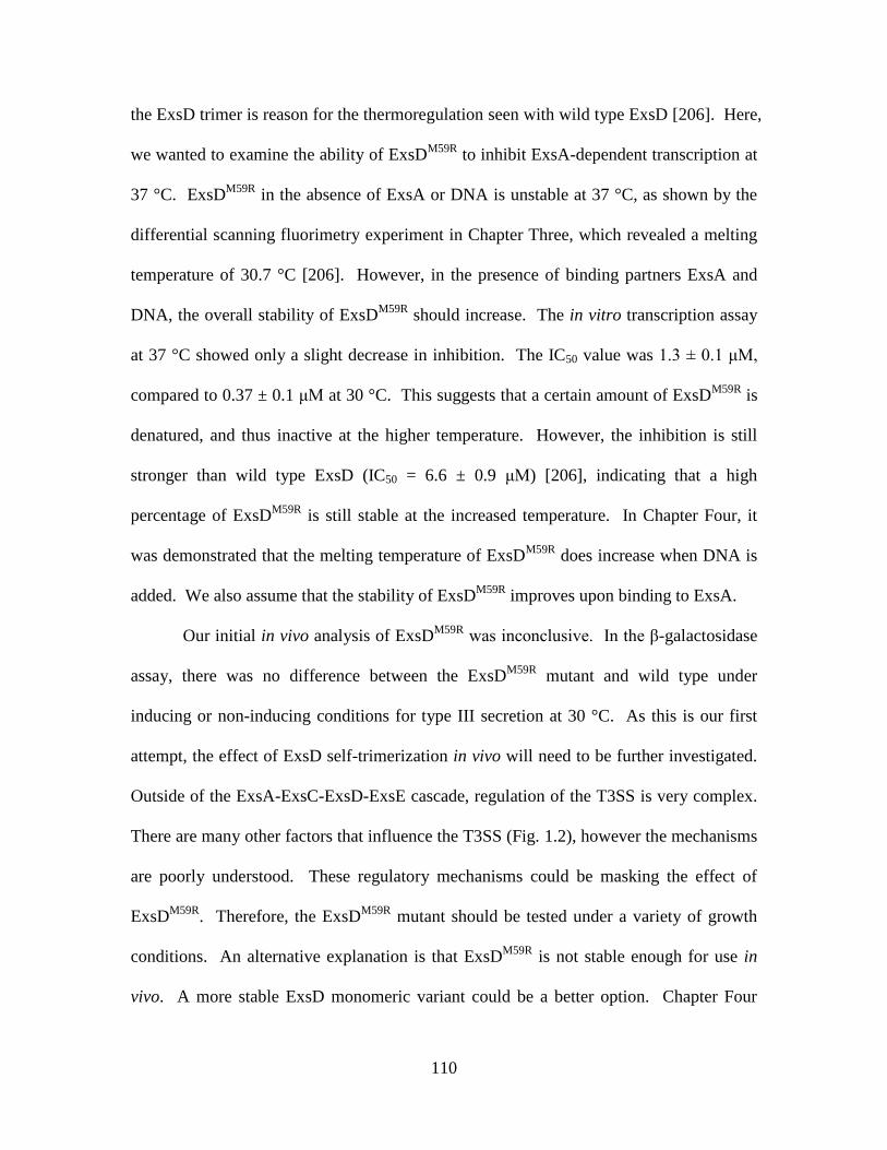
110
the ExsD trimer is reason for the thermoregulation seen with wild type ExsD [206]. Here,
we wanted to examine the ability of ExsDM59R
to inhibit ExsA-dependent transcription at
37 °C. ExsDM59R
in the absence of ExsA or DNA is unstable at 37 °C, as shown by the
differential scanning fluorimetry experiment in Chapter Three, which revealed a melting
temperature of 30.7 °C [206]. However, in the presence of binding partners ExsA and
DNA, the overall stability of ExsDM59R
should increase. The in vitro transcription assay
at 37 °C showed only a slight decrease in inhibition. The IC50 value was 1.3 ± 0.1 μM,
compared to 0.37 ± 0.1 μM at 30 °C. This suggests that a certain amount of ExsDM59R
is
denatured, and thus inactive at the higher temperature. However, the inhibition is still
stronger than wild type ExsD (IC50 = 6.6 ± 0.9 μM) [206], indicating that a high
percentage of ExsDM59R
is still stable at the increased temperature. In Chapter Four, it
was demonstrated that the melting temperature of ExsDM59R
does increase when DNA is
added. We also assume that the stability of ExsDM59R
improves upon binding to ExsA.
Our initial in vivo analysis of ExsDM59R
was inconclusive. In the β-galactosidase
assay, there was no difference between the ExsDM59R
mutant and wild type under
inducing or non-inducing conditions for type III secretion at 30 °C. As this is our first
attempt, the effect of ExsD self-trimerization in vivo will need to be further investigated.
Outside of the ExsA-ExsC-ExsD-ExsE cascade, regulation of the T3SS is very complex.
There are many other factors that influence the T3SS (Fig. 1.2), however the mechanisms
are poorly understood. These regulatory mechanisms could be masking the effect of
ExsDM59R
. Therefore, the ExsDM59R
mutant should be tested under a variety of growth
conditions. An alternative explanation is that ExsDM59R
is not stable enough for use in
vivo. A more stable ExsD monomeric variant could be a better option. Chapter Four

111
describes an ExsD variant lacking the coiled-coil region (ExsDΔC-C). This ExsD variant
showed strong, monomeric activity in the in vitro transcription assay. However,
ExsDΔC-C is not believed to be completely monomeric. At 30 °C, the activity of
ExsDΔC-C indicates that it is partially trimeric. Therefore, ExsDΔC-C could be more
stable than ExsDM59R
. A differential scanning fluorimetry experiment could be used to
determine the stability of ExsDΔC-C. If it shows increased stability, a chromosomal
ExsDΔC-C mutant could be constructed and used in β-galactosidase assays. We also
demonstrated in Chapter Four that the coiled-coil region is not important for ExsA
binding, so the mutation should not affect the ability of ExsD to inhibit ExsA.
Materials and Methods
In vitro transcription assay
The linear DNA template used in each assay encompassed positions -207 to 94 of
the PexsD promoter, relative to the transcription start site; and from this template, RNA
polymerase synthesizes an 82 base mRNA transcript. The template was produced by
PCR using forward primer 5´-CATCAGTTGCTGCTCAACAGCG-3´ and reverse primer
5´-CACCGCTTCTCGGGAGTACTGC-3´. The PCR product was run on a 2% agarose
gel and purified using the Wizard SV Gel and PCR Clean-up System (Promega, Madison,
WI, USA). Each 30 µL transcription assay reaction contained 4.4 fM of promoter
template, 50.4 µM bovine serum albumin (to eliminate non-specific protein-protein
interactions), 10 U purified RNA polymerase from P. aeruginosa (see Chapter Three), 1
U RiboGuard RNase Inhibitor (Epicentre Biotechnologies), 15 ng/µL
poly(deoxyinosinic-deoxycytidylic) acid (to prevent non-specific transcription initiation),

112
133 mM NaCl, 32 mM Tris-HCl (pH 7.4), 10 mM MgCl2, 25 µM EDTA, 0.9 mM TCEP,
0.2 mM DTT, and 15.5% glycerol. The time-course experiments contained 64 nM ExsA
and either no ExsD or 50 µM ExsD (no ExsA was added for the RNA-1 control
experiments). Samples were mixed and allowed to equilibrate at room temperature for
five min. Samples were then pre-incubated for 10 min at either 33 or 37°C, depending on
the experiment. Next, 3 µL NTPs (stock concentrations of 200 µM ATP, CTP, GTP and
40 µM UTP) mixed with 0.2 µL (0.2 µCi) of 3.3 mM P32
-alpha UTP was added to each
sample to start the reaction, and samples were incubated at either 33 or 37°C, depending
on the experiment. After the reactions were stopped by adding 12 µL 1X stop solution
(3M ammonium acetate, 50 mM EDTA, 0.11 mg/mL glycogen), 170 µL 100% cold
ethanol was added, and the samples were incubated at -20 °C for one hr. Following
centrifugation at 12,000 x g for 15 min, the supernatant was discarded and pellets were
resuspended in 12 μL 1X TBE (Tris/borate/EDTA)-urea sample buffer and heated at
70 °C for five min. After a brief centrifugation, the samples were loaded onto a 10%
TBE-urea gel and run at 200 mV for 60 min. Gels were exposed to a storage phosphor
screen (GE Healthcare) for 16 hr. The phosphor screen was scanned using a Typhoon
Trio Variable Mode Imager (GE Healthcare), and gel bands were quantified using Image
Quant TL v2005 (Amersham Biosciences, Piscataway, NJ, USA). The IC50 value for the
ExsDM59R
experiment was determined by analysis using Kaleidograph (Synergy
Software).
β-galactosidase assay
PA103-miniCTX-PexsD-lacZ (wild type) and PA103ExsDM59R
-miniCTX-PexsD-
lacZ strains were grown on Vogel-Bonner (VB) minimal medium plates with 4 µg/mL
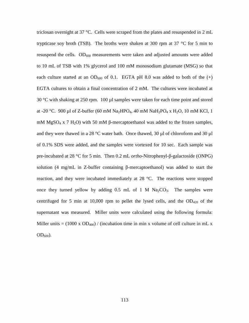
113
triclosan overnight at 37 °C. Cells were scraped from the plates and resuspended in 2 mL
trypticase soy broth (TSB). The broths were shaken at 300 rpm at 37 °C for 5 min to
resuspend the cells. OD600 measurements were taken and adjusted amounts were added
to 10 mL of TSB with 1% glycerol and 100 mM monosodium glutamate (MSG) so that
each culture started at an OD600 of 0.1. EGTA pH 8.0 was added to both of the (+)
EGTA cultures to obtain a final concentration of 2 mM. The cultures were incubated at
30 °C with shaking at 250 rpm. 100 µl samples were taken for each time point and stored
at -20 °C. 900 µl of Z-buffer (60 mM Na2HPO4, 40 mM NaH2PO4 x H2O, 10 mM KCl, 1
mM MgSO4 x 7 H2O) with 50 mM β-mercaptoethanol was added to the frozen samples,
and they were thawed in a 28 °C water bath. Once thawed, 30 µl of chloroform and 30 µl
of 0.1% SDS were added, and the samples were vortexed for 10 sec. Each sample was
pre-incubated at 28 °C for 5 min. Then 0.2 mL ortho-Nitrophenyl-β-galactoside (ONPG)
solution (4 mg/mL in Z-buffer containing β-mercaptoethanol) was added to start the
reaction, and they were incubated immediately at 28 °C. The reactions were stopped
once they turned yellow by adding 0.5 mL of 1 M Na2CO3. The samples were
centrifuged for 5 min at 10,000 rpm to pellet the lysed cells, and the OD420 of the
supernatant was measured. Miller units were calculated using the following formula:
Miller units = (1000 x OD400) / (incubation time in min x volume of cell culture in mL x
OD600).

114
Fig. A.1. Effect of ExsD on ExsA-dependent transcription at 33 °C. A 0-10 min time
course was performed with 64 nM ExsA with or without 50 μM ExsD. This experiment
was performed in triplicate. ExsD provides a 1.72x level of inhibition at this
temperature.
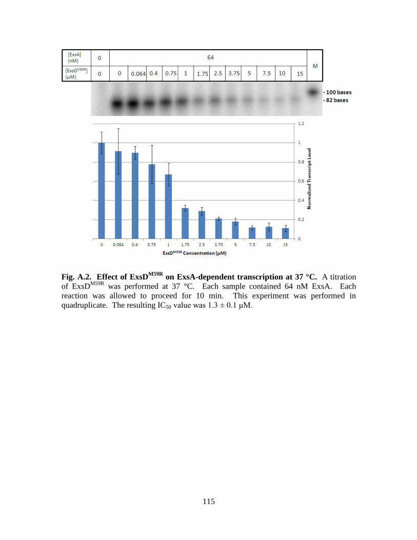
115
Fig. A.2. Effect of ExsDM59R
on ExsA-dependent transcription at 37 °C. A titration
of ExsDM59R
was performed at 37 °C. Each sample contained 64 nM ExsA. Each
reaction was allowed to proceed for 10 min. This experiment was performed in
quadruplicate. The resulting IC50 value was 1.3 ± 0.1 μM.
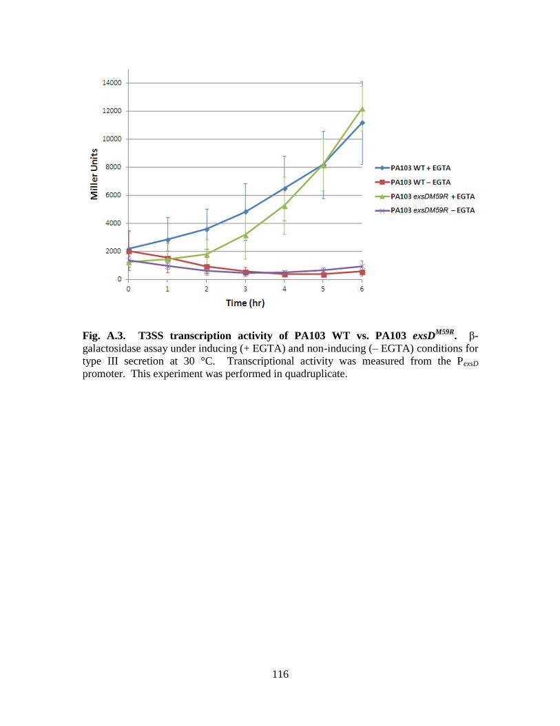
116
Fig. A.3. T3SS transcription activity of PA103 WT vs. PA103 exsDM59R
. β-
galactosidase assay under inducing (+ EGTA) and non-inducing (– EGTA) conditions for
type III secretion at 30 °C. Transcriptional activity was measured from the PexsD
promoter. This experiment was performed in quadruplicate.
117
REFERENCES
1. Haynes, W. C. & Rhodes, L. J. (1962) Comparative taxonomy of crystallogenic
strains of Pseudomonas aeruginosa and Pseudomonas chlororaphis, J Bacteriol.
84, 1080-4.
2. Propst, C. & Lubin, L. (1979) Light-mediated changes in pigmentation of
Pseudomonas aeruginosa cultures, Journal of General Microbiology. 113, 261-6.
3. Elrod, R. P. & Braun, A. C. (1942) Pseudomonas aeruginosa: Its role as a plant
pathogen, J Bacteriol. 44, 633-45.
4. Finkelstein, R., Raz, R., Stein, H., Reiss, D., Peleg, H., Merzbach, D. & Sobel, J. D.
(1989) Bone and joint infections due to Pseudomonas aeruginosa: clinical aspects
and treatment, Israel Journal of Medical Sciences. 25, 123-6.
5. Hopkins, R. S., Abbott, D. O. & Wallace, L. E. (1981) Follicular dermatitis outbreak
caused by Pseudomonas aeruginosa associated with a motel's indoor swimming
pool, Public Health Reports. 96, 246-9.
6. Stephenson, J. R., Heard, S. R., Richards, M. A. & Tabaqchali, S. (1985)
Gastrointestinal colonization and septicaemia with Pseudomonas aeruginosa due
to contaminated thymol mouthwash in immunocompromised patients, The
Journal of Hospital Infection. 6, 369-78.
7. Phillips, I. (1967) Pseudomonas aeruginosa respiratory tract infections in patients
receiving mechanical ventilation, The Journal of hygiene. 65, 229-35.
8. Church, D., Elsayed, S., Reid, O., Winston, B. & Lindsay, R. (2006) Burn wound
infections, Clinical Microbiology Reviews. 19, 403-34.
9. Mittal, R., Aggarwal, S., Sharma, S., Chhibber, S. & Harjai, K. (2009) Urinary tract
infections caused by Pseudomonas aeruginosa: a minireview, Journal of Infection
and Public Health. 2, 101-11.
10. Carruthers, M. M. & Kanokvechayant, R. (1973) Pseudomonas aeruginosa
endocarditis. Report of a case, with review of the literature, The American
Journal of Medicine. 55, 811-8.
11. Parrott, P. L., Terry, P. M., Whitworth, E. N., Frawley, L. W., Coble, R. S.,
Wachsmuth, I. K. & McGowan, J. E., Jr. (1982) Pseudomonas aeruginosa
peritonitis associated with contaminated poloxamer-iodine solution, Lancet. 2,
683-5.
12. Knight, V., Hardy, R. C. & Negrin, J., Jr. (1952) Meningitis due to Pseudomonas
aeruginosa; intrathecal treatment with streptokinase and streptodornase and
intramuscular and intrathecal treatment with neomycin, Journal of the American
Medical Association. 149, 1395-7.
13. Richardson, M. E. (1957) Pseudomonas aeruginosa septicemia, The Journal of the
American Osteopathic Association. 57, 135-7.
14. Shanson, D. C. (1990) Septicaemia in patients with AIDS, Transactions of the Royal
Society of Tropical Medicine and Hygiene. 84 Suppl 1, 14-6.
15. Rolston, K. V. & Bodey, G. P. (1992) Pseudomonas aeruginosa infection in cancer
patients, Cancer Investigation. 10, 43-59.
118
16. Gransden, W. R., Leibovici, L., Eykyn, S. J., Pitlik, S. D., Samra, Z., Konisberger, H.,
Drucker, M. & Phillips, I. (1995) Risk factors and a clinical index for diagnosis of
Pseudomonas aeruginosa bacteremia, Clinical Microbiology and Infection: The
Official Publication of the European Society of Clinical Microbiology and
Infectious Diseases. 1, 119-123.
17. Hashimoto, M., Hirouchi, S., Kosugi, J., Kohara, W. & Nakane, M. (1971)
Morphological studies on fimbriae of Pseudomonas aeruginosa and Aeromonas
hydrophila with special reference to their biological functions, The Bulletin of
Tokyo Medical and Dental University. 18, 105-22.
18. Doggett, R. G., Harrison, G. M. & Wallis, E. S. (1964) Comparison of some
properties of Pseudomonas aeruginosa isolated from infections in persons with
and without cystic fibrosis, J Bacteriol. 87, 427-31.
19. Linker, A. & Evans, L. R. (1984) Isolation and characterization of an alginase from
mucoid strains of Pseudomonas aeruginosa, J Bacteriol. 159, 958-64.
20. Cryz, S. J. & Iglewski, B. H. (1980) Production of alkaline protease by Pseudomonas
aeruginosa, Journal of Clinical Microbiology. 12, 131-3.
21. Morihara, K., Tsuzuki, H., Oka, T., Inoue, H. & Ebata, M. (1965) Pseudomonas
aeruginosa elastase. Isolation, crystallization, and preliminary characterization,
The Journal of Biological Chemistry. 240, 3295-304.
22. Berka, R. M., Gray, G. L. & Vasil, M. L. (1981) Studies of phospholipase C (heat
labile hemolysin) in Pseudomonas aeruginosa, Infection and Immunity. 34, 1071-
4.
23. Liu, P. V. (1966) The roles of various fractions of Pseudomonas aeruginosa in its
pathogenesis. II. Effects of lecithinase and protease, The Journal of Infectious
Diseases. 116, 112-6.
24. Scharmann, W. (1976) Cytotoxic effects of leukocidin from Pseudomonas
aeruginosa on polymorphonuclear leukocytes from cattle, Infection and Immunity.
13, 836-43.
25. Wilson, R., Pitt, T., Taylor, G., Watson, D., MacDermot, J., Sykes, D., Roberts, D. &
Cole, P. (1987) Pyocyanin and 1-hydroxyphenazine produced by Pseudomonas
aeruginosa inhibit the beating of human respiratory cilia in vitro, The Journal of
Clinical Investigation. 79, 221-9.
26. Miles, A. A. & Khimji, P. L. (1975) Enterobacterial chelators of iron: their
occurrence, detection, and relation to pathogenicity, Journal of Medical
Microbiology. 8, 477-90.
27. Naoi, M., Egami, F., Hamamura, N. & Homma, J. Y. (1958) [The toxic
lipopolysaccharides of Pseudomonas aeruginosa], Biochemische Zeitschrift. 330,
421-7.
28. Liu, P. V., Yoshii, S. & Hsieh, H. (1973) Exotoxins of Pseudomonas aeruginosa. II.
Concentration, purification, and characterization of exotoxin A, The Journal of
Infectious Diseases. 128, 514-9.
29. Iglewski, B. H., Sadoff, J., Bjorn, M. J. & Maxwell, E. S. (1978) Pseudomonas
aeruginosa exoenzyme S: an adenosine diphosphate ribosyltransferase distinct
from toxin A, Proceedings of the National Academy of Sciences of the United
States of America. 75, 3211-5.
119
30. Cowell, B. A., Chen, D. Y., Frank, D. W., Vallis, A. J. & Fleiszig, S. M. (2000)
ExoT of cytotoxic Pseudomonas aeruginosa prevents uptake by corneal epithelial
cells, Infection and Immunity. 68, 403-6.
31. Finck-Barbancon, V., Goranson, J., Zhu, L., Sawa, T., Wiener-Kronish, J. P.,
Fleiszig, S. M., Wu, C., Mende-Mueller, L. & Frank, D. W. (1997) ExoU
expression by Pseudomonas aeruginosa correlates with acute cytotoxicity and
epithelial injury, Molecular Microbiology. 25, 547-57.
32. Yahr, T. L., Vallis, A. J., Hancock, M. K., Barbieri, J. T. & Frank, D. W. (1998)
ExoY, an adenylate cyclase secreted by the Pseudomonas aeruginosa type III
system, Proceedings of the National Academy of Sciences of the United States of
America. 95, 13899-904.
33. Jean, D., Rezaiguia-Delclaux, S., Delacourt, C., Leclercq, R., Lafuma, C., Brun
Buisson, C., Harf, A. & Delclaux, C. (1998) Protective effect of endotoxin
instillation on subsequent bacteria-induced acute lung injury in rats, American
Journal of Respiratory and Critical Care Medicine. 158, 1702-8.
34. Meluleni, G. J., Grout, M., Evans, D. J. & Pier, G. B. (1995) Mucoid Pseudomonas
aeruginosa growing in a biofilm in vitro are killed by opsonic antibodies to the
mucoid exopolysaccharide capsule but not by antibodies produced during chronic
lung infection in cystic fibrosis patients, Journal of Immunology. 155, 2029-38.
35. Koch, C. & Hoiby, N. (1993) Pathogenesis of cystic fibrosis, Lancet. 341, 1065-9.
36. Goodman, A. L., Kulasekara, B., Rietsch, A., Boyd, D., Smith, R. S. & Lory, S.
(2004) A signaling network reciprocally regulates genes associated with acute
infection and chronic persistence in Pseudomonas aeruginosa, Developmental
Cell. 7, 745-54.
37. Burns, J. L., Gibson, R. L., McNamara, S., Yim, D., Emerson, J., Rosenfeld, M.,
Hiatt, P., McCoy, K., Castile, R., Smith, A. L. & Ramsey, B. W. (2001)
Longitudinal assessment of Pseudomonas aeruginosa in young children with
cystic fibrosis, The Journal of Infectious Diseases. 183, 444-52.
38. Richards, M. J., Edwards, J. R., Culver, D. H. & Gaynes, R. P. (1999) Nosocomial
infections in medical intensive care units in the United States. National
Nosocomial Infections Surveillance System, Critical Care Medicine. 27, 887-92.
39. Reuter, S., Sigge, A., Wiedeck, H. & Trautmann, M. (2002) Analysis of transmission
pathways of Pseudomonas aeruginosa between patients and tap water outlets,
Critical care medicine. 30, 2222-8.
40. Grieco, M. H. (1972) Pseudomonas arthritis and osteomyelitis, The Journal of Bone
and Joint Surgery American Volume. 54, 1693-704.
41. Crouch Brewer, S., Wunderink, R. G., Jones, C. B. & Leeper, K. V., Jr. (1996)
Ventilator-associated pneumonia due to Pseudomonas aeruginosa, Chest. 109,
1019-29.
42. Garau, J. & Gomez, L. (2003) Pseudomonas aeruginosa pneumonia, Current
Opinion in Infectious Diseases. 16, 135-43.
43. Ohmagari, N., Hanna, H., Graviss, L., Hackett, B., Perego, C., Gonzalez, V., Dvorak,
T., Hogan, H., Hachem, R., Rolston, K. & Raad, I. (2005) Risk factors for
infections with multidrug-resistant Pseudomonas aeruginosa in patients with
cancer, Cancer. 104, 205-12.

120
44. Aloush, V., Navon-Venezia, S., Seigman-Igra, Y., Cabili, S. & Carmeli, Y. (2006)
Multidrug-resistant Pseudomonas aeruginosa: risk factors and clinical impact,
Antimicrobial Agents and Chemotherapy. 50, 43-8.
45. Wisplinghoff, H., Bischoff, T., Tallent, S. M., Seifert, H., Wenzel, R. P. & Edmond,
M. B. (2004) Nosocomial bloodstream infections in US hospitals: analysis of
24,179 cases from a prospective nationwide surveillance study, Clinical infectious
Diseases: An Official Publication of the Infectious Diseases Society of America.
39, 309-17.
46. Arruda, E. A., Marinho, I. S., Boulos, M., Sinto, S. I., Caiaffa, H. H., Mendes, C. M.,
Oplustil, C. P., Sader, H., Levy, C. E. & Levin, A. S. (1999) Nosocomial
infections caused by multiresistant Pseudomonas aeruginosa, Infection Control
and Hospital Epidemiology: The Official Journal of the Society of Hospital
Epidemiologists of America. 20, 620-3.
47. Obritsch, M. D., Fish, D. N., MacLaren, R. & Jung, R. (2005) Nosocomial infections
due to multidrug-resistant Pseudomonas aeruginosa: epidemiology and treatment
options, Pharmacotherapy. 25, 1353-64.
48. Bryan, L. E., Kwan, S. & Godfrey, A. J. (1984) Resistance of Pseudomonas
aeruginosa mutants with altered control of chromosomal beta-lactamase to
piperacillin, ceftazidime, and cefsulodin, Antimicrobial Agents and Chemotherapy.
25, 382-4.
49. Reese, L. (1965) Nalidixic acid (Neggram) in the treatment of urinary infections,
Canadian Medical Association Journal. 92, 394-7.
50. Gershenfeld, L. & Guarini, J. R. (1951) The development of resistance by
Pseudomonas aeruginosa to neomycin, streptomycin and chloromycetin,
American Journal of Pharmacy and the Sciences Supporting Public Health. 123,
182-93.
51. Angus, B. L., Carey, A. M., Caron, D. A., Kropinski, A. M. & Hancock, R. E. (1982)
Outer membrane permeability in Pseudomonas aeruginosa: comparison of a wild-
type with an antibiotic-supersusceptible mutant, Antimicrobial Agents and
Chemotherapy. 21, 299-309.
52. Yoshimura, F. & Nikaido, H. (1982) Permeability of Pseudomonas aeruginosa outer
membrane to hydrophilic solutes, J Bacteriol. 152, 636-42.
53. Lebek, G. (1963) [The transmission of multiple resistance to antibiotics and
chemotherapeutic agents in their significance for hospitalism with multiple
resistant Gram-negative intestinal bacteria], Zentralblatt fur bakteriologie,
parasitenkunde, infektionskrankheiten und hygiene 1 abt medizinisch-hygienische
bakteriologie, Virusforschung und Parasitologie Originale. 191, 387-95.
54. Hooper, D. C. & Wolfson, J. S. (1989) Bacterial resistance to the quinolone
antimicrobial agents, The American Journal of Medicine. 87, 17S-23S.
55. Pagani, L., Landini, P., Luzzaro, F., Debiaggi, M. & Romero, E. (1990) Emergence
of cross-resistance to imipenem and other beta-lactam antibiotics in Pseudomonas
aeruginosa during therapy, Microbiologica. 13, 43-53.
56. Levy, S. B. (1992) Active efflux mechanisms for antimicrobial resistance,
Antimicrobial Agents and Chemotherapy. 36, 695-703.

121
57. Traub, W. H. (1970) Susceptibility of Pseudomonas aeruginosa to gentamicin sulfate
in vitro: lack of correlation between disc diffusion and broth dilution sensitivity
data, Applied Microbiology. 20, 98-102.
58. Chadwick, P. (1969) Effect of carbenicillin on Pseudomonas aeruginosa, Canadian
Medical Association Journal. 101, 74-80.
59. Frithz-Lindsten, E., Du, Y., Rosqvist, R. & Forsberg, A. (1997) Intracellular
targeting of exoenzyme S of Pseudomonas aeruginosa via type III-dependent
translocation induces phagocytosis resistance, cytotoxicity and disruption of actin
microfilaments, Molecular Microbiology. 25, 1125-39.
60. Coburn, B., Sekirov, I. & Finlay, B. B. (2007) Type III secretion systems and disease,
Clinical Microbiology Reviews. 20, 535-49.
61. Troisfontaines, P. & Cornelis, G. R. (2005) Type III secretion: more systems than
you think, Physiology. 20, 326-39.
62. Makino, K., Oshima, K., Kurokawa, K., Yokoyama, K., Uda, T., Tagomori, K.,
Iijima, Y., Najima, M., Nakano, M., Yamashita, A., Kubota, Y., Kimura, S.,
Yasunaga, T., Honda, T., Shinagawa, H., Hattori, M. & Iida, T. (2003) Genome
sequence of Vibrio parahaemolyticus: a pathogenic mechanism distinct from that
of V. cholerae, Lancet. 361, 743-9.
63. Garrity-Ryan, L., Kazmierczak, B., Kowal, R., Comolli, J., Hauser, A. & Engel, J. N.
(2000) The arginine finger domain of ExoT contributes to actin cytoskeleton
disruption and inhibition of internalization of Pseudomonas aeruginosa by
epithelial cells and macrophages, Infection and Immunity. 68, 7100-13.
64. Rocha, C. L., Coburn, J., Rucks, E. A. & Olson, J. C. (2003) Characterization of
Pseudomonas aeruginosa exoenzyme S as a bifunctional enzyme in J774A.1
macrophages, Infection and Immunity. 71, 5296-305.
65. Pederson, K. J. & Barbieri, J. T. (1998) Intracellular expression of the ADP
ribosyltransferase domain of Pseudomonas exoenzyme S is cytotoxic to
eukaryotic cells, Molecular Microbiology. 30, 751-9.
66. Fraylick, J. E., La Rocque, J. R., Vincent, T. S. & Olson, J. C. (2001) Independent
and coordinate effects of ADP-ribosyltransferase and GTPase-activating activities
of exoenzyme S on HT-29 epithelial cell function, Infection and Immunity. 69,
5318-28.
67. Lee, V. T., Smith, R. S., Tummler, B. & Lory, S. (2005) Activities of Pseudomonas
aeruginosa effectors secreted by the Type III secretion system in vitro and during
infection, Infection and Immunity. 73, 1695-705.
68. Vance, R. E., Rietsch, A. & Mekalanos, J. J. (2005) Role of the type III secreted
exoenzymes S, T, and Y in systemic spread of Pseudomonas aeruginosa PAO1 in
vivo, Infection and Immunity. 73, 1706-13.
69. Shaver, C. M. & Hauser, A. R. (2004) Relative contributions of Pseudomonas
aeruginosa ExoU, ExoS, and ExoT to virulence in the lung, Infection and
Immunity. 72, 6969-77.
70. Zaborina, O., Kohler, J. E., Wang, Y., Bethel, C., Shevchenko, O., Wu, L., Turner, J.
R. & Alverdy, J. C. (2006) Identification of multi-drug resistant Pseudomonas
aeruginosa clinical isolates that are highly disruptive to the intestinal epithelial
barrier, Annals of Clinical Microbiology and Antimicrobials. 5, 14.

122
71. Diaz, M. H., Shaver, C. M., King, J. D., Musunuri, S., Kazzaz, J. A. & Hauser, A. R.
(2008) Pseudomonas aeruginosa induces localized immunosuppression during
pneumonia, Infection and Immunity. 76, 4414-21.
72. Kurahashi, K., Kajikawa, O., Sawa, T., Ohara, M., Gropper, M. A., Frank, D. W.,
Martin, T. R. & Wiener-Kronish, J. P. (1999) Pathogenesis of septic shock in
Pseudomonas aeruginosa pneumonia, The Journal of Clinical Investigation. 104,
743-50.
73. Cowell, B. A., Evans, D. J. & Fleiszig, S. M. (2005) Actin cytoskeleton disruption by
ExoY and its effects on Pseudomonas aeruginosa invasion, FEMS Microbiology
Letters. 250, 71-6.
74. Sayner, S. L., Frank, D. W., King, J., Chen, H., VandeWaa, J. & Stevens, T. (2004)
Paradoxical cAMP-induced lung endothelial hyperpermeability revealed by
Pseudomonas aeruginosa ExoY, Circulation Research. 95, 196-203.
75. Yahr, T. L., Goranson, J. & Frank, D. W. (1996) Exoenzyme S of Pseudomonas
aeruginosa is secreted by a type III pathway, Molecular Microbiology. 22, 991-
1003.
76. Feltman, H., Schulert, G., Khan, S., Jain, M., Peterson, L. & Hauser, A. R. (2001)
Prevalence of type III secretion genes in clinical and environmental isolates of
Pseudomonas aeruginosa, Microbiology. 147, 2659-69.
77. Fleiszig, S. M., Wiener-Kronish, J. P., Miyazaki, H., Vallas, V., Mostov, K. E.,
Kanada, D., Sawa, T., Yen, T. S. & Frank, D. W. (1997) Pseudomonas
aeruginosa-mediated cytotoxicity and invasion correlate with distinct genotypes
at the loci encoding exoenzyme S, Infection and Immunity. 65, 579-86.
78. Schulert, G. S., Feltman, H., Rabin, S. D., Martin, C. G., Battle, S. E., Rello, J. &
Hauser, A. R. (2003) Secretion of the toxin ExoU is a marker for highly virulent
Pseudomonas aeruginosa isolates obtained from patients with hospital-acquired
pneumonia, The Journal of Infectious Diseases. 188, 1695-706.
79. Goehring, U. M., Schmidt, G., Pederson, K. J., Aktories, K. & Barbieri, J. T. (1999)
The N-terminal domain of Pseudomonas aeruginosa exoenzyme S is a GTPase-
activating protein for Rho GTPases, The Journal of Biological Chemistry. 274,
36369-72.
80. Pederson, K. J., Vallis, A. J., Aktories, K., Frank, D. W. & Barbieri, J. T. (1999) The
amino-terminal domain of Pseudomonas aeruginosa ExoS disrupts actin
filaments via small-molecular-weight GTP-binding proteins, Molecular
Microbiology. 32, 393-401.
81. Riese, M. J., Goehring, U. M., Ehrmantraut, M. E., Moss, J., Barbieri, J. T., Aktories,
K. & Schmidt, G. (2002) Auto-ADP-ribosylation of Pseudomonas aeruginosa
ExoS, The Journal of Biological Chemistry. 277, 12082-8.
82. Henriksson, M. L., Francis, M. S., Peden, A., Aili, M., Stefansson, K., Palmer, R.,
Aitken, A. & Hallberg, B. (2002) A nonphosphorylated 14-3-3 binding motif on
exoenzyme S that is functional in vivo, European Journal of Biochemistry / FEBS.
269, 4921-9.
83. Fu, H., Coburn, J. & Collier, R. J. (1993) The eukaryotic host factor that activates
exoenzyme S of Pseudomonas aeruginosa is a member of the 14-3-3 protein
family, Proceedings of the National Academy of Sciences of the United States of
America. 90, 2320-4.
123
84. Coburn, J., Kane, A. V., Feig, L. & Gill, D. M. (1991) Pseudomonas aeruginosa
exoenzyme S requires a eukaryotic protein for ADP-ribosyltransferase activity,
The Journal of Biological Chemistry. 266, 6438-46.
85. Hauser, A. R. (2009) The type III secretion system of Pseudomonas aeruginosa:
infection by injection, Nature reviews Microbiology. 7, 654-65.
86. Barbieri, A. M., Sha, Q., Bette-Bobillo, P., Stahl, P. D. & Vidal, M. (2001) ADP
ribosylation of Rab5 by ExoS of Pseudomonas aeruginosa affects endocytosis,
Infection and Immunity. 69, 5329-34.
87. Krall, R., Schmidt, G., Aktories, K. & Barbieri, J. T. (2000) Pseudomonas
aeruginosa ExoT is a Rho GTPase-activating protein, Infection and Immunity. 68,
6066-8.
88. Kazmierczak, B. I. & Engel, J. N. (2002) Pseudomonas aeruginosa ExoT acts in vivo
as a GTPase-activating protein for RhoA, Rac1, and Cdc42, Infection and
Immunity. 70, 2198-205.
89. Liu, S., Yahr, T. L., Frank, D. W. & Barbieri, J. T. (1997) Biochemical relationships
between the 53-kilodalton (Exo53) and 49-kilodalton (ExoS) forms of exoenzyme
S of Pseudomonas aeruginosa, J Bacteriol. 179, 1609-13.
90. Shafikhani, S. H. & Engel, J. (2006) Pseudomonas aeruginosa type III-secreted toxin
ExoT inhibits host-cell division by targeting cytokinesis at multiple steps,
Proceedings of the National Academy of Sciences of the United States of America.
103, 15605-10.
91. Deng, Q., Sun, J. & Barbieri, J. T. (2005) Uncoupling Crk signal transduction by
Pseudomonas exoenzyme T, The Journal of Biological Chemistry. 280, 35953-60.
92. Garrity-Ryan, L., Shafikhani, S., Balachandran, P., Nguyen, L., Oza, J., Jakobsen, T.,
Sargent, J., Fang, X., Cordwell, S., Matthay, M. A. & Engel, J. N. (2004) The
ADP ribosyltransferase domain of Pseudomonas aeruginosa ExoT contributes to
its biological activities, Infection and Immunity. 72, 546-58.
93. Geiser, T. K., Kazmierczak, B. I., Garrity-Ryan, L. K., Matthay, M. A. & Engel, J. N.
(2001) Pseudomonas aeruginosa ExoT inhibits in vitro lung epithelial wound
repair, Cellular Microbiology. 3, 223-36.
94. Shafikhani, S. H., Morales, C. & Engel, J. (2008) The Pseudomonas aeruginosa type
III secreted toxin ExoT is necessary and sufficient to induce apoptosis in
epithelial cells, Cellular Microbiology. 10, 994-1007.
95. Sato, H., Frank, D. W., Hillard, C. J., Feix, J. B., Pankhaniya, R. R., Moriyama, K.,
Finck-Barbancon, V., Buchaklian, A., Lei, M., Long, R. M., Wiener-Kronish, J. &
Sawa, T. (2003) The mechanism of action of the Pseudomonas aeruginosa-
encoded type III cytotoxin, ExoU, The EMBO Journal. 22, 2959-69.
96. Phillips, R. M., Six, D. A., Dennis, E. A. & Ghosh, P. (2003) In vivo phospholipase
activity of the Pseudomonas aeruginosa cytotoxin ExoU and protection of
mammalian cells with phospholipase A2 inhibitors, The Journal of Biological
Chemistry. 278, 41326-32.
97. Tamura, M., Ajayi, T., Allmond, L. R., Moriyama, K., Wiener-Kronish, J. P. & Sawa,
T. (2004) Lysophospholipase A activity of Pseudomonas aeruginosa type III
secretory toxin ExoU, Biochemical and Biophysical Research Communications.
316, 323-31.
124
98. Sato, H., Feix, J. B. & Frank, D. W. (2006) Identification of superoxide dismutase as
a cofactor for the Pseudomonas type III toxin, ExoU, Biochemistry. 45, 10368-75.
99. Stirling, F. R., Cuzick, A., Kelly, S. M., Oxley, D. & Evans, T. J. (2006) Eukaryotic
localization, activation and ubiquitinylation of a bacterial type III secreted toxin,
Cellular Microbiology. 8, 1294-309.
100. Rabin, S. D., Veesenmeyer, J. L., Bieging, K. T. & Hauser, A. R. (2006) A C
terminal domain targets the Pseudomonas aeruginosa cytotoxin ExoU to the
plasma membrane of host cells, Infection and Immunity. 74, 2552-61.
101. Hauser, A. R., Kang, P. J. & Engel, J. N. (1998) PepA, a secreted protein of
Pseudomonas aeruginosa, is necessary for cytotoxicity and virulence, Molecular
Microbiology. 27, 807-18.
102. Vallis, A. J., Finck-Barbancon, V., Yahr, T. L. & Frank, D. W. (1999) Biological
effects of Pseudomonas aeruginosa type III-secreted proteins on CHO cells,
Infection and Immunity. 67, 2040-4.
103. Yahr, T. L., Hovey, A. K., Kulich, S. M. & Frank, D. W. (1995) Transcriptional
analysis of the Pseudomonas aeruginosa exoenzyme S structural gene, J
Bacteriol. 177, 1169-78.
104. Shen, D. K., Quenee, L., Bonnet, M., Kuhn, L., Derouazi, M., Lamotte, D.,
Toussaint, B. & Polack, B. (2008) Orf1/SpcS chaperones ExoS for type three
secretion by Pseudomonas aeruginosa, Biomedical and Environmental Sciences:
BES. 21, 103-9.
105. Finck-Barbancon, V., Yahr, T. L. & Frank, D. W. (1998) Identification and
characterization of SpcU, a chaperone required for efficient secretion of the ExoU
cytotoxin, J Bacteriol. 180, 6224-31.
106. Schoehn, G., Di Guilmi, A. M., Lemaire, D., Attree, I., Weissenhorn, W. & Dessen,
A. (2003) Oligomerization of type III secretion proteins PopB and PopD precedes
pore formation in Pseudomonas, The EMBO Journal. 22, 4957-67.
107. Quinaud, M., Chabert, J., Faudry, E., Neumann, E., Lemaire, D., Pastor, A., Elsen,
S., Dessen, A. & Attree, I. (2005) The PscE-PscF-PscG complex controls type III
secretion needle biogenesis in Pseudomonas aeruginosa, The Journal of
Biological Chemistry. 280, 36293-300.
108. Urbanowski, M. L., Lykken, G. L. & Yahr, T. L. (2005) A secreted regulatory
protein couples transcription to the secretory activity of the Pseudomonas
aeruginosa type III secretion system, Proceedings of the National Academy of
Sciences of the United States of America. 102, 9930-5.
109. Rietsch, A., Vallet-Gely, I., Dove, S. L. & Mekalanos, J. J. (2005) ExsE, a secreted
regulator of type III secretion genes in Pseudomonas aeruginosa, Proceedings of
the National Academy of Sciences of the United States of America. 102, 8006-11.
110. Woestyn, S., Allaoui, A., Wattiau, P. & Cornelis, G. R. (1994) YscN, the putative
energizer of the Yersinia Yop secretion machinery, J Bacteriol. 176, 1561-9.
111. Blaylock, B., Riordan, K. E., Missiakas, D. M. & Schneewind, O. (2006)
Characterization of the Yersinia enterocolitica type III secretion ATPase YscN
and its regulator, YscL, J Bacteriol. 188, 3525-34.
125
112. Koster, M., Bitter, W., de Cock, H., Allaoui, A., Cornelis, G. R. & Tommassen, J.
(1997) The outer membrane component, YscC, of the Yop secretion machinery of
Yersinia enterocolitica forms a ring-shaped multimeric complex, Molecular
Microbiology. 26, 789-97.
113. Burghout, P., Beckers, F., de Wit, E., van Boxtel, R., Cornelis, G. R., Tommassen, J.
& Koster, M. (2004) Role of the pilot protein YscW in the biogenesis of the YscC
secretin in Yersinia enterocolitica, J Bacteriol. 186, 5366-75.
114. Journet, L., Agrain, C., Broz, P. & Cornelis, G. R. (2003) The needle length of
bacterial injectisomes is determined by a molecular ruler, Science. 302, 1757-60.
115. Burns, R. E., McDaniel-Craig, A. & Sukhan, A. (2008) Site-directed mutagenesis of
the Pseudomonas aeruginosa type III secretion system protein PscJ reveals an
essential role for surface-localized residues in needle complex function, Microbial
Pathogenesis. 45, 225-30.
116. Goure, J., Pastor, A., Faudry, E., Chabert, J., Dessen, A. & Attree, I. (2004) The V
antigen of Pseudomonas aeruginosa is required for assembly of the functional
PopB/PopD translocation pore in host cell membranes, Infection and Immunity.
72, 4741-50.
117. Mota, L. J. (2006) Type III secretion gets an LcrV tip, Trends in Microbiology. 14,
197-200.
118. Sawa, T., Yahr, T. L., Ohara, M., Kurahashi, K., Gropper, M. A., Wiener-Kronish,
J. P. & Frank, D. W. (1999) Active and passive immunization with the
Pseudomonas V antigen protects against type III intoxication and lung injury,
Nature Medicine. 5, 392-8.
119. Holder, I. A., Neely, A. N. & Frank, D. W. (2001) PcrV immunization enhances
survival of burned Pseudomonas aeruginosa-infected mice, Infection and
Immunity. 69, 5908-10.
120. Hovey, A. K. & Frank, D. W. (1995) Analyses of the DNA-binding and
transcriptional activation properties of ExsA, the transcriptional activator of the
Pseudomonas aeruginosa exoenzyme S regulon, J Bacteriol. 177, 4427-36.
121. Fuchs, E. L., Brutinel, E. D., Klem, E. R., Fehr, A. R., Yahr, T. L. & Wolfgang,
M. C. (2010) In vitro and in vivo characterization of the Pseudomonas aeruginosa
cyclic AMP (cAMP) phosphodiesterase CpdA, required for cAMP homeostasis
and virulence factor regulation, J Bacteriol. 192, 2779-90.
122. Dong, Y. H., Zhang, X. F. & Zhang, L. H. (2013) The global regulator Crc plays a
multifaceted role in modulation of type III secretion system in Pseudomonas
aeruginosa, MicrobiologyOpen. 2, 161-72.
123. Smith, R. S., Wolfgang, M. C. & Lory, S. (2004) An adenylate cyclase-controlled
signaling network regulates Pseudomonas aeruginosa virulence in a mouse model
of acute pneumonia, Infection and Immunity. 72, 1677-84.
124. Whitchurch, C. B., Beatson, S. A., Comolli, J. C., Jakobsen, T., Sargent, J. L.,
Bertrand, J. J., West, J., Klausen, M., Waite, L. L., Kang, P. J., Tolker-Nielsen, T.,
Mattick, J. S. & Engel, J. N. (2005) Pseudomonas aeruginosa fimL regulates
multiple virulence functions by intersecting with Vfr-modulated pathways,
Molecular Microbiology. 55, 1357-78.
126
125. Bange, G., Kummerer, N., Engel, C., Bozkurt, G., Wild, K. & Sinning, I. (2010)
FlhA provides the adaptor for coordinated delivery of late flagella building blocks
to the type III secretion system, Proceedings of the National Academy of Sciences
of the United States of America. 107, 11295-300.
126. Shen, D. K., Filopon, D., Chaker, H., Boullanger, S., Derouazi, M., Polack, B. &
Toussaint, B. (2008) High-cell-density regulation of the Pseudomonas aeruginosa
type III secretion system: implications for tryptophan catabolites, Microbiology.
154, 2195-208.
127. Anderson, G. G., Yahr, T. L., Lovewell, R. R. & O'Toole, G. A. (2010) The
Pseudomonas aeruginosa magnesium transporter MgtE inhibits transcription of
the type III secretion system, Infection and Immunity. 78, 1239-49.
128. Wu, W., Badrane, H., Arora, S., Baker, H. V. & Jin, S. (2004) MucA-mediated
coordination of type III secretion and alginate synthesis in Pseudomonas
aeruginosa, J Bacteriol. 186, 7575-85.
129. Van Alst, N. E., Wellington, M., Clark, V. L., Haidaris, C. G. & Iglewski, B. H.
(2009) Nitrite reductase NirS is required for type III secretion system expression
and virulence in the human monocyte cell line THP-1 by Pseudomonas
aeruginosa, Infection and Immunity. 77, 4446-54.
130. Shen, D. K., Filopon, D., Kuhn, L., Polack, B. & Toussaint, B. (2006) PsrA is a
positive transcriptional regulator of the type III secretion system in Pseudomonas
aeruginosa, Infection and Immunity. 74, 1121-9.
131. Wu, W. & Jin, S. (2005) PtrB of Pseudomonas aeruginosa suppresses the type III
secretion system under the stress of DNA damage, J Bacteriol. 187, 6058-68.
132. Jin, Y., Yang, H., Qiao, M. & Jin, S. (2011) MexT regulates the type III secretion
system through MexS and PtrC in Pseudomonas aeruginosa, J Bacteriol. 193,
399-410.
133. Hogardt, M., Roeder, M., Schreff, A. M., Eberl, L. & Heesemann, J. (2004)
Expression of Pseudomonas aeruginosa exoS is controlled by quorum sensing and
RpoS, Microbiology. 150, 843-51.
134. Pessi, G., Williams, F., Hindle, Z., Heurlier, K., Holden, M. T., Camara, M., Haas,
D. & Williams, P. (2001) The global posttranscriptional regulator RsmA
modulates production of virulence determinants and N-acylhomoserine lactones
in Pseudomonas aeruginosa, J Bacteriol. 183, 6676-83.
135. Mulcahy, H., O'Callaghan, J., O'Grady, E. P., Adams, C. & O'Gara, F. (2006) The
posttranscriptional regulator RsmA plays a role in the interaction between
Pseudomonas aeruginosa and human airway epithelial cells by positively
regulating the type III secretion system, Infection and Immunity. 74, 3012-5.
136. Ventre, I., Goodman, A. L., Vallet-Gely, I., Vasseur, P., Soscia, C., Molin, S.,
Bleves, S., Lazdunski, A., Lory, S. & Filloux, A. (2006) Multiple sensors control
reciprocal expression of Pseudomonas aeruginosa regulatory RNA and virulence
genes, Proceedings of the National Academy of Sciences of the United States of
America. 103, 171-6.
137. Zolfaghar, I., Angus, A. A., Kang, P. J., To, A., Evans, D. J. & Fleiszig, S. M.
(2005) Mutation of retS, encoding a putative hybrid two-component regulatory
protein in Pseudomonas aeruginosa, attenuates multiple virulence mechanisms,
Microbes and Infection / Institut Pasteur. 7, 1305-16.
127
138. O'Callaghan, J., Reen, F. J., Adams, C., Casey, P. G., Gahan, C. G. & O'Gara, F.
(2012) A novel host-responsive sensor mediates virulence and type III secretion
during Pseudomonas aeruginosa-host cell interactions, Microbiology. 158, 1057-
70.
139. Wolfgang, M. C., Lee, V. T., Gilmore, M. E. & Lory, S. (2003) Coordinate
regulation of bacterial virulence genes by a novel adenylate cyclase-dependent
signaling pathway, Developmental Cell. 4, 253-63.
140. Brencic, A., McFarland, K. A., McManus, H. R., Castang, S., Mogno, I., Dove, S. L.
& Lory, S. (2009) The GacS/GacA signal transduction system of Pseudomonas
aeruginosa acts exclusively through its control over the transcription of the RsmY
and RsmZ regulatory small RNAs, Molecular Microbiology. 73, 434-45.
141. Vallis, A. J., Yahr, T. L., Barbieri, J. T. & Frank, D. W. (1999) Regulation of ExoS
production and secretion by Pseudomonas aeruginosa in response to tissue
culture conditions, Infection and Immunity. 67, 914-20.
142. Rietsch, A. & Mekalanos, J. J. (2006) Metabolic regulation of type III secretion
gene expression in Pseudomonas aeruginosa, Molecular Microbiology. 59, 807-
20.
143. Rietsch, A., Wolfgang, M. C. & Mekalanos, J. J. (2004) Effect of metabolic
imbalance on expression of type III secretion genes in Pseudomonas aeruginosa,
Infection and Immunity. 72, 1383-90.
144. Zhou, L., Wang, J. & Zhang, L. H. (2007) Modulation of bacterial type III
secretion system by a spermidine transporter dependent signaling pathway, PloS
One. 2, e1291.
145. Bleves, S., Soscia, C., Nogueira-Orlandi, P., Lazdunski, A. & Filloux, A. (2005)
Quorum sensing negatively controls type III secretion regulon expression in
Pseudomonas aeruginosa PAO1, J Bacteriol. 187, 3898-902.
146. Singh, G., Wu, B., Baek, M. S., Camargo, A., Nguyen, A., Slusher, N. A.,
Srinivasan, R., Wiener-Kronish, J. P. & Lynch, S. V. (2010) Secretion of
Pseudomonas aeruginosa type III cytotoxins is dependent on Pseudomonas
quinolone signal concentration, Microbial Pathogenesis. 49, 196-203.
147. Thibault, J., Faudry, E., Ebel, C., Attree, I. & Elsen, S. (2009) Anti-activator ExsD
forms a 1:1 complex with ExsA to inhibit transcription of type III secretion
operons, The Journal of Biological Chemistry. 284, 15762-70.
148. Dasgupta, N., Lykken, G. L., Wolfgang, M. C. & Yahr, T. L. (2004) A novel anti
anti-activator mechanism regulates expression of the Pseudomonas aeruginosa
type III secretion system, Molecular Microbiology. 53, 297-308.
149. Vakulskas, C. A., Brady, K. M. & Yahr, T. L. (2009) Mechanism of Transcriptional
Activation by Pseudomonas aeruginosa ExsA, Journal of Bacteriology. 191,
6654-6664.
150. Gallegos, M. T., Schleif, R., Bairoch, A., Hofmann, K. & Ramos, J. L. (1997)
Arac/XylS family of transcriptional regulators, Microbiology and Molecular
Biology Reviews : MMBR. 61, 393-410.
151. Egan, S. M. (2002) Growing repertoire of AraC/XylS activators, J Bacteriol. 184,
5529-32.
152. Schleif, R. (2003) AraC protein: a love-hate relationship, BioEssays: News and
Reviews in Molecular, Cellular and Developmental Biology. 25, 274-82.

128
153. Martin, R. G. & Rosner, J. L. (2001) The AraC transcriptional activators, Current
Opinion in Microbiology. 4, 132-7.
154. Gralla, J. D. (1989) Bacterial gene regulation from distant DNA sites, Cell. 57,
193-5.
155. Brutinel, E. D., Vakulskas, C. A. & Yahr, T. L. (2009) Functional domains of ExsA,
the transcriptional activator of the Pseudomonas aeruginosa type III secretion
system, J Bacteriol. 191, 3811-21.
156. Rhee, S., Martin, R. G., Rosner, J. L. & Davies, D. R. (1998) A novel DNA-binding
motif in MarA: the first structure for an AraC family transcriptional activator,
Proceedings of the National Academy of Sciences of the United States of America.
95, 10413-8.
157. Kwon, H. J., Bennik, M. H., Demple, B. & Ellenberger, T. (2000) Crystal structure
of the Escherichia coli Rob transcription factor in complex with DNA, Nature
Structural Biology. 7, 424-30.
158. Kaniga, K., Bossio, J. C. & Galan, J. E. (1994) The Salmonella typhimurium
invasion genes invF and invG encode homologues of the AraC and PulD family
of proteins, Molecular Microbiology. 13, 555-68.
159. Lambert de Rouvroit, C., Sluiters, C. & Cornelis, G. R. (1992) Role of the
transcriptional activator, VirF, and temperature in the expression of the pYV
plasmid genes of Yersinia enterocolitica, Molecular Microbiology. 6, 395-409.
160. Hoe, N. P., Minion, F. C. & Goguen, J. D. (1992) Temperature sensing in Yersinia
pestis: regulation of yopE transcription by lcrF, J Bacteriol. 174, 4275-86.
161. Hoe, N. P. & Goguen, J. D. (1993) Temperature sensing in Yersinia pestis:
translation of the LcrF activator protein is thermally regulated, J Bacteriol. 175,
7901-9.
162. Savelkoul, P. H., Willshaw, G. A., McConnell, M. M., Smith, H. R., Hamers, A. M.,
van der Zeijst, B. A. & Gaastra, W. (1990) Expression of CFA/I fimbriae is
positively regulated, Microbial Pathogenesis. 8, 91-9.
163. DiRita, V. J., Parsot, C., Jander, G. & Mekalanos, J. J. (1991) Regulatory cascade
controls virulence in Vibrio cholerae, Proceedings of the National Academy of
Sciences of the United States of America. 88, 5403-7.
164. Zhou, X., Shah, D. H., Konkel, M. E. & Call, D. R. (2008) Type III secretion
system 1 genes in Vibrio parahaemolyticus are positively regulated by ExsA and
negatively regulated by ExsD, Molecular Microbiology. 69, 747-64.
165. Zhou, X., Konkel, M. E. & Call, D. R. (2010) Regulation of type III secretion
system 1 gene expression in Vibrio parahaemolyticus is dependent on interactions
between ExsA, ExsC, and ExsD, Virulence. 1, 260-72.
166. Kodama, T., Yamazaki, C., Park, K. S., Akeda, Y., Iida, T. & Honda, T. (2010)
Transcription of Vibrio parahaemolyticus T3SS1 genes is regulated by a dual
regulation system consisting of the ExsACDE regulatory cascade and H-NS,
FEMS Microbiology Letters. 311, 10-7.
167. Brutinel, E. D., Vakulskas, C. A., Brady, K. M. & Yahr, T. L. (2008)
Characterization of ExsA and of ExsA-dependent promoters required for
expression of the Pseudomonas aeruginosa type III secretion system, Molecular
Microbiology. 68, 657-71.
129
168. King, J. M., Brutinel, E. D., Marsden, A. E., Schubot, F. D. & Yahr, T. L. (2012)
Orientation of Pseudomonas aeruginosa ExsA monomers bound to promoter
DNA and base-specific contacts with the PexoT promoter, J Bacteriol. 194, 2573-
85.
169. Brutinel, E. D., King, J. M., Marsden, A. E. & Yahr, T. L. (2012) The distal ExsA
binding site in Pseudomonas aeruginosa type III secretion system promoters is
the primary determinant for promoter-specific properties, J Bacteriol. 194, 2564-
72.
170. Vakulskas, C. A., Brutinel, E. D. & Yahr, T. L. (2010) ExsA recruits RNA
polymerase to an extended -10 promoter by contacting region 4.2 of sigma-70, J
Bacteriol. 192, 3597-607.
171. McCaw, M. L., Lykken, G. L., Singh, P. K. & Yahr, T. L. (2002) ExsD is a
negative regulator of the Pseudomonas aeruginosa type III secretion regulon,
Molecular Microbiology. 46, 1123-33.
172. Brutinel, E. D., Vakulskas, C. A. & Yahr, T. L. (2010) ExsD inhibits expression of
the Pseudomonas aeruginosa type III secretion system by disrupting ExsA self-
association and DNA binding activity, J Bacteriol. 192, 1479-86.
173. Bernhards, R. C., Jing, X., Vogelaar, N. J., Robinson, H. & Schubot, F. D. (2009)
Structural evidence suggests that antiactivator ExsD from Pseudomonas
aeruginosa is a DNA binding protein, Protein Sci. 18, 503-13.
174. Khare, D., Ziegelin, G., Lanka, E. & Heinemann, U. (2004) Sequence-specific
DNA binding determined by contacts outside the helix-turn-helix motif of the
ParB homolog KorB, Nature Structural & Molecular Biology. 11, 656-63.
175. Bingle, L. E., Macartney, D. P., Fantozzi, A., Manzoor, S. E. & Thomas, C. M.
(2005) Flexibility in repression and cooperativity by KorB of broad host range
IncP-1 plasmid RK2, Journal of Molecular Biology. 349, 302-16.
176. Sosunova, E., Sosunov, V., Kozlov, M., Nikiforov, V., Goldfarb, A. & Mustaev, A.
(2003) Donation of catalytic residues to RNA polymerase active center by
transcription factor Gre, Proceedings of the National Academy of Sciences of the
United States of America. 100, 15469-74.
177. Lykken, G. L., Chen, G., Brutinel, E. D., Chen, L. & Yahr, T. L. (2006)
Characterization of ExsC and ExsD self-association and heterocomplex formation,
J Bacteriol. 188, 6832-40.
178. Vogelaar, N. J., Jing, X., Robinson, H. H. & Schubot, F. D. (2010) Analysis of the
crystal structure of the ExsC.ExsE complex reveals distinctive binding
interactions of the Pseudomonas aeruginosa type III secretion chaperone ExsC
with ExsE and ExsD, Biochemistry. 49, 5870-9.
179. Zheng, Z., Chen, G., Joshi, S., Brutinel, E. D., Yahr, T. L. & Chen, L. (2007)
Biochemical characterization of a regulatory cascade controlling transcription of
the Pseudomonas aeruginosa type III secretion system, The Journal of Biological
Chemistry. 282, 6136-42.
180. Nallamsetty, S., Austin, B. P., Penrose, K. J. & Waugh, D. S. (2005) Gateway
vectors for the production of combinatorially-tagged His6-MBP fusion proteins in
the cytoplasm and periplasm of Escherichia coli, Protein Sci. 14, 2964-71.
130
181. Yahr, T. L. & Frank, D. W. (1994) Transcriptional organization of the trans
regulatory locus which controls exoenzyme S synthesis in Pseudomonas
aeruginosa, J Bacteriol. 176, 3832-38.
182. Timmes, A., Rodgers, M. & Schleif, R. (2004) Biochemical and physiological
properties of the DNA binding domain of AraC protein, Journal of Molecular
Biology. 340, 731-8.
183. Kapust, R. B., Tozser, J., Fox, J. D., Anderson, D. E., Cherry, S., Copeland, T. D. &
Waugh, D. S. (2001) Tobacco etch virus protease: mechanism of autolysis and
rational design of stable mutants with wild-type catalytic proficiency, Protein Eng.
14, 993-1000.
184. Allan, B. & Kropinski, A. M. (1987) DNA-dependent RNA polymerase from
Pseudomonas aeruginosa, Biochem Cell Biol. 65, 776-82.
185. Finney, A. H., Blick, R. J., Murakami, K., Ishihama, A. & Stevens, A. M. (2002)
Role of the C-terminal domain of the alpha subunit of RNA polymerase in LuxR-
dependent transcriptional activation of the lux operon during quorum sensing, J
Bacteriol. 184, 4520-8.
186. Depuydt, P., Benoit, D., Vogelaers, D., Claeys, G., Verschraegen, G., Vandewoude,
K., Decruyenaere, J. & Blot, S. (2006) Outcome in bacteremia associated with
nosocomial pneumonia and the impact of pathogen prediction by tracheal
surveillance cultures, Intensive Care Med. 32, 1773-81.
187. Gaynes, R. & Edwards, J. R. (2005) Overview of nosocomial infections caused by
Gram-negative bacilli, Clinical Infectious Diseases: An Official Publication of the
Infectious Diseases Society of America. 41, 848-54.
188. Johnson, L. E., D'Agata, E. M., Paterson, D. L., Clarke, L., Qureshi, Z. A., Potoski,
B. A. & Peleg, A. Y. (2009) Pseudomonas aeruginosa bacteremia over a 10-year
period: multidrug resistance and outcomes in transplant recipients, Transpl Infect
Dis. 11, 227-34.
189. Trautmann, M., Lepper, P. M. & Haller, M. (2005) Ecology of Pseudomonas
aeruginosa in the intensive care unit and the evolving role of water outlets as a
reservoir of the organism, Am J Infect Control. 33, S41-9.
190. Wine, J. J. (1999) The genesis of cystic fibrosis lung disease, The Journal of
Clinical Investigation. 103, 309-12.
191. Hauser, A. R., Cobb, E., Bodi, M., Mariscal, D., Valles, J., Engel, J. N. & Rello, J.
(2002) Type III protein secretion is associated with poor clinical outcomes in
patients with ventilator-associated pneumonia caused by Pseudomonas
aeruginosa, Critical Care Medicine. 30, 521-8.
192. Hsu, D. I., Okamoto, M. P., Murthy, R. & Wong-Beringer, A. (2005)
Fluoroquinolone-resistant Pseudomonas aeruginosa: risk factors for acquisition
and impact on outcomes, J Antimicrob Chemother. 55, 535-41.
193. Roy-Burman, A., Savel, R. H., Racine, S., Swanson, B. L., Revadigar, N. S.,
Fujimoto, J., Sawa, T., Frank, D. W. & Wiener-Kronish, J. P. (2001) Type III
protein secretion is associated with death in lower respiratory and systemic
Pseudomonas aeruginosa infections, The Journal of Infectious Diseases. 183,
1767-74.

131
194. Angus, A. A., Evans, D. J., Barbieri, J. T. & Fleiszig, S. M. (2010) The ADP
ribosylation domain of Pseudomonas aeruginosa ExoS is required for membrane
bleb niche formation and bacterial survival within epithelial cells, Infection and
Immunity. 78, 4500-10.
195. Dacheux, D., Toussaint, B., Richard, M., Brochier, G., Croize, J. & Attree, I. (2000)
Pseudomonas aeruginosa cystic fibrosis isolates induce rapid, type III secretion-
dependent, but ExoU-independent, oncosis of macrophages and
polymorphonuclear neutrophils, Infection and Immunity. 68, 2916-24.
196. Evans, D. J., Frank, D. W., Finck-Barbancon, V., Wu, C. & Fleiszig, S. M. (1998)
Pseudomonas aeruginosa invasion and cytotoxicity are independent events, both
of which involve protein tyrosine kinase activity, Infection and Immunity. 66,
1453-9.
197. Maresso, A. W., Deng, Q., Pereckas, M. S., Wakim, B. T. & Barbieri, J. T. (2007)
Pseudomonas aeruginosa ExoS ADP-ribosyltransferase inhibits ERM
phosphorylation, Cellular Microbiology. 9, 97-105.
198. Saliba, A. M., Filloux, A., Ball, G., Silva, A. S., Assis, M. C. & Plotkowski, M. C.
(2002) Type III secretion-mediated killing of endothelial cells by Pseudomonas
aeruginosa, Microbial Pathogenesis. 33, 153-66.
199. Sato, H. & Frank, D. W. (2004) ExoU is a potent intracellular phospholipase,
Molecular Microbiology. 53, 1279-90.
200. Zolfaghar, I., Evans, D. J., Ronaghi, R. & Fleiszig, S. M. (2006) Type III secretion
dependent modulation of innate immunity as one of multiple factors regulated by
Pseudomonas aeruginosa RetS, Infection and Immunity. 74, 3880-9.
201. Minamino, T. & Namba, K. (2008) Distinct roles of the FliI ATPase and proton
motive force in bacterial flagellar protein export, Nature. 451, 485-8.
202. Paul, K., Erhardt, M., Hirano, T., Blair, D. F. & Hughes, K. T. (2008) Energy
source of flagellar type III secretion, Nature. 451, 489-92.
203. Wurtzel, O., Yoder-Himes, D. R., Han, K., Dandekar, A. A., Edelheit, S.,
Greenberg, E. P., Sorek, R. & Lory, S. (2012) The single-nucleotide resolution
transcriptome of Pseudomonas aeruginosa grown in body temperature, PLoS
Pathogens. 8, e1002945.
204. Dotsch, A., Eckweiler, D., Schniederjans, M., Zimmermann, A., Jensen, V., Scharfe,
M., Geffers, R. & Haussler, S. (2012) The Pseudomonas aeruginosa
transcriptome in planktonic cultures and static biofilms using RNA sequencing,
PloS One. 7, e31092.
205. Daines, D. A., Granger-Schnarr, M., Dimitrova, M. & Silver, R. P. (2002) Use of
LexA-based system to identify protein-protein interactions in vivo, Methods in
Enzymology. 358, 153-61.
206. Bernhards, R. C., Marsden, A. E., Esher, S. K., Yahr, T. L. & Schubot, F. D. (2013)
Self-trimerization of ExsD limits inhibition of the Pseudomonas aeruginosa
transcriptional activator ExsA in vitro, The FEBS Journal. 280, 1084-94.
207. Guilbault, C., Saeed, Z., Downey, G. P. & Radzioch, D. (2007) Cystic fibrosis
mouse models, American Journal of Respiratory Cell and Molecular Biology. 36,
1-7.