Embed Size (px)
Citation preview
BioMed CentralMolecular Neurodegeneration
ss
Open AcceResearch articleEvidence that the Amyloid beta Precursor Protein-intracellular domain lowers the stress threshold of neurons and has a "regulated" transcriptional roleLuca Giliberto1,4, Dawang Zhou1, Richard Weldon1, Elena Tamagno2, Pasquale De Luca3, Massimo Tabaton4 and Luciano D'Adamio*1,5Address: 1Department of Microbiology and Immunology, Albert Einstein College of Medicine, 1300 Morris Park Ave, Bronx, NY 10461, USA, 2Department of Experimental Medicine and Oncology, General Pathology Section, University of Turin, Turin, Italy, 3SZN-BioGeM, Ariano Irpino, Italy, 4Department of Neurosciences, Ophthalmology, and Genetics, University of Genova, Genova, Italy and 5Dipartimento di Biochimica e Biotecnologie Mediche, Università di Napoli Federico II, Naples, Italy
Email: Luca Giliberto - [email protected]; Dawang Zhou - [email protected]; Richard Weldon - [email protected]; Elena Tamagno - [email protected]; Pasquale De Luca - [email protected]; Massimo Tabaton - [email protected]; Luciano D'Adamio* - [email protected]
* Corresponding author
AbstractBackground: Regulated intramembrane proteolysis of the β-amyloid precursor protein by the γ-secretase yields two peptides. One, amyloid-β, is the major component of the amyloid plaquesfound in Alzheimer's disease patients. The other, APP IntraCellular Domain, has been involved inregulation of apoptosis, calcium flux and gene transcription. To date, a few potential target genestranscriptionally controlled by AID, alone or complexed with Fe65/Tip60, have been described.Although the reports are controversial: these include KAI1, Neprilysin, p53, EGFR, LRP and APP itself.Furthermore, p53 has been implicated in AID mediated susceptibility to apoptosis. To extend thesefindings, and assess their in vivo relevance, we have analyzed the expression of the putative targetgenes and of the total brain basal transriptoma in transgenic mice expressing AID in the forebrain.Also, we have studied the susceptibility of primary neurons from such mice to stress and pro-apoptotic agents.
Results: We found that AID-target genes and the mouse brain basal transcriptoma are notinfluenced by transgenic expression of AID alone, in the absence of Fe65 over-expression. Also,experiments conducted on primary neurons from AID transgenic mice, suggest a role for AID insensitizing these cells to toxic stimuli. Overall, these findings hint that a role for AID, in regulatinggene transcription, could be induced by yet undefined, and possibly stressful, stimuli in vivo.
Conclusion: Overall, these data suggest that the release of the APP intracellular domain maymodulate the sensitivity of neuronal cells to toxic stimuli, and that a transcriptional role of AIDcould be inscribed in signaling pathways thatare not activated in basal conditions.
Published: 2 September 2008
Molecular Neurodegeneration 2008, 3:12 doi:10.1186/1750-1326-3-12
Received: 19 July 2008Accepted: 2 September 2008
This article is available from: http://www.molecularneurodegeneration.com/content/3/1/12
© 2008 Giliberto et al; licensee BioMed Central Ltd. This is an Open Access article distributed under the terms of the Creative Commons Attribution License (http://creativecommons.org/licenses/by/2.0), which permits unrestricted use, distribution, and reproduction in any medium, provided the original work is properly cited.
Page 1 of 12(page number not for citation purposes)
Molecular Neurodegeneration 2008, 3:12 http://www.molecularneurodegeneration.com/content/3/1/12
BackgroundProcessing of Amyloid Precursor Protein (APP) by β- andγ-secretase produces Aβ peptides and APP IntraCellularDomain [1,2], the former being the major component ofAD amyloid plaques. Recent evidence indicates that AIDis a biologically active intracellular peptide. Initial find-ings indicated that AID could sensitize cells to apoptoticstimuli [3]. Subsequent studies have suggested a role ofAID in calcium release from endoplasmic reticulum stores[4]and in gene transcription [5]. The putative transcrip-tional role of AID has attracted most of the attentionbecause of the functional parallel with Notch signaling,another γ-secretase substrate. In the case of Notch, γ-processing releases NICD that, in the nucleus, binds tran-scription factors and activates transcription of specificgene targets [6,7]. For APP, similar models have been sug-gested, where AID travels to the nucleus bound to Fe65and Tip60 to activate transcription of target genes [5]; fur-thermore, Fe65 would also boost AID generation [8]. Theevidence that AID, Fe65 and Tip60 can all be found on theKAI1 [9] and Neprilysin (NEP) [10] promoters supportsthis model. AID gene targets that have been described sofar include KAI1 [9,11], GSK3 β [11,12], NEP [10], EGFR[13], LRP [14] and APP itself [15], and genes involved incell cycle control [16] and in tumorigenesis [13]. Agenome-wide approach to AID-mediated gene transcrip-tion has shown a possible effect of AID in regulating theexpression of proteins related to cytoskeletal organization[17] but failed to confirm previous target genes, as haveother studies [18,19]. Given this ambiguity in results, wehave reexamined the role of AID in transcription andapoptosis in vivo studying AID-transgenic (AIDtg) mice.We have found that AID does not univocally regulate thebasal expression of APP, NEP, KAI1 and p53 in vivo in themouse brain and that the brain transcriptome of AIDtgand littermate mice are identical. Altogether, these find-ings suggest that a transcriptional role for AID could beinducible. Nonetheless, toxicity tests performed on fore-brain primary cortical neurons from AIDtg mice show thatAID has the potential to sensitize neurons to toxic stimuli,possibly via a p53-dependent pathway [20,21].
Materials and methodsConstruction of the transgenic plasmidThe cDNA sequence corresponding to human AID 50, 57or 59 was subcloned into BamHI-XhoI sites of pHY12 vec-tor, which bears SV40 polyA signal. A NotI-NotI fragment,comprising the transgene, was then cloned into the pNNvector, downstream of the 8-kb CaMKIIα promoter. Thewhole plasmid was then linearized with SalI, run on aga-rose gel, purified and injected into oocytes of FVB micethat were than implanted in pseudo pregnant C57BL/6mice.
Mice breeding and handlingMice were maintained on a FVB background and handledaccording to the Ethical Guidelines for Treatment of Lab-oratory Animals of Albert Einstein College of Medicine.The procedures were described and approved in animalprotocol number 20040707.
Mice GenotypingGenomic DNA was extracted and purified from mice tailswith DNeasy Tissue Kit (Qiagen), according to the manu-facturer's protocol. PCR was conducted using Taq PCRCore Kit (Qiagen) and a Touchdown PCR protocol, start-ing at 60°C. Primers were constructed on the pNN (fw)and pHY (rev) vectors used for cloning, as follows: fw: 5'-CGAGTGGCCCCTAGTTC-3', rev: 5'-CACTGCAT-TCTAGTTGTGGTTTG-3'. Internal control primers for β-Actin are as follows: fw: 5'-ACCCACACTGTGCCCATCTA-3'; rev: 5'-CGGAACCGCTACTTGCC-3'. PCR productswere run on a 1.5% TBE agarose gel with 0.05% EthidiumBromide.
Mouse Brain DissectionBrains were dissected from sacrificed mice using a 3-diopter magnification lens, in ice-cold, RNase, DNase free1× PBS (Sigma) made in DEPC double distilled water.One hemisphere, for protein extraction, was shock frozenin liquid nitrogen and stored at-80°C, the other hemi-sphere was processed for RNA extraction as described.Forebrains only were utilized.
Primary Neuronal CulturesCulture plates were coated with 15 μg/mL Poly-L-Orni-thine (Low Molecular Weight, Sigma) for 45 minutes atroom temperature. Poly-L-Ornithine was the aspiratedand wells were soaked with 4 μg/mL mouse Laminin (Inv-itrogen), for 12–16 hours in a cell culture incubator at37°C, 95% humidity and 5% CO2. Eight weeks old FVBfemale mice were bred with age matched male mice for 3days. Pregnancy was ascertained according to vaginal plugand weight gain of the females. Females were sacrificed bycervical dislocation, after sedation with isoflurane, at 17.5days of gestation. Foetuses were processed separately, inorder to obtain pure transgenic cultures. Genotyping wascarried out as described, by isolating tail DNA. Forebrainswere dissected in ice cold HBSS (Invitrogen) + 0.5% w/vD-Glucose (Sigma) and 25 mM Hepes (Invitrogen), undera dissection microscope (Zoom 2000, Leica). Dissociationwas carried out in ice cold dissection medium plus0.01%w/v Papain (Worthington), 0.1%w/v Dispase(Roche) and 0.01% w/v DNase (Worthington), first bymeans of sterile razor blades, then by serial pipetting withflamed sterile glass Pasteur pipettes, and incubation at37°C twice for 15 minutes. Cells were then spun down at220 g for 5' at 4°C, resuspended in Neurobasal Mediumwith 2% B27, 1 mM Na Pyruvate, 100 units/ml penicillin,
Page 2 of 12(page number not for citation purposes)
Molecular Neurodegeneration 2008, 3:12 http://www.molecularneurodegeneration.com/content/3/1/12
100 μg/ml streptomycin, 2 mM Glutamax (all from Invit-rogen), filtered through a 40 μm cell strainer (Fisher),counted and plated on coated 6 well plates at a density ofabout 750.000 cells/well. Culture medium was com-pletely replaced after 16–20 hours, and new medium(30% of starting volume) was added every 3 days untilneeded. mRNA harvest was performed at 14 and 9 DIV.Also, at 9 DIV, neurons were treated for 3 hours with 500μM H2O2 in culture medium devoid of Na-Pyruvate, andfor 16 hours with either 700 μM Kainic Acid (Sigma), 7pg/μL FAS Ligand (Upstate), 1 μM Staurosporine (Sigma),1 μM Aβ 1–42 (Anaspec) or 500 μM Glutamic Acid(Sigma) in their regular culture medium. Also, sincereplacement of conditioned culture medium with freshmedium determines neuronal suffering in 8 h, and com-plete death in 30 h, medium was changed 16 hours previ-ous to cell damage and viability tests. All compounds wereresuspended, when necessary, according to the manufac-turer's instructions, and brought to the desired concentra-tion in sterile double distilled water. Aβ 1–42 wassolubilized in Hexafluoroisopropanol (HFIP, Sigma) to200 μM to prevent aggregation, and stored at -80°C inaliquots. The amount needed was then thawed, HFIP wasevaporated under the cell culture hood, and Aβ resus-pended in sterile double distilled water to the desired con-centration.
Immunostaining of cultured neurons and transgenic mice brainsCells, plated on Poly-D-Lysine coated coverslips (24 wellplates), were washed in TBS once and fixed with 4% PFAfor 30' at room temperature (RT), washed again and per-meabilized with 0.2% Triton X-100/TBS for 10' on ice,and cold methanol for 5' on ice. Blocking of aspecific anti-genic sites was performed with 10% Goat Serum/0.2%Triton X-100/TBS for 1 hr at RT. Primary antibodies were:anti MAP2 (Sigma, monoclonal clone HM-2, 1/500), antiNeuN (Chemicon, monoclonal clone A60, 1/500), antiGFAP (Abcam 7260, polyclonal 1/500). Secondary anti-bodies were anti-mouse Alexa Fluor 350 and anti rabbitAlexa Fluor 488, all in 5% Goat Serum/0.1% Triton X-100/TBS for 90' at room temperature. All washes inbetween and after antibodies incubations were 2 × 10'with TBS pH7.6/0.2% Triton X-100. Coverslips were thenmouted on Superfrost Plus(+) glass slides with a glycerolbased mount and stored at 4°C, shaded from light. Thisprocedure was optimized in order to obtain maximumreduction of background. Zeiss Axioskop, with fluores-cence filters, AxioCam and Axiovision software was usedfor images acquisition.
Assessment of neuronal toxicity and viabilityCell suffering was assessed by detecting LDH liberated inthe culture medium by damaged neurons treated withtoxic/pro-apoptotic stimuli (Roche), according to the
manufacturer's instructions. Cell viability was assayed byWST1 incorporation in lively cultured neurons after treat-ment with toxic/pro-apoptotic stimuli (Roche), accordingto the manufacturer's instructions.
AID peptide detection and western blotsFrozen hemispheres were homogenized through sonica-tion (3 × 30" cycles, with 5" pulses) in ice with HU 2–2575 Sonifier (Branson Sonic Power) at #4 power. Bufferis as follows: 2%SDS, 1× Roche Protease Inhibitors Com-plete Mini-tablets, with EDTA, 5 mM Na3VO4, 50 mMNaF, 1 mM DTT, 1 mM PMSF. Homogenates were spundown at 49000 rpm (100000 g) on a TLA110 rotor (Beck-man) at 4°C for 70'. Supernatants, corresponding to 1 mgof total proteins, quantified using BIORAD Smart Spec3000 and Protein Assay Reagent, were pre-cleared in Pro-tein A Plus (Pierce) for 4 hours at 4°C. Lysates were thenincubated with 1 μg of rabbit C-terminal APP antibody(Zymed) over night at 4°C. Finally, Protein A Plus(Pierce) was added again and incubated for 4 hours at4°C. Beads were washed, resuspended in NuPAGE LDSSample Buffer/β-MercaptoEthanol, boiled, and 10 μLwere loaded on a 4–12% NuPAGE gel. Proteins were thenblotted on a 0.2 μm nitrocellulose membrane (Schleicher& Schuell), blocked in 5% milk/PBS and probed witheither rabbit anti APP C-terminal antibody (Zymed, 1/500dilution) or with the rabbit C8 antibody (provided byDennis Selkoe, 1/500 dilution). Western Blots onhomogenates from AIDtg and littermate mice were car-ried out as described previously [22]. Secondary antibodywas a goat anti rabbit-HRP (Southern Biotech, 1/3000dilution). C8 was diluted in Superblock/PBS (Pierce),while secondary antibody was dilute in 5% milk/PBS.Blots were developed with SuperSignal West Pico Chemi-luminescent Substrate (Pierce) and SuperSignal WestDura Extended Duration Substrate (Pierce).
RT and Quantitative PCREach experiment was done in triplicate. Several primerpairs were tested, prior the experiments, to check forproper amplification and to rule out primer dimerization.Selected primers are as follows:
-hsAID: fw: 5'-GCATCGATTCTAGAATTCG-3'; rev: 5'-CCACCACACCATGATGAAT-3'
-hsAPP: fv: 5'-TCGGAAGTGAAGATGGATGC-3'; rev: 5'-CCTTTGTTCGAACCCACATC-3'
-mmKAI1: fv: 5'-CCTCTTCCTCTTCAACTTGCT-3'; rev: 5'-CGGAAATGAAGCTGTTCTTG-3'
-mmNeprilysin: fw: 5'-GGACATGAAATCACACATGG-3';rev: 5'-AAATTATTTGCCGACTGCTG-3'
Page 3 of 12(page number not for citation purposes)
Molecular Neurodegeneration 2008, 3:12 http://www.molecularneurodegeneration.com/content/3/1/12
-mmβ-actin: fv: 5'-AAATCGTGCGTGACATCAAA-3; 5'-TCTCCAGGGAGGAAGAGGAT-3'.
Mouse brain mRNA was extracted with Trizol reagent(Invitrogen), processed and purified with RNeasy ProtectKit (Qiagen) according to the manufacturers' protocols.Two μg of RNA were retro transcribed to cDNA usingSuperScript III First-Strand Synthesis System for RT-PCRkit (Invitrogen). Quantitative PCR was carried out usingPower Sybr Green PCR Master Mix on a ABI PRISM 7900HT Sequence Detection System (Applied Biosystems)according to the manufacturer's protocols. Data analysiswas conducted according to M. W. Pfaffl [23] and AppliedBiosystems references and protocols.
Sample preparation and hybridization for micro array analysisEach experimental point was performed in triplicate.Mouse Brains were homogenized in TRIZOL reagent (Inv-itrogen) and extracted following the manufacturer's pro-tocol. A further purification step with the PROTECT kit(Qiagen) was added. cRNA was generated by using theAffymetrix One-Cycle Target Labeling and Control Rea-gent kit (Affymetrix Inc., Santa Clara, California, USA),following the manufacturer's protocol. The biotinylatedcRNA was hybridized to the MOE 430 2.0 Affymetrix DNAchips, containing over 39000 genes and open readingframes from M. musculus Genome databases GenBank,dbEST and RefSeq. Chips were washed and scanned onthe Affymetrix Complete GeneChip® Instrument System,generating digitized image data files.
Micro array data analysisDAT files were analyzed by MAS 5.0 for detection calls(Affymetrix Inc.) and RMA for expression values. Theexpression values obtained were analyzed by using Gene-Spring GX (AgilentTechnologies). Results were filtered forflag (presence call), then for fold change > 1.5, obtaininga total of 5019 probe sets differentially expressed in thesamples versus the controls. Statistical analysis was ini-tially performed using the Two-Way ANOVA using Ageand Transgene Expression as parameters to test. As Agewas the only parameter to give significant results, we nextapplied a Welch T-Test on Age using as p-value cutoff0.001, multiple testing correction Bonferroni, obtaining aset of 380 genes statistically significant. Transgene Expres-sion didn't give any significant result even using a p-valuecut-off 0.05.
Statistical analysisAll quantified data represent an average of at least tripli-cate samples. Error bars represent standard errors of themean. Statistical significance was determined by Student'st test and a p < 0.05 was considered significant.
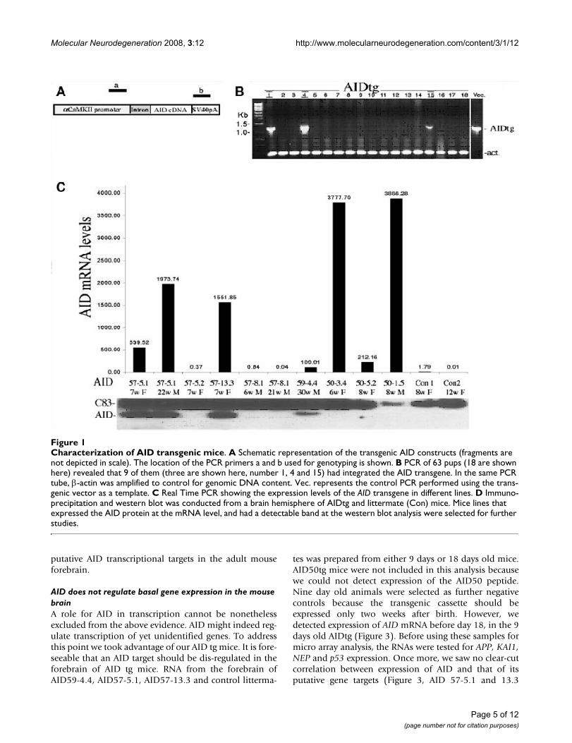
ResultsGeneration of AID transgenic animalsTo directly examine the effects of AID in vivo, and in thebrain, we generated transgenic mice expressing AID underthe control of the CaMKIIα promoter, targeting its expres-sion to the forebrain regions (which comprise the thala-mus, hypothalmus and the upper telencephalon) of thepostnatal mouse [24]. These areas are most relevant toAlzheimer's pathology. Endogenous AID is very shortlived and therefore virtually undetectable [25]. We gener-ated transgenic lines expressing either the 59- or 57-resi-due AID peptide, which would be produced by γ-cleavagetogether with either Aβ40 or Aβ42, respectively. In addi-tion, transgenic lines expressing a "ε-cleavage" AID of 50-residue [26,27] were also made. AID cDNAs were cloneddownstream of the 8-Kb CaMKIIα promoter and into aplasmid containing a mini-intron and the SV40 polyade-nylation sequence [24] (Figure 1A). The linearized plas-mids were injected into oocytes of FVB mice that werethan implanted in pseudo pregnant C57BL/6 mice. Tail-DNA PCR, showed that 9 out of 63 pups obtained hadintegrated the AID transgenes (samples are shown in Fig-ure 1B). More specifically, we obtained two AID59(AID59-4.4 and -1.1), four AID57 (AID57-13.3, -5.1, -5.2and -8.1) and three AID50 (AID50-3.4, -1.5 and 5.2)founder mice. Germline transmission was observed for allfounders. The expression levels of the AID transgenemRNA and protein were determined by real-time quanti-tative PCR and Immunoprecipitation followed by West-ern blot analysis, respectively. Total RNA and proteinlysates were isolated from the forebrain of adult AIDtganimals. Different levels of AID mRNA and AID peptidewere found in the different transgenic mice (compare Fig-ure 1C and 1D). Of note, AID50-1.5 and AID50-3.4 linesexpressed the highest levels of AID mRNA but no detecta-ble AID50 protein. This data suggests that AID50 is themore unstable AID peptide form.
All mice show, up to 24 months of age, a regular growthpattern and mating ability, and we cannot detect any grossdeficit or behavioral abnormality among the differentAIDtg lines compared to the wild type littermates.
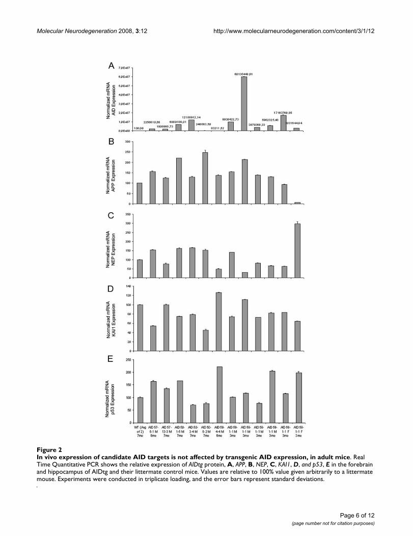
APP, KAI1, NEP and p53 gene expression is not altered in AID transgenic adult animalsTo determine whether AID affects APP, KAI1, NEP andp53 mRNA expression in vivo in the brain, RNA from theforebrain of adult (3–8 months) AID57-5.1, AID57-13.3,AID59-4.4, AID59-1.1, AID50-1.5, AID50-3.4, AID50-5.2and control littermates were analyzed by real-time quan-titative PCR. The data presented in Figure 2 show thatthere is no obvious correlation between AID mRNA andAPP, KAI1, NEP and p53 levels, considering also age, sexand AID transgene levels of expression. Overall, these datasuggest that AID is not involved in the basal expression of
Page 4 of 12(page number not for citation purposes)
Molecular Neurodegeneration 2008, 3:12 http://www.molecularneurodegeneration.com/content/3/1/12
putative AID transcriptional targets in the adult mouseforebrain.
AID does not regulate basal gene expression in the mouse brainA role for AID in transcription cannot be nonethelessexcluded from the above evidence. AID might indeed reg-ulate transcription of yet unidentified genes. To addressthis point we took advantage of our AID tg mice. It is fore-seeable that an AID target should be dis-regulated in theforebrain of AID tg mice. RNA from the forebrain ofAID59-4.4, AID57-5.1, AID57-13.3 and control litterma-
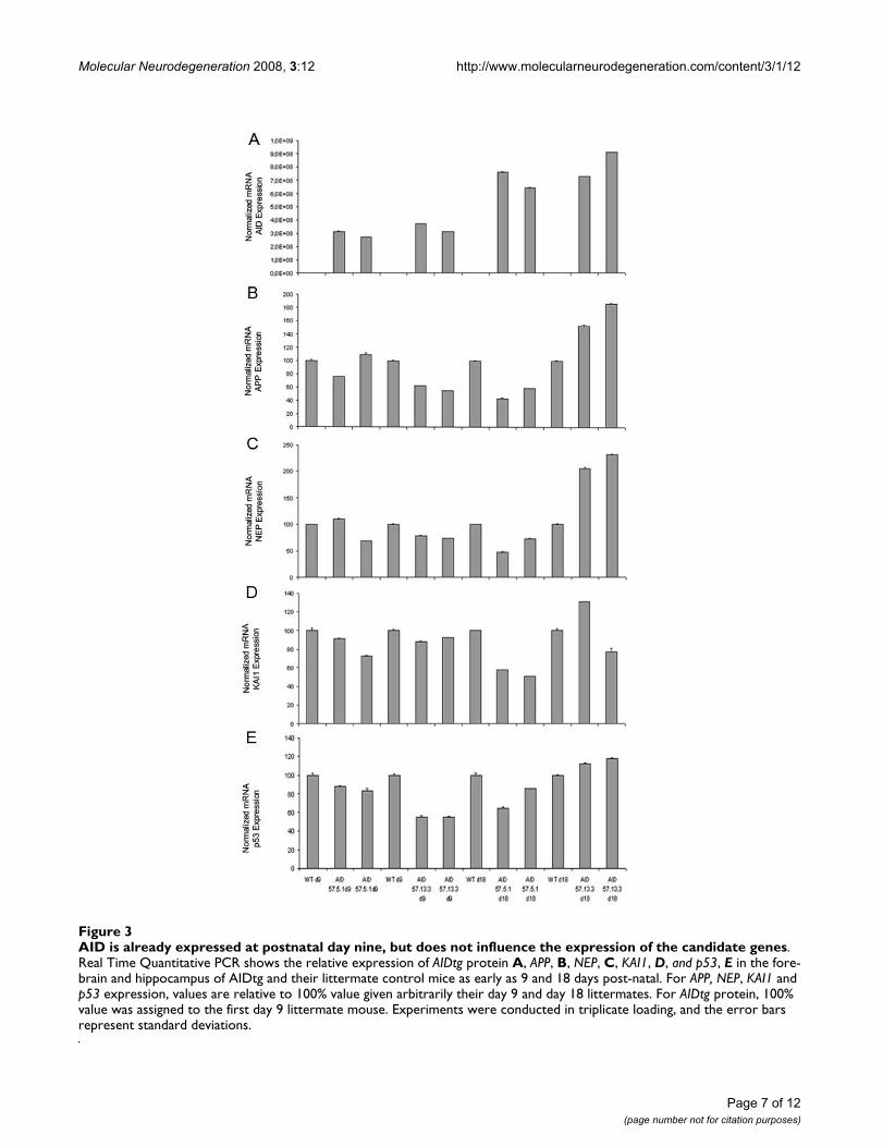
tes was prepared from either 9 days or 18 days old mice.AID50tg mice were not included in this analysis becausewe could not detect expression of the AID50 peptide.Nine day old animals were selected as further negativecontrols because the transgenic cassette should beexpressed only two weeks after birth. However, wedetected expression of AID mRNA before day 18, in the 9days old AIDtg (Figure 3). Before using these samples formicro array analysis, the RNAs were tested for APP, KAI1,NEP and p53 expression. Once more, we saw no clear-cutcorrelation between expression of AID and that of itsputative gene targets (Figure 3, AID 57-5.1 and 13.3
Characterization of AID transgenic miceFigure 1Characterization of AID transgenic mice. A Schematic representation of the transgenic AID constructs (fragments are not depicted in scale). The location of the PCR primers a and b used for genotyping is shown. B PCR of 63 pups (18 are shown here) revealed that 9 of them (three are shown here, number 1, 4 and 15) had integrated the AID transgene. In the same PCR tube, β-actin was amplified to control for genomic DNA content. Vec. represents the control PCR performed using the trans-genic vector as a template. C Real Time PCR showing the expression levels of the AID transgene in different lines. D Immuno-precipitation and western blot was conducted from a brain hemisphere of AIDtg and littermate (Con) mice. Mice lines that expressed the AID protein at the mRNA level, and had a detectable band at the western blot analysis were selected for further studies.
Page 5 of 12(page number not for citation purposes)
Molecular Neurodegeneration 2008, 3:12 http://www.molecularneurodegeneration.com/content/3/1/12
Page 6 of 12(page number not for citation purposes)
In vivo expression of candidate AID targets is not affected by transgenic AID expression, in adult miceFigure 2In vivo expression of candidate AID targets is not affected by transgenic AID expression, in adult mice. Real Time Quantitative PCR shows the relative expression of AIDtg protein, A, APP, B, NEP, C, KAI1, D, and p53, E in the forebrain and hippocampus of AIDtg and their littermate control mice. Values are relative to 100% value given arbitrarily to a littermate mouse. Experiments were conducted in triplicate loading, and the error bars represent standard deviations.
Molecular Neurodegeneration 2008, 3:12 http://www.molecularneurodegeneration.com/content/3/1/12
Page 7 of 12(page number not for citation purposes)
AID is already expressed at postnatal day nine, but does not influence the expression of the candidate genesFigure 3AID is already expressed at postnatal day nine, but does not influence the expression of the candidate genes. Real Time Quantitative PCR shows the relative expression of AIDtg protein A, APP, B, NEP, C, KAI1, D, and p53, E in the fore-brain and hippocampus of AIDtg and their littermate control mice as early as 9 and 18 days post-natal. For APP, NEP, KAI1 and p53 expression, values are relative to 100% value given arbitrarily their day 9 and day 18 littermates. For AIDtg protein, 100% value was assigned to the first day 9 littermate mouse. Experiments were conducted in triplicate loading, and the error bars represent standard deviations.
Molecular Neurodegeneration 2008, 3:12 http://www.molecularneurodegeneration.com/content/3/1/12
shown). Since we did not test EGFR and LRP mRNA levels,those genes (even their basal transcription) may still beregulated by AID alone, without over-expression of Fe65.Regardless, we analyzed the forebrain transcriptome ofthese mice using an Affymetrix DNA chips, containingover 39000 genes and open reading frames from M. mus-culus Genome databases GenBank, dbEST and RefSeq. Sta-tistical analysis performed using age and transgeneexpression as parameters to test, showed that age differ-ence was the only parameter to give significant results,yielding a set of 380 genes that were differentiallyexpressed between 9 and 18 day old mice (data notshown), indicating changes in the transcriptome duringpost-natal development. Transgene expression didn't giveany significant result even using a p-value cut-off 0.05indicating that the forebrain transcriptome was identicalin all age-matched mice analyzed. The above data argueagainst a role for AID in basal transcriptional regulation.
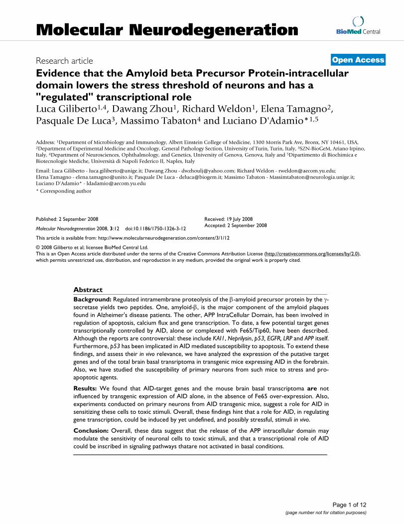
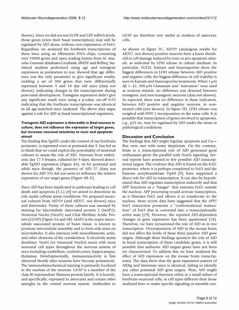
Transgenic AID expression is detectable in fetal neurons in culture, does not influence the expression of target genes, but increases neuronal sensitivity to toxic and apoptotic stimuliThe finding that TgAID, under the control of our forebrainpromoter, is expressed even at postnatal day 9, has led usto think that we could exploit the potentiality of neuronalcultures to assess the role of AID. Neurons from embry-onic day 17.5 fetuses, cultured for 9 days, showed detect-able TgAID expression (Figure 4A). As for postnatal andadult mice though, the presence of AID 57 (data notshown for AID 59) did not seem to influence the relativeexpression of our target genes (Figure 4B–E).
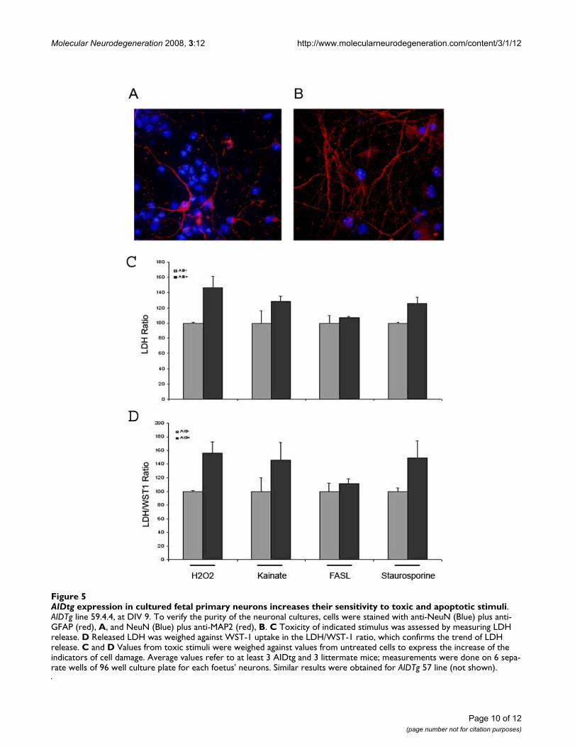
Since AID has been implicated in pathways leading to celldeath and apoptosis [3,12,28] we aimed to determine itsrole under cellular stress conditions. We prepared neuro-nal cultures from AID59 (and AID57, not shown) miceand littermates. Purity of these cultures was assessed bystaining for Microtubule Associated protein 2 (MAP2),Neuronal Nuclei (NeuN) and Glial fibrillary Acidic Pro-tein (GFAP) (Figure 5A and 5B). MAP2 is the major micro-tubule associated protein of brain tissue, is known topromote microtubule assembly and to form side-arms onmicrotubules. It also interacts with neurofilaments, actin,and other elements of the cytoskeleton. It electively stainsdendrites. NeuN (or Neuronal Nuclei) reacts with mostneuronal cell types throughout the nervous system ofmice including cerebellum, cerebral cortex, hippocampus,thalamus. Developmentally, immunoreactivity is firstobserved shortly after neurons have become postmitotic.The immunohistochemical staining is primarily localizedin the nucleus of the neurons. GFAP is a member of theclass III intermediate filament protein family. It is heavily,and specifically, expressed in astrocytes and certain otherastroglia in the central nervous system. Antibodies to
GFAP are therefore very useful as markers of astrocyticcells.
As shown in figure 5C, AID59 (analogous results forAID57, not shown) positive neurons have a lower thresh-old to cell damage induced by toxic or pro-apoptotic stim-uli, as indicated by LDH release in culture medium. Inparticular, H2O2, Kainate and Staurosporine show thebiggest differences in LDH release between AID positiveand negative cells; the biggest difference in cell viability isseen in Kainate and Staurosporine treatments. When 1 μMAβ 1–42, 500 μM Glutamate and "starvation" were usedas noxious stimuli, no difference was detected betweentransgenic and non-transgenic neurons (data not shown).As expected, there was no difference in these indicators,between AID positive and negative neurons, in non-treated cells (not shown). In figure 5D, LDH release wasweighed with WST-1 incorporation in the same cells. It ispossible that transcription of genes involved in apoptosis,e.g., p53 etc, may be regulated by AID under the stress orpathological conditions.
Discussion and ConclusionThe findings that AID might regulate apoptosis and Ca++flux were met with some skepticism. On the contrary,hints to a transcriptional role of AID generated greatenthusiasm given the parallel with Notch signaling. Sev-eral reports have pointed to few possible AID transcrip-tional targets. The evidence that AID is found on the KAI1promoter, where it is perhaps complexed to Fe65 and thehystone acetyltransferase Tip60 [9], have supported adirect role for AID in transcription. It can also be hypoth-esized that AID regulates transcription indirectly and thatAPP functions as a "hanger" that restrains Fe65 outsidethe nucleus: APP processing would activate transcription,as it liberates Fe65 and allows it to translocate to thenucleus. More recent data have suggested that the APP/Fe65 interaction promotes a "conformational matura-tion" of Fe65 that is converted into a transcriptionallyactive state [29]. However, the reported AID-dependentchanges in gene expression has been questioned [18].Therefore, we have reexamined the role of AID in in vivotranscription. Overexpression of AID in the mouse braindid not affect the levels of these three putative AID genetargets. Although these findings question the role of AIDin basal transcription of these candidate genes, it is stillpossible that authentic AID targets genes have not beenyet characterized. To address this we have analyzed theeffect of AID expression on the mouse brain transcrip-toma. The data show that the gene expression pattern ofAIDtg and littermate mice is identical, failing to identifyany other potential AID gene targets. Thus, AID mighthave a transcriptional function either in a small subset offorebrain neuronal cells, in cell types different than thoseanalyzed here or under specific signaling or stressful con-
Page 8 of 12(page number not for citation purposes)
Molecular Neurodegeneration 2008, 3:12 http://www.molecularneurodegeneration.com/content/3/1/12
Page 9 of 12(page number not for citation purposes)
AIDtg expression in cultured fetal primary neurons does not change the relative expression of APP, NEP, KAI1 and p53Figure 4AIDtg expression in cultured fetal primary neurons does not change the relative expression of APP, NEP, KAI1 and p53. A AIDtg expression was confirmed by tail genotyping of fetuses (not shown) and by QPCR data on cultured neurons (dark bars). Expression of AID, A, APP, B, NEP, C, KAI1, D, and p53, E, is relative to the 100% value given arbitrarily to the first AIDtg mouse. Experiments were conducted in triplicate loading, and the error bars represent standard deviations. Cultures were harvested at DIV 14. Similar results were achieved from younger cultures (DIV 9, not shown) and in the AIDTg 59 line (not shown).
Molecular Neurodegeneration 2008, 3:12 http://www.molecularneurodegeneration.com/content/3/1/12
Page 10 of 12(page number not for citation purposes)
AIDtg expression in cultured fetal primary neurons increases their sensitivity to toxic and apoptotic stimuliFigure 5AIDtg expression in cultured fetal primary neurons increases their sensitivity to toxic and apoptotic stimuli. AIDTg line 59.4.4, at DIV 9. To verify the purity of the neuronal cultures, cells were stained with anti-NeuN (Blue) plus anti-GFAP (red), A, and NeuN (Blue) plus anti-MAP2 (red), B. C Toxicity of indicated stimulus was assessed by measuring LDH release. D Released LDH was weighed against WST-1 uptake in the LDH/WST-1 ratio, which confirms the trend of LDH release. C and D Values from toxic stimuli were weighed against values from untreated cells to express the increase of the indicators of cell damage. Average values refer to at least 3 AIDtg and 3 littermate mice; measurements were done on 6 sepa-rate wells of 96 well culture plate for each foetus' neurons. Similar results were obtained for AIDTg 57 line (not shown).
Molecular Neurodegeneration 2008, 3:12 http://www.molecularneurodegeneration.com/content/3/1/12
ditions. Genome-wide analysis, conducted on neuronalcells expressing inducible AID, has shown that severalgenes involved in cytoskeletal dynamics can be regulatedby AID. The finding has been confirmed, by SYBR Greenreal-time PCR, in brains of AD patients for 2-Actin,IGFBP3, and TAGLN [17]. These target genes do not seemto be regulated by AID in our model. This might be due totwo reasons. Induction of the AID transgene was allowedfor 72 hours in culture before any effect could be detected.Our mice overexpressed AID for several days, as also evi-dent by AID mRNA detection in cultured neurons. It isforeseeable that any effect of AID overexpression, duringa longer period of time and in a more complex setting, asis the living mouse brain, would probably result in a dif-ferent expression arrangement, especially of genesdevoted to maintaining the integrity of the cell. Also, spo-radic AD brains are a much more complex and entropicsystem than ours, allowing for complex interactionsbetween different pathogenic entities. Thus, we cannotexclude that a dis-regulation of these target genes mayhappen later in the life of our mice or under differentstress conditions. The role of the intracellular fragment ofAPP, could possibly be understood by studying its effectunder specific stress situations, e.g. under apoptotic oroxidative stimuli, where it could play either a protective ora detrimental role for the cell, depending on other factorssuch as cell types and interaction with other proteins. Thiswould also explain the predisposition of some brainregions to Alzheimer's pathology. AID has been proposedas a possible mediator of cell death, via a reduction of thecellular threshold to apoptosis [3]. But recent findingshave also pointed to a possible protective effect of the APPc-terminal/Fe65 interaction, involving DNA damageresponse [28]. Our data show that over-expression of AIDin cultured neuronal cells predisposes them to a higherdegree of suffering, i.e. to a lower resistance to toxic andapoptotic stimuli. In our hands, only selective stimulicould reveal this peculiarity, possibly because of differentthreshold to cell damage for each experimental com-pound. Recent works show a role for AID in p53 associ-ated cell death [10]. In our model, under toxic stimuli,AID may lower the threshold to cell death through a p53-dependent mechanism by augmenting p53 expression.However, further experiments are required to test thishypothesis.
We believe that the key to understand the role of APPprocessing in gene regulation lays in the complex interac-tion of APP domains with other intra- or extra-cellular fac-tors, possibly having a role only in certain stressfulsituation or at a given "age". Further work will explore thenature of this complex network.
Competing interestsThe authors declare that they have no competing interests.
Authors' contributionsLG participated in the design of the study, handled themice colony and genotyping, designed the experiments,performed most of the experiments and cultures, partici-pated in the final analysis and draft preparation. DZ par-ticipated in the mice colony handling and genotyping,and characterization of tg mice. RW participated in themice colony handling and genotyping. ET participated inthe design of the study. PDL performed all the micro-arrayexperiments and analysis. MT participated in the design ofthe study. LD conceived and designed the study, designedthe tg mice, participated in the design of the experiments,participated in the handling of the mice colonies and gen-otyping, and in the analysis of the data, prepared the draft.
AcknowledgementsThis work was supported in part by Alzheimer Disease Research Grant A2003-076; National Institutes of Health Grants RO1 AG22024 and RO1 AG21588, the CARISA Foundation, the MIUR and the Regione Piemonte.
References1. Zheng H, Koo EH: The amyloid precursor protein: beyond
amyloid. Mol Neurodegener 2006/08/26 edition. 2006, 1:5.2. Selkoe DJ, Podlisny MB: Deciphering the genetic basis of Alzhe-
imer's disease. Annu Rev Genomics Hum Genet 2002, 3:67-99.3. Passer B, Pellegrini L, Russo C, Siegel RM, Lenardo MJ, Schettini G,
Bachmann M, Tabaton M, D'Adamio L: Generation of an apop-totic intracellular peptide by gamma-secretase cleavage ofAlzheimer's amyloid beta protein precursor. J Alzheimers Dis2000, 2(3-4):289-301.
4. Leissring MA, Murphy MP, Mead TR, Akbari Y, Sugarman MC, Jan-natipour M, Anliker B, Muller U, Saftig P, De Strooper B, Wolfe MS,Golde TE, LaFerla FM: A physiologic signaling role for thegamma -secretase-derived intracellular fragment of APP.Proc Natl Acad Sci U S A 2002/03/28 edition. 2002, 99(7):4697-4702.
5. Cao X, Sudhof TC: A transcriptionally [correction of transcrip-tively] active complex of APP with Fe65 and histone acetyl-transferase Tip60. Science 2001, 293(5527):115-120.
6. Artavanis-Tsakonas S, Rand MD, Lake RJ: Notch signaling: cell fatecontrol and signal integration in development. Science 1999/04/30 edition. 1999, 284(5415):770-776.
7. Annaert W, De Strooper B: Presenilins: molecular switchesbetween proteolysis and signal transduction. Trends Neurosci1999/09/11 edition. 1999, 22(10):439-443.
8. Wiley JC, Smith EA, Hudson MP, Ladiges WC, Bothwell M: Fe65stimulates proteolytic liberation of the beta-amyloid precur-sor protein intracellular domain. J Biol Chem 2007/09/15 edition.2007, 282(46):33313-33325.
9. Baek SH, Ohgi KA, Rose DW, Koo EH, Glass CK, Rosenfeld MG:Exchange of N-CoR corepressor and Tip60 coactivator com-plexes links gene expression by NF-kappaB and beta-amy-loid precursor protein. Cell 2002/08/02 edition. 2002,110(1):55-67.
10. Pardossi-Piquard R, Petit A, Kawarai T, Sunyach C, Alves da Costa C,Vincent B, Ring S, D'Adamio L, Shen J, Muller U, St George Hyslop P,Checler F: Presenilin-dependent transcriptional control of theAbeta-degrading enzyme neprilysin by intracellular domainsof betaAPP and APLP. Neuron 2005, 46(4):541-554.
11. Ryan KA, Pimplikar SW: Activation of GSK-3 and phosphoryla-tion of CRMP2 in transgenic mice expressing APP intracellu-lar domain. J Cell Biol 2005/10/19 edition. 2005, 171(2):327-335.
12. Kim HS, Kim EM, Lee JP, Park CH, Kim S, Seo JH, Chang KA, Yu E,Jeong SJ, Chong YH, Suh YH: C-terminal fragments of amyloidprecursor protein exert neurotoxicity by inducing glycogensynthase kinase-3beta expression. Faseb J 2003/08/19 edition.2003, 17(13):1951-1953.
13. Zhang YW, Wang R, Liu Q, Zhang H, Liao FF, Xu H: Presenilin/gamma-secretase-dependent processing of beta-amyloid
Page 11 of 12(page number not for citation purposes)
Molecular Neurodegeneration 2008, 3:12 http://www.molecularneurodegeneration.com/content/3/1/12
Publish with BioMed Central and every scientist can read your work free of charge
"BioMed Central will be the most significant development for disseminating the results of biomedical research in our lifetime."
Sir Paul Nurse, Cancer Research UK
Your research papers will be:
available free of charge to the entire biomedical community
peer reviewed and published immediately upon acceptance
cited in PubMed and archived on PubMed Central
yours — you keep the copyright
Submit your manuscript here:http://www.biomedcentral.com/info/publishing_adv.asp
BioMedcentral
precursor protein regulates EGF receptor expression. ProcNatl Acad Sci U S A 2007/06/09 edition. 2007, 104(25):10613-10618.
14. Liu Q, Zerbinatti CV, Zhang J, Hoe HS, Wang B, Cole SL, Herz J,Muglia L, Bu G: Amyloid precursor protein regulates brainapolipoprotein E and cholesterol metabolism through lipo-protein receptor LRP1. Neuron 2007/10/09 edition. 2007,56(1):66-78.
15. von Rotz RC, Kohli BM, Bosset J, Meier M, Suzuki T, Nitsch RM, Koni-etzko U: The APP intracellular domain forms nuclear multi-protein complexes and regulates the transcription of its ownprecursor. J Cell Sci 2004, 117(Pt 19):4435-4448.
16. Ahn KW, Joo Y, Choi Y, Kim M, Lee SH, Cha SH, Suh YH, Kim HS:Swedish amyloid precursor protein mutation increases cellcycle-related proteins in vitro and in vivo. J Neurosci Res 2008/04/29 edition. 2008, 86(11):2476-2487.
17. Muller T, Concannon CG, Ward MW, Walsh CM, Tirniceriu AL, TriblF, Kogel D, Prehn JH, Egensperger R: Modulation of gene expres-sion and cytoskeletal dynamics by the amyloid precursorprotein intracellular domain (AICD). Mol Biol Cell 2007,18(1):201-210.
18. Hebert SS, Serneels L, Tolia A, Craessaerts K, Derks C, Filippov MA,Muller U, De Strooper B: Regulated intramembrane proteolysisof amyloid precursor protein and regulation of expression ofputative target genes. EMBO Rep 2006/05/27 edition. 2006,7(7):739-745.
19. Waldron E, Isbert S, Kern A, Jaeger S, Martin AM, Hebert SS, Behl C,Weggen S, De Strooper B, Pietrzik CU: Increased AICD genera-tion does not result in increased nuclear translocation oractivation of target gene transcription. Exp Cell Res 2008/06/19edition. 2008, 314(13):2419-2433.
20. Ozaki T, Li Y, Kikuchi H, Tomita T, Iwatsubo T, Nakagawara A: Theintracellular domain of the amyloid precursor protein(AICD) enhances the p53-mediated apoptosis. Biochem Bio-phys Res Commun 2006, 351(1):57-63.
21. Checler F, Sunyach C, Pardossi-Piquard R, Sevalle J, Vincent B,Kawarai T, Girardot N, St George-Hyslop P, da Costa CA: Thegamma/epsilon-Secretase-Derived APP IntracellularDomain Fragments Regulate p53. Curr Alzheimer Res 2007,4(4):423-426.
22. Scheinfeld MH, Roncarati R, Vito P, Lopez PA, Abdallah M, D'AdamioL: Jun NH2-terminal kinase (JNK) interacting protein 1 (JIP1)binds the cytoplasmic domain of the Alzheimer's beta-amy-loid precursor protein (APP). J Biol Chem 2002,277(5):3767-3775.
23. Pfaffl MW: A new mathematical model for relative quantifica-tion in real-time RT-PCR. Nucleic Acids Res 2001/05/09 edition.2001, 29(9):e45.
24. Abel T, Nguyen PV, Barad M, Deuel TA, Kandel ER, BourtchouladzeR: Genetic demonstration of a role for PKA in the late phaseof LTP and in hippocampus-based long-term memory. Cell1997/03/07 edition. 1997, 88(5):615-626.
25. Cupers P, Orlans I, Craessaerts K, Annaert W, De Strooper B: Theamyloid precursor protein (APP)-cytoplasmic fragment gen-erated by gamma-secretase is rapidly degraded but distrib-utes partially in a nuclear fraction of neurones in culture. JNeurochem 2001, 78(5):1168-1178.
26. Yu C, Kim SH, Ikeuchi T, Xu H, Gasparini L, Wang R, Sisodia SS:Characterization of a presenilin-mediated amyloid precur-sor protein carboxyl-terminal fragment gamma. Evidencefor distinct mechanisms involved in gamma -secretaseprocessing of the APP and Notch1 transmembrane domains.J Biol Chem 2001/10/05 edition. 2001, 276(47):43756-43760.
27. Gu Y, Misonou H, Sato T, Dohmae N, Takio K, Ihara Y: Distinctintramembrane cleavage of the beta-amyloid precursor pro-tein family resembling gamma-secretase-like cleavage ofNotch. J Biol Chem 2001/08/03 edition. 2001, 276(38):35235-35238.
28. Minopoli G, Stante M, Napolitano F, Telese F, Aloia L, De Felice M, DiLauro R, Pacelli R, Brunetti A, Zambrano N, Russo T: Essentialroles for Fe65, Alzheimer amyloid precursor-binding pro-tein, in the cellular response to DNA damage. J Biol Chem2006/11/24 edition. 2007, 282(2):831-835.
29. Cao X, Sudhof TC: Dissection of amyloid-beta precursor pro-tein-dependent transcriptional transactivation. J Biol Chem2004/03/27 edition. 2004, 279(23):24601-24611.
Page 12 of 12(page number not for citation purposes)