Embed Size (px)
Citation preview
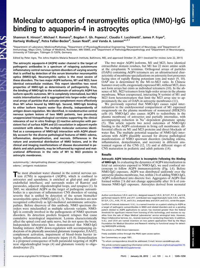
Molecular outcomes of neuromyelitis optica (NMO)-IgGbinding to aquaporin-4 in astrocytesShannon R. Hinsona, Michael F. Romerob, Bogdan F. Gh. Popescuc, Claudia F. Lucchinettic, James P. Fryera,Hartwig Wolburgd, Petra Fallier-Beckerd, Susan Noelle, and Vanda A. Lennona,c,f,1
aDepartment of Laboratory Medicine/Pathology, bDepartment of Physiology/Biomedical Engineering, cDepartment of Neurology, and fDepartment ofImmunology, Mayo Clinic, College of Medicine, Rochester, MN 55905; and dDepartment of Pathology/Neuropathology and eDepartment of Neurosurgery,University of Tübingen, Tübingen D-72076, Germany
Edited by Peter Agre, The Johns Hopkins Malaria Research Institute, Baltimore, MD, and approved October 31, 2011 (received for review June 22, 2011)
The astrocytic aquaporin-4 (AQP4) water channel is the target ofpathogenic antibodies in a spectrum of relapsing autoimmuneinflammatory central nervous system disorders of varying severitythat is unified by detection of the serum biomarker neuromyelitisoptica (NMO)-IgG. Neuromyelitis optica is the most severe ofthese disorders. The two major AQP4 isoforms, M1 and M23, haveidentical extracellular residues. This report identifies two novelproperties of NMO-IgG as determinants of pathogenicity. First,the binding of NMO-IgG to the ectodomain of astrocytic AQP4 hasisoform-specific outcomes. M1 is completely internalized, but M23resists internalization and is aggregated into larger-order orthog-onal arrays of particles that activate complement more effectivelythan M1 when bound by NMO-IgG. Second, NMO-IgG bindingto either isoform impairs water flux directly, independently ofantigen down-regulation. We identified, in nondestructive centralnervous system lesions of two NMO patients, two previouslyunappreciated histopathological correlates supporting the clinicalrelevance of our in vitro findings: (i) reactive astrocytes with per-sistent foci of surface AQP4 and (ii) vacuolation in adjacent myelinconsistent with edema. The multiple molecular outcomes identi-fied as a consequence of NMO-IgG interaction with AQP4 plausi-bly account for the diverse pathological features of NMO: edema,inflammation, demyelination, and necrosis. Differences in thenature and anatomical distribution of NMO lesions, and in theclinical and imaging manifestations of disease documented in pe-diatric and adult patients, may be influenced by regional and mat-urational differences in the ratio of M1 to M23 proteins inastrocytic membranes.
autoimmunity | demyelinating disease | astrocytopathy | intramyelinicedema | antigenic modulation
The most abundant water channel in the central nervous sys-tem (CNS) is aquaporin-4 (AQP4), which is confined to
astrocytes and ependyma; is enriched at glial–pial and glial–endothelial interfaces; and surrounds nodes of Ranvier andparanodes, adjacent oligodendroglial loops, and synapses (1). In2005, we identified AQP4 as the target of pathogenic autoanti-bodies in a spectrum of inflammatory CNS disorders of varyingseverity that is unified by detection of the serum biomarkerneuromyelitis optica (NMO)-IgG (2, 3). These disorders are nowrecognized collectively as IgG-mediated autoimmune astrocyto-pathies. Before discovery of this antibody, NMO spectrum dis-orders were misclassified as multiple sclerosis variants. NMO-IgG is centrally involved in the pathogenesis of NMO spectrumdisorders. Its detection predicts frequent relapses that causecumulative neurological impairment. Lesions characteristicallyaffect the spinal cord and optic nerve, but do not spare the brain.Independent laboratories have demonstrated that NMO-IgGbinding initiates AQP4 down-regulation with accompanying en-docytosis of its physically associated glutamate transporter, EAAT2,complement activation, impairment of blood–brain barrier in-tegrity, inflammation, and astrocyte injury (4–8). Demyelinationis a proposed consequence of both paranodal targeting of AQP4near oligodendroglial loops (4) and glutamate toxicity to oligo-dendrocytes (5).
The two major AQP4 isoforms, M1 and M23, have identicalextracellular domain residues, but M1 has 22 more amino acidsat the cytoplasmic N terminus. Orthogonal arrays of particles(OAPs) containing AQP4 tetramers are an ultrastructural char-acteristic ofmembrane specializations on astrocytic foot processesfacing sites of rapidly fluxing potassium ions and water (9, 10).OAP size is determined by the M1-to-M23 ratio. In Chinesehamster ovary cells, exogenously expressedM1,withoutM23, doesnot form arrays but exists as individual tetramers (10). In the ab-sence of M1, M23 tetramers form high-order arrays in the plasmamembrane. When coexpressed, interacting N termini of M1 andM23 form heterotetramers that limit the M23 array size to ap-proximately the size of OAPs in astrocytic membranes (11).We previously reported that NMO-IgG causes rapid inter-
nalization to the endolysosomal compartment of M1 expressedexogenously on the plasma membrane of nonneural (HEK293)cells (4). NMO-IgG also causes AQP4 to redistribute in theplasma membrane of astrocytes and partially internalize, withaccompanying reduction in Na+-dependent glutamate uptake(5). This article reports two novel outcomes of NMO-IgGbinding to the AQP4 ectodomain in living cell membranes: dif-ferential effects on M1 and M23 proteins and direct blockade ofwater flux. The multiple potential sequelae of NMO-IgG inter-actions with AQP4 plausibly account for diverse pathologicalmanifestations of NMO spectrum disorders (including edema,inflammation, demyelination, and necrosis) in different ana-tomical regions of the CNS (12, 13) and at different stages ofCNS maturation in pediatric and adult patients (14).
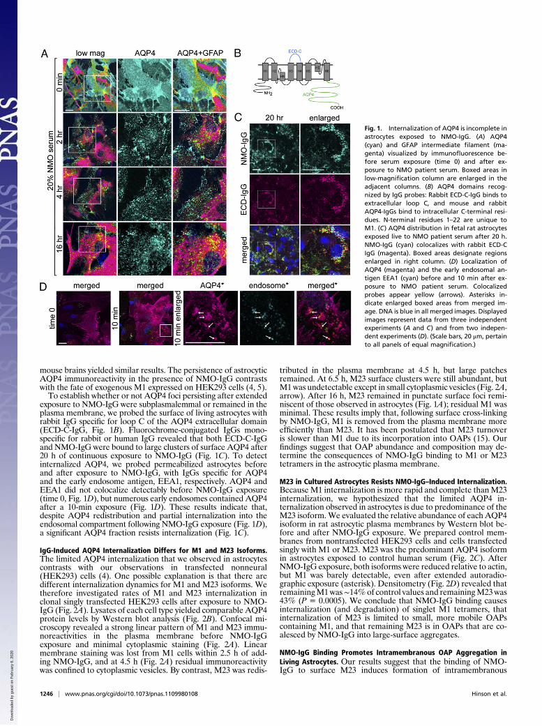
ResultsAstrocytic AQP4 Internalization Is Incomplete Following the Bindingof NMO-IgG. In evaluating the dynamics ofAQP4 internalization infetal rat astrocytes exposed to NMO-IgG, we used confocal mi-croscopy to follow AQP4 immunoreactivity (Fig. 1A). BeforeNMO-IgG exposure, AQP4 was distributed uniformly over theastrocytic plasma membrane, but, within 2 h of adding NMO-IgG,AQP4 redistributed into discrete foci. Aggregates of AQP4 thatformed within 2 h did not change appreciably after 16 h of con-tinuous NMO-IgG exposure. Astrocytes derived from neonatal
Author contributions: S.R.H. and V.A.L. designed research; S.R.H., B.F.G.P., P.F.-B., and S.N.performed research; M.F.R. and J.P.F. contributed new reagents/analytic tools; S.R.H., M.F.R.,B.F.G.P., C.F.L., H.W., P.F.-B., and V.A.L. analyzed data; and S.R.H. and V.A.L. wrote the paper.
Conflict of interest statement: V.A.L. is a named inventor on a patent relating to AQP4 asa target of pathogenic autoantibodies in NMO and related disorders and on a pendingpatent related to AQP4 applications to cancer; has received greater than the federalthreshold for significant interest from licensing of this technology; and receives no roy-alties from the sale of Mayo Medical Laboratories’ service serological tests. However,Mayo Collaborative Services, Inc., receives revenue for conducting these tests. In addition,V.A.L. and S.R.H are named inventors on two patent applications filed by the MayoFoundation for Medical Education and Research relating to functional assays for detect-ing NMO/AQP4 antibody.
This article is a PNAS Direct Submission.
Freely available online through the PNAS open access option.
See Commentary on page 1001.1To whom correspondence should be addressed. E-mail: [email protected].
This article contains supporting information online at www.pnas.org/lookup/suppl/doi:10.1073/pnas.1109980108/-/DCSupplemental.
www.pnas.org/cgi/doi/10.1073/pnas.1109980108 PNAS | January 24, 2012 | vol. 109 | no. 4 | 1245–1250
MED
ICALSC
IENCE
SSE
ECO
MMEN
TARY
Dow
nloa
ded
by g
uest
on
Feb
ruar
y 6,
202
0
mouse brains yielded similar results. The persistence of astrocyticAQP4 immunoreactivity in the presence of NMO-IgG contrastswith the fate of exogenous M1 expressed on HEK293 cells (4, 5).To establish whether or not AQP4 foci persisting after extended
exposure to NMO-IgG were subplasmalemmal or remained in theplasma membrane, we probed the surface of living astrocytes withrabbit IgG specific for loop C of the AQP4 extracellular domain(ECD-C-IgG, Fig. 1B). Fluorochrome-conjugated IgGs mono-specific for rabbit or human IgG revealed that both ECD-C-IgGand NMO-IgG were bound to large clusters of surface AQP4 after20 h of continuous exposure to NMO-IgG (Fig. 1C). To detectinternalized AQP4, we probed permeabilized astrocytes beforeand after exposure to NMO-IgG, with IgGs specific for AQP4and the early endosome antigen, EEA1, respectively. AQP4 andEEA1 did not colocalize detectably before NMO-IgG exposure(time 0, Fig. 1D), but numerous early endosomes contained AQP4after a 10-min exposure (Fig. 1D). These results indicate that,despite AQP4 redistribution and partial internalization into theendosomal compartment following NMO-IgG exposure (Fig. 1D),a significant AQP4 fraction resists internalization (Fig. 1C).
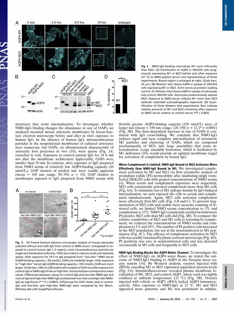
IgG-Induced AQP4 Internalization Differs for M1 and M23 Isoforms.The limited AQP4 internalization that we observed in astrocytescontrasts with our observations in transfected nonneural(HEK293) cells (4). One possible explanation is that there aredifferent internalization dynamics for M1 and M23 isoforms. Wetherefore investigated rates of M1 and M23 internalization inclonal singly transfected HEK293 cells after exposure to NMO-IgG (Fig. 2A). Lysates of each cell type yielded comparable AQP4protein levels by Western blot analysis (Fig. 2B). Confocal mi-croscopy revealed a strong linear pattern of M1 and M23 immu-noreactivities in the plasma membrane before NMO-IgGexposure and minimal cytoplasmic staining (Fig. 2A). Linearmembrane staining was lost from M1 cells within 2.5 h of add-ing NMO-IgG, and at 4.5 h (Fig. 2A) residual immunoreactivitywas confined to cytoplasmic vesicles. By contrast, M23 was redis-
tributed in the plasma membrane at 4.5 h, but large patchesremained. At 6.5 h, M23 surface clusters were still abundant, butM1was undetectable except in small cytoplasmic vesicles (Fig. 2A,arrow). After 16 h, M23 remained in punctate surface foci remi-niscent of those observed in astrocytes (Fig. 1A); residual M1 wasminimal. These results imply that, following surface cross-linkingby NMO-IgG, M1 is removed from the plasma membrane moreefficiently than M23. It has been postulated that M23 turnoveris slower than M1 due to its incorporation into OAPs (15). Ourfindings suggest that OAP abundance and composition may de-termine the consequences of NMO-IgG binding to M1 or M23tetramers in the astrocytic plasma membrane.
M23 in Cultured Astrocytes Resists NMO-IgG–Induced Internalization.BecauseM1 internalization is more rapid and complete thanM23internalization, we hypothesized that the limited AQP4 in-ternalization observed in astrocytes is due to predominance of theM23 isoform.We evaluated the relative abundance of each AQP4isoform in rat astrocytic plasma membranes by Western blot be-fore and after NMO-IgG exposure. We prepared control mem-branes from nontransfected HEK293 cells and cells transfectedsingly with M1 or M23. M23 was the predominant AQP4 isoformin astrocytes exposed to control human serum (Fig. 2C). AfterNMO-IgG exposure, both isoforms were reduced relative to actin,but M1 was barely detectable, even after extended autoradio-graphic exposure (asterisk). Densitometry (Fig. 2D) revealed thatremainingM1was∼14%of control values and remainingM23was43% (P = 0.0005). We conclude that NMO-IgG binding causesinternalization (and degradation) of singlet M1 tetramers, thatinternalization of M23 is limited to small, more mobile OAPscontaining M1, and that remaining M23 is in OAPs that are co-alesced by NMO-IgG into large-surface aggregates.
NMO-IgG Binding Promotes Intramembranous OAP Aggregation inLiving Astrocytes. Our results suggest that the binding of NMO-IgG to surface M23 induces formation of intramembranous
Fig. 1. Internalization of AQP4 is incomplete inastrocytes exposed to NMO-IgG. (A) AQP4(cyan) and GFAP intermediate filament (ma-genta) visualized by immunofluorescence be-fore serum exposure (time 0) and after ex-posure to NMO patient serum. Boxed areas inlow-magnification column are enlarged in theadjacent columns. (B) AQP4 domains recog-nized by IgG probes: Rabbit ECD-C-IgG binds toextracellular loop C, and mouse and rabbitAQP4-IgGs bind to intracellular C-terminal resi-dues. N-terminal residues 1–22 are unique toM1. (C) AQP4 distribution in fetal rat astrocytesexposed live to NMO patient serum after 20 h.NMO-IgG (cyan) colocalizes with rabbit ECD-CIgG (magenta). Boxed areas designate regionsenlarged in right column. (D) Localization ofAQP4 (magenta) and the early endosomal an-tigen EEA1 (cyan) before and 10 min after ex-posure to NMO patient serum. Colocalizedprobes appear yellow (arrows). Asterisks in-dicate enlarged boxed areas from merged im-age. DNA is blue in all merged images. Displayedimages represent data from three independentexperiments (A and C) and from two indepen-dent experiments (D). (Scale bars, 20 μm, pertainto all panels of equal magnification.)
1246 | www.pnas.org/cgi/doi/10.1073/pnas.1109980108 Hinson et al.
Dow
nloa
ded
by g
uest
on
Feb
ruar
y 6,
202
0
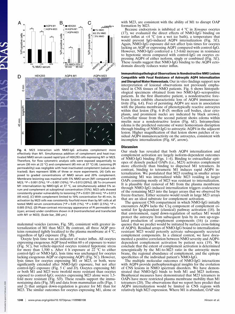
structures that resist internalization. To investigate whetherNMO-IgG binding changes the abundance or size of OAPs, weanalyzed neonatal mouse astrocytic membranes by freeze-frac-ture electron microscopy before and after in vitro exposure tohuman IgG. In the absence of human IgG, intramembranousparticles in the nonpolarized membranes of cultured astrocyteswere numerous, but OAPs, an ultrastructural characteristic ofastrocytic foot processes in vivo (10), were sparse (Fig. 3A,encircled in red). Exposure to control patient IgG for 24 h didnot alter the membrane architecture appreciably; OAPs weresmaller than 50 nm. In contrast, after exposure to IgG preparedfrom NMO serum of relatively low AQP4-binding capacity (20nmol/L), OAP clusters of modest size were readily apparent(mean = 104 nm; range, 30–350; n = 19). OAP clusters inmembranes exposed to IgG prepared from NMO serum with
fivefold greater AQP4-binding capacity (105 nmol/L) were oflarger size (mean = 190 nm; range, 120–350; n= 12, P= 0.0001)(Fig. 3B). The dose-dependent increase in size of OAPs is con-sistent with IgG cross-linking. We conclude that NMO-IgGinduces rapid and near complete internalization of tetramericM1 particles and clustering of OAPs, which are composedpredominantly of M23, into large assemblies that resist in-ternalization. Large assembly formation, which is facilitated byM1 deficiency (10), would provide an optimal membrane targetfor activation of complement by bound IgG.
When Complement is Limited, NMO-IgG Bound to M23 Activates MoreEffectively than NMO-IgG Bound to M1. We investigated comple-ment activation by M1 and M23 via flow cytometric analysis ofpropidium iodide (PI) permeability after incubating singly trans-fected HEK293 cells with graded concentrations of NMO serum(4). When serum and complement exposures were sequential,M23 cells consistently activated complement more than M1 cells(Fig. 4A). To minimize loss of M1 epitope density by IgG-inducedinternalization, we next exposed the cells to serum and comple-ment simultaneously. Again, M23 cells activated complementmore effectively than M1 cells (Fig. 4 B and C). To prevent frag-mentation of M23 cells and enable more accurate counting of le-sioned cells, we limited NMO serum concentration to 5% andcomplement to 15%.NMO-IgG consistently yielded twofoldmorePI-positive M23 cells than M1 cells did (Fig. 4B). To compare therelative sensitivities of M23 and M1 cells to lesioning by comple-ment, we reduced the concentrations of NMO serum and com-plement to 1%and10%.The number of PI-positive cells increasedin the M23 population, but not in the nontransfected or M1 pop-ulation (Fig. 4C). The efficacy of complement activation by M23cellswas readily visualized by phase-contrastmicroscopy (Fig. 4D).PI positivity was rare in nontransfected cells and was detectedoccasionally in M1 cells and frequently in M23 cells.
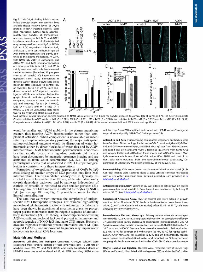
NMO-IgG Binding Blocks the AQP4 Water Channel. To investigate theeffect of NMO-IgG on AQP4 water fluxes, we tested the out-come of NMO-IgG binding to AQP4 in the Xenopus laevis oo-cyte system (16). By Western analysis, oocytes injected withcRNAs encoding M1 or M23 expressed equivalent protein levels(Fig. 5A). Immunofluorescence revealed plasma membrane lo-calization of M1, M23, and control AQP1, where each was tightlyconfined at ambient temperature (22 °C) (Fig. 5B). Oocytesinjected with vehicle or AQP1 cRNA lacked AQP4 immunore-activity. After exposure to NMO-IgG at 22 °C, M1 and M23appeared more punctate and M1 was prominent in subplas-
Fig. 2. NMO-IgG binding internalizes M1 more efficientlythan M23. (A) Distribution of AQP4 in HEK293 cells exog-enously expressing M1 or M23 before and after exposure(37 °C) to NMO patient serum and representative of threeexperiments. Boxed region is enlarged at right. (Scale bars,20 μm.) (B) Western blot shows AQP4 in lysates of HEK293cells expressing M1 or M23. Actin serves as protein loadingcontrol. (C) Western blot shows AQP4 in lysates of astrocytesand control HEK293 cells. Astrocytes predominantly expressM23. Exposure to NMO serum reduces M1 more than M23(asterisk: extended autoradiographic exposure). (D) Quan-tification of three Western blot experiments. Bars indicaterelative amounts of M1 and M23 remaining after exposureto NMO serum relative to control serum (*P = 0.003).
Fig. 3. (A) Freeze-fracture electron microscopic analysis of mouse astrocytescultured without and with IgG from control or NMO serum. Unexposed or ex-posed to control human IgG (∼2 mg/mL), most intramembranous particles aresingle and distributed uniformly. OAPs (encircled in red) are small and relativelysparse. After exposure for 24 h to IgG prepared from “low-titer” NMO serum(AQP4-binding capacity = 20 nmol/L), OAPs are modestly larger. After exposureto “high-titer” serum IgG (AQP4-binding capacity = 105 nmol/L), OAPs aremuchlarger. (Scale bars, 100nm.) (B) Scatter plotanalysis ofOAP sizeafter exposure tocontrol-IgGorNMO-IgGof loworhightiter.Horizontalbarscorrespondtomeanvalues. Differences between values for control-IgG and low-titer NMO-IgG andcontrol-IgGandhigh-titerNMO-IgGandbetween low-titer andhigh-titerNMO-IgG are significant (***P = 0.0001). Differences for OAP cluster sizes in control-IgG and low-titer and high-titer NMO-IgG were compared by the Mann–Whitney test with GraphPad software.
Hinson et al. PNAS | January 24, 2012 | vol. 109 | no. 4 | 1247
MED
ICALSC
IENCE
SSE
ECO
MMEN
TARY
Dow
nloa
ded
by g
uest
on
Feb
ruar
y 6,
202
0
malemmal vesicles (arrows, Fig. 5B), consistent with greater in-ternalization of M1 than M23. By contrast, all three AQP pro-teins remained tightly localized to the plasma membrane at 4 °C,regardless of IgG exposure (Fig. 5B).Oocyte lysis time was an indicator of water influx. All oocytes
expressing exogenous AQP lysed within 60 s of exposure to water(Fig. 5C), but vehicle-injected oocytes resisted hypotonic stressfor more than 1,500 s. After 4 h exposure at 22 °C to eithercontrol-IgG or NMO-IgG, lysis time was unchanged for oocyteslacking exogenous AQP or expressing AQP1 (Fig. 5C). However,lysis times for oocytes expressing M1 or M23, or both, weresignificantly extended after NMO-IgG exposure, but not aftercontrol-IgG exposure (Fig. 5 C and D). Oocytes expressing M1or both M1 and M23 were twofold more resistant than oocytesexposed to control-IgG; oocytes expressing M23 alone were 1.5-fold more resistant (Fig. 5D). These results support the immu-nostaining data (Fig. 5B) and data from mammalian cells (Figs. 1and 2) that antigen down-regulation is greater for M1 than forM23. The similar outcomes for oocytes expressing M1, alone or
with M23, are consistent with the ability of M1 to disrupt OAPformation by M23.Because endocytosis is inhibited at 4 °C in Xenopus oocytes
(17), we evaluated the direct effects of NMO-IgG binding onwater influx at <4 °C (on a wet ice bath), a temperature thatwould prevent IgG-induced AQP4 internalization (Fig. 5E).Again, NMO-IgG exposure did not affect lysis times for oocyteslacking an AQP or expressing AQP1 compared with control-IgG.However, NMO-IgG conferred a 1.5-fold increase in resistanceto hypotonic stress compared with control-IgG on oocytes ex-pressing AQP4 of either isoform, singly or combined (Fig. 5E).These results suggest that NMO-IgG binding to the AQP4 ecto-domain directly reduces water influx.
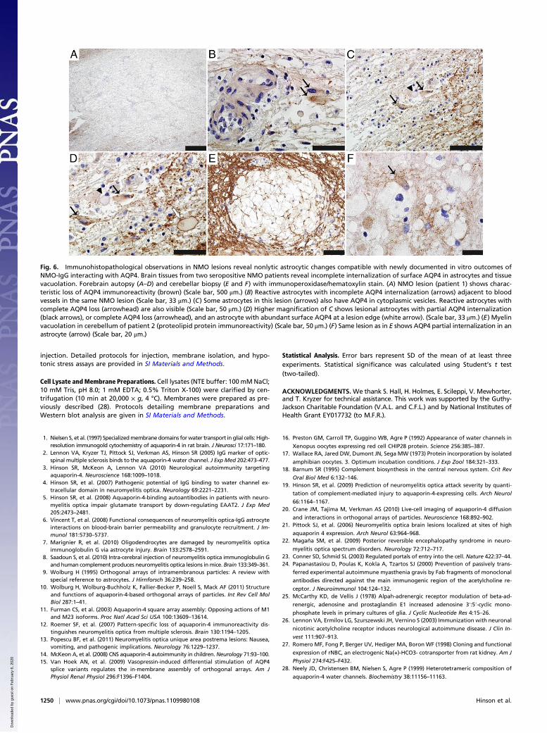
Immunohistopathological Observations in Nondestructive NMO LesionsCompatible with Focal Resistance of Astrocytic AQP4 Internalizationand DisruptedWater Homeostasis.Our in vitro findings support newinterpretation of lesional observations not previously empha-sized in CNS tissues of NMO patients. Fig. 6 shows histopath-ological specimens obtained from two NMO-IgG–seropositivepatients. In the first illustrative patient, a nondestructive fore-brain lesion exhibits characteristic loss of AQP4 immunoreac-tivity (Fig. 6A). Foci of persisting AQP4 are seen in associationwith the plasma membrane of phenotypically reactive astrocyteswithin that lesion (Fig. 6 B–D; swollen cell bodies, clear cyto-plasm, and prominent nuclei are indicated by black arrows).Cerebellar tissue from the second patient shows edema withinmyelin near a nondestructive lesion (Fig. 6E). Intramyelinicedema is the predicted outcome of water homeostasis disruptionthrough binding of NMO-IgG to astrocytic AQP4 in the adjacentlesion. Higher magnification of that lesion shows patches of re-sidual AQP4 immunoreactivity on the astrocytes, consistent withincomplete internalization (Fig. 6F, arrow).
DiscussionOur study has revealed that both AQP4 internalization andcomplement activation are largely isoform-dependent outcomesof NMO-IgG binding (Figs. 1–4). Binding to extracellular epit-opes of densely packed OAPs (i.e., M23) activates complementmore effectively than binding to dispersed M1 tetramers. Incontrast, binding to tetrameric M1 induces rapid AQP4 in-ternalization. We postulated that M23 residing in smaller arrayscontaining M1 was internalized while M23 residing in largerOAPs consisting mostly of M23 was resistant to internalization.It is plausible that removal of M1 from the plasma membranethrough NMO-IgG–induced internalization triggers coalescenceof the remaining M23 into the larger arrays that we observed byfreeze-fracture. Either scenario would result in large M23 arraysthat are an ideal substrate for complement activation.The quiescent CNS compartment in which NMO-IgG initially
encounters AQP4 lacks the C1q component of complement es-sential for Ig-dependent (classical) pathway activation (18). Inthat environment, rapid down-regulation of surface M1 wouldprotect the astrocyte from subsequent lysis by its own up-regu-lated production of complement components (16) (a conse-quence that we predict would be initiated by surface cross-linkingof AQP4). Residual arrays of NMO-IgG bound to internalization-resistant M23 would potently activate subsequently secretedcomplement components. In a clinical context, we have docu-mented a positive correlation between NMO severity and AQP4-dependent complement activation by patient sera (19). Weconclude that the extent of complement activation is determinedsynergistically by the M1-to-M23 ratio in the astrocytic mem-brane, the regional abundance of complement, and the epitopespecificities of the individual patient’s NMO-IgG.The multiple molecular outcomes of NMO-IgG interactions
with AQP4 provide pathophysiological insights for the evolutionof CNS lesions in NMO spectrum disorders. We have demon-strated that NMO-IgG binds to both M1 and M23 isoforms.Biophysical measures have demonstrated that M23 tetramers inOAPs have more restricted plasma membrane mobility than M1tetramers (20). The observations that we report here predict thatAQP4 internalization would be limited in CNS regions withrelatively high M23 expression. Where M1 is abundant, OAP size
Fig. 4. M23 interaction with NMO-IgG activates complement moreeffectively than M1. Simultaneous addition of complement and heat-inac-tivated NMO serum caused rapid lysis of HEK293 cells expressing M1 or M23.Therefore, for flow cytometric analysis cells were exposed sequentially toserum (30 min at 22 °C) and complement (45 min at 37 °C) (4). Lesioning (PIpermeability) was negligible with heat-inactivated complement (values sub-tracted). Bars represent SEMs of three or more experiments. (A) Cells ex-posed to graded concentrations of NMO serum and 20% complement.Membrane lesioning was maximal with 5% NMO serum [M1 compared withM23; *P < 0.001 (5%); *P < 0.001 (10%); *P = 0.013 (20%)]. (B) To circumventM1 internalization by NMO-IgG at 37 °C, we simultaneously added 5% se-rum and complement at suboptimal concentration (15%). M23 cells showedconsistently greater vulnerability to lesioning [*P = 0.031 (30 min); *P = 0.013(45 min)]. (C) With complement limited to 10% concentration for 45 min, itsactivation by M23 cells was consistently fourfold more than by M1 cells at alltested NMO serum concentrations [*P = 0.05 (1%); *P = 0.001 (2.5%); *P =0.005 (5%)]. (D) Phase-contrast microscopy appearance of PI permeable cells(cyan; arrows) under conditions shown in B (nontransfected and transfectedwith M1 or M23). (Scale bar, 200 μm.)
1248 | www.pnas.org/cgi/doi/10.1073/pnas.1109980108 Hinson et al.
Dow
nloa
ded
by g
uest
on
Feb
ruar
y 6,
202
0
would be smaller and AQP4 mobility in the plasma membranegreater, thus favoring AQP4 internalization rather than com-plement activation. When complement is unavailable or inacti-vated by complement-regulatory proteins, the major anticipatedpathophysiological outcome would be disruption of water ho-meostasis either by direct blockade of water flux and by AQP4internalization. NMO-characteristic periventricular abnormali-ties that rapidly reverse with high-dose corticosteroid therapyhave been documented by magnetic resonance imaging and areattributed to tissue water accumulation (21, 22). The strikingparenchymal edema that we observed in NMO histopathologicallesions is consistent with these interpretations.Formation of exceptionally large aggregates of OAPs by IgG
cross-linking of smaller arrays of M23 particles may limit M23internalization. Clathrin-mediated endocytosis is typically re-stricted to particles smaller than 120 nm, while internalization bycaveola-dependent pathways, and by pathways independent ofclathrin or caveolin, is restricted to even smaller particles (23).The large size of OAPs induced in cultured astrocytes by NMO-IgG (on average 190 nm; Fig. 3) plausibly accounts for theirlimited internalization.The data that we present increase the complexity of antigen-
specific NMO therapeutic strategies. For example, high-affinitymonoclonal IgG fragments reactive with autoantigen ectodomainshave been shown, in experimental myasthenia gravis, to protecttarget plasma membranes from pathogenic sequelae of autoanti-body interactions (24). In theory, a noncomplement-activatingAQP4-specific monoclonal IgG could prevent inflammatory andnecrotic sequelae of NMO-IgG binding to CNS targets. However,divalent fragments would not prevent internalization of M1 (andcoupled EAAT2), and monovalent fragments may impair waterhomeostasis in critical CNS locations.
Materials and MethodsAstrocytes, Cell Lines, and Transgenic Constructs. Astrocyte cultures wereestablished from cerebral cortices of fetal (embryonic days 18–21) rats ornewborn mice (25). M1 and M23 cDNAs and stably transfected clones ofHEK-293 were produced as described (2, 4). DNA encoding AQP4 extra-
cellular loop C was PCR amplified and cloned into pET 4T vector (Stratagene)to produce and purify GST-ECD-C fusion protein (26).
Antibodies and Sera. Fluorochrome-conjugated secondary antibodies werefrom Southern Biotechnology. Rabbit anti-AQP4 C terminus IgG and Cy3-MAbIgG anti-GFAPwere from Sigma, anti-EEA1MAb IgGwas from BD Biosciences,and rabbit anti-actin and anti-AQP1 C terminus IgGs were from Santa Cruzand Abcam. Rabbit anti-AQP4 loop C and mouse anti-AQP4 C terminus MAbIgGs were made in-house. Pools of ∼50 de-identified NMO and control pa-tient sera were obtained from the Neuroimmunology Laboratory, De-partment of Laboratory Medicine/Pathology, at the Mayo Clinic.
Immunostaining. Cells were grown and immunostained as described (4, 5).Confocal images were captured using a Zeiss LSM510 confocal microscopewith a 63× water immersion lens. Detailed procedures are provided in SIMaterials and Methods.
Antigen Modulation Assay. Serum or IgG was added to cells grown on coatedglass coverslips for at least 48 h. Complement was inactivated by holding 30min at 56 °C. See SI Materials and Methods.
Complement Activation Assay. NMO or control sera were added in growthmedium. After 30 min at 22 °C, fresh or heat-inactivated complement wasadded (Low–Tox-H, Cedarlane Laboratories). After 45 min at 37 °C, cells wereanalyzed by flow cytometry (4).
Freeze-Fracture Electron Microscopy. Primary mouse astrocyte monolayerswerefixed (2h, 22 °C)with 2.5%glutaraldehyde in0.1Mcacodylatebuffer (pH7.4), cryoprotected in30%glycerol, and quick-frozen in nitrogen slush (−210 °C).Specimenswere fractured in a freeze-fracture device (BAF400D; Balzers) at 5×10⁻⁶ mbar and −150 °C. Fracture faces were shadowed with platinum/carbon(2.5 nm, 45 °C) for contrast and with carbon (25 nm, 90 °C) for replica stabili-zation. After removing cell material in 12% sodium hypochlorite, replicaswere cleaned in double-distilled water and mounted on Pioloform-coatedcopper grids. Replicaswere examinedunder a Zeiss EM10electronmicroscope.
Oocyte Isolation and Injection. Oocytes were removed from X. laevis frogs(Xenopus Express), dissociated with collagenase (27), and studied 3–6 d after
Fig. 5. NMO-IgG binding inhibits waterinflux through AQP4. (A) Western blotanalysis shows relative levels of AQP4protein in cRNA-injected oocytes. Eachlane represents lysates from approxi-mately four oocytes. (B) Immunofluo-rescence staining of M1, M23, and AQP1in plasma membranes of cRNA-injectedoocytes exposed to control-IgG or NMO-IgG. At 4 °C, regardless of human IgG,and at 22 °C with control human IgG, allAQP immunoreactivities are tightly con-fined to the plasma membrane. At 22 °Cwith NMO-IgG, AQP1 is unchanged, butAQP4 M1 and M23 immunoreactivitiesare more punctate (asterisks), and M1 isvisibly associated with subplasmalemmalvesicles (arrows). (Scale bar, 10 μm, per-tains to all panels.) (C) Representativehypotonic stress assay (immersion indistilled water) shows oocyte lysis times(seconds) after exposure to control-IgGor NMO-IgG for 4 h at 22 °C. Each con-dition included 5–12 injected oocytes.Injected cRNAs are indicated below thegraph. Asterisks indicate significance incomparing oocytes exposed to control-IgG and NMO-IgG for M1 (P < 0.001),M23 (P = 0.005), and M1 + M23 (P <0.001). (D and E) Cumulative data fromfive to six hypotonic stress assays showfold increase in lysis times for oocytes exposed to NMO-IgG relative to lysis times for oocytes exposed to control-IgG at 22 °C or 4 °C. (D) Asterisks indicateP values relative to AQP1 controls: M1 (P < 0.001), M23 (P < 0.001), M1 + M23 (P < 0.001), and relative to M23—M1 (P = 0.032) and M1 + M23 (P = 0.016). (E)Comparisons are relative to AQP1: M1 (P = 0.028) and M23 (P = 0.001); differences between M1 and M23 were not significant.
Hinson et al. PNAS | January 24, 2012 | vol. 109 | no. 4 | 1249
MED
ICALSC
IENCE
SSE
ECO
MMEN
TARY
Dow
nloa
ded
by g
uest
on
Feb
ruar
y 6,
202
0
injection. Detailed protocols for injection, membrane isolation, and hypo-tonic stress assays are provided in SI Materials and Methods.
Cell Lysate andMembrane Preparations. Cell lysates (NTE buffer: 100 mMNaCl;10 mM Tris, pH 8.0; 1 mM EDTA; 0.5% Triton X-100) were clarified by cen-trifugation (10 min at 20,000 × g, 4 °C). Membranes were prepared as pre-viously described (28). Protocols detailing membrane preparations andWestern blot analysis are given in SI Materials and Methods.
Statistical Analysis. Error bars represent SD of the mean of at least threeexperiments. Statistical significance was calculated using Student’s t test(two-tailed).
ACKNOWLEDGMENTS. We thank S. Hall, H. Holmes, E. Scileppi, V. Mewhorter,and T. Kryzer for technical assistance. This work was supported by the Guthy-Jackson Charitable Foundation (V.A.L. and C.F.L.) and by National Institutes ofHealth Grant EY017732 (to M.F.R.).
1. Nielsen S, et al. (1997) Specializedmembrane domains forwater transport in glial cells: High-resolution immunogold cytochemistry of aquaporin-4 in rat brain. J Neurosci 17:171–180.
2. Lennon VA, Kryzer TJ, Pittock SJ, Verkman AS, Hinson SR (2005) IgG marker of optic-spinal multiple sclerosis binds to the aquaporin-4 water channel. J ExpMed 202:473–477.
3. Hinson SR, McKeon A, Lennon VA (2010) Neurological autoimmunity targetingaquaporin-4. Neuroscience 168:1009–1018.
4. Hinson SR, et al. (2007) Pathogenic potential of IgG binding to water channel ex-tracellular domain in neuromyelitis optica. Neurology 69:2221–2231.
5. Hinson SR, et al. (2008) Aquaporin-4-binding autoantibodies in patients with neuro-myelitis optica impair glutamate transport by down-regulating EAAT2. J Exp Med205:2473–2481.
6. Vincent T, et al. (2008) Functional consequences of neuromyelitis optica-IgG astrocyteinteractions on blood-brain barrier permeability and granulocyte recruitment. J Im-munol 181:5730–5737.
7. Marignier R, et al. (2010) Oligodendrocytes are damaged by neuromyelitis opticaimmunoglobulin G via astrocyte injury. Brain 133:2578–2591.
8. Saadoun S, et al. (2010) Intra-cerebral injection of neuromyelitis optica immunoglobulin Gand human complement produces neuromyelitis optica lesions inmice. Brain 133:349–361.
9. Wolburg H (1995) Orthogonal arrays of intramembranous particles: A review withspecial reference to astrocytes. J Hirnforsch 36:239–258.
10. Wolburg H, Wolburg-Buchholz K, Fallier-Becker P, Noell S, Mack AF (2011) Structureand functions of aquaporin-4-based orthogonal arrays of particles. Int Rev Cell MolBiol 287:1–41.
11. Furman CS, et al. (2003) Aquaporin-4 square array assembly: Opposing actions of M1and M23 isoforms. Proc Natl Acad Sci USA 100:13609–13614.
12. Roemer SF, et al. (2007) Pattern-specific loss of aquaporin-4 immunoreactivity dis-tinguishes neuromyelitis optica from multiple sclerosis. Brain 130:1194–1205.
13. Popescu BF, et al. (2011) Neuromyelitis optica unique area postrema lesions: Nausea,vomiting, and pathogenic implications. Neurology 76:1229–1237.
14. McKeon A, et al. (2008) CNS aquaporin-4 autoimmunity in children. Neurology 71:93–100.15. Van Hoek AN, et al. (2009) Vasopressin-induced differential stimulation of AQP4
splice variants regulates the in-membrane assembly of orthogonal arrays. Am JPhysiol Renal Physiol 296:F1396–F1404.
16. Preston GM, Carroll TP, Guggino WB, Agre P (1992) Appearance of water channels in
Xenopus oocytes expressing red cell CHIP28 protein. Science 256:385–387.17. Wallace RA, Jared DW, Dumont JN, Sega MW (1973) Protein incorporation by isolated
amphibian oocytes. 3. Optimum incubation conditions. J Exp Zool 184:321–333.18. Barnum SR (1995) Complement biosynthesis in the central nervous system. Crit Rev
Oral Biol Med 6:132–146.19. Hinson SR, et al. (2009) Prediction of neuromyelitis optica attack severity by quanti-
tation of complement-mediated injury to aquaporin-4-expressing cells. Arch Neurol
66:1164–1167.20. Crane JM, Tajima M, Verkman AS (2010) Live-cell imaging of aquaporin-4 diffusion
and interactions in orthogonal arrays of particles. Neuroscience 168:892–902.21. Pittock SJ, et al. (2006) Neuromyelitis optica brain lesions localized at sites of high
aquaporin 4 expression. Arch Neurol 63:964–968.22. Magaña SM, et al. (2009) Posterior reversible encephalopathy syndrome in neuro-
myelitis optica spectrum disorders. Neurology 72:712–717.23. Conner SD, Schmid SL (2003) Regulated portals of entry into the cell. Nature 422:37–44.24. Papanastasiou D, Poulas K, Kokla A, Tzartos SJ (2000) Prevention of passively trans-
ferred experimental autoimmune myasthenia gravis by Fab fragments of monoclonal
antibodies directed against the main immunogenic region of the acetylcholine re-
ceptor. J Neuroimmunol 104:124–132.25. McCarthy KD, de Vellis J (1978) Alpah-adrenergic receptor modulation of beta-ad-
renergic, adenosine and prostaglandin E1 increased adenosine 3′:5′-cyclic mono-
phosphate levels in primary cultures of glia. J Cyclic Nucleotide Res 4:15–26.26. Lennon VA, Ermilov LG, Szurszewski JH, Vernino S (2003) Immunization with neuronal
nicotinic acetylcholine receptor induces neurological autoimmune disease. J Clin In-
vest 111:907–913.27. Romero MF, Fong P, Berger UV, Hediger MA, BoronWF (1998) Cloning and functional
expression of rNBC, an electrogenic Na(+)-HCO3- cotransporter from rat kidney. Am J
Physiol 274:F425–F432.28. Neely JD, Christensen BM, Nielsen S, Agre P (1999) Heterotetrameric composition of
aquaporin-4 water channels. Biochemistry 38:11156–11163.
Fig. 6. Immunohistopathological observations in NMO lesions reveal nonlytic astrocytic changes compatible with newly documented in vitro outcomes ofNMO-IgG interacting with AQP4. Brain tissues from two seropositive NMO patients reveal incomplete internalization of surface AQP4 in astrocytes and tissuevacuolation. Forebrain autopsy (A–D) and cerebellar biopsy (E and F) with immunoperoxidase/hematoxylin stain. (A) NMO lesion (patient 1) shows charac-teristic loss of AQP4 immunoreactivity (brown) (Scale bar, 500 μm.) (B) Reactive astrocytes with incomplete AQP4 internalization (arrows) adjacent to bloodvessels in the same NMO lesion (Scale bar, 33 μm.) (C) Some astrocytes in this lesion (arrows) also have AQP4 in cytoplasmic vesicles. Reactive astrocytes withcomplete AQP4 loss (arrowhead) are also visible (Scale bar, 50 μm.) (D) Higher magnification of C shows lesional astrocytes with partial AQP4 internalization(black arrows), or complete AQP4 loss (arrowhead), and an astrocyte with abundant surface AQP4 at a lesion edge (white arrow). (Scale bar, 33 μm.) (E) Myelinvacuolation in cerebellum of patient 2 (proteolipid protein immunoreactivity) (Scale bar, 50 μm.) (F) Same lesion as in E shows AQP4 partial internalization in anastrocyte (arrow) (Scale bar, 20 μm.)
1250 | www.pnas.org/cgi/doi/10.1073/pnas.1109980108 Hinson et al.
Dow
nloa
ded
by g
uest
on
Feb
ruar
y 6,
202
0