Embed Size (px)
Citation preview

1
Molecular Pathways - INVITED ARTICLE
Molecular Pathways: Mucins and Drug Delivery in Cancer
Chinthalapally V. Rao*, Naveena B. Janakiram, and Altaf Mohammed*
Center for Cancer Prevention and Drug Development, Hematology and Oncology Section, Stephenson Cancer Center, University of Oklahoma Health Sciences Center, Oklahoma City, OK, USA
Running Title: Mucin synthesis inhibition
Keywords: molecular pathways, mucins, drug delivery, cancer, GCNT3, talniflumate, prevention,
treatment
*Corresponding Author: Center for Cancer Prevention and Drug Development, 975 NE 10th Street, BRC II, Room 211, OUHSC, Oklahoma City, OK, 73104, USA. Phone: +1 405-271-3224, Fax: +1 405-271-3225
Email: [email protected] (Altaf Mohammed); [email protected] (C.V. Rao)
Conflict of interest: The authors declare no conflicts of interest.
Grant Support: This work was supported by the College of Medicine Alumni Association Grant and
Stephenson Cancer Center Research Support Fund to A.M. and the Kerley-Cade Endowed Chair Fund
and National Cancer Institute NCI-N01-CN53300 to C.V.R.
Word count: Abstract – 171, Text (excluding references & supplementary data): 2632
Total number of figures: 1
Research. on May 29, 2020. © 2016 American Association for Cancerclincancerres.aacrjournals.org Downloaded from
Author manuscripts have been peer reviewed and accepted for publication but have not yet been edited. Author Manuscript Published OnlineFirst on December 30, 2016; DOI: 10.1158/1078-0432.CCR-16-0862

2
Abstract:
Over the past few decades, clinical and preclinical studies have clearly demonstrated the role of mucins
in tumor development. It is well established that mucins form a barrier impeding drug access to target
sites, leading to cancer chemoresistance. Recently gained knowledge regarding core enzyme synthesis
has opened avenues to explore the possibility of disrupting mucin synthesis to improve drug efficacy.
Cancer cells exploit aberrant mucin synthesis to efficiently mask the epithelial cells and ensure survival
under hostile tumor microenvironment conditions. However, O-glycan synthesis enzyme core 2 beta 1,6
N-acetylglucosaminyltransferase (GCNT3/C2GnT-2) is overexpressed in Kras-driven mouse and human
cancer, and inhibition of GCNT3 has been shown to disrupt mucin synthesis. This previously
unrecognized developmental pathway might be responsible for aberrant mucin biosynthesis and
chemoresistance. In this molecular pathways article, we briefly discuss the potential role of mucin
synthesis in cancers, ways to improve drug delivery and disrupt mucin mesh to overcome
chemoresistance by targeting mucin synthesis, and the unique opportunity to target the GCNT3
pathway for the prevention and treatment of cancers.
Research. on May 29, 2020. © 2016 American Association for Cancerclincancerres.aacrjournals.org Downloaded from
Author manuscripts have been peer reviewed and accepted for publication but have not yet been edited. Author Manuscript Published OnlineFirst on December 30, 2016; DOI: 10.1158/1078-0432.CCR-16-0862

3
Background
Mucins: Mucin biology, synthesis, and functions are complex. Mucins form a mucous gel barrier and
protect the epithelia of most organs from physical and chemical damage and infection (1). In 1835,
Nicolas Theodore de Saussure first used the term “mucin” to describe these substances (2). Two years
later, Eichwald identified that mucin is a combination of carbohydrates and proteins (3). Mucins are
comprised of amino- and carboxy-terminal protein regions with a large central region formed by amino
acid tandem repeats rich in proline, threonine, and serine residues with O-linked oligosaccharides or N-
linked oligosaccharides (1, Fig 1A). Most mucin core glycans are O-linked oligosaccharides composed of
N-acetyl galactosamine (GalNAc), N-acetyl glucosamine (GlcNAc), galactose (Gal), fucose (Fuc), and
neuraminic acid (sialic acid, NeuNAc) linked to serine or threonine.
There are eight O-GalNAc glycan core structures, designated cores 1- 8: core T antigen, Galβ1-
3GalNAcαSer/Thr (core 1), GlcNAcβ1-6(Galβ1-3)GalNAcαSer/Thr (core 2), GlcNAcβ1-3GalNAcαSer/Thr
(core 3), (GlcNAcβ1-6(GlcNAcβ1-3)GalNAcαSer/Thr (core 4), GalNAcα1-3GalNAcαSer/Thr (core 5),
GlcNAcβ1-6GalNAcαSer/Thr (core 6), GalNAcα1-6GalNAcαSer/Thr (core 7), and Galα1-3GalNAcαSer/Thr
(core 8; Fig. 1B). Starting from GalNAc on serine or threonine residues in a polypeptide, Core 1 synthase
(C1GnT) transfers galactose to make the Core 1 structure, and Core 3 synthase (C3GnT) transfers GlcNAc
to form a Core 3 structure. Core 1 is converted to Core 2 by C2GnT-1, C2GnT-2, and C2GnT-3, whereas
Core 3 is converted to Core 4 by C2GnT-2. Mannose, fucose, glucose, and GlcNAc are directly linked to
Ser/Thr; they form core 5-8 structures. Many sugars modified by acetylation or sulfation on the mucin
O-glycans are antigenic. Mucin O-glycans are involved in biological processes, including embryonic
development, immune responses, protein folding, cell signaling, and malignancies. These compounds
are often heterogeneous.
Over 20 mucins have been identified; they are classified as either membrane-bound mucins or
secreted mucins (1,4). MUC2, MUC5AC, MUC5B, MUC6, MUC7, MUC9, and MUC19 are secreted mucins.
Research. on May 29, 2020. © 2016 American Association for Cancerclincancerres.aacrjournals.org Downloaded from
Author manuscripts have been peer reviewed and accepted for publication but have not yet been edited. Author Manuscript Published OnlineFirst on December 30, 2016; DOI: 10.1158/1078-0432.CCR-16-0862
4
Their primary function is to participate in mucus formation to protect the underlying epithelia against
injuries related to inflammation and infection. Membrane-bound mucins include MUC1, MUC3A/B,
MUC4, MUC12, MUC13, MUC15, MUC16, MUC17, MUC20, and MUC21. They are thought to play
important roles in cellular interactions, molecular cell signaling, and biological processes. Recent studies
of secretory and transmembrane mucins focused on their role in malignancies. Glycosylation in mucins
may restrict their cellular and tissue-level expression. However, information regarding which cores are
attached to mucins of known MUC backbones is limited. O-glycans purified from normal and diseased
tissues have different core types. Whether the complexity of O-glycan structures reflects altered levels
of specific mucins or alterations in glycosylation of specific MUC proteins in disease or inflammation is
unknown. Multiple core type O-glycans may be attached to a single MUC backbone, especially in mucins
that have more than one TR-type domain.
Differential roles of mucins in cancer: Mucin deregulation is observed, and MUC1 is overexpressed, in
pancreatic, lung, breast, colon, ovarian, and prostate cancers (5). MUC4 is overexpressed in colon
adenocarcinoma and pancreatic cancer. MUC16 is elevated in ovarian and pancreatic cancers. Mucins
have been identified as significant components of the glycocalyx in various tumors (6,7). Large
glycoproteins are abundantly expressed in tumor cells, the microenvironment, and on circulating tumor
cells from patients with advanced disease (7). Because of their specific pattern of expression during
tumor progression, mucins remain under intense investigation as biomarkers and therapeutic targets.
For example, full-length MUC13 expressed in MUC13-null pancreatic cancer cell lines significantly
increases cell motility, invasion, proliferation, and clonogenicity. Exogenous MUC13 expression
significantly enhanced pancreatic tumor growth and reduced survival in a xenograft mouse model.
These characteristics were correlated with the upregulation/phosphorylation of HER2, p21-activated
kinase 1 (PAK1), extracellular signal-regulated kinase (ERK), Akt and metastasin (S100A4), and p53
suppression (8). Similarly, esophageal cancer cells lacking MUC1 proliferated, migrated, and invaded
Research. on May 29, 2020. © 2016 American Association for Cancerclincancerres.aacrjournals.org Downloaded from
Author manuscripts have been peer reviewed and accepted for publication but have not yet been edited. Author Manuscript Published OnlineFirst on December 30, 2016; DOI: 10.1158/1078-0432.CCR-16-0862

5
less. Subcutaneous xenografts were significantly smaller when cells did not express MUC1 (9). Thus,
mucin subtypes play a key role in cancer cell proliferation, migration, invasion, and tumor growth.
The modified forms of glycosylated tumor-associated mucins promote tumor cell invasion,
migration, intravasation, and extravasation which help in immunosuppression or immune evasion (10-
15). Galectin-3 binding to Major histocompatibility complex class I-related chain A carrying core2 O-
glycans (MICAC2) through poly-N-acetyllactosamine impair NK cell activation by reducing IFN-g and
granzyme B secreation (10-14). Studies are warranted to identify if these molecules are immunogenic
under these situations. MUC16 and mesothelin were co-expressed in infiltrating components, promoting
pancreatic cancer invasion (16). The presence of MUC-1-specific CD8 cytotoxic lymphocytes in breast
and ovarian cancer and PDAC prompted the design of a MUC1-targeting vaccine (17,18). Preclinical
studies with MUC1 transgenic animals demonstrated its efficacy in stimulating immune responses
against this antigen (19).
The mucin cores are deregulated during tumor cell transformation, leading to aberrant
expression. MUC1, MUC4, MUC5AC, and MUC16 are strongly up-regulated in patients with PDAC,
pancreatic intraepithelial neoplasia (PanINs), and intrapapillary mucinous neoplasia (IPMNs; 20).
However, some mucins play controversial roles. While most mucins are involved in tumor progression,
MUC2 suppresses tumors. In colon cancer, MUC2 helps protect the barrier functions of normal colonic
crypts. Loss of MUC2 leads to colonization of the intestinal tract with different microbial flora, which
may have pro-carcinogenic or bystander effects (21). Similarly, MUC17 is highly expressed on the
intestinal epithelial surface and helps in epithelial restitution and protection against E. Coli infection
(22). Mucins vary depending on the glycan moieties present on the peptide backbone.
Targeting mucin synthesis: Clinical and preclinical studies elucidated the tumor-promoting roles of
mucins in cancer. Based on these findings, several mucins were individually targeted. Little knowledge
exists about potential intervention approaches involving mucin-glycan synthesizing genes. All mucins
Research. on May 29, 2020. © 2016 American Association for Cancerclincancerres.aacrjournals.org Downloaded from
Author manuscripts have been peer reviewed and accepted for publication but have not yet been edited. Author Manuscript Published OnlineFirst on December 30, 2016; DOI: 10.1158/1078-0432.CCR-16-0862
6
contain one or more of the core glycans. C1GNT is involved in forming core 1, and C3GNT forms core 3.
C2GNT is involved in forming core 2 from core 1 and core 4 from core 3. The core 2 beta 1,6 N-
acetylglucosaminyltransferase (GCNT3/ C2GNT) plays a significant role in mucin glycan biosynthesis.
Aberrant GCNT3 expression leads to mucin overexpression (23-26). GCNT3 activity plays an important
role in physiological processes, including inflammatory and immune responses. Core 3-derived glycans, a
major type of O-glycan expressed by normal gastrointestinal epithelial cells, are downregulated during
malignancy, due to loss of functional β3-N-acetylglucosaminyltransferase-6 (C3GnT, core 3 synthase)
expression. Expression of core 3-derived O-glycans in pancreatic cancer cells suppressed tumor growth
and metastasis through modulation of mucin glycosylation and other cell surface and extracellular
matrix proteins (27).
CLINICAL-TRANSLATIONAL ADVANCES
For several decades, researchers focused on the protective role of mucins in epithelial cells and their
role in cancer progression. Less attention was given to glycan synthesis, which comprises more than 50%
of the mucin structure, and to enzymes or genes involved in core glycan synthesis. Recent studies have
explored the aberrant expression of mucin-glycan synthesis genes, suggesting the possibility of evading
chemoresistance caused by mucins and improving existing therapies, or developing novel glycan
synthesis enzyme inhibitors or combined strategies to simultaneously disrupt mucin synthesis and
inhibit tumor growth (Fig. 1C,D). In this section, we discuss how to improve drug delivery and disrupt
mucin mesh to overcome chemoresistance by targeting mucin synthesis.
Drug Delivery and Disruption of Mucin Barrier: Targeting mucinous cancer cells with drugs is
challenging. Although several drugs inhibit tumor cells in vitro, the effect is seldom replicated in vivo.
There are several reasons for treatment failure. One such factor for chemoresistance and poor prognosis
is the mucin mesh barrier (28-38). Aberrant Muc1 expression is associated with poor disease-free and
Research. on May 29, 2020. © 2016 American Association for Cancerclincancerres.aacrjournals.org Downloaded from
Author manuscripts have been peer reviewed and accepted for publication but have not yet been edited. Author Manuscript Published OnlineFirst on December 30, 2016; DOI: 10.1158/1078-0432.CCR-16-0862

7
overall survival in non-small-cell lung cancer (39-41). Although the involvement of mucins is unclear,
evidence supports the involvement of a physical barrier, resistance to apoptosis, drug metabolism, cell
stemness, and EMT as factors responsible for chemoresistance (42).
Most mucins carry an electronegative potential that may create electrostatic interactions with
positively charged drugs, thereby decreasing their diffusion (42). Understanding the mechanisms of
resistance involving mucins will contribute to the development of next-generation targeted therapy
molecules. For example, in vitro and in vivo MUC1 knockdown reduced tumor cell growth, cell
proliferation, MAPK, cell migration, and invasion via MMP13, and cell survival and apoptosis via Akt and
Bcl2 (43). Researchers have suggested a correlation between mucin overexpression and cancer, and
demonstrated a link between the aberrant and differential overexpression of mucin glycoproteins and
disease initiation, progression, and poor prognosis (35-38, 44-46).
Previous research has demonstrated aberrant expression of membrane-bound and secreted
mucins in ductal adenocarcinomas, pancreatic intraepithelial neoplasia, IPMNs, and mucinous cystic
neoplasms (35-38, 44-46). The extent to which the dense mucin mesh influences the antiproliferative
activity of 5-fluorouracil (5-FU) was investigated in human pancreatic cancer cells (5, 47). MUC1 vaccine
in vivo and a small molecule inhibitor drug (GO-201) that inhibits MUC1-cytoplasmic tail oligomerization
have shown promising results (48-54). Administration of GO-201 to nude mice bearing human breast
tumor xenografts was associated with loss of tumorigenicity and extensive necrosis, resulting in
prolonged tumor growth regression.
Most reports focus on mucin peptide gene expression or aspects of the glycosylated mucins,
with little mention of the enzymes that catalyze mucin biosynthesis. Little knowledge exists regarding
potential intervention approaches involving mucin-glycan-synthesizing genes. Core mucin genes were
recently evaluated as targets to disrupt mucin mesh formation, leading to targeted drug delivery for
pancreatic cancer. Using survival data from human patients with pancreatic cancer, next-generation
Research. on May 29, 2020. © 2016 American Association for Cancerclincancerres.aacrjournals.org Downloaded from
Author manuscripts have been peer reviewed and accepted for publication but have not yet been edited. Author Manuscript Published OnlineFirst on December 30, 2016; DOI: 10.1158/1078-0432.CCR-16-0862
8
sequencing of genetically engineered Kras-driven mouse pancreatic tumors, and human pancreatic
cancer cells, a novel core mucin-synthesizing enzyme, GCNT3 (core 2 beta 1,6 N-
acetylglucosaminyltransferase), was identified. In mouse pancreatic tumors, GCNT3 upregulation was
correlated with increased expression of mucins. GCNT3 was significantly overexpressed in human
pancreatic cancer and reduced patient survival by 7 months. High GCNT3 expression was also associated
with patients’ drinking, smoking habits, and diabetes diagnosis (26). Aberrant GCNT3 expression was
linked with increased mucin production, aggressive tumorigenesis, and reduced patient survival; CRISPR-
mediated GCNT3 knockout in pancreatic cancer cells reduced proliferation and spheroid formation (26).
A small molecule inhibitor that selectively binds to GCNT3, talniflumate, was identified via in silico small
molecular docking simulation approaches (26). Talniflumate enhanced drug delivery and the antitumor
effects of EGFR inhibitor (26). Docking predictions suggested that three hydrogen bonds between
talniflumate and GCNT3 contribute to a docking affinity of -8.3 kcal/mol. Furthermore, talniflumate
alone and in combination with low-dose gefitinib reduced GCNT3 expression, leading to disrupted mucin
production in vivo and in vitro (26). Thus, targeting mucin biosynthesis through GCNT3 may improve
drug responsiveness (Fig. 1C). Further development of agents and their mechanisms targeting mucin
synthesis and permitting access of drugs to epithelial cells is needed.
Developmental state of the pathway: Many data suggest the role of mucins in the acquisition of
chemoresistance. Thus, there is the possibility of interfering with the pathway responsible for mucin
production as a treatment strategy. Current strategies, however, focus on individual mucins. One
promising approach is targeting the key pathway leading to core glycan synthesis. However, there are no
reports evaluating the mucin-glycan-synthesizing genes as targets. C2GNT-1, 2, and 3 are important core
glycan-synthesizing enzymes. These core enzymes form basic structure of most mucins. C2GNT2/GCNT3
have been studied in vitro and in vivo and are reported as attractive novel targets for pancreatic cancer
treatment (26). GCNT3 activity plays an important role in physiological processes, including
Research. on May 29, 2020. © 2016 American Association for Cancerclincancerres.aacrjournals.org Downloaded from
Author manuscripts have been peer reviewed and accepted for publication but have not yet been edited. Author Manuscript Published OnlineFirst on December 30, 2016; DOI: 10.1158/1078-0432.CCR-16-0862
9
inflammatory and immune responses. Furthermore, the expression of distinct oligosaccharide
structures, together with differential glycosylation of mucin core proteins, confers on tumor cells an
enormous range of potential ligands for interaction with other cell surface receptors. The process of
natural selection within growing tumor cell populations creates cells that express novel combinations
and forms of mucins, which contribute to the survival of these tumor cells during invasion and
metastasis. Mucin core enzymes might promote biological properties, like inflammation and immune
suppression, leading to enhanced tumor growth.
Drug development, strategies to overcome resistance, and opportunities for treatment: Since mucin
glycan synthesis and signaling is involved in cancer cell immunosuppression and growth and metastasis,
there is significant interest in developing therapies targeted against this glycan synthesis pathway.
Applying novel technologies and utilizing crystal structures of core enzymes, specific inhibitors for
GCNT3 or other core enzymes can be designed and evaluated. GCNT3’s crystal structure has been
reported, enabling the development of small molecule inhibitor talniflumate. However, the
development of direct small-molecule GCNT3 inhibitors is underway. Development of glycan core
enzyme inhibitors will enable chemoprevention and pancreatic cancer treatment. GCNT3 core glycan
enzyme synthesis increases in correlation with the stepwise progression of PanIN lesions to ductal
adenocarcinoma. Hence, using GCNT3 inhibitors blocks mucin synthesis and might help in delaying
PanIN progression (Fig 1D). During the adenocarcinoma stage, it appears possible to disrupt the mucin
mesh with specific core enzyme inhibitors, thereby potentially preventing tumor progression into
metastasis and helping with delivery of standard drugs to target sites (Fig. 1C,D). GCNT3 inhibitors may
act independently to inhibit tumor growth or in combination with other standard drugs, like
gemcitabine, to synergistically inhibit tumors. However, the mechanisms underlying GCNT3 inhibitor
activity remain poorly defined. Genome-editing technologies, such as CRISPR/CAS9, should enable
precise and efficient mutation of core enzymes to study mechanisms that are induced by the tumor
Research. on May 29, 2020. © 2016 American Association for Cancerclincancerres.aacrjournals.org Downloaded from
Author manuscripts have been peer reviewed and accepted for publication but have not yet been edited. Author Manuscript Published OnlineFirst on December 30, 2016; DOI: 10.1158/1078-0432.CCR-16-0862

10
microenvironment. In proof-of-concept experiments, studies using GCNT3 KO mice aim to demonstrate
its role in mucins and tumor progression. Cutting-edge genetic methods for targeting GCNT3 signaling
may enhance the efficacy of current pancreatic cancer therapies. GCNT3 overexpression was also seen
in hepatocellular carcinoma cell lines and orthotopic xenograft tumors (55). However, in colon cancer
like MUC2 subtype, GCNT3 expression is low (56). As per human protein atlas, most cancer tissues
displayed weak to strong granular cytoplasmic GCNT3 staining. Strong staining was seen in pancreatic,
stomach and ovarian cancers. Gliomas, lymphomas and skin cancers were negative (57).
Mucins have been investigated as therapeutic targets for pancreatic and other cancers. MUC1
vaccine was well tolerated, with no adverse effects. It was effective in animal models, alone and in
combination with COX-2 inhibitors and gemcitabine (58). Several clinical trials (Clinical trials.gov,
NCT00669734, NCT00008099, NCT00597129, NCT00603863) involving MUC1 vaccines for pancreatic
cancer are underway or have been completed. A Phase I/II Trial in patients with relapsed or refractory
acute myeloid leukemia is studying the targeted MUC1 inhibitor, GO-203-2C, alone and in combination
with gemcitabine. GO-203-2c targets cancer cells, while leaving healthy cells unaffected. MUC2 vaccine,
combined with QS21, was used to treat patients with prostate cancer. A preclinical study showed that a
peptide vaccine of MUC1 inhibited tumor growth, and significantly regressed breast cancer tumors (48).
One-third of patients with pancreatic and biliary cancer in a Phase I/II clinical trial of a MUC1 peptide-
loaded DC vaccine administered post-resection survived, without evidence of recurrence (54). Many
formulations of MUC1 vaccine were tested in patients with pancreatic cancer showing increased
disease-free survival with mucin-specific humoral and T-cell responses (50, 51). A follow-up of a phase III
study with patients with stage II breast cancer showed that those who were using oxidized mannan-
MUC1 (M-FP) vaccine survived longer, without evidence of toxicity or autoimmunity (59). A phase III
START study with tecemotide, a MUC1-antigen-specific cancer immunotherapy, reported a notable
survival benefit for drug-treated patients with unresectable stage III non-small-cell lung cancer versus
Research. on May 29, 2020. © 2016 American Association for Cancerclincancerres.aacrjournals.org Downloaded from
Author manuscripts have been peer reviewed and accepted for publication but have not yet been edited. Author Manuscript Published OnlineFirst on December 30, 2016; DOI: 10.1158/1078-0432.CCR-16-0862
11
those treated with placebo (60). A 21-mer peptide MUC1 vaccine induced T and B cell MUC1-specific
immunity in patients with multiple myeloma (61).
These findings suggest that mucins play a significant role in tumor growth and metastases, and
blocking mucins with vaccines can control tumor growth and spread. Immunological Targeting core
enzymes for decreased mucin production or disrupting mucin synthesis are attractive strategies.
Conclusions and Perspectives: The GCNT3 branch of core glycan synthesis is a novel and ill-
characterized pathway with significant therapeutic relevance in human cancer. This molecular pathway
controls unique biologic processes in mucin synthesis and tumor-infiltrating immune cells to promote
tumor progression. Although mucin synthesis signaling can be targeted through various methods (Fig 1C,
D), potent small-molecule inhibitors are an attractive strategy to disrupt mucin synthesis, permitting
drug access to target sites, thereby overcoming chemoresistance.
Acknowledgements: We thank Ms. Agata Bien and Ms. Kathy Kyler for editing this review.
References:
1. Hollingsworth MA, Swanson BJ. Mucins in cancer: protection and control of the cell surface. Nat
Rev Cancer 2004;4:45–60.
2. Gottschalk A. In The Chemistry and Biology of Sialic Acids and Related Substances; Gottschalk,
A., Ed.; Cambridge University Press: New York, 1960, pp 1–11.
3. Montreuil J, Vliegenthart JFG, Schachter H. In Glycoproteins and Disease; Elsevier: Amsterdam,
1995; Vol. 30.
4. Kufe DW. Mucins in cancer: function, prognosis and therapy. Nat Rev Cancer 2009;9:874–85.
5. Jonckheere N, Skrypek N, Van Seuningen I. Mucins and tumor resistance to chemotherapeutic
drugs. Biochim Biophys Acta 2014;1846(1):142-51.
Research. on May 29, 2020. © 2016 American Association for Cancerclincancerres.aacrjournals.org Downloaded from
Author manuscripts have been peer reviewed and accepted for publication but have not yet been edited. Author Manuscript Published OnlineFirst on December 30, 2016; DOI: 10.1158/1078-0432.CCR-16-0862
12
6. Kalra AV, Campbell RB. Mucin overexpression limits the effectiveness of 5-FU by reducing
intracellular drug uptake and antineoplastic drug effects in pancreatic tumours. European
Journal of Cancer 2009;45:164-73.
7. Paszek MJ, DuFort CC, Rossier O, Bainer R, Mouw JK, Godula K, et al. The cancer glycocalyx
mechanically primes integrin-mediated growth and survival. Nature 2014;511:319-25.
8. Chauhan SC, Ebeling MC, Maher DM, Koch MD, Watanabe A, Aburatani H, et al. MUC13 mucin
augments pancreatic tumorigenesis. Mol Cancer Ther 2012;11(1):24-33.
9. Gronnier C, Bruyère E, Lahdaoui F, Jonckheere N, Perrais M, Leteurtre E, et al. The MUC1 mucin
regulates the tumorigenic properties of human esophageal adenocarcinomatous cells. Biochim
Biophys Acta 2014;1843(11):2432-7.
10. Komatsu M, Tatum L, Altman NH, Carothers Carraway CA, Carraway KL. Potentiation of
metastasis by cell surface sialomucin complex (rat MUC4), a multifunctional anti-adhesive
glycoprotein. Int J Cancer 2000;87(4):480-6.
11. Komatsu M, Yee L, Carraway KL. Overexpression of sialomucin complex, a rat homologue of
MUC4, inhibits tumor killing by lymphokine-activated killer cells. Cancer Res 1999;59(9):2229-36.
12. Tinder TL, Subramani DB, Basu GD, Bradley JM, Schettini J, Million A, et al. MUC1 enhances
tumor progression and contributes toward immunosuppression in a mouse model of
spontaneous pancreatic adenocarcinoma. J Immunol 2008;181(5):3116-25.
13. Tsuboi S, Sutoh M, Hatakeyama S, Hiraoka N, Habuchi T, Horikawa Y, et al. A novel strategy for
evasion of NK cell immunity by tumours expressing core2 O-glycans. EMBO J 20118;30(15):3173-
85.
14. Senapati S, Chaturvedi P, Chaney WG, Chakraborty S, Gnanapragassam VS, Sasson AR, et al.
Novel INTeraction of MUC4 and galectin: potential pathobiological implications for metastasis in
lethal pancreatic cancer. Clin Cancer Res 2011;17(2):267-74.
Research. on May 29, 2020. © 2016 American Association for Cancerclincancerres.aacrjournals.org Downloaded from
Author manuscripts have been peer reviewed and accepted for publication but have not yet been edited. Author Manuscript Published OnlineFirst on December 30, 2016; DOI: 10.1158/1078-0432.CCR-16-0862

13
15. Swanson BJ, McDermott KM, Singh PK, Eggers JP, Crocker PR, Hollingsworth MA. MUC1 is a
counter-receptor for myelin-associated glycoprotein (Siglec-4a) and their interaction contributes
to adhesion in pancreatic cancer perineural invasion. Cancer Res 2007;67(21):10222-9.
16. Shimizu A, Hirono S, Tani M, Kawai M, Okada K, Miyazawa M, et al. Coexpression of MUC16 and
mesothelin is related to the invasion process in pancreatic ductal adenocarcinoma. Cancer Sci
2012;103(4):739-46.
17. Karanikas V, Hwang LA, Pearson J, Ong CS, Apostolopoulos V, Vaughan H, et al. Antibody and T
cell responses of patients with adenocarcinoma immunized with mannan-MUC1 fusion protein.
J Clin Invest 1997;100:2783–92.
18. Ioannides CG, Fisk B, Jerome KR, Irimura T, Wharton JT, Finn OJ. Cytotoxic T cells from ovarian
malignant tumors can recognize polymorphic epithelial mucin core peptides. J Immunol
1993;151:3693–703.
19. Soares MM, Mehta V, Finn OJ. Three different vaccines based on the 140-amino acid MUC1
peptide with seven tandemly repeated tumor-specific epitopes elicit distinct immune effector
mechanisms in wild-type versus MUC1-transgenic mice with different potential for tumor
rejection. J Immunol 2001;166(11):6555-63.
20. Nissim S, Idos GE, Wu B. Genetic markers of malignant transformation in intraductal papillary
mucinous neoplasm of the pancreas: a meta-analysis. Pancreas 2012;41:1195–1205.
21. Velcich A, Yang W, Heyer J, Fragale A, Nicholas C, Viani S, et al. Colorectal cancer in mice
genetically deficient in the mucin Muc2. Science 2002;295:1726–29.
22. Resta-Lenert S, Das S, Batra SK, Ho SB. Muc17 protects intestinal epithelial cells from
enteroinvasive E. coli infection by promoting epithelial barrier integrity. Am J Physiol
Gastrointest Liver Physiol 2011;300:1144-55.
Research. on May 29, 2020. © 2016 American Association for Cancerclincancerres.aacrjournals.org Downloaded from
Author manuscripts have been peer reviewed and accepted for publication but have not yet been edited. Author Manuscript Published OnlineFirst on December 30, 2016; DOI: 10.1158/1078-0432.CCR-16-0862

14
23. Yonezawa S, Nakamura A, Horinouchi M, Sato E. The expression of several types of mucin is
related to the biological behavior of pancreatic neoplasms. J Hepatobiliary Pancreat Surg
2002;9:328-41.
24. Kim GE, Bae HI, Park HU, Kuan SF, Crawley SC, Ho JJ, et al. Aberrant expression of MUC5AC and
MUC6 gastric mucins and sialyl Tn antigen in intraepithelial neoplasms of the pancreas.
Gastroenterology 2002;123:1052-60.
25. Jones S, Zhang X, Parsons DW, Lin JC, Leary RJ, Angenendt P, et al. Core Signaling Pathways in
Human Pancreatic Cancers Revealed by Global Genomic Analyses. Science 2008;321:1801-06.
26. Rao CV, Janakiram NB, Madka V, Kumar G, Scott EJ, Pathuri G, et al. Small-Molecule Inhibition of
GCNT3 Disrupts Mucin Biosynthesis and Malignant Cellular Behaviors in Pancreatic Cancer.
Cancer Res 2016;76:1965-74.
27. Radhakrishnan P, Grandgenett PM, Mohr AM, Bunt SK, Yu F, Chowdhury S, Hollingsworth MA.
Expression of core 3 synthase in human pancreatic cancer cells suppresses tumor growth and
metastasis. Int J Cancer 2013;133:2824-33.
28. Jonckheere N, Skrypek N, Seuningen IV. Mucins and Pancreatic Cancer. Cancers 2010;2:1794-
1812.
29. Skrypek N, Duchêne B, Hebbar M, Leteurtre E, Seuningen IV, Jonckheere N. The MUC4 mucin
mediates gemcitabine resistance of human pancreatic cancer cells via the Concentrative
Nucleoside Transporter family. Oncogene 2013;32:1714-23.
30. Jonckheere N, Skrypek N, Seuningen IV. Mucins and tumor resistance to chemotherapeutic
drugs. Biochimica et Biophysica Acta (BBA) - Reviews on Cancer 2014;1846:142-51.
31. Mimeault M, Johansson SL, Senapati S, Momi N, Chakraborty S, Batra SK. MUC4 down-
regulation reverses chemoresistance of pancreatic cancer stem/progenitor cells and their
progenies. Cancer Lett 2010;295:69-84.
Research. on May 29, 2020. © 2016 American Association for Cancerclincancerres.aacrjournals.org Downloaded from
Author manuscripts have been peer reviewed and accepted for publication but have not yet been edited. Author Manuscript Published OnlineFirst on December 30, 2016; DOI: 10.1158/1078-0432.CCR-16-0862

15
32. Wissniowski TT, Meister S, Hahn EG, Kalden JR, Voll R, Ocker M. Mucin production determines
sensitivity to bortezomib and gemcitabine in pancreatic cancer cells. Int J Oncol 2012;40:1581-
89.
33. Nath S, Daneshvar K, Roy LD, Grover P, Kidiyoor A, Mosley L, et al. MUC1 induces drug
resistance in pancreatic cancer cells via upregulation of multidrug resistance genes. Oncogenesis
2013;2:e51.
34. Mekenkamp LJ, Heesterbeek KJ, Koopman M, Tol J, Teerenstra S, Venderbosch S, et al .
Mucinous adenocarcinomas: poor prognosis in metastatic colorectal cancer. Eur J Cancer
2012;48:501–09
35. Nagao T, Kinoshita T, Hojo T, Tsuda H, Tamura K, Fujiwara Y. The differences in the histological
types of breast cancer and the response to neoadjuvant chemotherapy: the relationship
between the outcome and the clinicopathological characteristics. Breast 2012;21:289–95
36. Oberholzer K, Menig M, Kreft A, Schneider A, Junginger T, Heintz A, et al. Rectal cancer:
mucinous carcinoma on magnetic resonance imaging indicates poor response to neoadjuvant
chemoradiation. Int J Radiat Oncol Biol Phys 2012;82:842–48
37. Poujade O, Morice P, Rouzier R, Madelenat P, Lecuru F, Muray JM, et al. Pathologic response
rate after concomitant neo-adjuvant radiotherapy and chemotherapy for adenocarcinoma of
the uterine cervix: a retrospective multicentric study. Int J Gynecol Cancer 2010;20:815–20
38. Messager M, Lefevre JH, Pichot-Delahaye V, Souadka A, Piessen G, Mariette C. The impact of
perioperative chemotherapy on survival in patients with gastric signet ring cell adenocarcinoma:
a multicenter comparative study. Ann Surg 2011;254:684–93.
39. Situ D, Wang J, Ma Y, Zhu Z, Hu Y, Long H, et al. Expression and prognostic relevance of MUC1 in
stage IB non-small cell lung cancer. Med Oncol 2010;28:596–604.
Research. on May 29, 2020. © 2016 American Association for Cancerclincancerres.aacrjournals.org Downloaded from
Author manuscripts have been peer reviewed and accepted for publication but have not yet been edited. Author Manuscript Published OnlineFirst on December 30, 2016; DOI: 10.1158/1078-0432.CCR-16-0862

16
40. Khodarev N, Pitroda S, Beckett M, MacDermed D, Huang L, Kufe D, et al. MUC1-induced
transcriptional programs associated with tumorigenesis predict outcome in breast and lung
cancer. Cancer Res 2009;69:2833–7.
41. MacDermed DM, Khodarev NN, Pitroda SP, Edwards DC, Pelizzari CA, Huang L, et al. MUC1-
associated proliferation signature predicts outcomes in lung adenocarcinoma patients. BMC
Medical Genomics 2010;3:16.
42. Khanvilkar K, Donovan MD, Flanagan DR. Drug transfer through mucus. Adv Drug Deliv Rev
2001;48:173–93.
43. Tréhoux S, Duchêne B, Jonckheere N, Van Seuningen I. The MUC1 oncomucin regulates
pancreatic cancer cell biological properties and chemoresistance. Implication of p42-44 MAPK,
Akt, Bcl-2 and MMP13 pathways. Biochem Biophys Res Commun 2015;456(3):757-62.
44. Nagata K, Horinouchi M, Saitou M, Higashi M, Nomoto M, Goto M, et al. Mucin expression
profile in pancreatic cancer and the precursor lesions. Journal of hepatobiliarypancreatic surgery
2007;14:243-54.
45. Moniaux N, Andrianifahanana M, Brand RE, Batra SK. Multiple roles of mucins in pancreatic
cancer, a lethal and challenging malignancy. Br J Cancer 2004;91:1633-8.
46. Torres MP, Chakraborty S, Souchek J, Batra SK. Mucin-based targeted pancreatic cancer therapy.
Curr Pharm Des 2012;18:2472-81.
47. Kalr AV, Campbell RB. Mucin impedes cytotoxic effect of 5-FU against growth of human
pancreatic cancer cells: overcoming cellular barriers for therapeutic gain. Br J Cancer
2007;8:910-18.
48. Bitler BG, Menzl I, Huerta CL, Sands B, Knowlton W, Chang A, Schroeder JA. Intracellular MUC1
peptides inhibit cancer progression. Clin Cancer Res 2009;15:100–09.
Research. on May 29, 2020. © 2016 American Association for Cancerclincancerres.aacrjournals.org Downloaded from
Author manuscripts have been peer reviewed and accepted for publication but have not yet been edited. Author Manuscript Published OnlineFirst on December 30, 2016; DOI: 10.1158/1078-0432.CCR-16-0862

17
49. Cheever MA, Allison JP, Ferris AS, Finn OJ, Hastings BM, Hecht TT, et al. The Prioritization of
Cancer Antigens: A National Cancer Institute Pilot Project for the Acceleration of Translational
Research. Clin Cancer Res, 2009;15:5323-37.
50. Ramanathan RK, Lee KM, McKolanis J, Hitbold E, Schraut W, Moser AJ, et al. Phase I study of a
MUC1 vaccine composed of different doses of MUC1 peptide with SB-AS2 adjuvant in resected
and locally advanced pancreatic cancer. Cancer Immunol Immunotherapy 2005;54:254-64.
51. Yamamoto K, Ueno T, Kawaoka T, Hazama S, Fukui M, Suehiro Y, et al. MUC1 Peptide
Vaccination in Patients with Advanced Pancreas or Biliary Tract Cancer. Anticancer Res
2005;25:3575-80.
52. Soares MM, Mehta V, Finn OJ. Three Different Vaccines Based on the 140-Amino Acid MUC1
Peptide with Seven Tandemly Repeated Tumor-Specific Epitopes Elicit Distinct Immune Effector
Mechanisms in Wild-Type Versus MUC1-Transgenic Mice with Different Potential for Tumor
Rejection. Journal of Immunology 2001;166:6555-63.
53. Rowse GJ, Tempero RM, VanLith ML, Hollingsworth MA, Gendler SJ. Tolerance and Immunity to
MUC1 in a Human MUC1 Transgenic Murine Model. Cancer Research 1998;58:315-21.
54. Lepisto AJ, Moser AJ, Zeh H, Lee K, Bartlett D, McKolanis JR, et al. A phase I/II study of a MUC1
peptide pulsed autologous dendritic cell vaccine as adjuvant therapy in patients with resected
pancreatic and biliary tumors. Cancer Therapy 2008;6:955-64.
55. Liu T, Zhang S, Chen J, Jiang K, Zhang Q, Guo K, et al. The transcriptional profiling of glycogenes
associated with hepatocellular carcinoma metastasis. PLoS One 2014;9(9):e107941.
56. González-Vallinas M, Vargas T, Moreno-Rubio J, Molina S, Herranz J, Cejas P, et al. Clinical
relevance of the differential expression of the glycosyltransferase gene GCNT3 in colon cancer.
Eur J Cancer 2015;51(1):1-8.
57. http://www.proteinatlas.org/ENSG00000140297-GCNT3/cancer
Research. on May 29, 2020. © 2016 American Association for Cancerclincancerres.aacrjournals.org Downloaded from
Author manuscripts have been peer reviewed and accepted for publication but have not yet been edited. Author Manuscript Published OnlineFirst on December 30, 2016; DOI: 10.1158/1078-0432.CCR-16-0862
18
58. Mukherjee P, Basu GD, Tinder TL, Subramani DB, Bradley JM, Arefayene M, et al. Progression of
Pancreatic Adenocarcinoma Is Significantly Impeded with a Combination of Vaccine and COX-2
Inhibition. Journal of Immunology 2009;182:216-24.
59. Vassilaros S, Tsibanis A, Tsikkinis A, Pietersz GA, McKenzie IF, Apostolopoulos V. Up to 15-year
clinical follow-up of a pilot Phase III immunotherapy study in stage II breast cancer patients
using oxidized mannan-MUC1. Immunotherapy 2013;5(11):1177-82.
60. Carmon L, Avivi I, Kovjazin R, Zuckerman T, Dray L, Gatt ME, et al. Phase I/II study exploring
ImMucin, a pan-major histocompatibility complex, anti-MUC1 signal peptide vaccine, in multiple
myeloma patients. Br J Haematol 2015;169(1):44-56.
61. Mitchell P, Thatcher N, Socinski MA, Wasilewska-Tesluk E, Horwood K, Szczesna A, et al.
Tecemotide in unresectable stage III non-small-cell lung cancer in the phase III START study:
updated overall survival and biomarker analyses. Ann Oncol 2015;26(6):1134-42.
Figure Legends
Figure 1. A. General structure of mucin, B. mucin glycan synthesis, C. aberrant expression of GCNT3 leading to mucin mesh and drug resistance in tumors. Inhibition of mucin synthesis by targeting GCNT3 enhances drug uptake, D. schematic representation showing the GCNT3 pathway inhibition by small molecules and combination treatments to overcome chemoresistance caused by mucins.
Research. on May 29, 2020. © 2016 American Association for Cancerclincancerres.aacrjournals.org Downloaded from
Author manuscripts have been peer reviewed and accepted for publication but have not yet been edited. Author Manuscript Published OnlineFirst on December 30, 2016; DOI: 10.1158/1078-0432.CCR-16-0862
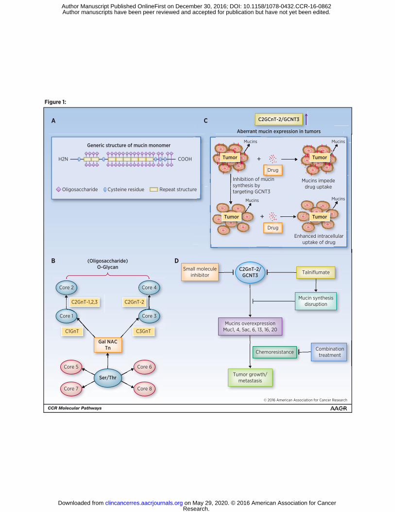
Figure 1:
© 2016 American Association for Cancer Research
H2N
(Oligosaccharide)O-Glycan
C2GCnT-2/GCNT3
Aberrant mucin expression in tumors
Core 2
C2GnT-1,2,3
C1GnT
C2GnT-2
Core 1
Core 4
Mucins Mucins
Mucins Mucins
Tumor Tumor
Tumor Tumor
Drug
Drug
Inhibition of mucinsynthesis bytargeting GCNT3
Mucins impededrug uptake
+
+
Enhanced intracellularuptake of drug
Core 3
A C
B D
Generic structure of mucin monomer
COOH
Oligosaccharide Cysteine residue Repeat structure
C3GnT
Gal NACTn
Ser/Thr
Core 5
Core 7
Core 6
Core 8
C2GnT-2/GCNT3
Tumor growth/metastasis
Mucins overexpressionMuc1, 4, 5ac, 6, 13, 16, 20
Small moleculeinhibitor
Chemoresistance Combinationtreatment
Mucin synthesisdisruption
Talniflumate
Research. on May 29, 2020. © 2016 American Association for Cancerclincancerres.aacrjournals.org Downloaded from
Author manuscripts have been peer reviewed and accepted for publication but have not yet been edited. Author Manuscript Published OnlineFirst on December 30, 2016; DOI: 10.1158/1078-0432.CCR-16-0862
Published OnlineFirst December 30, 2016.Clin Cancer Res Chinthalapally V Rao, Naveena B. Janakiram and Altaf Mohammed Molecular Pathways: Mucins and drug delivery in cancer
Updated version
10.1158/1078-0432.CCR-16-0862doi:
Access the most recent version of this article at:
Manuscript
Authoredited. Author manuscripts have been peer reviewed and accepted for publication but have not yet been
E-mail alerts related to this article or journal.Sign up to receive free email-alerts
Subscriptions
Reprints and
To order reprints of this article or to subscribe to the journal, contact the AACR Publications
Permissions
Rightslink site. Click on "Request Permissions" which will take you to the Copyright Clearance Center's (CCC)
.http://clincancerres.aacrjournals.org/content/early/2016/12/30/1078-0432.CCR-16-0862To request permission to re-use all or part of this article, use this link
Research. on May 29, 2020. © 2016 American Association for Cancerclincancerres.aacrjournals.org Downloaded from
Author manuscripts have been peer reviewed and accepted for publication but have not yet been edited. Author Manuscript Published OnlineFirst on December 30, 2016; DOI: 10.1158/1078-0432.CCR-16-0862