Embed Size (px)
Citation preview

Molecules of Life
Chapter 3

3.1 Molecules of Life
Molecules of life are synthesized by living cells• Carbohydrates• Lipids • Proteins • Nucleic acids

Structure to Function
Molecules of life differ in three-dimensional structure and function• Carbon backbone• Attached functional groups
Structures give clues to how they function

Organic Compounds
Consist primarily of carbon and hydrogen atoms• Carbon atoms bond covalently with up to four
other atoms, often in long chains or rings
Functional groups attach to a carbon backbone • Influence organic compound’s properties

An Organic Compound: Glucose
Four models

Functional Groups

Fig. 3.3, p. 36
In alcohols (e.g.,sugars, amino acids);water soluble
In fatty acid chains;insoluble in water
In sugars, amino acids,nucleotides; watersoluble. An aldehydeif at end of a carbonbackbone; a ketone ifattached to an interiorcarbon of backbone
In amino acids, fattyacids, carbohydrates;water soluble. Highlypolar; acts as an acid(releases H+)
carboxyl
methyl
hydroxyl
carbonyl
(ionized)(non-ionized)
(ketone)(aldehyde)

Fig. 3.3, p. 36
In amino acids andcertain nucleotidebases; water soluble,acts as a weak base(accepts H+)
In nucleotides (e.g.,ATP), also in DNA,RNA, many proteins,phospholipids; watersoluble, acidic
amino
phosphate
icon
(ionized)(non-ionized)

Functional Groups: The Importance of Position

one of the estrogens
Fig. 3.4, p. 37
testosterone
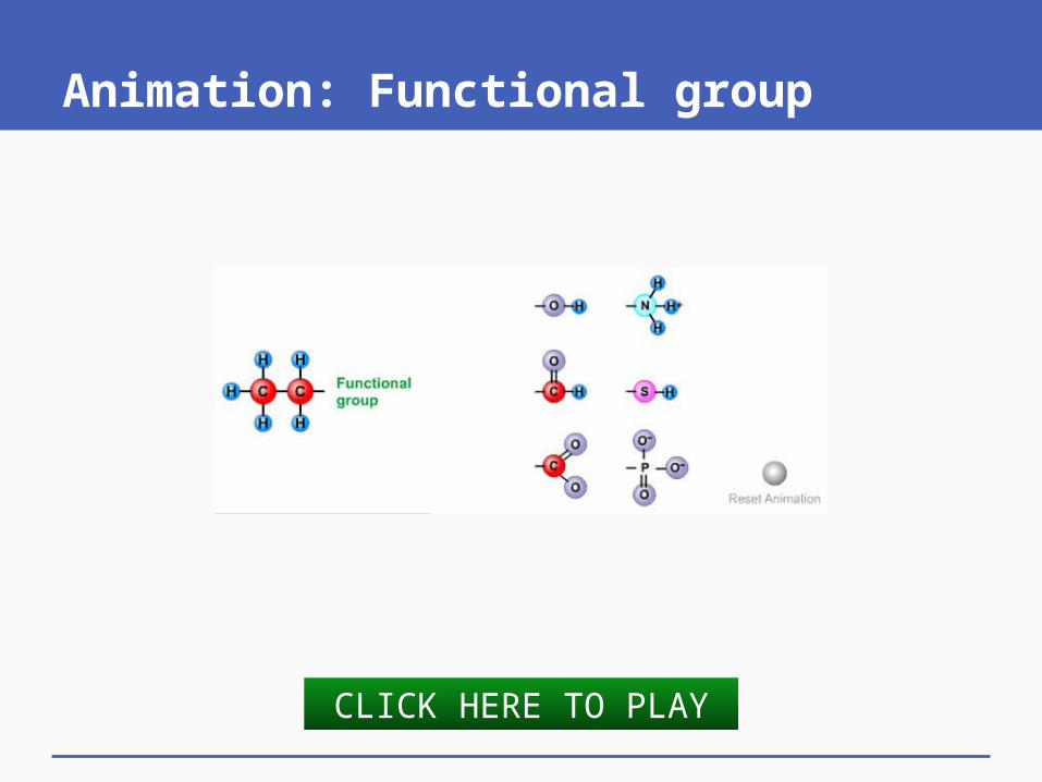
Animation: Functional group
CLICK HERE TO PLAY

Processes of Metabolism
Cells use energy to grow and maintain themselves
Enzyme-driven reactions build, rearrange, and split organic molecules
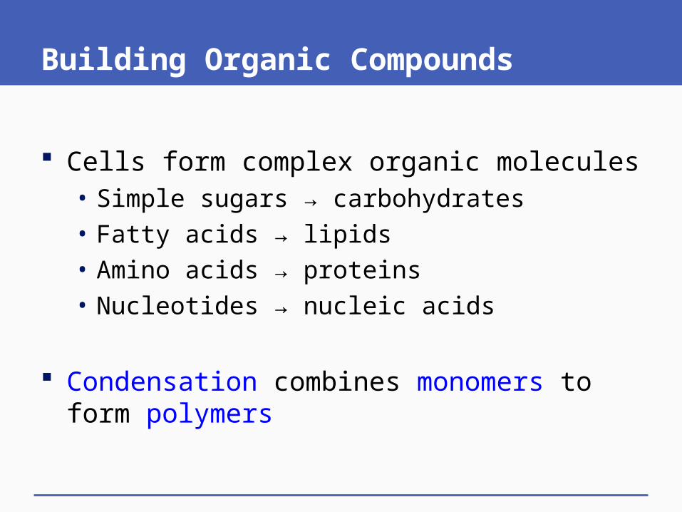
Building Organic Compounds
Cells form complex organic molecules• Simple sugars → carbohydrates• Fatty acids → lipids• Amino acids → proteins• Nucleotides → nucleic acids
Condensation combines monomers to form polymers

What Cells Do to Organic Compounds

Condensation and Hydrolysis

Fig. 3.5, p. 37
enzyme action at functional groups
enzyme action at functional groups
Condensation Hydrolysis

Animation: Condensation and hydrolysis
CLICK HERE TO PLAY
Key Concepts: STRUCTURE DICTATES FUNCTION
We define cells partly by their capacity to build complex carbohydrates and lipids, proteins, and nucleic acids
The main building blocks are simple sugars, fatty acids, amino acids, and nucleotides
These organic compounds have a backbone of carbon atoms with functional groups attached

3.2 Carbohydrates – The Most Abundant Ones
Three main types of carbohydrates• Monosaccharides (simple sugars)• Oligosaccharides (short chains)• Polysaccharides (complex carbohydrates)
Carbohydrate functions• Instant energy sources • Transportable or storable forms of energy• Structural materials

Simple Sugars: Glucose and Fructose

Oligosaccharides: Sucrose

glucose fructose
sucrose
Fig. 3.6, p. 38
c Formation of a sucrose molecule

Complex Carbohydrates: Bonding Patterns

Complex Carbohydrates: Starch, Cellulose, and Glycogen

Complex Carbohydrates: Starch, Cellulose, and Glycogen

Fig. 3.8, p. 39
c Glycogen. In animals, thispolysaccharide is a storage form for excess glucose. It is especially abundant in the liver and muscles of highly active animals, including fishes and people.
Structure of cellulose

Animation: Structure of starch and cellulose
CLICK HERE TO PLAY

Complex Carbohydrates: Chitin

Key Concepts:CARBOHYDRATES
Carbohydrates are the most abundant biological molecules
Simple sugars function as transportable forms of energy or as quick energy sources
Complex carbohydrates are structural materials or energy reservoirs

3.3 Greasy, Oily – Must Be Lipids
Lipids • Fats, phospholipids, waxes, and sterols• Don’t dissolve in water• Dissolve in nonpolar substances (other lipids)
Lipid functions• Major sources of energy• Structural materials• Used in cell membranes

Fats
Lipids with one, two, or three fatty acid tails• Saturated• Unsaturated (cis and trans)
Triglycerides (neutral fats )• Three fatty acid tails• Most abundant animal fat (body fat)• Major energy reserves

Fatty Acids

Animation: Fatty acids
CLICK HERE TO PLAY

Trans and Cis Fatty Acids

Triglyceride Formation

glycerol
three fatty acid tails Triglyceride, a neutral fat
Fig. 3.11, p. 40
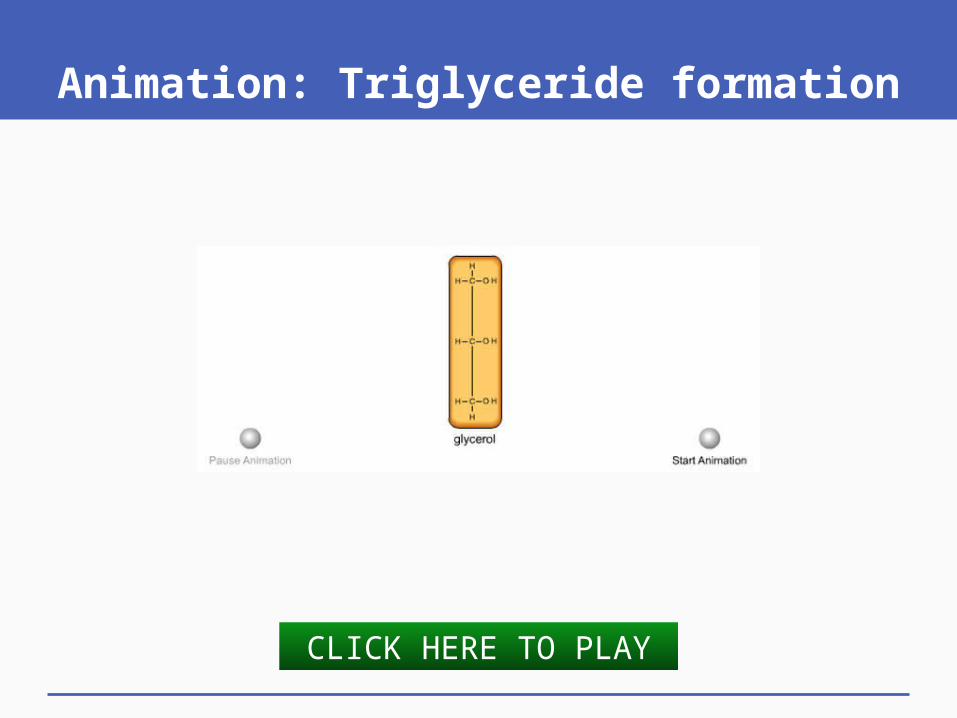
Animation: Triglyceride formation
CLICK HERE TO PLAY

Phospholipids
Main component of cell membranes • Hydrophilic head,
hydrophobic tails

hydrophilic head
two hydrophilic tails
Fig. 3.13, p. 41
b

Fig. 3.13, p. 41
c Cell membrane section

Animation: Phospholipid structure
CLICK HERE TO PLAY

Waxes
Firm, pliable, water repelling, lubricating

Sterols: Cholesterol
Membrane components; precursors of other molecules (steroid hormones)

Animation: Cholesterol
CLICK HERE TO PLAY

Key Concepts:LIPIDS
Complex lipids function as energy reservoirs, structural materials of cell membranes, signaling molecules, and waterproofing or lubricating substances

3.4 Proteins – Diversity in Structure and Function
Proteins have many functions• Structures• Nutrition• Enzymes• Transportation• Communication• Defense

Protein Structure
Built from 20 kinds of amino acids

Fig. 3.15, p. 42

Fig. 3.15, p. 42
carboxylgroup
aminogroup

Fig. 3.15, p. 42
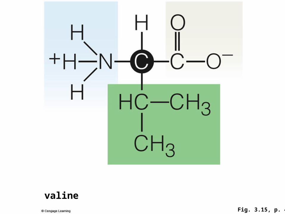
Fig. 3.15, p. 42
valine

Protein Synthesis

Protein Synthesis

Protein Synthesis

Four Levels of Protein Structure
1. Primary structure • Amino acids joined by peptide bonds form a
linear polypeptide chain
2. Secondary structure• Polypeptide chains form sheets and coils
3. Tertiary structure• Sheets and coils pack into functional domains

Four Levels of Protein Structure
4. Quaternary structure• Many proteins (e.g. enzymes) consist of two or
more chains
Other protein structures• Glycoproteins• Lipoproteins• Fibrous proteins

Levels of Protein Structure

Fig. 3.17, p. 43
a Protein primarystructure: Aminoacids bonded in apolypeptide chain.
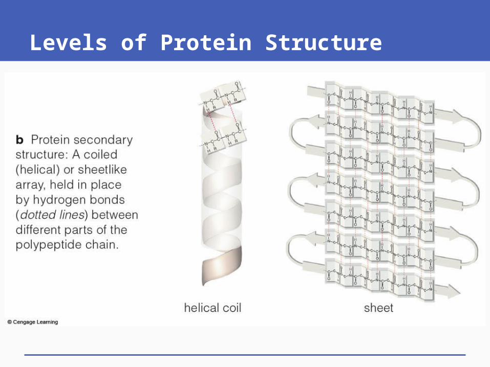
Levels of Protein Structure
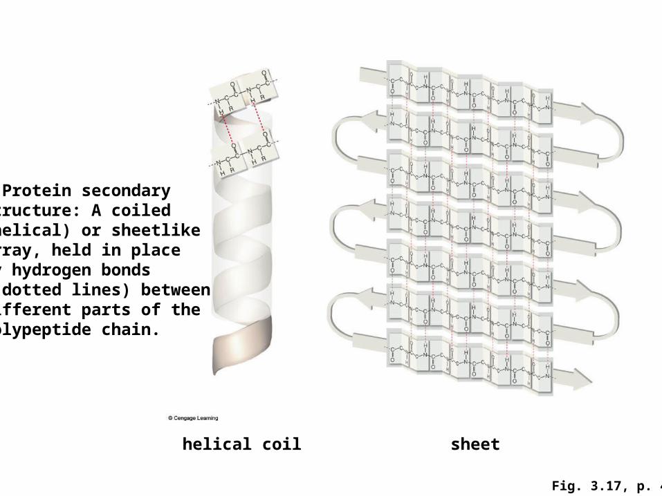
Fig. 3.17, p. 43
b Protein secondarystructure: A coiled(helical) or sheetlikearray, held in placeby hydrogen bonds( dotted lines) betweendifferent parts of thepolypeptide chain.
helical coil sheet

Levels of Protein Structure

Fig. 3.17, p. 43
c Protein tertiary structure: A chain’s coiled parts, sheetlikearrays, or both have folded and twisted into stable, functionaldomains, including clusters, pockets, and barrels.
barrel

Levels of Protein Structure

Fig. 3.17, p. 43
d Protein quaternarystructure: Many weakinteractions hold twoor more polypeptidechains together asa single molecule.

Animation: Structure of an amino acid
CLICK HERE TO PLAY

Animation: Peptide bond formation
CLICK HERE TO PLAY

Animation: Secondary and tertiary structure
CLICK HERE TO PLAY

Animation: Globin and hemoglobin structure
CLICK HERE TO PLAY

3.5 Why is Protein StructureSo Important?
Protein structure dictates function
Sometimes a mutation in DNA results in an amino acid substitution that alters a protein’s structure and compromises its function• Example: Hemoglobin and sickle-cell anemia

Normal Hemoglobin Structure

Fig. 3.18, p. 44
alpha globin
heme
a Globin. The secondarystructure of this polypeptideincludes several helixes. Thecoils fold up to form a pocketthat cradles heme, a functionalgroup with an iron atom at itscenter. The kind of molecularrepresentation shown here iscalled a ribbon model, after itsappearance. Appendix V hasmore details about such models.

Normal Hemoglobin Structure

alpha globin
beta globin beta globin
Fig. 3.18, p. 44
alpha globin
b Hemoglobin is one of the proteins with quaternary structure. Itconsists of four globin molecules held together by hydrogen bonds.To help you distinguish among them, the two alpha globin chainsare shown here in green, and the two beta globins are in brown.

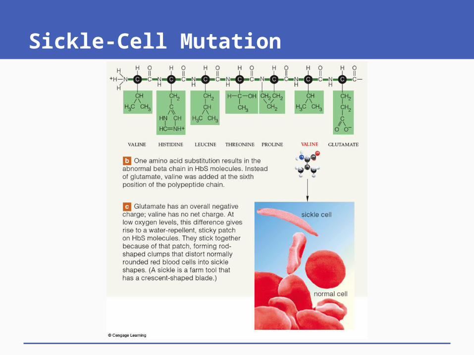
Sickle-Cell Mutation

Fig. 3.19, p. 45
THREONINE VALINE HISTIDINE LEUCINE GLUTAMATEPROLINE GLUTAMATE
a Normal amino acid sequence at the start of a beta chain for hemoglobin.
Sickle-Cell Mutation

Fig. 3.19, p. 45
VALINE HISTIDINE LEUCINE GLUTAMATEVALINETHREONINE PROLINE
sickle cell
normal cell
b One amino acid substitution results in theabnormal beta chain in HbS molecules. Insteadof glutamate, valine was added at the sixthposition of the polypeptide chain.
c Glutamate has an overall negative charge; valine has no net charge. At low oxygen levels, this difference gives rise to a water-repellent, sticky patch on HbS molecules. They stick togetherbecause of that patch, forming rodshaped clumps that distort normally rounded red blood cells into sickle shapes. (A sickle is a farm tool that has a crescent-shaped blade.)

Sickle-Cell Mutation

Clumping of cells in bloodstream
Circulatory problems, damage to brain, lungs, heart, skeletal muscles, gut, and kidneys
Heart failure, paralysis, pneumonia, rheumatism, gut pain, kidney failure
Spleen concentrates sickle cells
Spleen enlargement
Immune system compromised
Rapid destruction of sickle cells
Anemia, causing weakness,fatigue, impaired development,heart chamber dilation
Impaired brain function, heart failure Fig. 3.19, p. 45
d Melba Moore, celebrity spokes-person for sickle-cell anemia organizations. Right, range of symptoms for a person with two mutated genes for hemoglobin’s beta chain.

Clumping of cells in bloodstream
Spleen concentrates sickle cells
Rapid destruction of sickle cells
Circulatory problems, damage to brain, lungs, heart, skeletal muscles, gut, and kidneys
Heart failure, paralysis, pneumonia, rheumatism, gut pain, kidney failure
Spleen enlargement
Immune system compromised
Anemia, causing weakness,fatigue, impaired development,heart chamber dilation
Impaired brain function, heart failure Fig. 3-19, p. 45
d Melba Moore, celebrity spokes-person for sickle-cell anemia organizations. Right, range of symptoms for a person with two mutated genes for hemoglobin’s beta chain.
Stepped Art

Animation: Sickle-cell anemia
CLICK HERE TO PLAY

Denatured Proteins
If a protein unfolds and loses its three-dimensional shape (denatures), it also loses its function
Caused by shifts in pH or temperature, or exposure to detergent or salts • Disrupts hydrogen bonds and other molecular
interactions responsible for protein’s shape

Key Concepts:PROTEINS
Structurally and functionally, proteins are the most diverse molecules of life
They include enzymes, structural materials, signaling molecules, and transporters

Animation: Molecular models of the protein hemoglobin
CLICK HERE TO PLAY

3.6 Nucleotides, DNA, and RNAs
Nucleotide structure, 3 parts:• Sugar• Phosphate group• Nitrogen-containing base

Fig. 3.20, p. 46
three phosphate groups
base (blue)
sugar (orange)

Nucleotide Functions: Reproduction, Metabolism, and Survival
DNA and RNAs are nucleic acids, each composed of four kinds of nucleotide subunits
ATP energizes many kinds of molecules by phosphate-group transfers
Other nucleotides function as coenzymes or as chemical messengers

Nucleotides of DNA
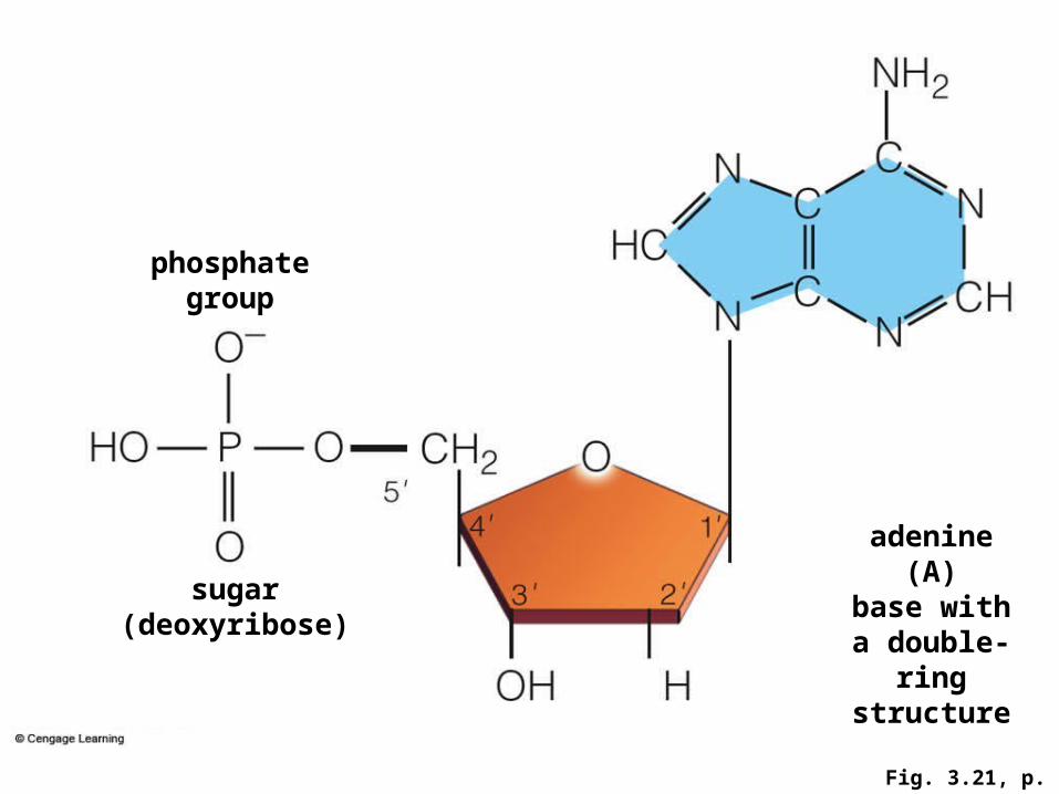
Fig. 3.21, p. 46
adenine(A)
base with a double-ring
structure
phosphate group
sugar (deoxyribose)

Fig. 3.21, p. 46
THYMINE(T)
base with a single-ring structure

Fig. 3.21, p. 46
GUANINE(C)
base with a double-ring
structure

Fig. 3.21, p. 46
CYTOSINE(C)
base with a single-ring structure

DNA, RNAs, and Protein Synthesis
DNA (double-stranded)• Encodes information about the primary structure
of all cell proteins in its nucleotide sequence
RNA molecules (usually single stranded)• Different kinds interact with DNA and one another
during protein synthesis

The DNA Double-Helix

Fig. 3.22, p. 47
covalentbonding incarbonbackbone
hydrogen bondingbetween bases

Key Concepts:NUCLEOTIDES AND NUCLEIC ACIDS
Nucleotides have major metabolic roles and are building blocks of nucleic acids
Two kinds of nucleic acids, DNA and RNA, interact as the cell’s system of storing, retrieving, and translating information about building proteins

Animation: Nucleotide subunits of DNA
CLICK HERE TO PLAY

Animation: Structure of ATP
CLICK HERE TO PLAY